Embed Size (px)
Citation preview
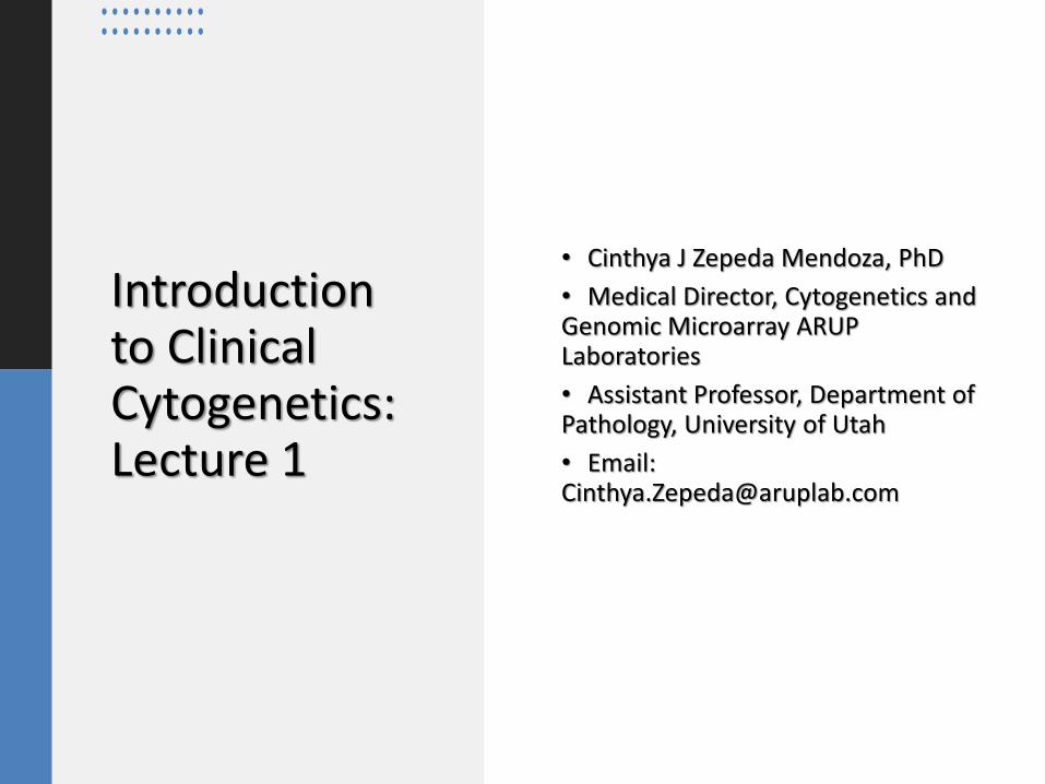
Introduction to Clinical Cytogenetics: Lecture 1
• Cinthya J Zepeda Mendoza, PhD
• Medical Director, Cytogenetics and Genomic Microarray ARUP Laboratories
• Assistant Professor, Department of Pathology, University of Utah
• Email: [email protected]
Cytogenetics Textbooks
• Gardner and Amor. (2018) Gardner and Sutherland’s Chromosome Abnormalities and Genetic Counseling. 5th edition
• Gersen and Keagle. (2013) The Principles of Clinical Cytogenetics. 3rd edition
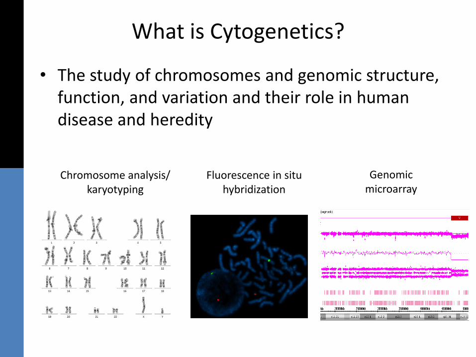
What is Cytogenetics?
• The study of chromosomes and genomic structure, function, and variation and their role in human disease and heredity
Chromosome analysis/karyotyping
Fluorescence in situ hybridization
Genomic microarray
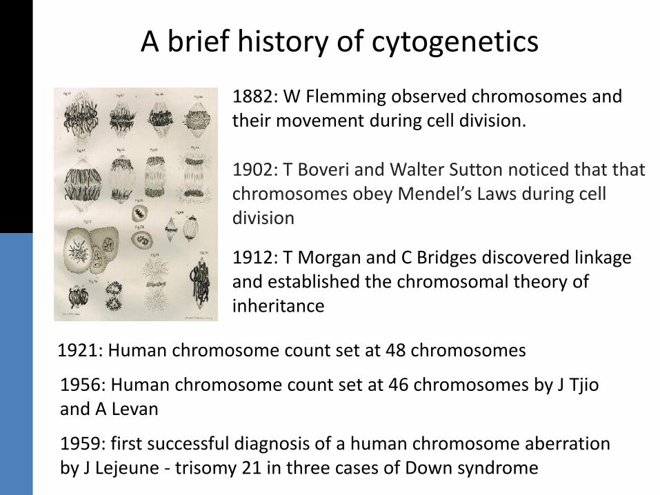
A brief history of cytogenetics
1882: W Flemming observed chromosomes and their movement during cell division.
1902: T Boveri and Walter Sutton noticed that that chromosomes obey Mendel’s Laws during cell division
1912: T Morgan and C Bridges discovered linkage and established the chromosomal theory of inheritance
1921: Human chromosome count set at 48 chromosomes
1956: Human chromosome count set at 46 chromosomes by J Tjioand A Levan
1959: first successful diagnosis of a human chromosome aberration by J Lejeune - trisomy 21 in three cases of Down syndrome
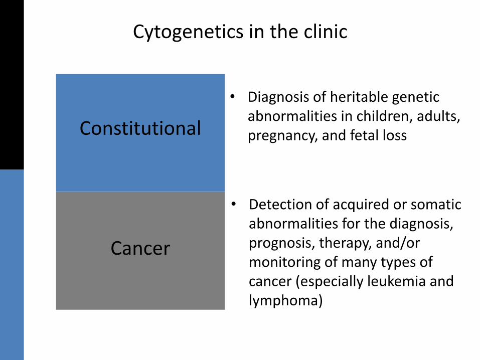
Cytogenetics in the clinic
Cancer
Constitutional
• Diagnosis of heritable genetic abnormalities in children, adults, pregnancy, and fetal loss
• Detection of acquired or somatic abnormalities for the diagnosis, prognosis, therapy, and/or monitoring of many types of cancer (especially leukemia and lymphoma)
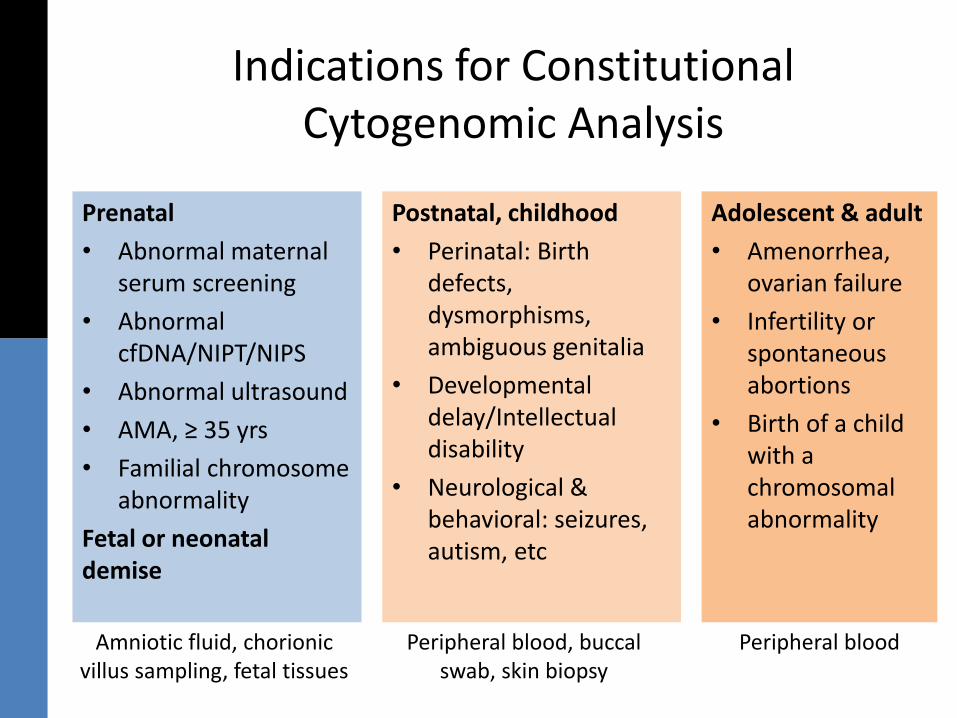
Indications for Constitutional Cytogenomic Analysis
Prenatal
• Abnormal maternal serum screening
• Abnormal cfDNA/NIPT/NIPS
• Abnormal ultrasound
• AMA, ≥ 35 yrs
• Familial chromosome abnormality
Fetal or neonatal demise
Amniotic fluid, chorionic villus sampling, fetal tissues
Postnatal, childhood
• Perinatal: Birth defects, dysmorphisms, ambiguous genitalia
• Developmental delay/Intellectual disability
• Neurological & behavioral: seizures, autism, etc
Peripheral blood, buccal swab, skin biopsy
Adolescent & adult
• Amenorrhea, ovarian failure
• Infertility or spontaneous abortions
• Birth of a child with a chromosomal abnormality
Peripheral blood
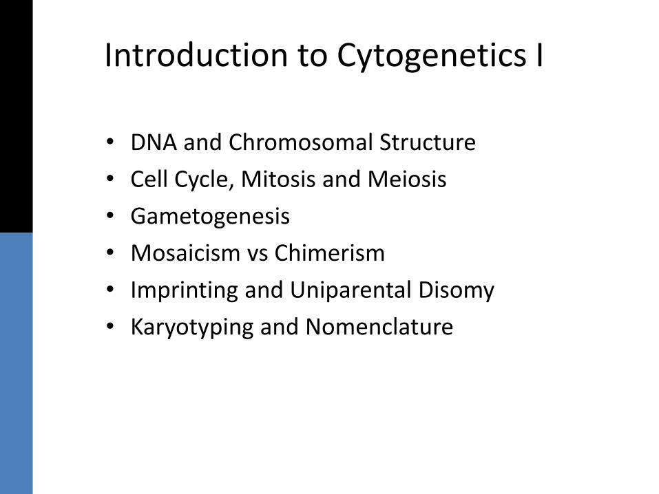
Introduction to Cytogenetics I
• DNA and Chromosomal Structure
• Cell Cycle, Mitosis and Meiosis
• Gametogenesis
• Mosaicism vs Chimerism
• Imprinting and Uniparental Disomy
• Karyotyping and Nomenclature
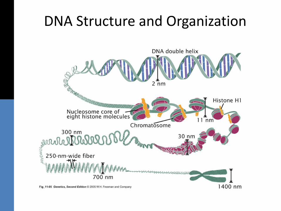
DNA Structure and Organization
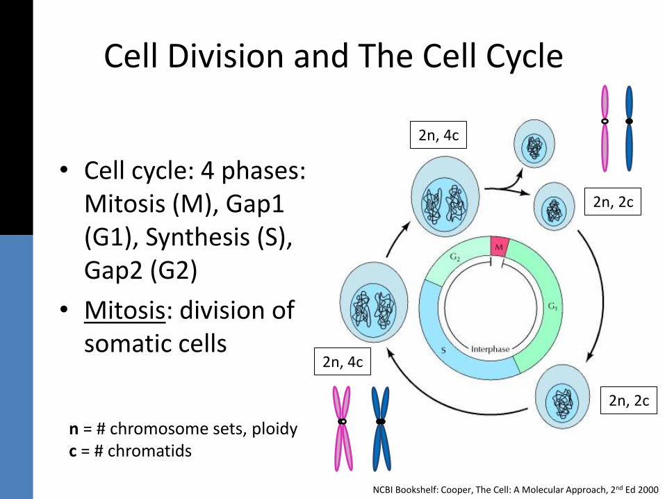
Cell Division and The Cell Cycle
• Cell cycle: 4 phases: Mitosis (M), Gap1 (G1), Synthesis (S), Gap2 (G2)
• Mitosis: division of somatic cells
NCBI Bookshelf: Cooper, The Cell: A Molecular Approach, 2nd Ed 2000
n = # chromosome sets, ploidyc = # chromatids
2n, 4c
2n, 2c
2n, 2c
2n, 4c
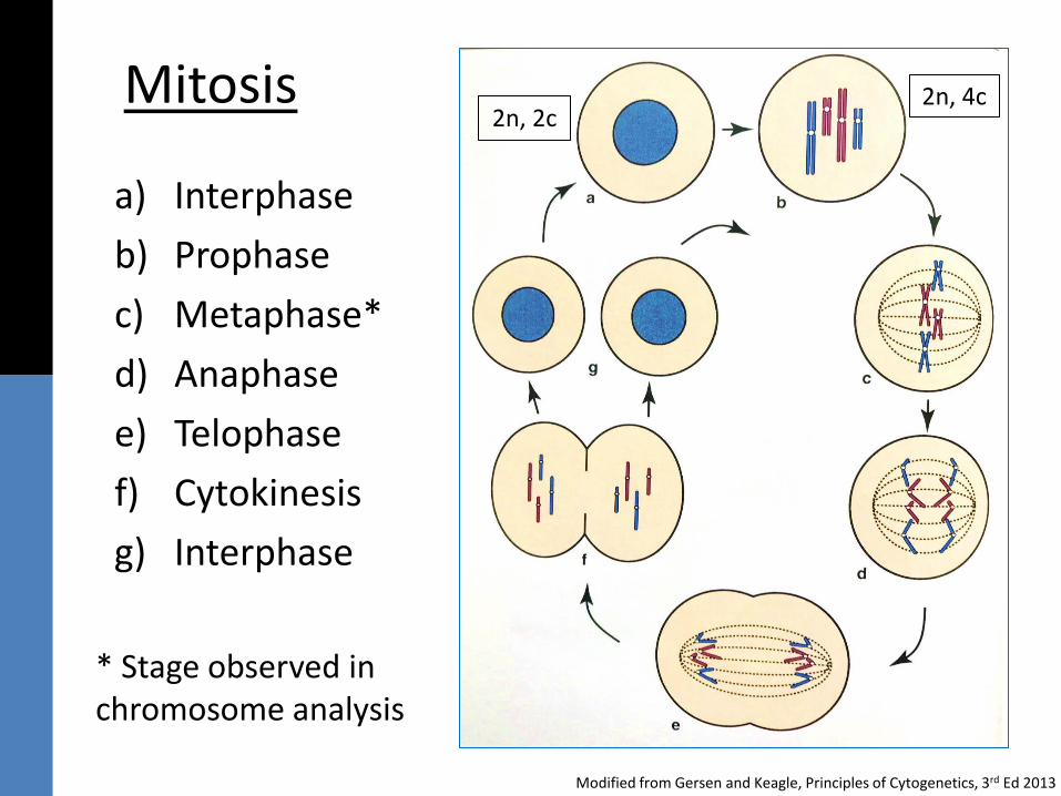
Mitosis
a) Interphase
b) Prophase
c) Metaphase*
d) Anaphase
e) Telophase
f) Cytokinesis
g) Interphase
Modified from Gersen and Keagle, Principles of Cytogenetics, 3rd Ed 2013
* Stage observed in chromosome analysis
2n, 4c2n, 2c
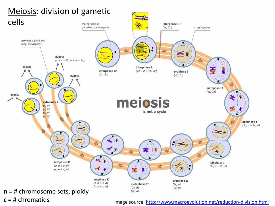
Image source: http://www.macroevolution.net/reduction-division.html
2n, 4c
1n, 2c
n = # chromosome sets, ploidyc = # chromatids
Meiosis: division of gametic cells
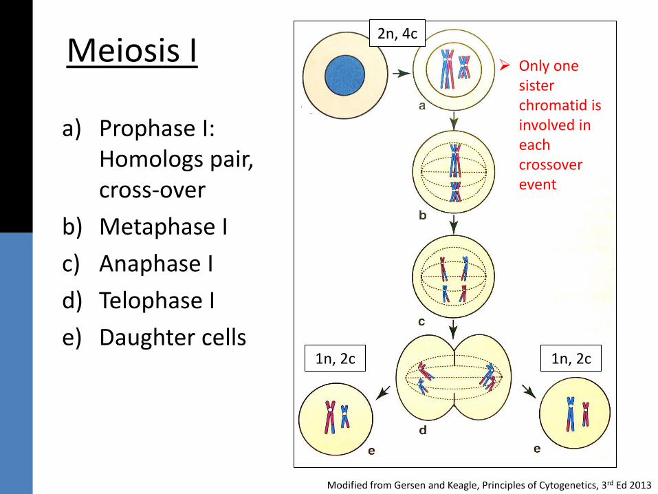
Meiosis I
a) Prophase I: Homologs pair, cross-over
b) Metaphase I
c) Anaphase I
d) Telophase I
e) Daughter cells
Modified from Gersen and Keagle, Principles of Cytogenetics, 3rd Ed 2013
2n, 4c
1n, 2c 1n, 2c
➢ Only one sister chromatid is involved in each crossover event
2n, 4c
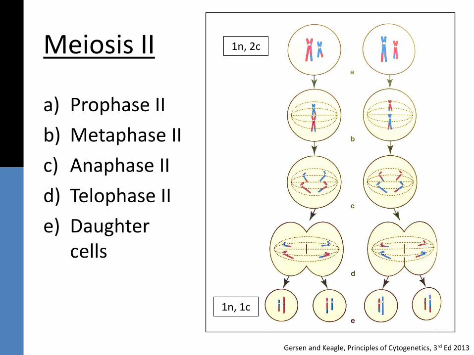
Meiosis II
a) Prophase II
b) Metaphase II
c) Anaphase II
d) Telophase II
e) Daughter cells
Gersen and Keagle, Principles of Cytogenetics, 3rd Ed 2013
1n, 2c
1n, 1c
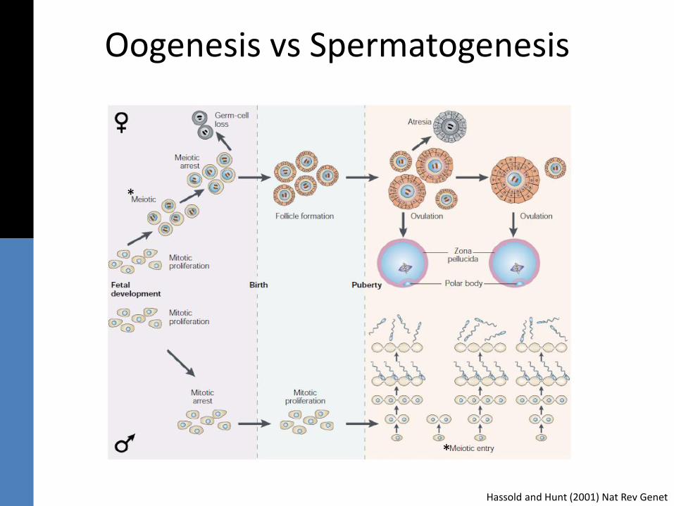
Oogenesis vs Spermatogenesis
Hassold and Hunt (2001) Nat Rev Genet
*
*
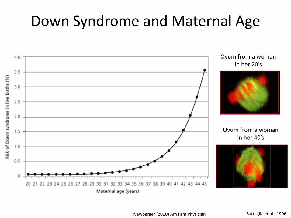
Down Syndrome and Maternal Age
Newberger (2000) Am Fam Physician Battaglia et al., 1996
Ovum from a woman in her 20’s
Ovum from a woman in her 40’s
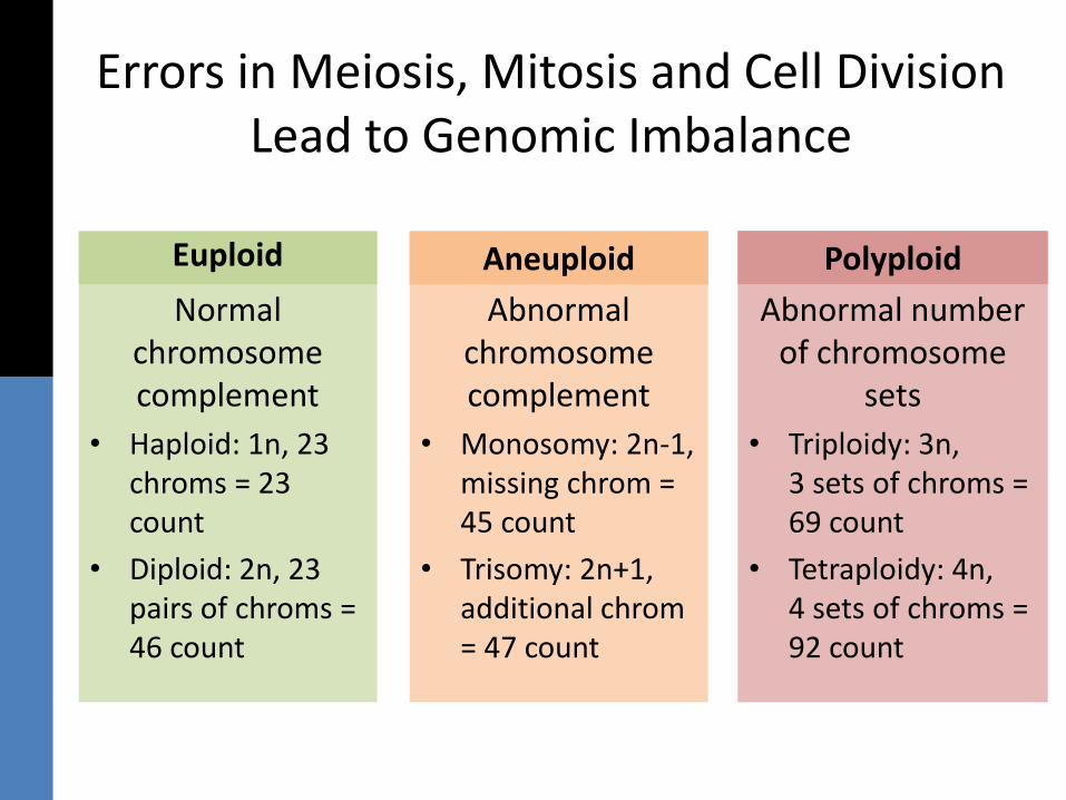
Errors in Meiosis, Mitosis and Cell Division Lead to Genomic Imbalance
Euploid
Normal chromosome complement
• Haploid: 1n, 23 chroms = 23 count
• Diploid: 2n, 23 pairs of chroms = 46 count
Euploid Aneuploid
Abnormal chromosome complement
• Monosomy: 2n-1, missing chrom = 45 count
• Trisomy: 2n+1, additional chrom= 47 count
Aneuploid Polyploid
Abnormal number of chromosome
sets
• Triploidy: 3n, 3 sets of chroms = 69 count
• Tetraploidy: 4n, 4 sets of chroms = 92 count
Polyploid
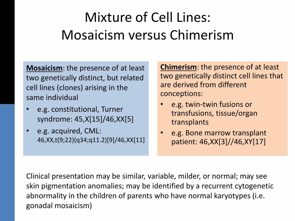
Mixture of Cell Lines: Mosaicism versus Chimerism
Mosaicism: the presence of at least two genetically distinct, but related cell lines (clones) arising in the same individual
• e.g. constitutional, Turner syndrome: 45,X[15]/46,XX[5]
• e.g. acquired, CML: 46,XX,t(9;22)(q34;q11.2)[9]/46,XX[11]
Clinical presentation may be similar, variable, milder, or normal; may see skin pigmentation anomalies; may be identified by a recurrent cytogenetic abnormality in the children of parents who have normal karyotypes (i.e. gonadal mosaicism)
Chimerism: the presence of at least two genetically distinct cell lines that are derived from different conceptions:
• e.g. twin-twin fusions or transfusions, tissue/organ transplants
• e.g. Bone marrow transplant patient: 46,XX[3]//46,XY[17]
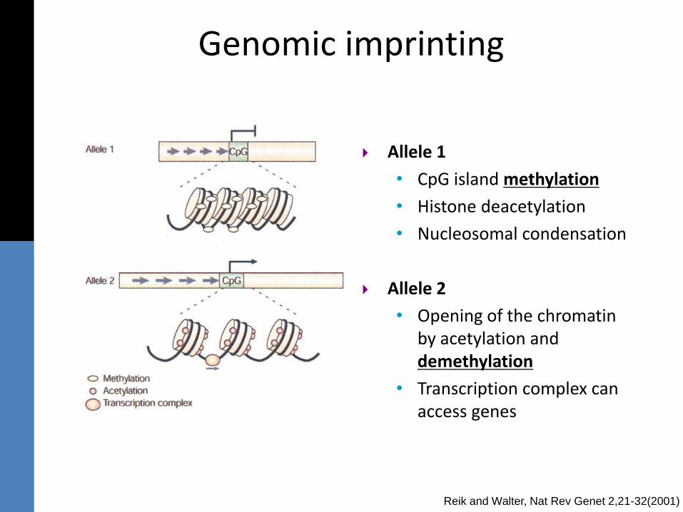
Allele 1
• CpG island methylation
• Histone deacetylation
• Nucleosomal condensation
Allele 2
• Opening of the chromatin by acetylation and demethylation
• Transcription complex can access genes
Genomic imprinting
Reik and Walter, Nat Rev Genet 2,21-32(2001)
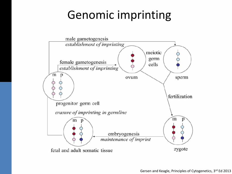
Genomic imprinting
Gersen and Keagle, Principles of Cytogenetics, 3rd Ed 2013
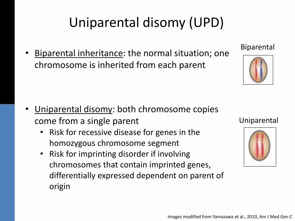
Uniparental disomy (UPD)
• Biparental inheritance: the normal situation; one chromosome is inherited from each parent
• Uniparental disomy: both chromosome copies come from a single parent• Risk for recessive disease for genes in the
homozygous chromosome segment• Risk for imprinting disorder if involving
chromosomes that contain imprinted genes, differentially expressed dependent on parent of origin
Biparental
Uniparental
Images modified from Yamazawa et al., 2010, Am J Med Gen C
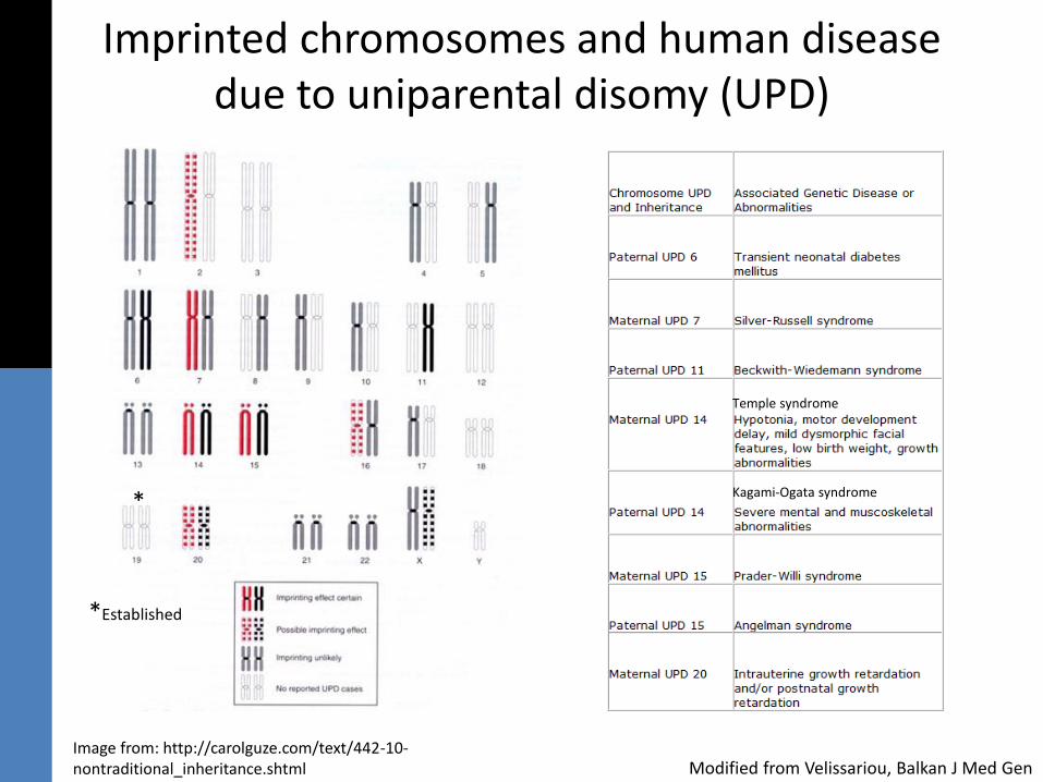
Imprinted chromosomes and human disease due to uniparental disomy (UPD)
Image from: http://carolguze.com/text/442-10-nontraditional_inheritance.shtml Modified from Velissariou, Balkan J Med Gen
Kagami-Ogata syndrome
Temple syndrome
*
*Established
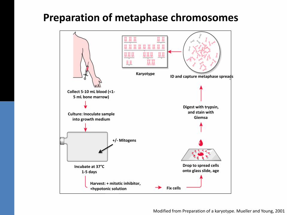
Preparation of metaphase chromosomes
Modified from Preparation of a karyotype. Mueller and Young, 2001
+/- Mitogens
Harvest: + mitotic inhibitor, +hypotonic solution
Culture: Inoculate sample into growth medium
Incubate at 37°C 1-5 days
Collect 5-10 mL blood (<1-5 mL bone marrow)
Fix cells
Drop to spread cells onto glass slide, age
Digest with trypsin, and stain with
Giemsa
KaryotypeID and capture metaphase spreads
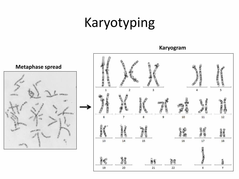
Karyotyping
Metaphase spread
Karyogram
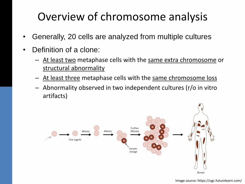
Overview of chromosome analysis
• Generally, 20 cells are analyzed from multiple cultures
• Definition of a clone:
– At least two metaphase cells with the same extra chromosome or structural abnormality
– At least three metaphase cells with the same chromosome loss
– Abnormality observed in two independent cultures (r/o in vitro artifacts)
Image source: https://ugc.futurelearn.com/

Chromosome Structure and Classification
Images modified, source: http://learn.genetics.utah.edu/content/chromosomes/readchromosomes/
Telomere
Telomere
Centromere
p a
rmq
arm
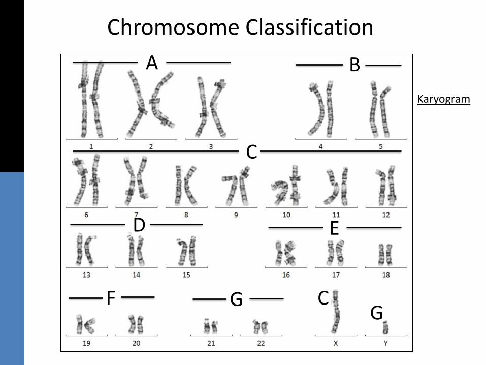
Chromosome Classification
A B
C
D E
F G
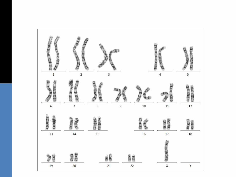
Karyogram
CG
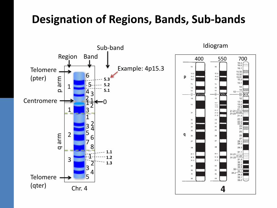
Designation of Regions, Bands, Sub-bands
Telomere(pter)
Telomere(qter)
Centromere
p a
rmq
arm
1
2
3
Region Band
1
6
54 32 1
5.35.25.1
Sub-band
6
1231
23 45
78
12
34
5
1.11.21.3
0
Chr. 4
Idiogram
400 550 700
Example: 4p15.3
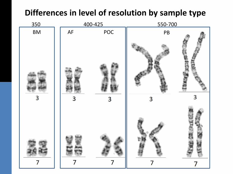
Differences in level of resolution by sample type350 400-425 550-700
BM AF POC PB
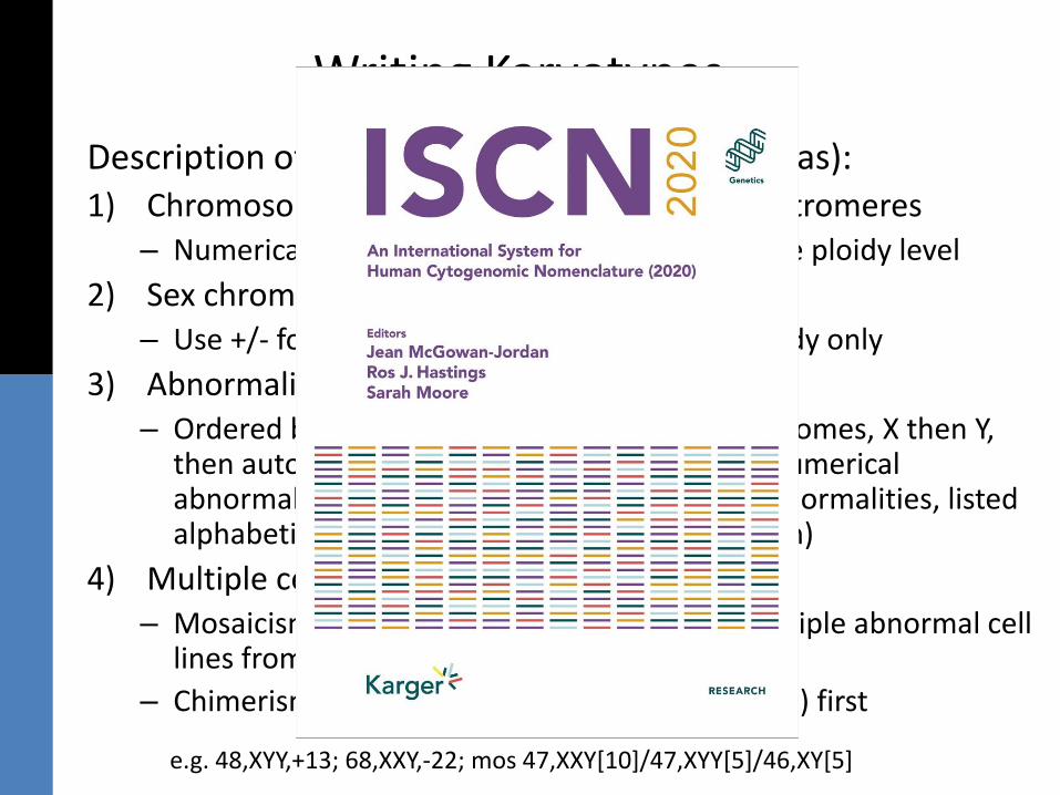
Writing Karyotypes
Description of… (in order, separated by commas):1) Chromosome number (count)-based on #centromeres
– Numerical changes are expressed relative to the ploidy level
2) Sex chromosome constitution– Use +/- for acquired sex chromosome aneuploidy only
3) Abnormalities present– Ordered by chromosome number (sex chromosomes, X then Y,
then autosomes 1-22) and abnormality type (numerical abnormalities/aneuploidies, then structural abnormalities, listed alphabetically and by location/band, low to high)
4) Multiple cell lines– Mosaicism: List abnormal clone(s) first, list multiple abnormal cell
lines from largest to smallest clone size
– Chimerism: List recipient (individual’s karyotype) first
e.g. 48,XYY,+13; 68,XXY,-22; mos 47,XXY[10]/47,XYY[5]/46,XY[5]
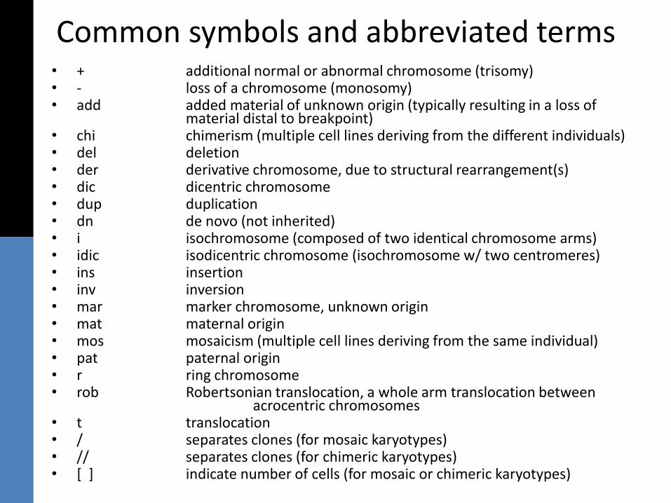
Common symbols and abbreviated terms• + additional normal or abnormal chromosome (trisomy)• - loss of a chromosome (monosomy)• add added material of unknown origin (typically resulting in a loss of
material distal to breakpoint)• chi chimerism (multiple cell lines deriving from the different individuals)• del deletion• der derivative chromosome, due to structural rearrangement(s)• dic dicentric chromosome• dup duplication• dn de novo (not inherited)• i isochromosome (composed of two identical chromosome arms)• idic isodicentric chromosome (isochromosome w/ two centromeres)• ins insertion• inv inversion• mar marker chromosome, unknown origin• mat maternal origin• mos mosaicism (multiple cell lines deriving from the same individual)• pat paternal origin• r ring chromosome• rob Robertsonian translocation, a whole arm translocation between
acrocentric chromosomes• t translocation• / separates clones (for mosaic karyotypes)• // separates clones (for chimeric karyotypes)• [ ] indicate number of cells (for mosaic or chimeric karyotypes)
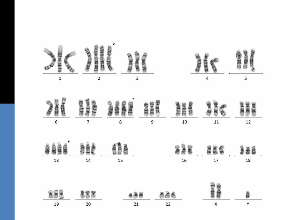
Nomenclature Practice:Numerical Abnormalities
(Constitutional)
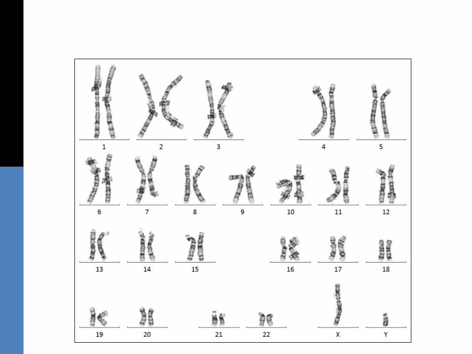
a) Normal
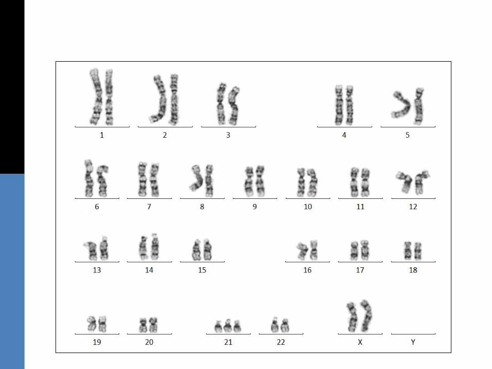
b) Abnormal
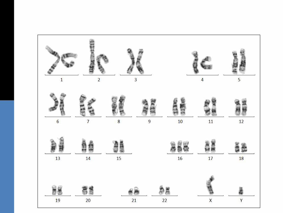
c) Abnormal
d) Abnormal
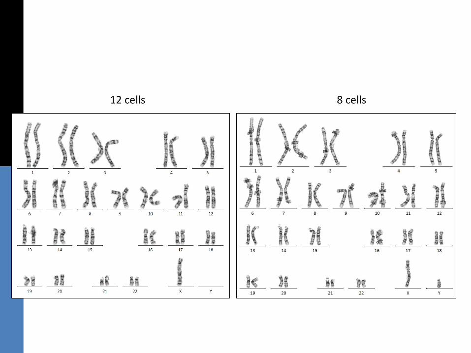
e) Abnormal
12 cells 8 cells
f) Abnormal