Embed Size (px)
Citation preview

Registered as a specially authorised society under the Friendly Societies Act (1974). Registration No. 200SA.. Registered Office: British Society for Human Genetics, Clinical Genetics Unit, Birmingham Women’s Hospital, Birmingham, B15 2TG.
PROFESSIONAL GUIDELINES FOR CLINICAL CYTOGENETICS
GENERAL BEST PRACTICE GUIDELINES (2007) v1.01
March 2007

Association for Clinical Cytogenetics GENERAL BEST PRACTICE GUIDELINES (2007) v1.01
page 2 of 25
CONTENTS 1 INTRODUCTION.......................................................................................... 3 1.1 ANALYSIS AND CHECKING ................................................................... 3 1.1.1 Constitutional work.......................................................................... 3 1.1.2 Oncology work. ............................................................................... 4
1.2 TABLE 1. G- BANDING EVALUATION SCORE ........................................... 5 1.3 TABLE 2. MINIMUM G BANDING SCORE FOR REFERRAL REASON............... 6 1.4 REPORTING........................................................................................ 7 1.5 TABLE 3. CYTOGENETIC GUIDELINE REPORTING TARGETS....................... 8 1.6 ARCHIVING AND STORAGE................................................................... 8
2 POST-NATAL BLOOD SAMPLES ..................................................................... 9 2.1 Reasons for Referral ............................................................................ 9 2.2 Techniques......................................................................................... 9 2.3 Analysis............................................................................................. 9 2.3.1 Syndromes with anomalous chromosome behaviour and instability .......10
3 PRENATAL DIAGNOSIS SAMPLES .................................................................12 3.1 Reasons for Referral ...........................................................................12
4 SOLID TISSUE SAMPLES.............................................................................13 4.1 Reasons for Referral ...........................................................................13 4.2 Techniques........................................................................................13 4.3 Analysis............................................................................................13
5 HAEMATOLOGICAL DISORDERS...................................................................15 5.1 Reasons for Referral ...........................................................................15 5.2 Sample Type .....................................................................................15 5.3 Techniques........................................................................................15 5.4 Analysis............................................................................................16
6 SOLID TUMOURS .......................................................................................17 6.1 Reasons for Referral ...........................................................................17 6.2 Techniques........................................................................................17 6.3 Analysis............................................................................................17
7 FLUORESCENCE IN SITU HYBRIDISATION (FISH)...........................................19 7.1 Techniques........................................................................................19 7.2 Analysis of constitutional abnormalities ................................................19 7.3 Analysis of acquired abnormalities .......................................................20
8 MOSAICISM ..............................................................................................21 8.1 Postnatal constitutional analysis ..........................................................21 8.2 Prenatal diagnosis analysis..................................................................22 8.3 Oncology and acquired chromosome abnormalities.................................22
9 DEFINITIONS OF TERMS.............................................................................23 10 REFERENCES ........................................................................................24 11 Version Control .....................................................................................25

Association for Clinical Cytogenetics GENERAL BEST PRACTICE GUIDELINES (2007) v1.01
page 3 of 25
1 INTRODUCTION Professional guidelines for Cytogenetics laboratories incorporate the standards imposed by regulatory bodies (Clinical Pathology Accreditation (CPA) [1] and by statute (Clinical Governance) while taking into account current practice in the U.K. Elements of the service not subject to statute may be varied in order to comply with local constraints and agreements. It must be noted that these guidelines are minimum requirements and that professional judgement is of paramount importance for many circumstances. The use of ‘shall’ in this document indicates a requirement and the use of ‘should’ indicates a recommendation. Where there appears to be contradiction between available guidelines, the most recently published should be taken to apply to all. All diagnostic Cytogenetics laboratories shall be accredited to nationally or internationally accepted standards. Laboratories shall participate in an External Quality Assessment Scheme for all aspects of their service for which a scheme is available [2].
1.1 ANALYSIS AND CHECKING Either the analyst, or the independent checker of analysis, shall be a registered Clinical Scientist. Analytical procedures and the checking systems used for each type of analysis shall be documented and specify the minimum level and experience of the staff involved, with reference to relevant scopes of practice for clinical scientists and genetic technologists in clinical cytogenetics. 1.1.1 Constitutional work. The minimum recommended quality of constitutional preparations will depend on the reason for referral (See Tables 1 and 2). Standard analysis shall be of a minimum of two metaphases and shall consist of every pair of homologues being cleared in full at least twice at the minimum quality level appropriate for the referral reason. It is recognised that additional cells of varying quality may be examined in the analysis process without affecting the overall case quality score. Independent checking is an essential part of the analytical process. A minimum of one cell shall be analysed by the

Association for Clinical Cytogenetics GENERAL BEST PRACTICE GUIDELINES (2007) v1.01
page 4 of 25
checker, with reference made to other cells when obscured regions of the karyotype need to be clarified, so that every pair of homologues is analysed at least once at the minimum quality level appropriate for the referral reason. In mosaic cases, one cell shall be checked from each cell line. The following aspects shall be checked before authorisation of the final report:
• Identification details on slides, clinical referral form, record of analysis and any hard copy or computerised images must concur.
• The level of analysis carried out is adequate with reference to laboratory and professional standards and the quality level reported concurs between analyst and checker.
• The karyotype result concurs between checker and analyst and is written in correct ISCN when practicable.
• Patient details on the report to be issued concur with the referral form and the record of analysis.
• The information in the text of the report concurs with the record of analysis.
FISH analysis checking, on metaphase or interphase, should be carried out down the microscope or from an unenhanced image. If checking is from the latter, there shall be a system in place for verification that the image checked is from the patient’s slides and preparations. 1.1.2 Oncology work. A single cell will normally be representative of the karyotype in constitutional work but this is not applicable to oncology preparations. Checking of oncology cytogenetics and fluorescence in situ hybridisation (FISH) preparations will be included with the professional guidelines for each area of investigation.

Association for Clinical Cytogenetics GENERAL BEST PRACTICE GUIDELINES (2007) v1.01
page 5 of 25
1.2 TABLE 1. G- BANDING EVALUATION SCORE At least three of the criteria to be obtained to apply banding scores 3-9
0 No banding
1 Identification of some chromosomes by morphology and major landmarks
2 POOR <300 band
Unequivocal identification of chromosomes due to major landmarks
3 300 band
2 bands on 8p (8p12 & 8p22) 3 bands on 10q (10q21, 10q23, 10q25) 20p12 visible 22q12 distinct
4 MODERATE 400 band
3 bands on mid-4q (q22-28) 3 bands mid-5q (5q14, 5q21, 5q23) 2 bands on 9p (9p21 & 9p23) 13q33 distinct
5 500 band
7q33 & 7q35 distinct 3 bands on 11p (11p12, 11p14, 11p15.4) 14q32.2 distinct 4 bands on 18q (18q12.1, 18q12.3, 18q21.2, 18q22)
6 GOOD 550 band
5q31.2 distinct 8p21.2 visible 2 bands on 11pter (11p15.2 & 11p15.4) 22q13.2 distinct
7 700 band
2p25.2 2q37.2 distinct 10q21.1 and 10q21.3 resolve 17q22-q24 resolves into 3 bands
8 EXCELLENT 850 band
4p15.31 & 4p15.33 distinct 5p15.32 distinct 11q24.1 and 11q24.3 distinct 19p13.12 and 19p13.2 distinct
9 900 band
11p14.1 visible 20p12.1 & 20p12.3 distinct 22q11.22 distinct 22q13.32 distinct
10 Banding Resolution higher than level 9 with additional bands to those seen at the 900bphs level (ISCN 2005)[3] seen consistently on both homologues.

Association for Clinical Cytogenetics GENERAL BEST PRACTICE GUIDELINES (2007) v1.01
page 6 of 25
1.3 TABLE 2. MINIMUM G BANDING SCORE FOR REFERRAL REASON
The recommended scores given below are defined as the lowest standard acceptable for a given reason for referral in constitutional analysis without issuing a qualified report.
MINIMUM QUALITY G-Banding SCORE
Reason for referral
Confirmation of aneuploidy e.g. direct lymphocyte, direct CV or solid tissue culture preparation.
2
Exclusion of known large structural rearrangements. e.g lymphocyte, solid tissue, CVS direct preparation or amniotic fluid cell preparation
3
Identification and exclusion of small expected structural rearrangements e.g. lymphocyte, solid tissue, CVS culture or amniotic fluid preparation
4
Routine amniotic fluid and CV culture preparations 4
Abnormal ultrasound scan associated with AF and CV referrals. 5
Blood sample referrals 6
For microdeletion syndromes (when no FISH probe is available) 7

Association for Clinical Cytogenetics GENERAL BEST PRACTICE GUIDELINES (2007) v1.01
page 7 of 25
1.4 REPORTING
It is the responsibility of the clinical scientist to provide a clear and unambiguous description of the cytogenetic findings and an explanation of the clinical implications of the results [4]. The report will be inserted into the patient’s notes and may be seen, not only by the referring clinician, but also by other healthcare workers, some of whom may not have a clear understanding of cytogenetics. When writing a report it is important to remember that it may also be made available to the patient. Handwritten alterations should never be made to the report; accreditation standards will insist that validation procedures are in place to ensure no alteration of reports can be made after issue. It is not necessary to include details of culture procedures, unless relevant, e.g. from direct or cultured CVS, direct or cultured tumour. Validation of reports shall be carried out by a clinical scientist at the professional level of at least Band 8a. The report of an abnormal case shall include the following:
� karyotype designation using correct current ISCN nomenclature where practicable
� a clear written description of the abnormality, and whether the karyotype is balanced or unbalanced
� the name of any associated syndrome � methods used in establishing the result � clinical interpretation to include (as appropriate): a) whether the cytogenetics result is consistent with the clinical
findings, and/or an indication of the expected consequences of the abnormality
b) request for follow up of family members at risk of the same or related abnormality, starting with closest available relatives
c) an assessment of risk/recurrence d) recommendation for consideration of prenatal diagnosis in
future pregnancies e) onward referral for genetic counselling
All laboratories should endeavour to maintain adequate reporting times (see Table 3). It is recognised that local clinical need may influence the reporting times for non-urgent work. The ACC Professional Standards Committee collects annual audit data from UK laboratories to inform the profession of workload activity. (5)

Association for Clinical Cytogenetics GENERAL BEST PRACTICE GUIDELINES (2007) v1.01
page 8 of 25
1.5 TABLE 3. CYTOGENETIC GUIDELINE REPORTING TARGETS
Referral category Sample Type
Urgent referral Routine referral
Prenatal diagnosis Rapid aneuploidy QF/PCR/FISH testing
95% within 3 working days
n/a
Karyotype result
Amnio CVS Fetal Blood
95% within 14 calendar days
n/a
Postnatal diagnosis
Rapid aneuploidy testing 95% within 3 working days
Karyotype result Blood
95% within 10 calendar days
95% within 28 calendar days
Karyotype result Tissue 95% within 28 calendar days
Haematology/Leukaemia
Rapid PCR/FISH testing 95% within 3 working days
Karyotype result
Bone marrow /blood
95% within 14 calendar days (NOTE: A diagnostic FISH
result is adequate in this
category, with confirmatory
cytogenetics treated as for
routine referrals)
95% within 21 calendar days
1.6 ARCHIVING AND STORAGE Guidelines published by the Royal College of Pathologists (2006) for "The retention and storage of pathological records and archives" should be followed [6], including those for the retention of request forms, daybooks, worksheets, correspondence, photographs, computer images and slides.

Association for Clinical Cytogenetics GENERAL BEST PRACTICE GUIDELINES (2007) v1.01
page 9 of 25
2 POST-NATAL BLOOD SAMPLES Please refer to related Professional Guidelines for Clinical Cytogenetics: Postnatal Best Practice Guidelines (2007).
2.1 Reasons for Referral
Blood samples submitted for constitutional chromosome analysis should be prioritised according to urgency. Urgent referrals should include:
• Patient presenting in pregnancy with family history of chromosome abnormality
• Indeterminate gender at birth • New born babies with a suspected chromosome abnormality • Parents of a structural abnormality or unusual variant, found • during prenatal diagnosis • Request for a specific clinical need
2.2 Techniques
Laboratories should have Standard Operating Procedures for all relevant techniques to produce banded preparations of a quality appropriate to the reason for referral and for a repertoire of additional techniques, which should be available for further investigations when required. Fragile X analysis can be done by a variety of molecular and cytogenetics techniques, and decided by local policy. The laboratory should have a policy in place covering onward referral to specialised centres, for cases for which it does not have the relevant expertise or appropriate facilities, including breakage syndromes (see below). All techniques employed should be subject to internal quality control.
2.3 Analysis
See section 1.1.1 for description of a standard analysis. In the majority of cases a minimum of two banded metaphases, of a quality suitable for the reason for referral should be fully analysed. One cell must be independently checked. (Also refer to section 8 on Mosaicism).
Association for Clinical Cytogenetics GENERAL BEST PRACTICE GUIDELINES (2007) v1.01
page 10 of 25
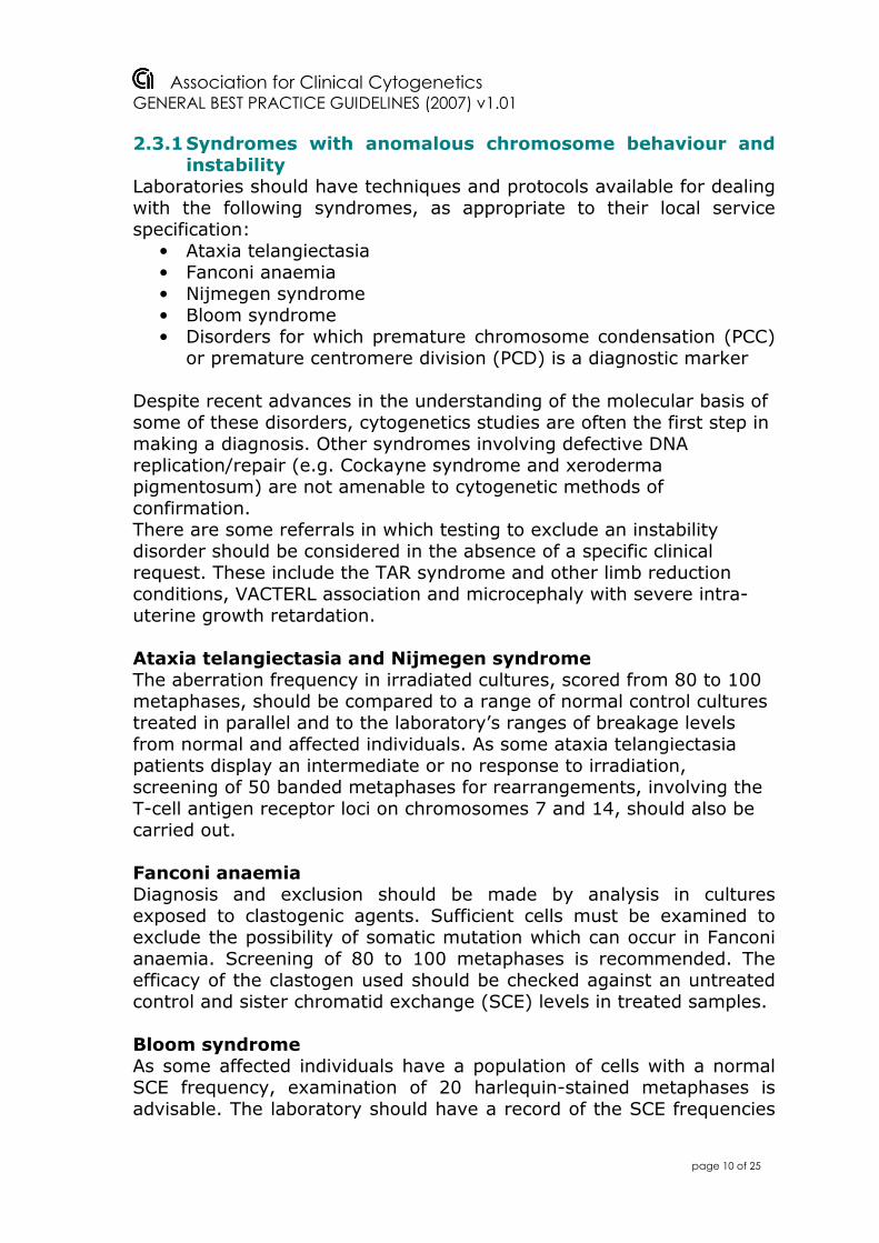
2.3.1 Syndromes with anomalous chromosome behaviour and instability
Laboratories should have techniques and protocols available for dealing with the following syndromes, as appropriate to their local service specification:
• Ataxia telangiectasia • Fanconi anaemia • Nijmegen syndrome • Bloom syndrome • Disorders for which premature chromosome condensation (PCC)
or premature centromere division (PCD) is a diagnostic marker Despite recent advances in the understanding of the molecular basis of some of these disorders, cytogenetics studies are often the first step in making a diagnosis. Other syndromes involving defective DNA replication/repair (e.g. Cockayne syndrome and xeroderma pigmentosum) are not amenable to cytogenetic methods of confirmation. There are some referrals in which testing to exclude an instability disorder should be considered in the absence of a specific clinical request. These include the TAR syndrome and other limb reduction conditions, VACTERL association and microcephaly with severe intra-uterine growth retardation. Ataxia telangiectasia and Nijmegen syndrome The aberration frequency in irradiated cultures, scored from 80 to 100 metaphases, should be compared to a range of normal control cultures treated in parallel and to the laboratory’s ranges of breakage levels from normal and affected individuals. As some ataxia telangiectasia patients display an intermediate or no response to irradiation, screening of 50 banded metaphases for rearrangements, involving the T-cell antigen receptor loci on chromosomes 7 and 14, should also be carried out. Fanconi anaemia Diagnosis and exclusion should be made by analysis in cultures exposed to clastogenic agents. Sufficient cells must be examined to exclude the possibility of somatic mutation which can occur in Fanconi anaemia. Screening of 80 to 100 metaphases is recommended. The efficacy of the clastogen used should be checked against an untreated control and sister chromatid exchange (SCE) levels in treated samples. Bloom syndrome As some affected individuals have a population of cells with a normal SCE frequency, examination of 20 harlequin-stained metaphases is advisable. The laboratory should have a record of the SCE frequencies

Association for Clinical Cytogenetics GENERAL BEST PRACTICE GUIDELINES (2007) v1.01
page 11 of 25
found when the same methods are applied to a range of normal control samples. Roberts syndrome, PCC and PCD Fifty block-stained or C-banded metaphases should be scored for paired centromeres (evidence of premature centromere division), centromere puffing and tramline chromosomes. Fifty banded metaphases should be counted for evidence of aneuploidy. For PCC, the entire slide should be screened under low power for the anomalous appearance of metaphases. ICF syndrome Fifty banded metaphases should be scored for anomalies of the heterochromatic regions of chromosomes 1, 9 and 16 and for multibranched configurations.

Association for Clinical Cytogenetics GENERAL BEST PRACTICE GUIDELINES (2007) v1.01
page 12 of 25
3 PRENATAL DIAGNOSIS SAMPLES Please refer to related Professional Guidelines for Clinical Cytogenetics: Prenatal Diagnosis Best Practice Guidelines (2005) Prenatal Diagnosis Best Practice Guidelines: Chorionic Villi (CVS) (2007) QF-PCR Best Practice Guidelines (2005) For fetal blood samples, refer to Postnatal Best Practice Guidelines (2007).
3.1 Reasons for Referral
Prenatal diagnosis is offered to patients at risk of chromosome anomalies. Reasons for referral will include the following:
• Abnormal ultrasound scan • Carrier of a structural rearrangement • Elevated risk of a chromosome abnormality indicated by
biochemical and/or ultrasound maternal screening. • Previous chromosome anomaly.
Prenatal diagnosis is normally carried out using one or more of the following sample types:
• Amniotic fluid • Chorionic villi • Fetal blood

Association for Clinical Cytogenetics GENERAL BEST PRACTICE GUIDELINES (2007) v1.01
page 13 of 25
4 SOLID TISSUE SAMPLES
4.1 Reasons for Referral
A variety of tissues are suitable for establishing long-term cultures. For fibroblast cultures, skin samples and either placenta or other fetal tissues are normally used. Care should be taken to dissect off maternal deciduas from placental tissue. Maternal cell contamination can be a significant problem, particularly from products of conception [7]. Because of the sporadic nature of the majority of chromosome abnormalities, parental blood samples should be karyotyped in preference to fetal loss samples in cases where there are three or more miscarriages as the referral reason. This may be a matter for local agreement. Consent issues: It is important to be aware that referrals following fetal loss should a) have consent for genetic testing from the parent and b) that disposal of fetal tissue is consistent with parental wishes. This consent is the responsibility of the referring clinician [8] Referral reasons will include: Prenatal
• Abnormal ultrasound scan, including hydatidiform mole and “blighted ovum”
• Known familial chromosome rearrangement • Previous chromosomally abnormal child • Unexplained miscarriages and stillbirths with congenital
anomalies Postnatal
• Confirmation of prenatal diagnosis • Investigation of mosaicism in dysmorphic/developmentally
delayed individuals • Tissue specific syndromes
4.2 Techniques
At least two independent cultures should be set up.
4.3 Analysis See section 1.1.1 for description of a standard analysis.

Association for Clinical Cytogenetics GENERAL BEST PRACTICE GUIDELINES (2007) v1.01
page 14 of 25
In the majority of cases a minimum of two banded metaphases, of a quality suitable for the reason for referral should be fully analysed. One cell must be independently checked. (Also refer to section 8 on Mosaicism). The cells should be sampled from independent colonies if using the in situ harvesting technique. Where a normal female result is obtained and there is reason to suspect maternal cell contamination, more extended analysis should be considered or a comment added to the final report [6]. For confirmation of trisomy detected at prenatal diagnosis a limited analysis, using cytogenetics, QF-PCR or FISH techniques on touch imprints, is sufficient. QF-PCR on DNA, or interphase FISH on touch imprints can replace chromosome analysis for exclusion of specific aneuploidies in other appropriate circumstances.

Association for Clinical Cytogenetics GENERAL BEST PRACTICE GUIDELINES (2007) v1.01
page 15 of 25
5 HAEMATOLOGICAL DISORDERS Please refer to related Professional Guidelines for Clinical Cytogenetics: Haemato-Oncology Best Practice Guidelines (2005) Recommendations for FISH Scoring in Oncology (2003)
5.1 Reasons for Referral
All laboratories offering a service should be able to provide an analytical and interpretative service for a range of haematological disorders. The diseases commonly undertaken are given below although, in practice, the service specification is often agreed locally with referring clinicians:
• Chronic Myeloid Leukaemia • Other myeloproliferative disorders • Acute Lymphocytic Leukaemia (ALL) • Acute Myeloid Leukaemia (AML) • Aplastic Anaemia • Chronic B and T cell disorders • Infiltrating tumours • Myelodysplasia
Referral can be at:
• Diagnosis • Follow up after treatment, including transplantation • Relapse/Transformation • As part of a national or locally agreed trial
5.2 Sample Type
Heparinised bone marrow samples are preferred, but if blasts appear to be present in the circulation, then heparinised blood can be adequate. Alternative tissue may be required in some situations.
5.3 Techniques
The number and type of cultures established should take into account the reason for referral in order to maximise the chance of detecting an abnormal clone. If appropriate, additional stimulated cultures should be set up, using mitogens specific to the suspected condition.

Association for Clinical Cytogenetics GENERAL BEST PRACTICE GUIDELINES (2007) v1.01
page 16 of 25
5.4 Analysis
The quality of metaphases obtained from unstimulated blood and from bone marrow samples is generally poor, particularly in leukaemia. As normal cells with better chromosome morphology may be present, it is important to analyse cells of varying quality in order to maximise the likelihood of detecting a clone. Adequate numbers of metaphases should be analysed or examined before the report of a normal karyotype or of the existence of an abnormal clone is given. If a sample yields fewer than twenty normal cells, the report should be suitably qualified. If a sample yields fewer than ten normal cells analysed, the case should usually be regarded as failed. With all abnormal findings, sufficient cells should be analysed to establish the clonality of the abnormality(see section 8), although it might not be appropriate to expend undue effort if the abnormalities are very complex. Analysing, interpreting and reporting the results of leukaemia work is a specialised area, where close co-operation between the laboratory and the referring clinician is vital. For instance, a laboratory will often have local agreements on the types of investigations to be implemented (e.g. FISH and/or cytogenetics) and the levels of analysis required for individual patients. It is important to maintain flexibility in the application of investigations and recognise when adequate cytogenetics information has been accrued for the clinical management of the patient.

Association for Clinical Cytogenetics GENERAL BEST PRACTICE GUIDELINES (2007) v1.01
page 17 of 25
6 SOLID TUMOURS
6.1 Reasons for Referral
There are relatively few situations where the chromosome analysis of solid tumours can be considered as a routine diagnostic service. In practice, there is often a strong research component and the service provided is best determined by local demand. Chromosome analysis, when combined with FISH protocols, is of most significant clinical relevance in studies of paediatric tumours, particularly neuroblastoma. Currently, it may provide useful diagnostic and prognostic information in the following instances:
• Ewing's Sarcoma/PNET • Lymphoma • Neuroblastoma • Rhabdomyosarcoma • Synovial Sarcoma • Wilms tumours • Breast cancer, bladder cancer and glioma
6.2 Techniques
Both mechanical and enzymatic disruption may be required to dissociate the tumour cells for culture. The adoption of more than one culture regime is recommended, to maximise the chance of detecting abnormal clones. Information on the sample quality from the referring clinician may be important in deciding the culture strategy. The use of both direct preparations and short-term culture methods is recommended, because normal tissue may overgrow abnormal clones as culture time increases. As a supplementary or alternative approach, biopsy imprints can often be used for FISH.
6.3 Analysis
The quality of metaphases obtained from tumour samples is variable, often with a low mitotic index. Recommending specific minimum analyses is therefore unhelpful. In general, culturing achieves similar quality to solid tissue cultures, whereas direct harvests yield chromosomes of similar quality to bone marrows. Adequate numbers of metaphases of varying quality should be analysed or examined before the report of a normal karyotype or of the existence of an abnormal clone is given. For biopsy imprints, confirmation of the proportion or presence of tumour cells can be beneficial to the analyst. If a sample

Association for Clinical Cytogenetics GENERAL BEST PRACTICE GUIDELINES (2007) v1.01
page 18 of 25
yields fewer than ten normal cells, the report should be failed. The application of FISH techniques provides an essential adjunct to cytogenetic analysis. Reporting and interpreting the results of tumour work is a specialised area, where close co-operation between the laboratory and the referring clinician is vital.

Association for Clinical Cytogenetics GENERAL BEST PRACTICE GUIDELINES (2007) v1.01
page 19 of 25
7 FLUORESCENCE IN SITU HYBRIDISATION (FISH)
Refer to related Professional Guidelines for Clinical Cytogenetics: Postnatal Best Practice Guidelines (2007) Recommendations for FISH Scoring in Oncology (2003)
7.1 Techniques
Most of the established FISH methods should be within the routine repertoire of the diagnostic laboratory, using commercially available probes, or probes validated in house. If neither is available, the laboratory should have defined strategies for onward referral of samples requiring FISH analysis. Routine techniques include:
• Chromosome painting • Identification of telomeric and sub-telomeric regions • Interphase analysis for aneuploidy • Locus-specific identification for microdeletion and other • syndromes • Dual or multi-probe analysis
Laboratories dealing with haematological referrals should be able to undertake:
• Rearrangement analysis, using locus-specific probe combinations • Interphase analysis for the detection of low level clones and
graft/host chimaerism The laboratory may not be able to undertake some more specialised techniques e.g. M-FISH and CGH, but again should be prepared to forward samples to an expert laboratory in the event of analysis being required. Reports of FISH analysis should be composed using the main reporting guidelines as appropriate and, in particular, include:
• information on the limitations of the test • details of the probe used including a locus identification and
manufacturer • implications of the result • follow up advice
7.2 Analysis of constitutional abnormalities
Refer to Professional Guidelines for Clinical Cytogenetics: Postnatal Best Practice Guidelines (2007)
Association for Clinical Cytogenetics GENERAL BEST PRACTICE GUIDELINES (2007) v1.01
page 20 of 25
The laboratory should have protocols for both metaphase and interphase FISH analysis, not only for cultured cells but also for interphase analysis in a variety of uncultured tissues, such as uncultured blood preparations, amniocytes and buccal mucosa. Protocols should be based on probe validation and the requirements of a particular test i.e. probe specificity and sensitivity should be confirmed before diagnostic use.
7.3 Analysis of acquired abnormalities
Refer to Professional Guidelines for Clinical Cytogenetics: Recommendations for FISH Scoring in Oncology (2003)

Association for Clinical Cytogenetics GENERAL BEST PRACTICE GUIDELINES (2007) v1.01
page 21 of 25
8 MOSAICISM
8.1 Postnatal constitutional analysis
Referring clinicians should be made aware that it is not possible to reliably exclude mosaicism from any analysis and specifically those not targeted for extended counts. More than one cell line may be present for a variety of reasons, including:
• Age related sex chromosome aneuploidy • Chimaerism • Cultural artefact • True constitutional mosaicism
Scoring strategies should take into account knowledge of the conditions under which each type is most likely to occur, to allow a distinction to be made between them. Routine analytical protocols are not designed to confirm or exclude mosaicism. Where clinically significant mosaicism is suspected, extended scoring or analysis protocols should be applied. These include:
• A minimum of 30 cells (giving appropriate confidence limits) • Duplicate cultures • More than one tissue type
FISH analysis may be the most suitable method of confirming suspected mosaicism, if suitable probes are available. Extended analysis should be considered in cases presenting with the following reasons for referral:
• Ambiguous genitalia /indeterminate sex • Clinical details suggestive of known autosomal mosaic syndromes
e.g. trisomy 8; + i(12)(p10); +dic(15)(q12) • Clinical features suggestive of a specific aneuploidy syndrome,
but which have a normal karyotype on standard analysis • Diagnosed or suspected cases of sex chromosome aneuploidy,
known to be associated with mosaicism e.g. Turner syndrome • Follow-up of a prenatal diagnosis of a possible mosaicism of
clinical significance in an affected child after birth • Karyotypically normal parents of more than one child with the
same, or a related chromosome abnormality • Variation in skin pigmentation • Hemi-hypertrophy

Association for Clinical Cytogenetics GENERAL BEST PRACTICE GUIDELINES (2007) v1.01
page 22 of 25
8.2 Prenatal diagnosis analysis
Refer to related Professional Guidelines for Clinical Cytogenetics: Prenatal Diagnosis Best Practice Guidelines (2005)
8.3 Oncology and acquired chromosome abnormalities Refer to related Professional Guidelines for Clinical Cytogenetics: Haemato-Oncology Best Practice Guidelines (2005) The early detection of originating or evolving clones with specific acquired chromosome abnormalities is of particular importance in the study of neoplasia, because of the diagnostic and prognostic significance of such findings. Culture methods, analytical procedures and independent checking, in such cases, needs to be tailored to the specific type of referral and the stage of development of the condition, with the intention of maximising clone detection.

Association for Clinical Cytogenetics GENERAL BEST PRACTICE GUIDELINES (2007) v1.01
page 23 of 25
9 DEFINITIONS OF TERMS Analyse: To count a metaphase and compare every chromosome, band for band, with its homologue and to verify the banding pattern of the X and Y-chromosomes in male karyotypes. Clear: To confirm that a chromosome, or region of a chromosome, is normal by comparison with its homologue. Case: For constitutional karyotypes, a case can be equated to any tissue from an individual patient. Clone: A cell population originally derived from a single progenitor cell. Such cells will have an identical chromosome constitution. Generally, in Cytogenetics, a clone is said to exist if three cells have lost the same chromosome, or two cells contain the same extra or rearranged chromosome. Count: To enumerate the total number of chromosomes in any given metaphase, or in FISH analysis to enumerate the number of signals in an interphase nucleus. Examine: To look for the presence or absence of any abnormality in a case. Score/Screen: To check for the presence or absence of abnormalities in a cell or metaphase without full analysis. Validation: Final authorisation for a report to be sent out and protection of the computer record.

Association for Clinical Cytogenetics GENERAL BEST PRACTICE GUIDELINES (2007) v1.01
page 24 of 25
10 REFERENCES 1. Standards for the Medical Laboratory. Version 1.03, (2004). Clinical Pathology Accreditation (UK) Ltd. 2. UK NEQAS in Clinical Cytogenetics: Participants’ Manual 1999. UKNEQAS Executive Office: Sheffield S10 2PB. 3. ISCN 2005: An International System for Human Cytogenetic Nomenclature, Shaffer L.G., Tommerup N. (eds): S Karger, Basel 2005 4. Guidelines for Clinical Cytogenetics. The Association of Clinical Cytogeneticists and NEQAS 1994. 5. Association of Clinical Cytogeneticists Professional Standards Committee Data Collection 2003-2004 6. The Retention and Storage of Pathological Records and Archives. 3rd Edition. 2006 The Royal College of Pathologists, London. 7. Rodgers, C.S., Creasy, M.R., Fitchett, M., Maliszewska, C.T., Pratt, N.R., and Waters, J.J. (1996) Solid tissue culture for cytogenetic analysis: a collaborative survey for the Association of Clinical Cytogeneticists. J. Clin. Pathol. 49: 638-641. 8. Human Tissue Act 2004 HM Stationery Office. ISBN 010 543004 8.

Association for Clinical Cytogenetics GENERAL BEST PRACTICE GUIDELINES (2007) v1.01
page 25 of 25
11 Version Control issue date Current document summary of changes Version
replaced
03/09/2007
General Best Practice Guidelines 2007 v1.01
- updated referencing between ACC Best Practice Documents - Section 1.3, Table 2 Minimum G-banding scores; ‘Postnatal Referrals’ changed to ‘Blood sample referrals’
General Best Practice Guidelines 2007
Previous versions
07/03/2007
General Best Practice Guidelines 2007
Professional Guidelines for Clinical Cytogenetics 2001
Produced by ACC Professional Standards Committee