Embed Size (px)
Citation preview

See discussions, stats, and author profiles for this publication at: https://www.researchgate.net/publication/265417879
INTRAOSSEOUS HEMANGIOMA OF THE MANDIBLE: A CASE REPORT
Article · March 2010
CITATION
1
READS
312
6 authors, including:
Junaid Ahmed
Manipal University
64 PUBLICATIONS 152 CITATIONS
SEE PROFILE
Hemant Mathur
Pacific Dental College
20 PUBLICATIONS 62 CITATIONS
SEE PROFILE
Saurabh Goel
Pacific University India
10 PUBLICATIONS 36 CITATIONS
SEE PROFILE
Mohit Pal Singh
Pacific University India
23 PUBLICATIONS 238 CITATIONS
SEE PROFILE
All content following this page was uploaded by Hemant Mathur on 09 September 2014.
The user has requested enhancement of the downloaded file.

B a k u , A z e r b a i j a n | 163
INTERNATIONAL JOURNAL Of ACADEMIC RESEARCH Vol. 3. No. 1. January, 2011, Part I
INTRAOSSEOUS HEMANGIOMA OF THE MANDIBLE: A CASE REPORT
Dr. Junaid Ahmed, Dr. Hemant Mathur*, Dr. Payal Tripathi,
Dr. Saurabh Goel, Dr. Mohit Pal Singh
Department of Oral Medicine and Radiology, Pacific Dental College and Hospital, Udaipur, Rajasthan (INDIA)
*Corresponding author: [email protected] ABSTRACT Intraosseous vascular lesions are rare conditions, comprising only 0.5% to 1% of all intraosseous tumors.
They mainly occur in the second decade of life especially in women. The most common location is vertebral column and skull, although rare instances of its occurrence in the mandible have also been reported. Central hemangioma of the mandible and maxilla is extremely rare. Although the mucosal and soft tissue lesions are readily suspected by their clinical appearance, the infrabony lesions may be difficult to distinguish visually. Clinically, the patient may be completely symptom-free or may present characteristic symptoms. The most frequent radiographic finding is a multilocular radiolucent image with honeycomb or soap bubble appearance. There are various therapeutic alternatives, although surgical excision remains as the gold standard.
This article reports a case of a 17 year-old female who presented the clinical, radiological and histological features of intraosseous hemangioma involving mandible.
Key words: Hemangioma, Honeycomb, Embolization 1. INTRODUCTION Hemangiomas are benign tumors characterized by the proliferation of blood vessels (1). They are often
present at birth or appear soon after, and grow rapidly by endothelial proliferation. Hemangioma of bone is histologically classified by Thoma as peripheral type (arising from periosteum) and central or intraosseous type (arising from central spongiosa) (2). Most central hemangiomas are the cavernous type (large vessels), but can be of capillary type (small vessels) also. Capillary hemangiomas are composed of many small capillaries lined by a single layer of endothelial cells supported in a connective tissue stroma of varying density whereas cavernous hemangiomas are formed by large, thin-walled vessels or sinusoids lined with a single layer of endothelium which are separated by thin septa of connective tissues. The central or intraosseous type is frequently found in the vertebrae and skull and rarely develops in the jaws (3). Mandible is a very infrequent location although possible. The female: male ratio is 2:1 and the peak of incidence is between the second and fifth decades of life (4). Though its origin is not defined some authors believe that it is a true neoplasm, whereas others state it to be a hamartoma resulting from proliferation of intraosseous mesodermal cells that undergo endothelial differentiation. Clinically, the patient may be completely symptom-free or may present discomfort, pulsatile bleeding, bluish discoloration, mobile teeth, and derangement of the arch form or accelerated dental exfoliation. The most frequent radiographic finding is a multilocular radiolucent image with honeycomb or soap bubble appearance (5).
2. CASE REPORT A 17 year old female was referred to the Department of Oral medicine and Radiology with a complaint of
painless swelling of right lower back side of the face since one month (Fig.1). Patient was aware of the swelling, but presented without symptoms. Facial asymmetry was present on the right side of face. Patient reported no increase in size of swelling. The overlying skin appeared normal without increase in temperature. Swelling was bony hard, round, nontender and size approximately 1.5 x 2 cm, extending from the right side of angle of the mouth to middle of body of the mandible. The rest of head & neck examination were unremarkable. Intraorally, 42 and 45 were displaced, and there was bony hard swelling causing obliteration of right buccal vestibule in relation to 42 to 46 (Fig.2).
Fig.1. Extraoral swelling Fig. 2. Obliteration of right buccal vestibule

164 | www.ijar.lit.az
INTERNATIONAL JOURNAL Of ACADEMIC RESEARCH Vol. 3. No.1. January, 2011, Part I
Radiographic investigations Mandibular occlusal radiograph revealed buccal cortical plate expansion with bone appearing to be emanating from the periphery giving a typical sunburst appearance (Fig 3). Panoramic radiograph revealed coarse trabeculation crossing areas of increased radiodensity with ground-glass appearance in relation to 43 & 44 (Fig.4). Ultrasonography (color Doppler) showed increased vascular flow in the region of 43, 44, 45 & 46 (Fig.5). Fine Needle Aspiration Cytology (FNAC) showed abundant RBC’S without malignant & giant cells. Computed tomography (C.T) revealed breaking of right buccal cortical plate, extending into soft tissue (Fig.6). External carotid arteriogram shows a vascular mass supplied by the lingual artery overlying the posterior body and angle of the mandible (Fig.7).
Fig. 3. Buccal cortical plate expansion giving a typical sunburst appearance
Fig. 4. Coarse trabeculation crossing areas of increased radiodensity with ground-glass
appearance in relation to 43 & 44.
Fig. 5: Showed increased vascular flow in the region of 43, 44, 45 & 46
Fig. 6. (Coronal C. T scan) Breaking of right buccal cortical plate, extending into soft tissue
Fig.7. External carotid arteriogram shows a vascular mass supplied by the lingual artery overlying the posterior body and
angle of the mandible
Fig. 8. Lesion was resected intotal & illiac bone graft was placed with mini plates
B a k u , A z e r b a i j a n | 165
INTERNATIONAL JOURNAL Of ACADEMIC RESEARCH Vol. 3. No. 1. January, 2011, Part I
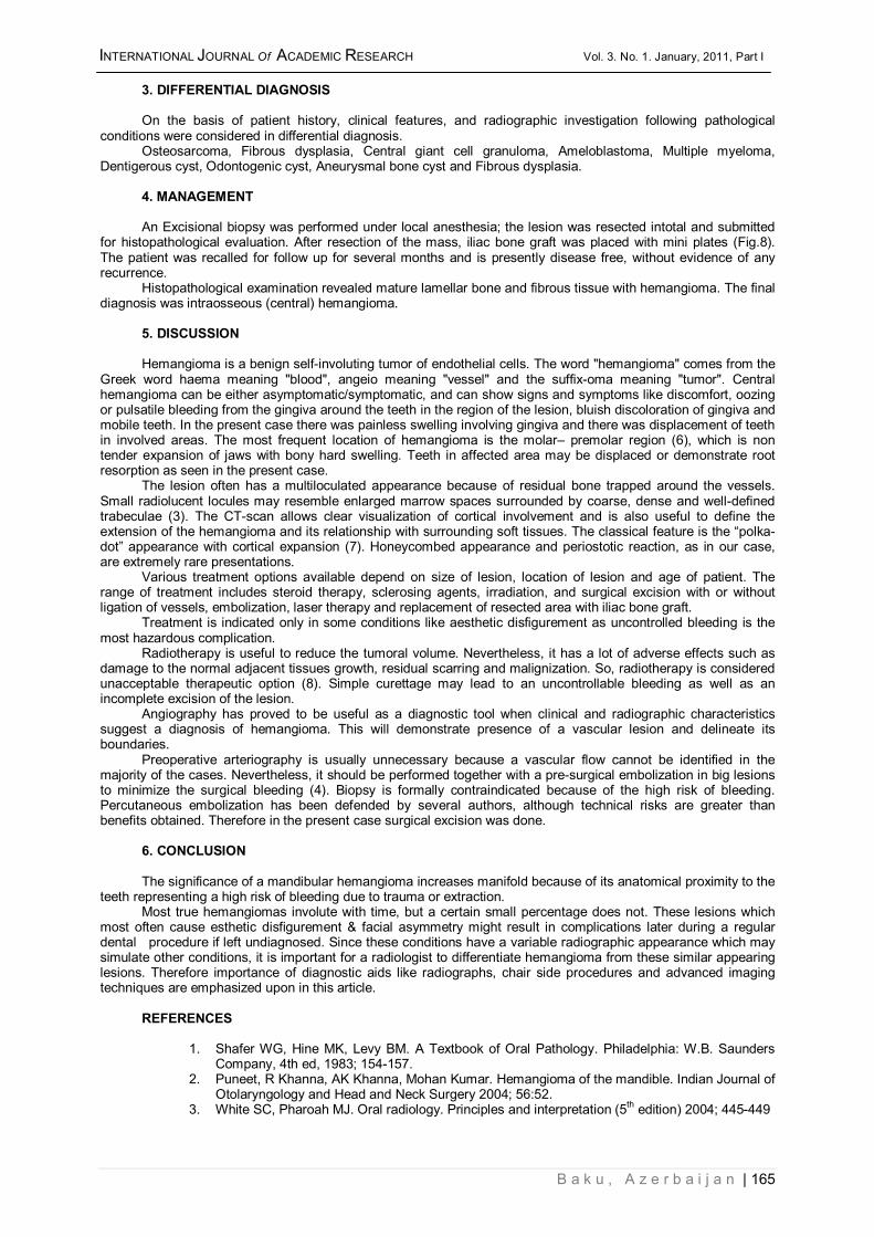
3. DIFFERENTIAL DIAGNOSIS On the basis of patient history, clinical features, and radiographic investigation following pathological
conditions were considered in differential diagnosis. Osteosarcoma, Fibrous dysplasia, Central giant cell granuloma, Ameloblastoma, Multiple myeloma,
Dentigerous cyst, Odontogenic cyst, Aneurysmal bone cyst and Fibrous dysplasia. 4. MANAGEMENT An Excisional biopsy was performed under local anesthesia; the lesion was resected intotal and submitted
for histopathological evaluation. After resection of the mass, iliac bone graft was placed with mini plates (Fig.8). The patient was recalled for follow up for several months and is presently disease free, without evidence of any recurrence.
Histopathological examination revealed mature lamellar bone and fibrous tissue with hemangioma. The final diagnosis was intraosseous (central) hemangioma.
5. DISCUSSION Hemangioma is a benign self-involuting tumor of endothelial cells. The word "hemangioma" comes from the
Greek word haema meaning "blood", angeio meaning "vessel" and the suffix-oma meaning "tumor". Central hemangioma can be either asymptomatic/symptomatic, and can show signs and symptoms like discomfort, oozing or pulsatile bleeding from the gingiva around the teeth in the region of the lesion, bluish discoloration of gingiva and mobile teeth. In the present case there was painless swelling involving gingiva and there was displacement of teeth in involved areas. The most frequent location of hemangioma is the molar– premolar region (6), which is non tender expansion of jaws with bony hard swelling. Teeth in affected area may be displaced or demonstrate root resorption as seen in the present case.
The lesion often has a multiloculated appearance because of residual bone trapped around the vessels. Small radiolucent locules may resemble enlarged marrow spaces surrounded by coarse, dense and well-defined trabeculae (3). The CT-scan allows clear visualization of cortical involvement and is also useful to define the extension of the hemangioma and its relationship with surrounding soft tissues. The classical feature is the “polka-dot” appearance with cortical expansion (7). Honeycombed appearance and periostotic reaction, as in our case, are extremely rare presentations.
Various treatment options available depend on size of lesion, location of lesion and age of patient. The range of treatment includes steroid therapy, sclerosing agents, irradiation, and surgical excision with or without ligation of vessels, embolization, laser therapy and replacement of resected area with iliac bone graft.
Treatment is indicated only in some conditions like aesthetic disfigurement as uncontrolled bleeding is the most hazardous complication.
Radiotherapy is useful to reduce the tumoral volume. Nevertheless, it has a lot of adverse effects such as damage to the normal adjacent tissues growth, residual scarring and malignization. So, radiotherapy is considered unacceptable therapeutic option (8). Simple curettage may lead to an uncontrollable bleeding as well as an incomplete excision of the lesion.
Angiography has proved to be useful as a diagnostic tool when clinical and radiographic characteristics suggest a diagnosis of hemangioma. This will demonstrate presence of a vascular lesion and delineate its boundaries.
Preoperative arteriography is usually unnecessary because a vascular flow cannot be identified in the majority of the cases. Nevertheless, it should be performed together with a pre-surgical embolization in big lesions to minimize the surgical bleeding (4). Biopsy is formally contraindicated because of the high risk of bleeding. Percutaneous embolization has been defended by several authors, although technical risks are greater than benefits obtained. Therefore in the present case surgical excision was done.
6. CONCLUSION The significance of a mandibular hemangioma increases manifold because of its anatomical proximity to the
teeth representing a high risk of bleeding due to trauma or extraction. Most true hemangiomas involute with time, but a certain small percentage does not. These lesions which
most often cause esthetic disfigurement & facial asymmetry might result in complications later during a regular dental procedure if left undiagnosed. Since these conditions have a variable radiographic appearance which may simulate other conditions, it is important for a radiologist to differentiate hemangioma from these similar appearing lesions. Therefore importance of diagnostic aids like radiographs, chair side procedures and advanced imaging techniques are emphasized upon in this article.
REFERENCES
1. Shafer WG, Hine MK, Levy BM. A Textbook of Oral Pathology. Philadelphia: W.B. Saunders Company, 4th ed, 1983; 154-157.
2. Puneet, R Khanna, AK Khanna, Mohan Kumar. Hemangioma of the mandible. Indian Journal of Otolaryngology and Head and Neck Surgery 2004; 56:52.
3. White SC, Pharoah MJ. Oral radiology. Principles and interpretation (5th edition) 2004; 445-449

166 | www.ijar.lit.az
INTERNATIONAL JOURNAL Of ACADEMIC RESEARCH Vol. 3. No.1. January, 2011, Part I
4. Alves S, Junqueira JL, De Oliveira EM, Pieri SS, De Magalhães MH. Condylar hemangioma: report of a case and review of the literature. Oral Surg Oral Med Oral Pathol Oral Radiol Endod 2006; 102(5): 23-27.
5. Schajowicz F, Rebecchini AC, Bosch-Mayol G. Intracortical haemangioma simulating osteoid osteoma. J Bone Joint Surg 1979; 61B: 94-95.
6. Nikhil Marwah, Archna Agnihotri, Samir Dutta. Central Hemangioma: An Overview and Case Report. Pediatr Dent 2006; 28: 460-466.
7. Willinsky RA, Rubenstein JD, Cruickshank B. Case report. Intracortical hemangioma of tibia. Skeletal Radiol 1982; 9: 137-139.
8. Kenan S, Abdelwahab IF, Klein MJ, Lewis MM. Hemangiomas of the long tubular bone. Clin Orthop Rel Res 1992; 256-260.
View publication statsView publication stats