Embed Size (px)
Citation preview

INNATE AND ADAPTIVE IMMUNITY
IMMUNE SYSTEM AND DISEASE

MECHANISMS OF PATHOGENICITY
• Pathogenicity: The ability to cause • disease• Virulence: The extent of pathogenicity
PORTALS OF ENTRY• Mucous membranes• Skin• Blood• Respiratory tract• Gastrointestinal tract

MECHANISMS OF PATHOGENICITY
Figure 15.9

THE CONCEPT OF IMMUNITY
• Susceptibility: Lack of resistance to a disease• Immunity: Ability to ward off disease• Innate immunity: Defenses against any pathogen• Adaptive immunity: Immunity, resistance to a
specific pathogen
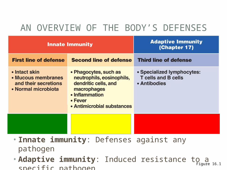
AN OVERVIEW OF THE BODY’S DEFENSES
Figure 16.1
• Innate immunity: Defenses against any pathogen• Adaptive immunity: Induced resistance to a
specific pathogen
FIRST LINE OF DEFENSEINNATE (NON-SPECIF IC) IMMUNITY

PHYSICAL FACTORS
• Skin• Epidermis consists
of tightly packed cells with• Keratin, a protective
waterproof protein
Figure 16.2
• Mucous membranes• Mucus: Traps microbes• Ciliary escalator:
Microbes trapped in mucus are transported away from the lungs
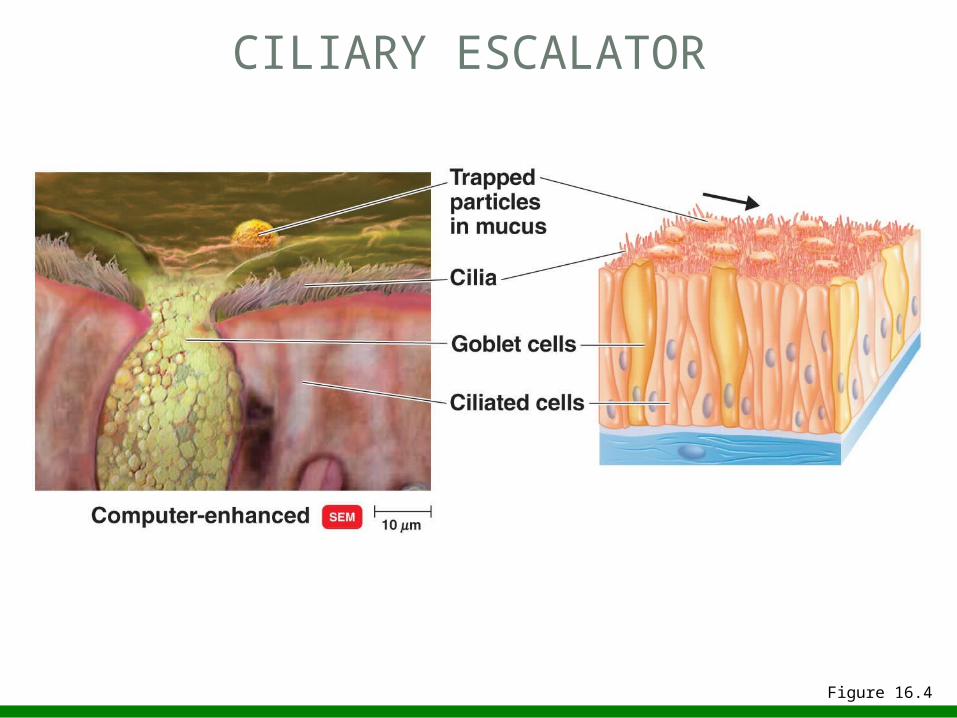
CILIARY ESCALATOR
Figure 16.4

CILIARY ESCALATOR
Figure 24.7
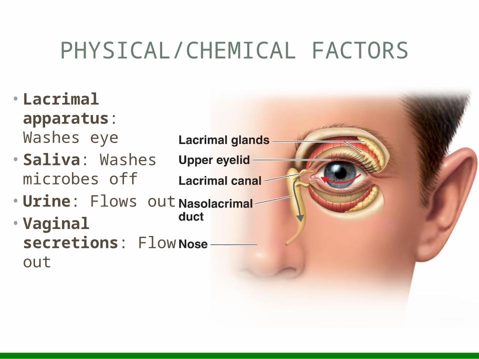
PHYSICAL/CHEMICAL FACTORS
• Lacrimal apparatus: Washes eye
• Saliva: Washes microbes off
• Urine: Flows out• Vaginal
secretions: Flow out
CHEMICAL FACTORS
• Fungistatic fatty acid in sebum• Low pH (3–5) of skin• Lysozyme in perspiration, tears, saliva, and urine• Low pH (1.2–3.0) of gastric juice• Low pH (3–5) of vaginal secretions
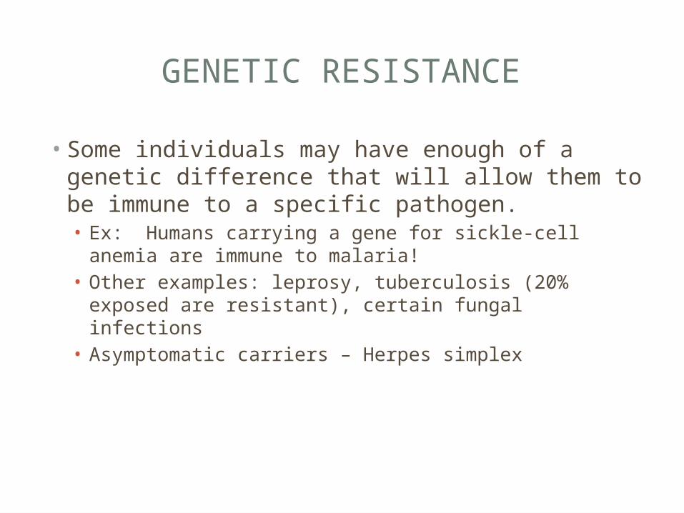
GENETIC RESISTANCE
• Some individuals may have enough of a genetic difference that will allow them to be immune to a specific pathogen. • Ex: Humans carrying a gene for sickle-cell anemia are
immune to malaria!• Other examples: leprosy, tuberculosis (20% exposed are
resistant), certain fungal infections• Asymptomatic carriers – Herpes simplex
SECOND LINE OF DEFENSEINNATE (MAINLY NON-SPECIF IC)


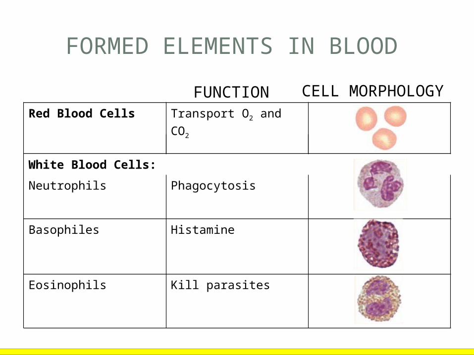
Red Blood Cells Transport O2 and CO2
White Blood Cells:Neutrophils Phagocytosis
Basophiles Histamine
Eosinophils Kill parasites
FORMED ELEMENTS IN BLOOD
FUNCTION CELL MORPHOLOGY
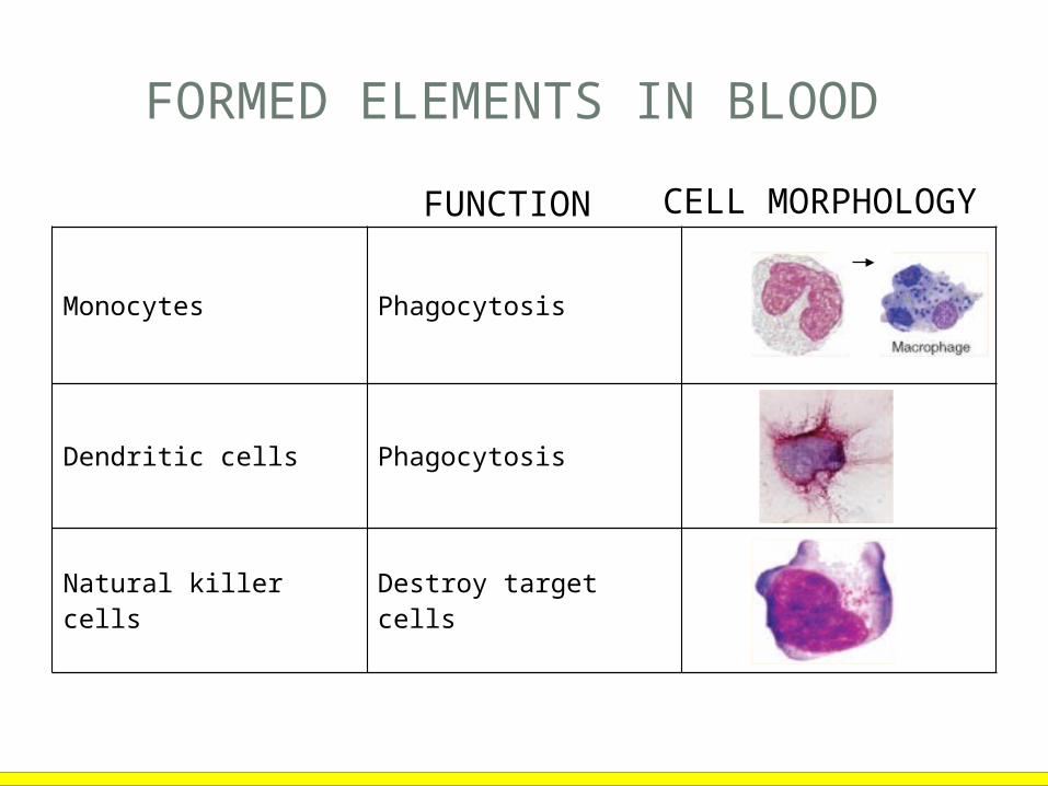
FORMED ELEMENTS IN BLOOD
Monocytes Phagocytosis
Dendritic cells Phagocytosis
Natural killer cells Destroy target cells
FUNCTION CELL MORPHOLOGY

FORMED ELEMENTS IN BLOOD
T cells Cell-mediated immunity
B cells Produce antibodies
Platelets Blood clotting
FUNCTION CELL MORPHOLOGY

• Percentage of each type of white cell in a sample of 100 white blood cells
Neutrophils 60–70%
Basophils 0.5–1%
Eosinophils 2–4%
Monocytes 3–8%
Lymphocytes (T and B cells) 20–25%
DIFFERENTIAL WHITE CELL COUNT

DEVELOPMENT OF A MACROPHAGE

PHAGOCYTOSIS
• Phago: From Greek, meaning eat
• Cyte: From Greek, meaning cell
• Ingestion of microbes or particles by a cell, performed by phagocytes
Figure 16.6
• Neutrophils• Fixed macrophages• Wandering macrophages VIDEO

Figure 16.7
PHAGOCYTOSIS

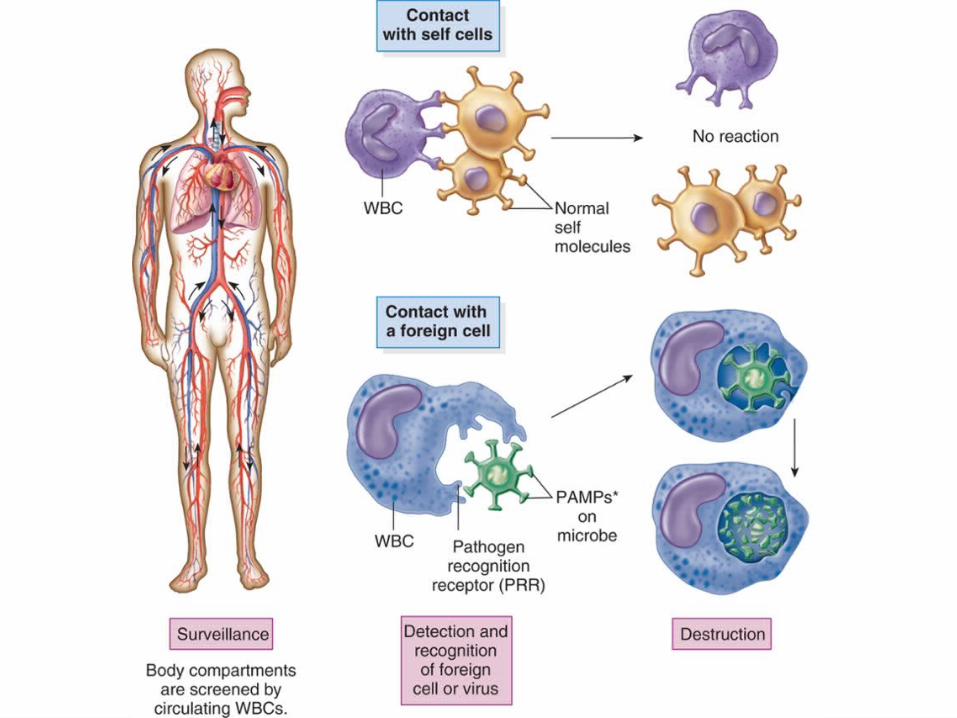
HOW DO WBC SURVEY AND RECOGNIZE?
• PRRs – Patten Recognition Receptors• Protein located on surface of WBC
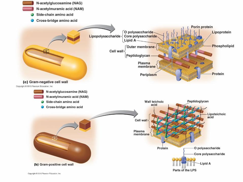
• PAMPs – Pathogen Associated Molecular Pattern• Proteins, lipids, or carbs located on the pathogen that are
distinguishable from other non-pathogenic cells such as:• Peptidoglycan, lipoteichoic acid, lipopolysaccharides,
flagellin, zymosan, double stranded RNA, etc…
• Adherence

COMPONENTS OF LYMPHATIC SYSTEM
Figure 16.5a
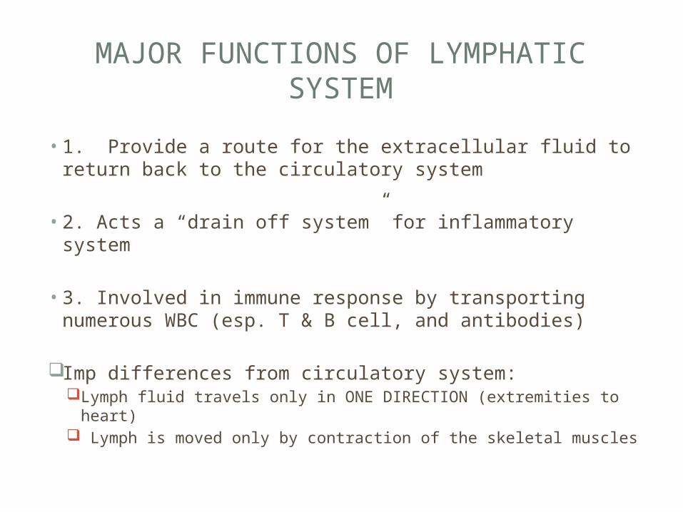
MAJOR FUNCTIONS OF LYMPHATIC SYSTEM
• 1. Provide a route for the extracellular fluid to return back to the circulatory system
• 2. Acts a “drain off system” for inflammatory system
• 3. Involved in immune response by transporting numerous WBC (esp. T & B cell, and antibodies)
Imp differences from circulatory system:Lymph fluid travels only in ONE DIRECTION (extremities to
heart) Lymph is moved only by contraction of the skeletal muscles
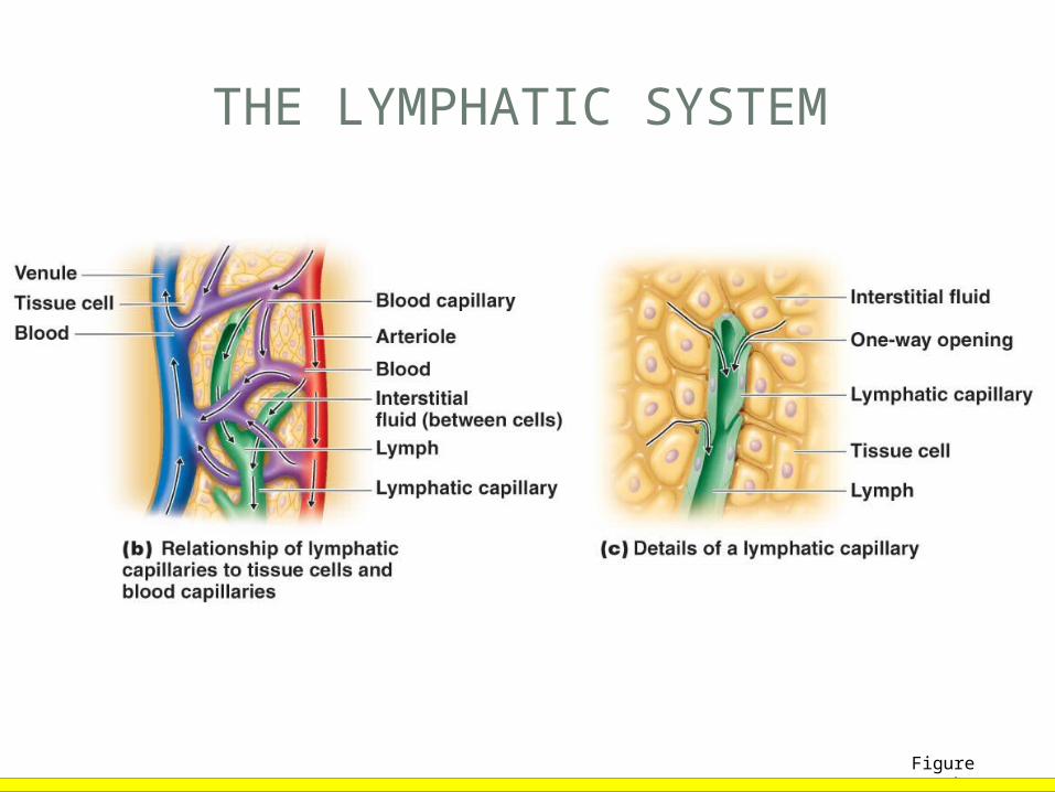
THE LYMPHATIC SYSTEM
Figure 16.5b–c

INFLAMMATION
Identifiable signs of inflammation:• Redness (rubor)• Swelling (tumor)• Pain (dolor)• Warmth (calor)
• Acute-phase proteins activated (complement, cytokine, and kinins)
• Vasodilation (histamine, prostaglandins, and leukotrienes)

WHAT CAUSES AN INFLAMMATORY RESPONSE?
• Tissue injury or death (physical – bump, fall, etc..)• Trauma from infection• Allergic reactions (diet or environmental factors)

PRIMARY FUNCTION OF THE INFLAMMATORY RESPONSE
• 1. mobilize and attract immune response to site of injury
• 2. sets scene to repair tissue damage & clear away harmful substances
• 3. destroy and block microbes from further invasion


THE PROCESS OF INFLAMMATION
Figure 16.8a, b

PHAGOCYTE MIGRATION AND PHAGOCYTOSIS
Figure 16.8c
[Insert Animation Inflammation: Overview, Steps.]


FEVER
• Hypothalamus normally set at 37°C
• Toxins from bacteria trigger the deregulation of the hypothalamus (exogenous pyrogen). Examples are endotoxins (remember them?).• Pyrogen – substance that causes a rise in body temp
• Monocytes, neutrophils, and macrophages endogenous pyrogens as part of inflammatory response. (ex: macrophages -> interleukin 1 (IL-1) & tumor necrosis factor (TNF)).
• Vasodilation and sweating: Body temperature falls

FEVER
• Advantages• Inhibits multiplication of
temp sensitive microbes• Lower iron concentrations
(nutrient used by some microbes that can limit their growth)
• Speeds up immune response such as phagocytosis
• Produces Interferons
• Disadvantages• Tachycardia• Acidosis• Dehydration• 44–46°C fatal
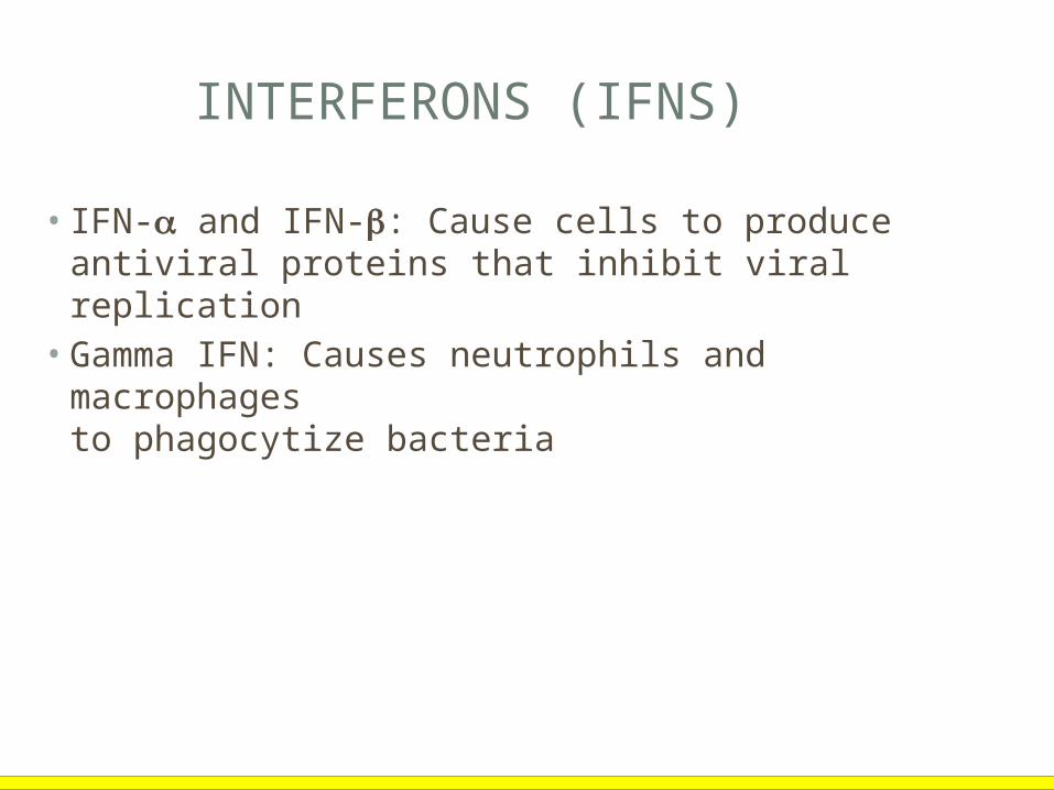
INTERFERONS (IFNS)
• IFN- and IFN-: Cause cells to produce antiviral proteins that inhibit viral replication
• Gamma IFN: Causes neutrophils and macrophages to phagocytize bacteria

ANTIVIRAL ACTIONS OF INTERFERONS
Figure 16.15
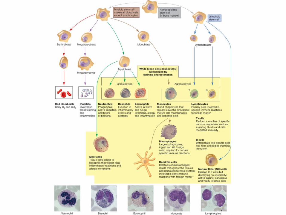
IMMUNE CELL TYPES (AGAIN…)