Embed Size (px)
Citation preview

Inducible Heme Oxygenase in the Kidney: a Model for
the Homeostatic Control of Hemoglobin Catabolism
NEVILLE R. PIMSTONE, PETERENGEL, RAIMOTENHUNEN,PAuL T. SEITZ,HARVEYS. MARvER, and RuDI SHMID
From the Department of Medicine, University of California, San FranciscoMedical Center, San Francisco, California 94122, and the Department ofClinical Chemistry, Meilahti Hospital, University of Helsinki,Helsinki 29, Finland
A B S T R A C T We have recently identified and charac-terized NADPH-dependent microsomal heme oxygenaseas the major enzymatic mechanism for the conversionof hemoglobin-heme to bilirubin-IXa in vivo. Enzymeactivity is highest in tissues normally involved in redcell breakdown, that is, spleen, liver, and bone marrow,but it usually is negligible in the kidney. However,renal heme oxygenase activity may be transiently in-creased 30- to 100-fold following hemoglobinemia thatexceeded the plasma haptoglobin-binding capacity andconsequently resulted in hemoglobinuria. Maximal stim-ulation of enzyme activity in rats is reached 6-16 hrfollowing a single intravenous injection of 30 mg ofhemoglobin per 100 g body weight; activity returns tobasal levels after about 48 hr. At peak level, total enzymeactivity in the kidneys exceeds that of the spleen or liver.Cyclohexamide, puromycin, or actinomycin D, givenjust before, or within a few hours after, a single intra-venous injection of hemoglobin minimizes or preventsthe rise in renal enzyme activity; this suggests that theincrease in enzyme activity is dependent on continuedsynthesis of ribonucleic acid and protein. The apparentbiological half-life of renal heme oxygenase is about
This work was presented in part at the Annual Meeting ofthe American Society for Clinical Investigation, AtlanticCity, N. J., May 1970 (1). Dr. Pimstone's present addressis the Department of Medicine, University of Cape Town,P. 0. Box 594, Cape Town, Republic of South Africa.
Dr. Marver died 11 July 1971.Peter Engel was a Predoctoral Research Fellow from
Duke University.Dr. Pimstone is the recipient of an International Postdoc-
toral Research Fellowship F05-TW-1360; Dr. Marver wasthe recipient of Research Career Development Award K04-AM-14301 from the National Institutes of Health; and Dr.Tenhunen's work was supported by the National ResearchCouncil for Medical Sciences, Finland.
Received for publication 19 February 1971.
6 hr. These observations indicate that functional adapta-tion of renal heme oxygenase activity reflects enzymeinduction either directly or indirectly by the substrate,hemoglobin.
Filtered rather than plasma hemoglobin appears toregulate renal heme oxygenase activity. Thus, stabiliza-tion of plasma hemoglobin in its tetrameric form withbis (N-maleimidomethyl) ether, which diminishes itsglomerular filtration and retards it plasma clearance,results in reduced enzyme stimulation in the kidney,but enhances its activity in the liver. These findings sug-gest that the enzyme is localized in the tubular epithelialcells rather than in the glomeruli and is activated by lu-minal hemoglobin. Direct support for this concept wasobtained by the demonstration of heme oxygenase ac-tivity in renal tubules isolated from rabbits that had beeninjected with hemoglobin.
INTRODUCTIONRecent studies have defined an enzymatic mechanism bywhich the heme moiety of hemoglobin is converted tobile pigment (2-4). The rate-limiting enzyme is hemeoxygenase, which is a microsomal enzyme containingcytochrome P450 as the terminal oxidase (4, 5). Itcatalyzes the oxidation of heme at the a-methene bridge(3) to form equimolar amounts of biliverdin and ofcarbon monoxide (5). The biliverdin then is reduced tobilirubin by the NADPH-dependent soluble enzyme,biliverdin reductase (6, 7).
Heme oxygenase is most active in organs normallyengaged in the sequestration and breakdown of senes-cent red cells, namely spleen, liver, and bone marrow(5). Its activity in rat spleen, as determined in vitro,is in good agreement with the requirements for thenormal turnover of erythrocyte-hemoglobin in the intact
2042 The Journal of Clinical Investigation Volume 50 1971

animals (2). After splenectomy, or in conditions wherethe turnover of hemoglobin is accelerated, for instancein hemolytic anemia (4, 5), hepatic heme oxygenase ac-tivity is elevated reflecting the increased amount ofhemoglobin that is removed by the liver (8). Similarly,enzyme activity is enhanced in isolated peritoneal or al-veolar macrophages that, in vivo, have ingested heminor hemoglobin before being harvested (9). These find-ings leave little doubt that the activity of heme oxv-genase is controlled by the amount of heme that is of-fered for catabolism. The nature of this regulatorymechanism is described in the present report, using theresponse of the kidney to hemoglobinuria as the ex-perimental model.
As the kidney normally is not involved in the break-down of red cells or hemoglobin, it expectedly has verylow levels of heme oxygenase activity (5). However, insevere intravascular hemolysis, when hemoglobin re-leased from circulating red cells exceeds the bindingcapacity of the plasma haptoglobin (10), unbound he-moglobin may be filtered by the renal glomeruli and ap-pear in the urine (11). From the study of Bunn andJandl (12), it would appear that in hemoglobinemiawith hemoglobinuria, the kidney may be able to catabo-lize part of the filtered hemoglobin that has been reab-sorbed by proximal convoluted tubules (13-16). If thiswere the case, one would expect the kidney to respondto hemoglobinuria with increased heme oxygenase ac-tivity and one would further predict that this adaptivemechanism is localized in the tubular epithelial cells.We have investigated this relationship under optimalexperimental conditions in rats made hemoglobinuricwith intravenous injections of hemoglobin.
METHODSMost experiments were carried out in female Sprague-Dawley rats, weighing 250-350 g. The animals had freeaccess to water and to Berkeley Diet Chow (Berkeley Bio-logicals, Berkeley, Calif.). In studies requiring analysis ofurine, rats were housed in metabolic cages permitting unin-terrupted and quantitative urine collection.
Rat hemoglobin was obtained as outlined by Bunn, Esham,and Bull (11). Isotopically labeled rat hemoglobin was pre-pared by the method of Keene and Jandl (17) using 'FeCl3(Tracer Lab, Waltham, Mass.) as isotopic precursor. Thehemoglobin was complexed with bis (N-maleimidomethyl)ether (BME)' as described by Bunn et al. (11). Titrationof the reactive sulfhydryl groups of hemoglobin solutionstreated with BME2 (18) indicated that the reaction hadgone to completion. The concentration of hemoglobin inthese solutions, measured as cyanmethemoglobin (19), wasusually between 40 and 60 mg per ml. In rat urine, thehemoglobin concentration was measured by the benzidinemethod (20). Radioactivity in samples containing 'Fe wasmeasured in a well-type scintillation counter (Nuclear-
'Kindly supplied by Uniroyal Inc., Wayne, N. J.2Abbreviations used in this paper: BME, bis(N-maleimi-
domethyl) ether.
Chicago Corp., Des Plaines, Ill.) for a minimum of either10 min or 10,000 counts. For these determinations, onewhole kidney, or 1 ml of blood or urine, or suitable por-tions thereof, diluted to 1 ml with distilled water, wereused. Microsomal heme oxygenase activity was determinedon 20,000 g supernatant fractions of tissue homogenates(2, 3, 5) and expressed as mnumoles bilirubin formed per10 mg protein in 1 min. For computation of total organenzyme activity, the assumption was made that all micro-somal heme oxygenase activity was recovered in the 20,000 gsupernatant fraction of the homogenate (2).
All experiments were carried out on groups of rats ofsimilar age matched by weight. Hemoglobin was adminis-tered by a single injection into the tail vein after the ani-mals had been lightly anesthetized with ether. The volumeof fluid injected was usually about 1 ml and never exceeded3 ml. Control animals received an intravenous injection ofisotonic saline. In most experiments, 30 mg of hemoglobinper 100 g body weight was given; this dose exceeded byfar the haptoglobin-binding capacity of the plasma (10),regularly led to hemoglobinuria and generally resulted inmaximal stimulation of renal heme oxygenase activity. Inthe dose-effect studies, 5-50 mg of hemoglobin was in-jected per 100 g body weight.
Cyclohexamide (Sigma Chemical Co., St. Louis, Mo.),puromycin (Nutritional Biochemical Corporation, Cleve-land, Ohio), or actinomycin D (Sigma Chemical Co.),were given intraperitoneally either 1 hr before or 2 (actino-mycin) or 4 hr (cyclohexamide) after the intravenous ad-ministration of 30 mg of hemoglobin per 100 g body weight;puromycin was inj ected hourly. Control animals receivedisotonic saline, given intraperitoneally at comparable times.The doses of these inhibitors used were similar to thoseemployed in studies of 5-aminolevulinic acid synthetase inrat liver (21), i.e. cyclohexamide, 1.8 mg, and actinomycin,1.5 mg per kg; puromycin was given every hour in a doseof 100 mg per kg. To test the effectiveness of cyclohexa-mide in suppressing renal protein synthesis, 50 ACi of leu-cine-14C (New England Nuclear Corp, Boston, Mass.) wasinjected intravenously 1 hr after the intraperitoneal ad-ministration of cyclohexamide given at doses that rangedfrom 0.3 to 2.1 mg per kg body weight. 4 hr after the iso-tope administration, the kidneys were removed and the "C-activity in the trichloroacetic acid-precipitable fraction wasdetermined (12). A dose of 1.8 mg per kg of cyclohexamidereduced leucine-"C incorporation into soluble kidney pro-teins by two-thirds.
In a series of experiments, the response of the kidneysto injected native hemoglobin was compared with that ofBME-treated hemoglobin; the latter is poorly filtered bythe glomeruli (11) because of its tetrameric form (22). 40rats received unlabeled hemoglobin or BME-hemoglobinintravenously at a dose of 15 mg per 100 g body weight,and the hemoglobin excreted in the urine during the en-suing 4 hr was measured. In 12 rats that had been given30 mg per 100 g body weight of hemoglobin-59Fe or BME-hemoglobin-'9Fe, plasma radioactivity was measured at1, 4, and 8 hr after the intravenous injection. From thesevalues and the plasma volume estimated from the bodyweight (23), the total circulating radioactivity was calcu-lated. Radioactivity excreted in the urine was compared withthe excreted amount of hemoglobin, determined colorimetri-cally. Nine rats were depleted of their plasma haptoglobin(10, 11) by two preliminary intravenous injections of 10mg native hemoglobin spaced 1 hr apart. 1 hr later, eachanimal received intravenously, 2 mg of BME-treated or 2
Heme Oxygenase in the Kidney 2043
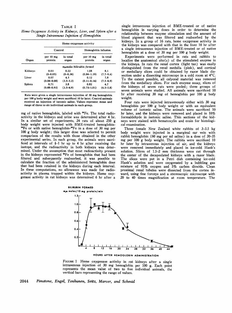
TABLE IHemeOxygenase Activity in Kidneys, Liver, and Spleen after a
Single Intravenous Injection of Hemoglobin
Hemeoxygenase activity
Control Hemoglobin infusion
per 10 mg in total per 10 mg in totalOrgan protein organ protein organ
mEumoles bilirubin formedKidneys 0.03 0.22 1.08 8.1
(0-0.05) (0-0.36) (0.86-1.19) (7.7-9.4)Liver 0.07 4.5 0.12 7.8
(0.06-0.08) (3.5-5.2) (0.11-0.14) (7.5-8.9)Spleen 0.71 3.8 0.82 4.7
(0.68-0.93) (3.5-4.9) (0.75-1.01) (4.5-5.8)
Rats were given a single intravenous injection of 30 mg hemoglobinper 100 g body weight and were sacrificed 10 hr later. Control animalsreceived an injection of isotonic saline. Values represent mean andrange of three to six individual animals in each group.
mg of native hemoglobin, labeled with 'Fe. The total radio-activity in the kidneys and urine was determined after 4 hr.In a similar set of experiments, 24 rats of about 250 gbody weight were injected with BME-treated hemoglobin-'Fe or with native hemoglobin-'mFe in a dose of 30 mg per100 g body weight; this larger dose was selected to permitcomparison of the results with those obtained in the otherexperimental series. In each group, the animals were sacri-ficed at intervals of i-1 hr up to 4 hr after receiving theisotope, and the radioactivity in both kidneys was deter-mined. Under the assumption that most radioactivity presentin the kidneys represented 'mFe of hemoglobin that had beenfiltered and subsequently reabsorbed, it was possible tocalculate the fraction of the administered hemoglobin dosethat had been retained in the kidneys during each interval.In these computations, no allowance was made for radio-activity in plasma trapped within the kidneys. Heme oxy-genase activity in rat kidneys was determined 6 hr after a
BILIRUBIN FORMEDmA moles/10 mg protein/min
single intravenous injection of BME-treated or of nativehemoglobin in varying doses in order to determine therelationship between enzyme stimulation and the amount ofblood pigment that was filtered and reabsorbed by thekidneys. In a group of 16 rats, heme oxygenase activity inthe kidneys was compared with that in the liver 10 hr aftera single intravenous injection of BME-treated or of nativehemoglobin at a dose of 30 mg per 100 g body weight.
Experiments were performed in rats and rabbits tolocalize the anatomical site(s) of the stimulated enzyme inthe kidneys. In rats the renal cortex (light tan) was easilydistinguished from the renal medulla (pink), and corticalor medullary slices could be obtained by razor blade dis-section under a dissecting microscope in a cold room at 4VC.To the extent possible, all calyceal material was removedfrom the medullary slices. For each enzyme assay, slices ofthe kidneys of seven rats were pooled; three groups ofseven animals were studied. All animals were sacrificed 10hr after receiving 30 mg of hemoglobin per 100 g bodyweight.
Four rats were injected intravenously either with 30 mghemoglobin per 100 g body weight or with an equivalentvolume of isotonic saline. The animals were sacrificed 10hr later, and the kidneys were removed and placed in 10%formaldehyde in isotonic saline. Thin sections of the kid-neys were stained with hematoxylin and eosin for histologi-cal examination.
Three female New Zealand white rabbits of 3-3.5 kgbody weight were injected in a marginal ear vein withrabbit hemoglobin (60 mg per ml saline) in a dose of 30-35mg per 100 g body weight. The rabbits were sacrificed 10hr later by intravenous injection of air, and the kidneyswere removed immediately and placed in ice-cold Hank'ssolution. Slices of 1.5-2 mm thickness were cut throughthe center of the decapsulated kidneys with a razor blade.The slices were put in a Petri dish containing ice-coldHank's solution and were oxygenated by a bubbling gasmixture of 95% oxygen and 5%o carbon dioxide. Cleanproximal renal tubules were dissected from the cortex in-ward, using fine forceps and a stereoscopic microscope with20 to 40 times magnification at room temperature. The
HOURSAFTER HEMOGLOBINADMINISTRATION
FIGURE 1 Heme oxygenase activity in rat kidneys after a singleintravenous injection of 30 mg hemoglobin per 100 g. Each pointrepresents the mean value of two to five individual animals, thevertical bars representing the range of values.
2044 Pimstone, Engel, Tenhunen, Seitz, Marver, and Schmid
1.0I
a
0.8a -
O ° 0.6
Z X'o 0.4
=
-o 0.2
EB
1i 2
7/ 203 30 410
DOSE OF HEMOGLOBINmg/1009 rat
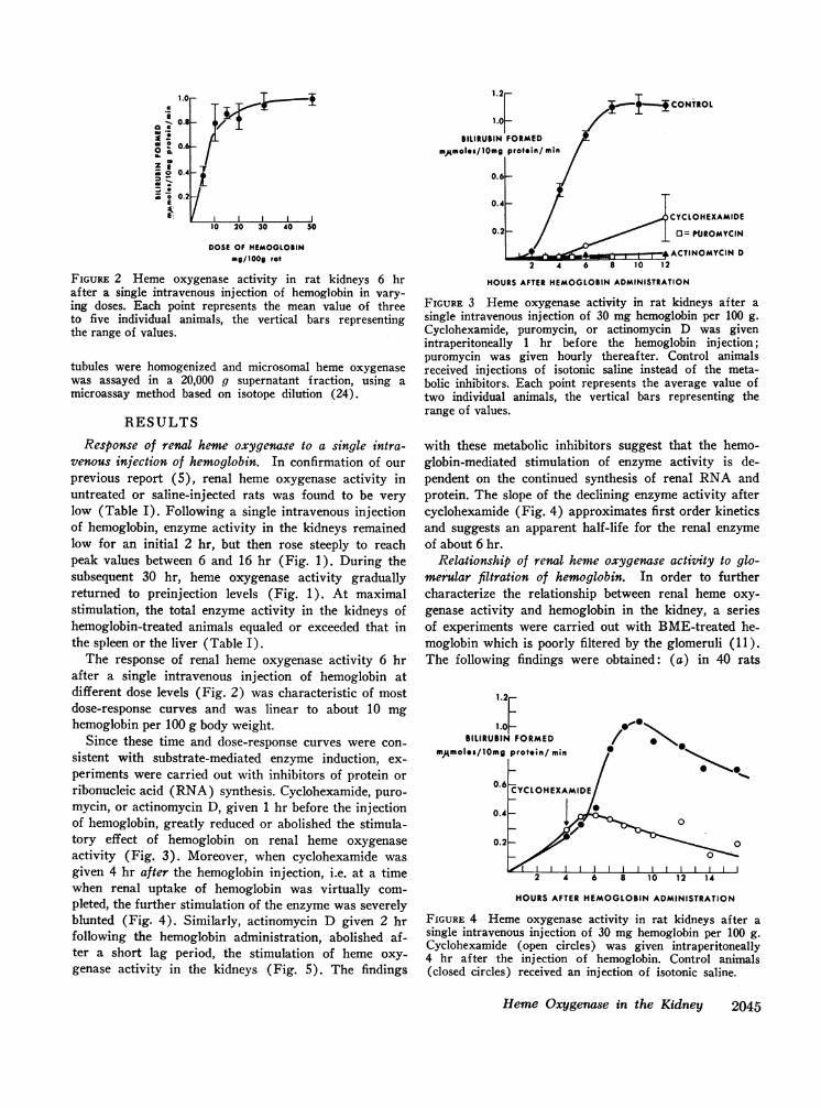
FIGuRE 2 Heme oxygenase activity in rat kidneys 6 hrafter a single intravenous injection of hemoglobin in vary-ing doses. Each point represents the mean value of threeto five individual animals, the vertical bars representingthe range of values.
tubules were homogenized and microsomal heme oxygenasewas assayed in a 20,000 g supernatant fraction, using amicroassay method based on isotope dilution (24).
RESULTS
Response of renal heme oxygenase to a single intra-venous injection of hemoglobin. In confirmation of ourprevious report (5), renal heme oxygenase activity inuntreated or saline-injected rats was found to be verylow (Table I). Following a single intravenous injectionof hemoglobin, enzyme activity in the kidneys remainedlow for an initial 2 hr, but then rose steeply to reachpeak values between 6 and 16 hr (Fig. 1). During thesubsequent 30 hr, heme oxygenase activity graduallyreturned to preinjection levels (Fig. 1). At maximalstimulation, the total enzyme activity in the kidneys ofhemoglobin-treated animals equaled or exceeded that inthe spleen or the liver (Table I).
The response of renal heme oxygenase activity 6 hrafter a single intravenous injection of hemoglobin atdifferent dose levels (Fig. 2) was characteristic of mostdose-response curves and was linear to about 10 mghemoglobin per 100 g body weight.
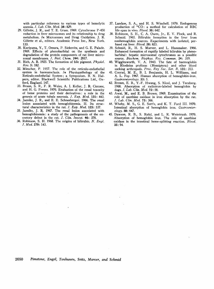
Since these time and dose-response curves were con-sistent with substrate-mediated enzyme induction, ex-periments were carried out with inhibitors of protein orribonucleic acid (RNA) synthesis. Cyclohexamide, puro-mycin, or actinomycin D, given 1 hr before the injectionof hemoglobin, greatly reduced or abolished the stimula-tory effect of hemoglobin on renal heme oxygenaseactivity (Fig. 3). Moreover, when cyclohexamide wasgiven 4 hr after the hemoglobin injection, i.e. at a timewhen renal uptake of hemoglobin was virtually com-pleted, the further stimulation of the enzyme was severelyblunted (Fig. 4). Similarly, actinomycin D given 2 hrfollowing the hemoglobin administration, abolished af-ter a short lag period, the stimulation of heme oxy-genase activity in the kidneys (Fig. 5). The findings
BILIRUBIN FORMED
mAmoles/10mg protein/ min
)CYCLOHEXAMIDE
0= NIJROMYCIN
ACTINOMYCIN D2 4 6 8 10 12
HOURSAFTER HEMOGLOBINADMINISTRATION
FIGURE 3 Heme oxygenase activity in rat kidneys after asingle intravenous injection of 30 mg hemoglobin per 100 g.Cyclohexamide, puromycin, or actinomycin D was givenintraperitoneally 1 hr before the hemoglobin injection;puromycin was given hourly thereafter. Control animalsreceived injections of isotonic saline instead of the meta-bolic inhibitors. Each point represents the average value oftwo individual animals, the vertical bars representing therange of values.
with these metabolic inhibitors suggest that the hemo-globin-mediated stimulation of enzyme activity is de-pendent on the continued synthesis of renal RNA andprotein. The slope of the declining enzyme activity aftercyclohexamide (Fig. 4) approximates first order kineticsand suggests an apparent half-life for the renal enzymeof about 6 hr.
Relationship of renal heme oxygenase activity to glo-merular filtration of hemoglobin. In order to furthercharacterize the relationship between renal heme oxy-genase activity and hemoglobin in the kidney, a seriesof experiments were carried out with BME-treated he-moglobin which is poorly filtered by the glomeruli (11).The following findings were obtained: (a) in 40 rats
12F1.0 z-
SILIRUBIN FORMED / | *
mAmolIs/10mg protein/ min
0,6 CYCLOHEXAMIDE
0.4
0.2-
2 4 6 8 10 12 14
HOURSAFTER HEMOGLOBINADMINISTRATION
FIGURE 4 Heme oxygenase activity in rat kidneys after asingle intravenous injection of 30 mg hemoglobin per 100 g.Cyclohexamide (open circles) was given intraperitoneally4 hr after the injection of hemoglobin. Control animals(closed circles) received an injection of isotonic saline.
Heme Oxygenase in the Kidney 2045

1.0C
0.8
_0
0.
Z E, 2 0.4
_
o 0. 2E
t
4 0
CONTROL
b / | t\2~~ ACTINOMYCIl4 D
ACTINOMYCINVi I I I I I I4 8 12 1 6
HOURSAFTER HEMOGLOBINADMINISTRATION
FIGURE 5 Heme oxygenase activity in rat kidneys after asingle intravenous injection of 30 mg hemoglobin per 100 g.Actinomycin D (open circles) was given intraperitoneally2 hr after the injection of hemoglobin; control animals(closed circles) received an injection of isotonic saline.One group of animals was sacrificed approximately 5 minafter the administration of the actinomycin or saline. Eachpoint represents the mean of two to five individual animals,the vertical bars representing the range of values.
that were injected with native or BME-treated hemo-globin in doses of 15 mg per 100 g rat, hemoglobinuriawas significantly less with the BME-hemoglobin (0.5mg, SD 0.05, in 4 hr) than with the native unreactedhemoglobin (4.4 mg, SD 1.95, in 4 hr) (25). (b) Therate of disappearance of BME-treated hemoglobin fromthe circulation was much slower than that of native he-moglobin (Table II). (c) In five haptoglobin-depletedrats, 49% (range 48-52%) of a small dose (2 mg) ofhemoglobin-'Fe was taken up by the kidneys in 4 hr.Comparable values with four rats given BME-treatedhemoglobin were less than 5% (range 1.6-4.5%).Almost no radioactivity was detectable in the urine ofthese rats during the 4 hr of the study. (d) When native
TABLE I IHemoglobin-59Fe Retained in Circulating Plasma after a Single
Intravenous Injection of RadioactiveHemoglobin in Rats
Dose retained in circulation
Hemoglobin injected 1 hr 4 hr 8 hr
Hemoglobin-"9Fe 18 4 1(14-22) (3-5) (0-2)
BME-treated hemoglobin-59Fe 50 30 14(42-58) (28-32) (12-16)
Each value represents the average of two individual experi-ments. The range of values in each group is given in brackets.
hemoglobin-'Fe or BME-treated hemoglobin-59Fe wasinjected in a dose of 30 mg per 100 g body weight, theradioactivity after 4 hr in the kidneys of the rats givenBME-hemoglobin was about one-third less than in ani-mals that had received native hemoglobin (Fig. 6). Inall instances, the rate of renal isotope uptake was rapid,and was nearly completed in 4 hr.
These observations confirm the report by Bunn et al.(11) that complexing of hemoglobin with the sulfhydrylreagent BMEgreatly reduces its glomerular filtration,minimizes hemoglobinuria, and prolongs the hemo-globinemia.
When rats were injected with varying doses of BME-treated hemoglobin, the stimulatory effect on renal hemeoxygenase activity was significantly less at all doselevels than with native hemoglobin (Fig. 7). By contrast,the pattern of enzyme stimulation in the liver was re-versed, in that hepatic heme oxygenase activity washigher after injection of BME-treated hemoglobin thanafter native hemoglobin. Thus, in eight rats given 30 mgper 100 g body weight of BME-hemoglobin, enzymeactivity (msmoles bilirubin/10 mg protein per min) af-ter 10 hr in the liver was 0.44 (0.04 SE) and in thekidney 0.68 (0.11 SE). The respective values after ad-ministration of a comparable amount of native hemo-globin to eight rats were: liver 0.13 (0.007 SE), andkidneys 1.07 (0.06 SE). These results are significantlydifferent at the P < 0.005 level (25).
Anatomical localization of stimulated renal heme oxy-genase activity. When renal cortex and medulla ofhemoglobin-injected rats were assayed separately forheme oxygenase activity, most of the enzyme activitywas found in the cortical tissue, with only traces pres-ent in the medulla (Fig. 8). On histological examina-
40
zau
z
00
09
0
zan
- 160.
12a
-00, 8
; 4
NATIVE-x ...5ma. 9-
. I. HEMOGLOBIN-"' F
_ 3E.i' )E ~~BME-HEMOGLOBIN59Fel />B
v I1 2 3 4
HOURSAFTER HEMOGLOBINADMINISTRATION
FIGuRE 6 Hemoglobin-69Fe in rat kidneys after intravenousinjection of native hemoglobin-'Fe (closed circles) orBME-treated hemoglobin-'Fe (open circles) at a dose of30 mg per 100 g. The results, measured as 'Fe in the twokidneys, are expressed as milligrams hemoglobin. Eachpoint represents the average of two individual experiments,the vertical bars representing the range of values.
2046 Pimstone, Engel, Tenhunen, Seitz, Marver, and Schmid
31
$I
11

0E1J0. 20 3 0 S
OD BME-EMOGLOBIN0.0.6-
0.4
0
E 0.2
10 20 30 40 50
DOSE OF HEMOGLOBINmg/100g rat
FIGURE 7 Heme oxygenase activity in rat kidneys 6 hrafter a single intravenous injection of native hemoglobin(closed circles) or BME-treated hemoglobin (open circles)in varying doses. Each point represents the mean value ofthree to five individual animals, the vertical bars repre-senting the range of values.
tion of hematoxylin and eosin-stained sections of kidneysobtained 10 hr after hemoglobin injection, the morphol-ogy of the nephron and the surrounding vessels ap-peared normal and there was no evidence of mononuclearcell infiltration.
The kidneys of the three rabbits injected with hemo-globin exhibited heme oxygenase activity (Table III)comparable with that of stimulated rat kidney (Table I).In the proximal tubules dissected from these kidneys(equivalent to 0.04-0.07 mg of protein), heme oxygenaseactivity of the microsomal fraction was slightly higherthan in the comparable fraction obtained from the wholekidneys (Table III). In tubular preparations obtainedfrom control rabbits, no heme oxygenase activity wasdetectable.
DISCUSSIONThe present results establish that the kidneys of ratsand rabbits possess an enzymatic mechanism for theconversion of heme to bilirubin. This finding might havebeen anticipated, as Ostrow, Jandl, and Schmid (26)had shown that after infusion of 'Fe-, "C-labeled hemo-globin in rats, even when nearly half of the isotopic ironwas retained in the kidneys, the heme-"C activity ad-ministered was almost quantitatively converted to bili-rubin-"C excreted in the bile. Furthermore, the obser-vations of Keene and Jandl (17) indicated that whenplasma hemoglobin levels exceed the haptoglobin-bindingcapacity, the kidneys become a major site of hemoglobinuptake in the rat. More recently, Bunn and Jandl (12)demonstrataed by means of serial extractions, that hemo-globin, absorbed by the renal parenchyma, rapidly dis-appears from the kidneys leaving behind the iron moiety,which is mobilized more slowly. Finally, in chronic
0.8
E
Mi 0.6
Mi
004
-0.2-
E
* CORTEX MEDULLA
FIGURE 8 Heme oxygenase activity in renal cortex andmedulla of rats injected 10 hr earlier with 30 mg hemo-globin per 100 g. Bracket represents range of values inthree groups of seven animals each.
intravascular hemolysis, such as paroxysmal nocturnalhemoglobinuria, hemosiderin, presumably derived fromiron-laden tubular cells (27), frequently appears in theurine (28). These observations suggested that in hemo-globinemia resulting in hemoglobinuria, the kidneys mayplay a major role in the catabolism of hemoglobin.
In tissues ordinarily concerned with the removal anddegradation of senescent red cells, the heme of hemo-globin is converted to bile pigment by a microsomalheme oxygenase system (2-4). In vitro studies indicatethat this enzyme can utilize as substrates only hemeor easily dissociable heme-protein complexes (3). Thus,the enzymatic degradation of hemoglobin or of intra-cellular hemoproteins is probably governed first by fac-tors that modify the hemoprotein to increase its dissoci-ation constant. Heme oxygenase activity must then becoordinated with this process. An enzymatic apparatusexhibiting very similar properties and subcellular locali-zation has now been demonstrated in the kidneys. How-ever, under physiological conditions, renal heme oxy-genase activity is very low but is increased manyfoldfollowing the filtration of plasma hemoglobin into theurine. This is reminiscent of the elevated heme oxy-genase activity in the liver after splenectomy or experi-mental hemolysis (4, 5) and of the stimulation of theenzyme in macrophages that have ingested heme or he-
TABLE I I IHeme Oxygenase Activity in Whole Kidneys and in Isolated
Renal Tubules of Rabbits Injected Intravenouslywith Hemoglobin
Hemeoxygenase activity
Rabbit Whole kidney Renal tubules
mjsmoles bilirubin formed/10 mg protein per min
1 1.2 1.5*2 0.9 1.23 0.8 1.0
* Average of duplicate determinations.
Heme Oxygenase in the Kidney 2047

moglobin in vivo before being harvested (9). In thepresent study, the characteristics of the time and dose-response curves of renal heme oxygenase after a singleintravenous injection of hemoglobin, and the inhibitoryeffects of cyclohexamide, puromycin, and actinomycin Dstrongly suggest that this functional adaptation of theenzyme reflects induction of heme oxygenase by its sub-strate, hemoglobin. The effect of these metabolic inhibi-tors cannot be ascribed solely to impaired glomerularfiltration or tubular reabsorption of hemoglobin as theinhibitors effectively thwarted further stimulation of en-zyme activity when they were given after the hemo-globin had been taken up by the kidneys.
The apparent half-life of microsomal heme oxygenasein the kidney, as calculated from these curves, is about6 hr. This is significantly less than that reported forother microsomal enzymes involved in mixed functionoxidations, such as NADPH-dependent cytochrome creductase (29) for which a half-life of as low as 30 hrhas been reported (30). The short biological half-life of microsomal heme oxygenase is surprising, but mayserve to underscore the previously reported differences(3, 5) between the microsomal enzyme systems thatcatalyze the oxidation of drugs and of heme.
In the previous studies of the enzymatic conversionof heme to bilirubin (2-5), heme oxygenase activity wasidentified in tissues that were known to contain mesenchy-mal cells with phagocytic properties such as the spleen(2-5) or liver (4, 5) or in isolated macrophages har-vested from the peritoneal cavity or the lungs of rodents(9). These findings were consistent with the postulatethat hemoglobin catabolism is primarily a function of thereticuloendothelial system (31, 32). It might have beenexpected, therefore, that heme oxygenase activity in thekidney also would be localized in phagocytic cells ofmesenchymal origin located perhaps in the glomerularcapillary loop or in the interstitial tissue of the renalmedulla. However, the following findings point to therenal tubules as a major site of enzyme localization.In hemoglobinuric rats, heme oxygenase activity in thekidneys is confined almost entirely to the renal cortex(Fig. 8) which on histological examination showed nomononuclear cell infiltration. Functional studies withBME-treated hemoglobin also were in line with the con-cept that the enzyme activity is in the tubules rather thanin the glomeruli. When hemoglobin was stabilized in itstetrameric form with BME, its glomerular filtration wasminimized, less was taken up by the kidneys (Fig. 6) orlost in the urine, and consequently, the plasma levelremained elevated for a longer time (Table II). Whileadministration of both BME-treated hemoglobin andnative hemoglobin resulted in increased renal hemeoxygenase activity (Fig. 7), the stimulation in rats re-ceiving the BME-treated hemoglobin was significantly
less than in animals given the native hemoglobin. This isconsistent with the postulate that heme oxygenase ac-tivity in the kidney is regulated by filtered hemoglobinthat has been reabsorbed by the renal tubules, ratherthan by the plasma hemoglobin level. That BME-treatedhemoglobin is a potent stimulator of heme oxygenasewas demonstrated in the liver, where enzyme activitywas considerably higher after the infusion of BME-treated hemoglobin than after equivalent amounts ofnative hemoglobin, reflecting the delayed plasma clear-ance of the former.
Direct confirmation that heme oxygenase activity canbe stimulated in proximal tubular cells was obtained byenzyme assay of tubular preparations isolated from therenal cortex of rabbits that had been given intravenousinjections of hemoglobin. The measurement of hemeoxygenase activity in the small amount of protein avail-able with these preparations required the use of anewly developed microassay for the enzyme which isdescribed by one of us (R. T.) in detail elsewhere. Itwas noted that the specific activity of heme oxygenase inisolated renal-tubular preparations was only slightlyhigher than that in the whole organ. This may not besurprising in view of the fact that clean dissection ofrenal tubules in amounts sufficient for enzyme assay re-quired approximately 2 hr, during which time consider-able enzyme activity may have been lost. This probablyshould be taken into account in comparing heme oxy-genase activity in whole kidneys with that in isolatedtubular preparations. Moreover, the possible lability ofthe enzyme system makes it difficult to exclude thepresence of some degree of heme oxygenase activity inany tissue structure whose isolation requires prolongedexposure to room temperature. Since hemoproteins areubiquitous and turn over continuously, the existence ofenzymatic mechanisms capable of degrading heme mayindeed by postulated for all cell types.
The demonstration of heme oxygenase activity in theproximal tubules of the kidney extends previous morpho-logic (12, 13) and functional (14, 15) observationswhich indicated that filtered hemoglobin in part may bereabsorbed and catabolized in the renal tubular appa-ratus. Although in hemoglobinuria this catabolic mecha-nism may be adaptively increased, the functional sig-nificance of this adaptive response for the preservationof the body's iron stores appears to be limited. In chronicmild hemoglobinemia, when the plasma haptoglobin-binding capacity is low or exhausted, the inducible hemeoxygenase system in the renal tubules may serve to re-duce hemoglobinuria thereby minimizing renal iron loss.It is apparent, however, that when the filtered load ofhemoglobin is increased, this homeostatic mechanism iseasily overwhelmed and thus rendered relatively inef-fective. The role of the adaptive heme oxygenase system
2048 Pimstone, Engel, Tenhunen, Seitz, Marver, and Schmid

of the kidney in the multifactorial pathogenesis of hemo-globinuric nephropathy (33-35) is unclear.
The presence of heme oxygenase activity in the tubu-lar cells of the kidney, which are of epithelial origin,indicates that the enzymatic capacity to convert heme tobilirubin is not limited to phagocytic cells of mesenchymalorigin. This is consistent with the previously presentedinferential evidence that bilirubin may be formed in thehepatic parenchymal cells (5). Thus, "early-labeled"bilirubin excreted in the bile (36), appears to reflectlargely, turnover of heme and hemoproteins that areformed and broken down in the hepatocytes (37, 38); thisturnover and consequently, the rate of bilirubin forma-tion may be enhanced significantly by treatment withdrugs such as phenobarbital (39). It also is attractiveto speculate that heme oxygenase may be present in theepithelial cells of the gut mucosa. In the intestines ofblood sucking arthropods, the cells lining the mucosalsurface have been observed to contain biliverdin (40).In carnivorous and omnivorous mammals, hemoglobin-iron that has reached the intestine may in part be ab-sorbed and reutilized (41-45); the ferroprotoporphyrinappears to be cleaved after it has been absorbed by theintestinal mucosa but before the iron is released into theplasma (42-45). Although various mechanisms havebeen proposed to explain this phenomenon (43-45),heme oxygenase in the epithelial cells of the intestinalmucosa would appear to be a likely possibility.
ACKNOWLEDGMENTSThis study was supported in part by U. S. Public HealthService Research Grant AM-11275, U. S. Public HealthService Training Grant AM-05598, and the Walter C.Pew Fund for Gastrointestinal Research.
REFERENCES1. Pimstone, N. R., P. Engel, P. T. Seitz, H. S. Marver,
and R. Schmid. 1970. Inducible heme oxygenase in thekidney: a model for the homeostatic control of hemo-globin catabolism. J. Clin. Invest. 49: 74a. (Abstr.)
2. Tenhunen, R., H. S. Marver, and R. Schmid. 1968. Theenzymatic conversion of heme to bilirubin by microsomalheme oxygenase. Proc. Nat. Acad. Sci. U. S. A. 61: 748.
3. Tenhunen, R., H. S. Marver, and R. Schmid. 1969.Microsomal heme oxygenase. Characterization of theenzyme. J. Biol. Chem. 244: 6388.
4. Tenhunen, R., H. S. Marver, and R. Schmid. 1969. Theenzymatic conversion of hemoglobin to bilirubin. Tranis.Ass. Amer. Physicians Philadelphia. 82: 363.
5. Tenhunen, R., H. S. Marver, and R. Schmid. 1970. Theenzymatic catabolism of hemoglobin: stimulation ofmicrosomal heme oxygenase by hemin. J. Lab. Clin.Med. 75: 410.
6. Tenhunen, R., M. E. Ross, H. S. Marver, and R.Schmid. 1970. Reduced nicotinamide-adenine dinucleotidephosphate-dependent biliverdin reductase. Partial purifi-cation and characterization. Biochemistry. 9: 298.
7. Colleran, E., and P. OCarra. 1970. Specificity of bili-verdin reductase. Biochem. J. 119: 16p. (Abstr.)
8. Jandl, J. H., A. R. Jones, and W. B. Castle. 1957. De-struction of red cells by antibodies in man. I. Observa-tions on the sequestration and lysis of red cells alteredby immune mechanisms. J. Clin. Invest. 36: 1428.
9. Pimstone, N. R., R. Tenhunen, P. T. Seitz, R. Schmid,and H. S. Marver. 1971. The enzymatic degradation ofhemoglobin to bile pigment by macrophages. J. Exp. Med.133: 1264.
10. Gabrieli, E. R., P. Heckert, and A. Elliott. 1962. Kineticsof plasma hemoglobin catabolism in the rat. I. Bloodclearance of I'-tagged hemoglobin. Proc. Soc. Exp.Biol. Med. 109: 787.
11. Bunn, H. F., W. T. Esham, and R. W. Bull. 1969. Therenal handling of hemoglobin. I. Glomerular filtration.J. Exp. Med. 129: 909.
12. Bunn, H. F., and J. H. Jandl. 1969. The renal handlingof hemoglobin. II. Catabolism. J. Exp. Med. 129: 925.
13. Ericsson, J. L. E. 1964. Absorption and decomposition ofhomologous hemoglobin in renal proximal tubular cells.Acta Pathol. Microbiol. Scand. Suppl. 68: 1.
14. Miller, F. 1960. Hemoglobin absorption by the cells ofthe proximal convoluted tubule in mouse kidney. J.Biophys. Biochem. Cytol. 8: 689.
15. Lathem, W., B. B. Davis, P. H. Zweig, and R. Dew.1960. The demonstration and localization of renal tubularreabsorption of hemoglobin by stop flow analysis. J.Clin. Invest. 39: 840.
16. Malmendier, C. L., J. P. DeKoster, F. Vander Veiken, H.Brauman, M. D. Mayttenaere, and P. Lambert. 1960.Stop-flow analysis applied to the excretion of hemo-globin. Amer. J. Physiol. 199: 292.
17. Keene, W. R., and J. H. Jandl. 1965. The sites ofhemoglobin catabolism. Blood. 26: 705.
18. Boyer, P. D. 1954. Spectrophotometric study of the re-action of protein sulfhydryl groups with organic mer-curials. J. Amer. Chem. Soc. 76: 4331.
19. Crosby, W. H., and D. N. Houchin. 1957. Preparingstandard solutions of cyanmethemoglobin. Blood. 12:1132.
20. Crosby, W. H., and F. W. Furth. 1956. A modificationof the benzidine method for measurement of hemoglobinin plasma and urine. Blood. 11: 380.
21. Marver, H. S., A. Collins, D. P. Tschudy, and M.Rechcigl, Jr. 1966. 8-Aminolevulinic acid synthetase. II.Induction in rat liver. J. Biol. Chem. 241: 4323.
22. Simon, S. R., and W. H. Konigsberg. 1966. Chemicalmodification of hemoglobins: a study of conformationrestraint by internal bridging. Proc. Nat. Acad. Sci.U. S. A. 56: 749.
23. Keene, W. R., and J. H. Jandl. 1965. Studies of thereticuloendothelial mass and sequestering function ofrat bone marrow. Blood. 26: 157.
24 Tenhunen, R. 1971. Method for microassay of microsomalheme oxygenase activity. Anal. Biochem. In press.
25. Snedecor, G. W., and W. G. Cochran. 1967. In Statis-tical Methods. Iowa State University Press, Ames, Iowa.6th edition.
26. Ostrow, J. D., J. H. Jandl, and R. Schmid. 1962. Theformation of bilirubin from hemoglobin in vitro. J.Clin. Invest. 41: 1628.
27. Crosby, W. H. 1953. Paroxysmal nocturnal hemoglobin-uria. Relation of the clinical manifestations to underly-ing pathogenic mechanisms. Blood. 8: 769.
28. Crosby, W. H., and W. Dameshek. 1951. The signifi-cance of hemoglobinemia and associated hemosiderinuria
Heme Oxygenase in the Kidney 2049
with particular reference to various types of hemolyticanemia. J. Lab. Clin. Med. 38: 829.
29. Gillette, J. R., and T. E. Gram. 1969. Cytochrome P-450reduction in liver microsomes and its relationship to drugmetabolism. In Microsomes and Drug Oxidation. J. R.Gillette et al., editors. Academic Press Inc., New York.133.
30. Kuriyama, Y., T. Omura, P. Siekevitz, and G. E. Palade.1969. Effects of phenobarbital on the synthesis anddegradation of the protein components of rat liver micro-somal membranes. J. Biol. Chem. 244: 2017.
31. Rich, A. R. 1925. The formation of bile pigment. Physiol.Rev. 5: 182.
32. Miescher, P. 1957. The role of the reticulo-endothelialsystem in haematoclasia. In Physiopathology of theReticulo-endothelial System; a Symposium. B. N. Hal-pern, editor. Blackwell Scientific Publications Ltd., Ox-ford, England. 147.
33. Braun, S. R., F. R. Weiss, A. I. Keller, J. R. Ciccone,and H. G. Preuss. 1970. Evaluation of the renal toxocityof heme proteins and their derivatives: a role in thegenesis of acute tubule necrosis. J. Exp. Med. 131: 443.
34. Jaenike, J. R., and E. E. Schneeberger. 1966. The renallesion associated with hemoglobinemia. II. Its struc-tural characteristics in the rat. J. Exp. Med. 123: 537.
35. Jaenike, J. R. 1967. The renal lesion associated withhemoglobinemia: a study of the pathogenesis of the ex-cretory defect in the rat. J. Clin. Invest. 46: 378.
36. Robinson, S. H. 1968. The origins of bilirubin. N. Engl.J. Med. 279: 143.
37. Landaw, S. A., and H. S. Winchell. 1970. Endogenousproduction of 14CO: a method for calculation of RBClife-span in vivo. Blood. 36: 642.
38. Robinson, S. H., C. A. Owen, Jr., E. V. Flock, and R.Schmid. 1965. Bilirubin formation in the liver fromnonhemoglobin sources. Experiments with isolated, per-fused rat liver. Blood. 26: 823.
39. Schmid, R., H. S. Marver, and L. Hammaker. 1966.Enhanced formation of rapidly labeled bilirubin by pheno-barbital: hepatic microsomal cytochromes as a possiblesource. Biochem. Biophys. Res. Commun. 24: 319.
40. Wigglesworth, V. A. 1943. The fate of haemoglobinin Rhodnius prolixus (Hemiptera) and other bloodsucking arthropods. Proc. Roy Soc. Ser. B. 131: 313.
41. Conrad, M. E., B. I. Benjamin, H. L. Williams, andA. L. Foy. 1967. Human absorption of hemoglobin-iron.Gastroenterologv. 53: 5.
42. Brown, E. B., Y.-F. Hwang, S. Nicol, and J. Ternberg.1968. Absorption of radioiron-labeled hemoglobin bydogs. J. Lab. Clin. Med. 72: 58.
43. Awai, M., and E. B. Brown. 1969. Examination of therole of xanthine oxidase in iron absorption by the rat.J. Lab. Clin. Med. 73: 366.
44. Wheby, M. S., G. E. Suttle, and K. T. Ford III. 1970.Intestinal absorption of hemoglobin iron. Gastroenter-ology. 58: 647.
45. Dawson, R. B., S. Rafal, and L. R. Weintraub. 1970.Absorption of hemoglobin iron. The role of xanthineoxidase in the intestinal heme-splitting reaction. Blood.35: 94.
2050 Pimstone, Engel, Tenhunen, Seitz, Marver, and Schmid