Embed Size (px)
Citation preview
IAEA-TECDOC-1257
Implementation of microsource high dose rate (mHDR)
brachytherapy in developing countries
November 2001

The originating Section of this publication in the IAEA was:
Applied Radiation Biology and Radiotherapy Section International Atomic Energy Agency
Wagramer Strasse 5 P.O. Box 100
A-1400 Vienna, Austria
IMPLEMENTATION OF MICROSOURCE HIGH DOSE RATE (mHDR)
BRACHYTHERAPY IN DEVELOPING COUNTRIES IAEA, VIENNA, 2001 IAEA-TECDOC-1257
ISSN 1011–4289 © IAEA, 2001
Printed by the IAEA in Austria November 2001

FOREWORD
Brachytherapy using remote afterloading of a single high dose rate 192Ir microsource was developed in the 1970s. After its introduction to clinics, this system has spread rapidly among developed Member States and has become a highly desirable modality in cancer treatment. This technique is now gradually being introduced to the developing Member States. The 192Ir sources are produced with a high specific activity. This results in a high dose rate (HDR) to the tumour and shorter treatment times. The high specific activity simultaneously results in a much smaller source (so-called micro source, around 1 mm in diameter) which may be easily inserted into tissue through a thin delivery tube, the so-called interstitial treatment, as well as easily inserted into body cavities, the so-called intracavitary or endoluminal treatment. Another advantage is the ability to change dwell time (the time a source remains in one position) of the stepping source which allows dose distribution to match the target volume more closely. The purpose of this TECDOC is to advise radiation oncologists, medical physicists and hospital administrators in hospitals which are planning to introduce 192Ir microsource HDR (mHDR) remote afterloading systems. The document supplements IAEA-TECDOC-1040, Design and Implementation of a Radiotherapy Programme: Clinical, Medical Physics, Radiation Protection and Safety Aspects, and will facilitate implementation of this new brachytherapy technology, especially in developing countries. The operation of the system, “how to use the system”, is not within the scope of this document.
This TECDOC is based on the recommendations of an Advisory Group meeting held in Vienna in April 1999. The IAEA staff member responsible for this publication was H. Tatsuzaki of the Division of Human Health.

EDITORIAL NOTE
The use of particular designations of countries or territories does not imply any judgement by the publisher, the IAEA, as to the legal status of such countries or territories, of their authorities and institutions or of the delimitation of their boundaries.
The mention of names of specific companies or products (whether or not indicated as registered) does not imply any intention to infringe proprietary rights, nor should it be construed as an endorsement or recommendation on the part of the IAEA.
CONTENTS
1. SUMMARY FOR ADMINISTRATORS ...........................................................................1
2. DEVELOPMENTS IN MICRO HDR TECHNOLOGY.....................................................1
3. CURRENT USE OF MICRO HDR IN DEVELOPING COUNTRIES..............................2
3.1. Cervical cancer ...........................................................................................................3 3.2. Oesophageal cancer ....................................................................................................6 3.3. Head and neck cancer .................................................................................................7 3.4. Lung cancer ................................................................................................................7 3.5. Other sites...................................................................................................................8 3.6. Clinical advances........................................................................................................8 3.7. Treatment planning.....................................................................................................8
4. COMPONENTS OF TREATMENT UNIT ........................................................................9
4.1. mHDR source .............................................................................................................9 4.2. Afterloader device (treatment unit) ............................................................................9 4.3. Control console.........................................................................................................10 4.4. Applicators ...............................................................................................................10 4.5. Treatment planning system.......................................................................................11
5. INFRASTRUCTURE (BUILDING, IMAGING, PLANNING SYSTEM) REQUIRED.......................................................................................................................12
5.1. Building ....................................................................................................................12 5.1.1. Infrastructure required for applicator/catheter placement (procedure room) ..........................................................................................12
5.1.2. Infrastructure required for localisation radiographs .....................................12 5.1.3. Infrastructure required for the treatment planning room ..............................13 5.1.4. Infrastructure required for the treatment room .............................................13 5.2. Images.......................................................................................................................14 5.3. Treatment planning procedure..................................................................................15 5.4. Equipment for radiation safety and source handling ................................................15 5.5. Spare parts ................................................................................................................15 5.6. Other requirements ...................................................................................................16
6. PERSONNEL REQUIREMENTS AND TRAINING..........................................................16
6.1. Personnel requirements ............................................................................................16 6.1.1. Radiation oncologist .....................................................................................16 6.1.2. Medical physicist ..........................................................................................16 6.1.3. Technician/brachytherapy technician ...........................................................17 6.1.4. Nurse.............................................................................................................17
6.2. Training ....................................................................................................................17 6.2.1. Radiation oncologist training........................................................................17 6.2.2. Physicist training ..........................................................................................18 6.2.3. Technician and nurse training.......................................................................18 6.2.4. Emergency procedures..................................................................................18
7. QUALITY ASSURANCE (QA) .......................................................................................18
7.1. QA on the treatment unit ..........................................................................................19 7.2. QA on the planning system ......................................................................................19 7.3. QA on the patient treatment procedure ....................................................................22
8. RADIATION SAFETY.....................................................................................................23
8.1. The relevant radiation safety standards and related documents ...............................23 8.1.1. Authorizations ..............................................................................................23 8.1.2. Responsibilities.............................................................................................24 8.1.3. Radiation protection programme and committee .........................................24 8.2. Specific remarks with regard to the application of the
requirements to high dose rate brachytherapy ..........................................................24 8.2.1. Double checks in the quality assurance programme.....................................25 8.2.2. Prevention from accidental exposure............................................................25
8.2.3. Mitigation of accidental exposure: Emergency plan and response...............25 8.2.4. Investigation of accidental exposure.............................................................26
8.2.5. Identified causes of and contributing factors to accidental exposure in radiotherapy .............................................................26
9. COST–UTILISATION FACTORS ...................................................................................27
9.1. Cost–strategic perspective ........................................................................................27 9.2. Cost–operational perspective....................................................................................27 9.3. Capital cost ...............................................................................................................28 9.3.1. Direct capital costs incurred in setting up a mHDR treatment facility.........28 9.3.2. LDR brachytherapy remote afterloading units..............................................28 9.4. Ongoing costs ...........................................................................................................29 9.4.1. Source replacement.......................................................................................29 9.4.2. Maintenance contract....................................................................................29 9.4.3. Applicator replacement.................................................................................30 9.4.4. Staffing .........................................................................................................30 9.4.5. Anaesthesia...................................................................................................30
9.5. Comparison of LDR remote after loading with mHDR remote afterloading practice and associated ongoing costs ..................................................30
9.5.1. Clinical practice............................................................................................30 9.5.2. Bed stay ........................................................................................................31 9.5.3. Case mix .......................................................................................................31 9.5.4. Source replacement.......................................................................................32 9.5.5. Staffing .........................................................................................................32 9.5.6. Special requirements for interstitial brachytherapy ......................................32 9.5.7. Patient throughput.........................................................................................33 9.5.8. Comparison of cost .......................................................................................33 REFERENCES .........................................................................................................................35 CONTRIBUTORS TO DRAFTING AND REVIEW ..............................................................39
1
1. SUMMARY FOR ADMINISTRATORS
Micro high dose rate system (mHDR) is a highly versatile brachytherapy system for enhancing cure and achieving palliation in many common cancers of developing countries.
The mHDR treatment system is necessarily purchased as a complete unit, comprising the 192Ir radioactive source, source loading unit, applicators, treatment planning system and control console.
Infrastructure support may require additional or improved buildings and procurement of or access to new imaging facilities.
A supportive budget is needed for quarterly source replacement and annual maintenance cost without which the system rapidly becomes non-operational.
Specialised training is required for the radiation oncologist, medical physicist, and technician before mHDR can be introduced. Training for the oncologist and medical physicist is an ongoing process as new techniques or sites of treatment are introduced.
Procedures for quality assurance (QA) of patient treatment, the treatment and the planning system must be introduced. Emergency procedures with adequate training of all associated personnel must be in place.
The decision to select mHDR in preference to alternate methods of brachytherapy is influenced by the versatility of the machine to treat a wide variety of clinical sites. In departments with personnel and budgetary resources to support this equipment appropriately, economic advantage only becomes evident if large numbers of patients are treated. Intangible benefits of source safety, personnel safety and easy adaptation to fluctuating demand for treatments also require consideration when evaluating the need to introduce this treatment system.
Readers should refer to the IAEA-TECDOC-1040 [1] for general guidelines for mHDR brachytherapy facilities.
2. DEVELOPMENTS IN MICRO HDR TECHNOLOGY
Brachytherapy came into use soon after the discovery of radium by Marie Curie in 1898. Goldberg and London used it for the treatment of facial basal cell carcinomas in 1903 with surface applicators. Interstitial afterloading techniques were developed in the same year. Before the 1950s, the radioactive material was generally inserted directly into the tumour, "hot loading.” Although brachytherapy was effective, it suffered from a major disadvantage of radiation exposure to medical caregivers. This and the advent of high voltage teletherapy for deep tumours led to a decline in the use of brachytherapy in the 1950s.
“Manual afterloading” was introduced to reduce the radiation exposure hazard by first inserting hollow needles or tubes into the tumour and then loading the radioactive material through the tubes, thus increasing the accuracy and reducing the radiation exposure to the caregivers.
2
Sievert first proposed the concept of “remote controlled afterloading” in 1937 [2]. In this technique, hollow tubes are inserted into or close to the tumour and are connected to the radioactive material that is housed in a shielded container. By remote control, the radiation source is driven through the transfer cables into the tumour, thus eliminating radiation exposure to the personnel.
Low dose rate (LDR), medium dose rate (MDR), or high dose rate (HDR) techniques can be used to perform remote controlled brachytherapy. The ICRU report #38 [3] categorised the dose rate, which is rather arbitrary as follows:
LDR: 0.4 to 2.0 Gy per hour
MDR: 2.0 to 12.0 Gy per hour
HDR: >12.0 Gy per hour
By the ICRU definition, HDR is >12 Gy/h, although the usual dose rate employed in current HDR brachytherapy units is about 100–300 Gy per hour. Remote controlled afterloading eliminates the hazards of radiation exposure, regardless of whether LDR, MDR, or HDR brachytherapy is used; however, the use of HDR has the added advantage that the treatments can be performed in only a few minutes. This allows the treatments to be given in an outpatient setting with minimal risk of applicator movement and minimal patient discomfort.
Walstam in Stockholm developed a system using the concept of remote controlled brachytherapy in 1964 [4]. Henschke and Hilaris devised the oscillating source system in 1965 [5]. In the same year, O'Connell in London developed a system using cobalt-60 sources [6]. Wakabayashi in Hokkaido also used an afterloading system with cobalt-60 in 1965 [7]. Mundinger and Sauerwein introduced a remote afterloader using a single 192Ir source mainly for intracranial implants in 1966 [8]. Although the earlier HDR machines had a limited number of channels (1–3), current models usually have 12–24 channels to allow treatment of larger tumour volumes at one time. Another development was the introduction of stepping source radiation in some systems to allow optimisation of treatment plans by varying the dwell times [9]. Currently, more than 1000 units exist in the world, including almost 400 in the developing countries.
3. CURRENT USE OF MICRO HDR IN DEVELOPING COUNTRIES
Surgery, chemotherapy and radiation therapy or a combination thereof can be used to treat cancer. mHDR brachytherapy is just one of the radiation modalities that can be used for these treatments. The other radiation modalities include conventional external beam, conformal external beam, proton beam, LDR manually afterloading brachytherapy, LDR remote afterloading brachytherapy, and MDR brachytherapy. The choice of the radiation modality used depends on the efficacy, site, equipment availability, treatment duration, expertise and radiation safety considerations. How often mHDR brachytherapy is used depends on how common a particular cancer is in that country and whether that site can be effectively treated by mHDR brachytherapy.
3
The incidence of various types of cancer is different in each country. Cancer of the cervix is the commonest type of cancer in many developing countries. Cervix is also a site that is very accessible to brachytherapy devices. The cure rates increase markedly when brachytherapy is added. Therefore, cervix is the commonest site treated by mHDR brachytherapy in developing countries. This is not true in the developed countries, where other sites than cervix such as lung are commonly treated by mHDR brachytherapy because of the high incidence. Other sites where mHDR brachytherapy can be used include oesophagus, endometrium, breast, bile duct, soft tissue, head and neck, nasopharynx, prostate, and rectum.
There are other factors to be considered in choosing the treatment modality. A major factor is the reduction of staff doses from normal operating conditions. In this respect, remote afterloaders, both LDR and HDR have advantages over manual afterloading techniques. If a site can be treated equally well by manually afterloading LDR and by mHDR brachytherapy, the mHDR option is preferred because of the radiation protection advantage. In manually afterloading LDR brachytherapy, a regular supply of the 192Ir is stocked and cut as needed or the 192Ir ribbons or wires can be ordered on a case by case basis. Stringent inventory control is required to eliminate loss especially of the cut segments. The risk of source loss is extremely small in mHDR, since there is only one source that is house inside the afterloader, which is kept in a locked and controlled area.
Another significant advantage of stepped mHDR is the possibility of dose optimisation. Variation of the dwell times at each dwell position of the stepping mHDR source allows optimisation of the dose distribution within the target volume. However, it should be cautioned that optimisation cannot compensate for sub-optimal catheter placement.
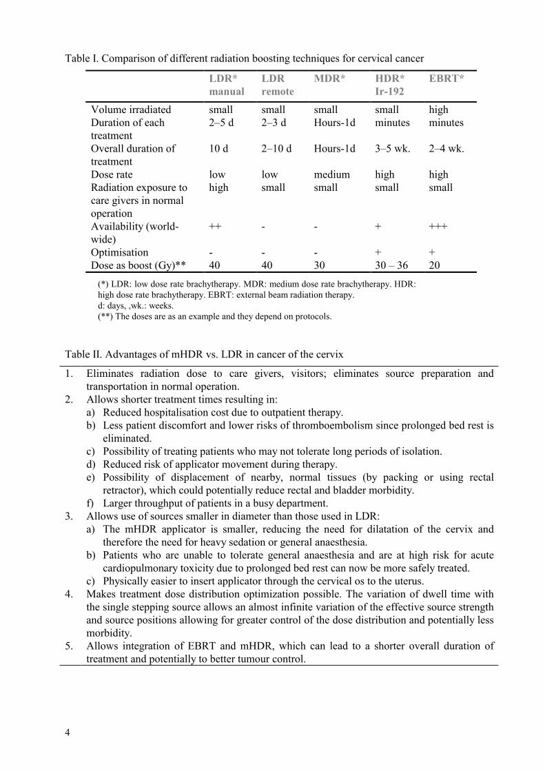
The radiation oncologist must evaluate each patient carefully weighing the risks with the benefits of mHDR brachytherapy in each circumstance. Table I summarises the comparison between the different radiation modalities as applied to cervical cancers. If cases are properly selected and a proper dose and fractionation scheme is devised, mHDR brachytherapy has wide applicability. It is convenient not only because of the short treatment time but also because of its potential for outpatient therapy and because it can be used to treat large numbers of patients (as staffing allows). The advantages of mHDR brachytherapy are enumerated in Table II.
The summary of the common clinical indications of mHDR provided here is necessarily brief. Treatment protocols are outside of the scope of this document and the reader is directed to standard textbooks or reviews for further details [10–14].
3.1. Cervical cancer
The incidence of cervical cancer is high, exceeding 30/100,000 in many developing countries, and constitutes the most common malignancy in some of these countries. Early stage cervical cancer is treated either by surgery (radical hysterectomy) or by radiation therapy, achieving similar cure rates in excess of 80%. Advanced stage cervical cancers are generally treated by radiation therapy alone. Brachytherapy is an essential component of the curative treatment of this disease [15–16]. When cervical cancer is treated by external beam radiation therapy (EBRT) alone, the cure rate is minimal. Advanced cervical cancer (Stage IIB–IIIB) treated by a combination of EBRT and LDR or mHDR brachytherapy can achieve
4
Table I. Comparison of different radiation boosting techniques for cervical cancer LDR*
manual LDR remote
MDR* HDR* Ir-192
EBRT*
Volume irradiated small small small small high Duration of each treatment
2–5 d 2–3 d Hours-1d minutes minutes
Overall duration of treatment
10 d 2–10 d Hours-1d 3–5 wk. 2–4 wk.
Dose rate low low medium high high Radiation exposure to care givers in normal operation
high small small small small
Availability (world-wide)
++ - - + +++
Optimisation - - - + + Dose as boost (Gy)** 40 40 30 30 – 36 20
(*) LDR: low dose rate brachytherapy. MDR: medium dose rate brachytherapy. HDR: high dose rate brachytherapy. EBRT: external beam radiation therapy. d: days, ,wk.: weeks. (**) The doses are as an example and they depend on protocols.
Table II. Advantages of mHDR vs. LDR in cancer of the cervix
1. Eliminates radiation dose to care givers, visitors; eliminates source preparation and transportation in normal operation.
2. Allows shorter treatment times resulting in: a) Reduced hospitalisation cost due to outpatient therapy. b) Less patient discomfort and lower risks of thromboembolism since prolonged bed rest is
eliminated. c) Possibility of treating patients who may not tolerate long periods of isolation. d) Reduced risk of applicator movement during therapy. e) Possibility of displacement of nearby, normal tissues (by packing or using rectal
retractor), which could potentially reduce rectal and bladder morbidity. f) Larger throughput of patients in a busy department.
3. Allows use of sources smaller in diameter than those used in LDR: a) The mHDR applicator is smaller, reducing the need for dilatation of the cervix and
therefore the need for heavy sedation or general anaesthesia. b) Patients who are unable to tolerate general anaesthesia and are at high risk for acute
cardiopulmonary toxicity due to prolonged bed rest can now be more safely treated. c) Physically easier to insert applicator through the cervical os to the uterus.
4. Makes treatment dose distribution optimization possible. The variation of dwell time with the single stepping source allows an almost infinite variation of the effective source strength and source positions allowing for greater control of the dose distribution and potentially less morbidity.
5. Allows integration of EBRT and mHDR, which can lead to a shorter overall duration of treatment and potentially to better tumour control.
5
cure rates of 60% and 30%, respectively. Because of the high incidence and good cure rates, cervical cancer is the most common indication for brachytherapy. Gynaecological brachytherapy can account for up to 100% of the brachytherapy practice in some developing countries. mHDR machines are capable of treating larger numbers of patients, due to the short treatment times needed. Implementation of mHDR should therefore be considered for developing countries with a high incidence of this disease.
When treating cervical cancer with radiation therapy, the goals are to treat Point A to at least a total LDR equivalent dose of 80–85 Gy for early stage disease and 85–90 Gy for advanced stage [17]. The pelvic side-wall dose recommendations are 50–55 Gy for early lesions and 55–60 Gy for advanced ones. The relative proportion of the total dose given by EBRT and brachytherapy depend upon the initial volume of disease, the ability to displace the bladder and rectum, the degree of tumour regression during pelvic irradiation, and institutional preference. Every attempt should be made to keep the ICRU bladder and rectal dose below 70–80% of point A dose. This equates to a total LDR equivalent dose below 75–80 Gy and 70–75 Gy for bladder and rectum, respectively. Interstitial brachytherapy, if available, should be considered for patients with disease that cannot be optimally encompassed by intracavitary brachytherapy [17].
In treating cancer of the cervix by LDR brachytherapy, the intracavitary insertion is typically performed after EBRT to the pelvis. In mHDR, the EBRT and brachytherapy are commonly integrated, the mHDR beginning after about 2 weeks (20 Gy) of EBRT. Typically the brachytherapy is performed once a week, while the pelvic EBRT is continued (with a midline block in some centres) to about 40–50 Gy. In some institutes, the EBRT is therefore given four times a week (EBRT is not given on the day of brachytherapy), while some institutes keep EBRT with five times a week. There are some differences in the brachytherapy technique for mHDR compared to that for LDR since the former uses narrower applicators that can be inserted on an outpatient basis under intravenous sedation or without sedation (see Table I and II). General or spinal anaesthesia is generally not used since little or no dilation of the cervical os is required. A variety of mHDR applicators (Fletcher, Henschke, Ring, etc.) are available. The use of fixed geometry applicators is encouraged in developing countries since it simplifies and speeds up the treatment planning process and reduces the chance of error. It is very important to use packing spacer or retractors to temporarily displace the rectum and bladder from the high dose region for the duration of treatment. Since the treatment duration is short, an external immobilisation device can be used to fix the position of the applicators and to minimise applicator movement. The type of applicator used is dependent on the preference of the radiation oncologist and the competence of the medical physicist.
Ideally the applicator insertion, radiograph generation, and treatment should take place in a dedicated brachytherapy suite, which makes patient movement unnecessary. However, it is recognised that it is not possible for all institutions to have a dedicated brachytherapy suite with an operating room and imaging equipment. Hence it may be necessary to transfer patients between the operating room, the simulator, and the treatment room; however, moving the patient after the radiographs for treatment planning have been obtained should be minimised.
The mHDR dose used (prescribed to point A) is dependent on the stage of the disease and the dose of EBRT used. The ratio of EBRT to brachytherapy is dependent on stage, with more emphasis being placed on EBRT for the more advanced stages, which are commonly seen in the developing countries. Although there is marked variation in the dose and fractionation employed, most centres are comfortable using a schedule of about 6–8 Gy per
6
fraction per week and 3–6 fractions (the smaller number of fractions being used by those using larger doses per fraction). While recognising that many efficacious mHDR fractionation schedules exist, the American Brachytherapy Society (ABS) has suggested a schedule of 45 Gy pelvic EBRT and 5 mHDR fractions of 6 Gy for patients with early stage cervical cancer and 6.5 Gy for advanced stage cervical cancer, respectively [17]. While mHDR allows optimisation, it should be realised that incorrect optimisation can be worse than no optimisation at all. Hence it is suggested that institutions begin by using the standard treatment plans that have been used in a large centre and then optimise the treatment plans for individual patient circumstances as they gain more experience.
Analysis of world-wide reviews (retrospective studies as well as prospective randomised clinical trials) suggests that LDR and HDR treatments are probably equivalent in terms of survival, local control, and morbidity [18–26]. Overall survivals of about 60% are obtained with 70–90% five-year survivals achieved for early (stage I-II) disease and 20–50% for advanced (stage III-IV) disease, respectively. Severe (grade III-IV) complications range from 0–10% (overall 3.5%) [18-26].
3.2. Oesophageal cancer
HDR brachytherapy has been used for the treatment of oesophageal cancer either alone or in combination with external beam radiation therapy [27–29]. Oesophageal cancer is a common problem in developing countries around the Caspian Sea (Turkmenistan, Kazakhstan, Uzbekistan and the Islamic Republic of Iran), Southern Africa (Malawi, South Africa, Lesotho and Botswana), and parts of China and Mongolia. As most cases are advanced in developing countries, the results of radical treatment of cancer of the oesophagus are dismal (5 year survival =6%); hence treatment is essentially palliative.
Surgical palliation techniques (intubation, bypass surgery, and resection) have been used with small survival benefit and at a cost of major morbidity, mortality, and major requirements for the limited surgical facilities. Intubation results in a survival equivalent to laser treatment alone of about 4 months and is accompanied by mortality and morbidity of 10% and 17% respectively. [30]. Palliative resection is used in fitter, selected patients with accordingly varying mortality up to 12% and morbidity of up to 71%, but it achieves longer survival.
The micro HDR technique is relatively simple since a single line catheter is used for the brachytherapy. The insertion is performed after surgical dilatation and biopsy, with sedation. Treatment is usually given using an intraoral approach and a nasogastric tube or a special oesophageal applicator. The largest diameter applicator that can be inserted easily should be used to minimise dose to the mucosa relative to the dose at depth. The site to be irradiated can be confirmed by fluoroscopy or endoscopy. The length treated includes the tumour and a margin of 2–5 cm. The dose is prescribed at 1 cm from the source, and doses of 5–15 Gy per fraction have been given for 1–4 fractions [28]. mHDR brachytherapy can be given before, concurrently with, or after EBRT. The advantage of giving brachytherapy after EBRT is that a more uniform dose can be delivered to the residual tumour after it has been reduced by treatment. The advantage of giving the brachytherapy initially is to have rapid relief of the major symptom, dysphagia.
The EBRT given is generally 40–60 Gy to the tumour with a 5 cm margin. Chemotherapy may also be added, but in these cases the external beam dose is usually

7
reduced. Brachytherapy at doses of 16 Gy in 2 treatments or 18 Gy in 3 treatments have been used to palliate oesophageal cancers without additional EBRT [29]. Retrospective studies as well as prospective, randomised clinical trials show that there is improved local control and survival when HDR brachytherapy is added to EBRT [31]. Since a high dose is delivered to the oesophageal mucosa, possible side effects include oesophageal ulcer, oesophageal fistula, and oesophageal stricture [31].
3.3. Head and neck cancer
Head and neck cancer is a common problem in some developing countries (e.g. nasopharyngeal cancers in China, oral cancers in India). Brachytherapy, especially using manually afterloading LDR 192Ir, has been widely used in these areas. mHDR brachytherapy may be used in selected cases to reduce radiation exposure and permit optimisation. However, these advantages are offset by the need for multiple fractionation, especially since the head and neck area does not tolerate high dose per fraction. mHDR can also be used to treat these tumours in institutions without manual afterloading facilities.
The nasopharynx is easily accessed by an intracavitary mHDR applicator. In Rotterdam, doses of 18 Gy in 6 fractions are delivered by a special nasopharynx applicator to boost 46–60 Gy of EBRT [32]. The use of mHDR brachytherapy catheters in removable dental moulds allows highly reproducible, repeated fractionated outpatient brachytherapy applications to superficial tumours [33]. Doses of about 15–20 Gy in 3–5 fractions can be delivered in this manner to boost 45–50 Gy EBRT. Data on the use of mHDR alone as salvage in tumours recurrent after EBRT are sparse. Doses of 50–55 Gy at 3 Gy per fraction have been used.
3.4. Lung cancer
In the developed countries, the lung is probably the most common site of the current use of mHDR brachytherapy. This is not so in developing countries, probably because of the relatively lower incidence of lung cancer. Even with aggressive therapies, locoregional failure occurs in a significant number of patients. The use of mHDR brachytherapy is well established for the palliation of endobronchial obstruction recurrent after external beam radiation therapy or in combination with external beam irradiation for palliation of metastatic lung cancers.
According to statistics, persistence or local relapse after standard external beam treatment for non small cell lung cancer occurs in 60% of patients. Endobronchial brachytherapy significantly improves the quality of life of these patients. Literature review shows palliation rates over 65%.
One or two catheters inserted through the brush channel of a flexible bronchoscope are used to deliver the mHDR treatment. This procedure can also be done using a rigid bronchoscope under general anaesthesia. Rapid response is seen when mHDR is used for palliation of severe haemoptysis. It can also be used with curative intent as a boost to external beam radiation therapy. The dose and fractionation used vary widely, ranging from 15 Gy in 1 fraction to 4 Gy x 5 fractions [14]. The intervals between fractions also vary, although a one-week interval is usual. The results from various centres show symptomatic improvement in over 50% of patients and bronchoscopic response from 59% to 100% [34]. Comparison of these results is difficult because of differences in patient population, dose, and fractionation. Common complications of therapy include haemoptysis, radiation bronchitis, and stenosis.
8
3.5. Other sites
HDR brachytherapy has also been used to treat carcinoma of the endometrium, vagina, breast, bile duct, brain, skin, sarcomas, prostate, and rectum. However, since the use of brachytherapy in these organs is uncommon in developing countries, they are not included in this TECDOC.
3.6. Clinical advances
While the current indications for mHDR have been mentioned in the previous section, some of the recent clinical advances in mHDR should be considered when planning a new mHDR program. One way to improve the therapeutic ratio of mHDR brachytherapy is to deliver the irradiation during surgery while the patient is still anaesthetised. This technique (intraoperative mHDR brachytherapy) allows radiosensitive normal tissues to be retracted or shielded during surgery, thus lowering the radiation dose to normal tissue [35]. Additionally, since the irradiation is given under direct vision, the risk of a geographical miss is reduced. Maximum surgical debulking is attempted whenever possible. The tumour bed is irradiated using special intraoperative applicators with parallel mHDR catheters embedded in them 1 cm apart. The use of a fixed geometry applicator allows the patient to be treated without delay, using pre-planned dosimetry for the selected applicator. Doses of 10–20 Gy are usually given as a single fraction over 10–60 minutes. Ideally, the surgery should be performed in a shielded operating room with remote anaesthesia and a television monitoring system. Hence, when starting a new program, a shielded operating room should be incorporated into the plans if intraoperative mHDR brachytherapy is contemplated. Unfortunately, due to the limited availability of shielded operating rooms, only a few institutions have used intraoperative mHDR brachytherapy.
Some of the mHDR machines are certified as transportable radioactive containers. This allows the machines to be transported between hospitals to be used on a shared basis when one centre does not have sufficient patient load to justify the purchase of a dedicated mHDR afterloader.
The development of thin diameter sources allows percutaneous interstitial brachytherapy through very thin needles (21 G). This may be of particular advantage for lip, nose, eye lid tumours and for percutaneous, image-guided treatment of intrathoracic or intraabdominal tumours.
3.7. Treatment planning
The simplest treatment planning uses a single catheter with the dose prescribed at a specified radius, as used in treating cancer of the oesophagus. Fixed geometry applicator, intracavitary treatment using a standard treatment plan is next in complexity, followed by intracavitary treatment with non-fixed geometry applicators and optimised treatment plans. Multi-plane rigid interstitial application with optimised treatment planning is used for breast or prostate. Multi-plane, flexible, interstitial application with optimised treatment planning (as in breast implants) is the most complicated. Optimally, clinical examinations supplemented by one or several imaging modalities (CT, MRI, ultrasound) are used to define the target volume and optimise the treatment plan to deliver a high dose to the tumour while minimising the dose to normal tissues.
9
4. COMPONENTS OF TREATMENT UNIT
Brachytherapy has been used as an integral part of cancer treatment for almost a century. It has been enhanced with the development of after-loading devices and new radioisotopes as described in the previous section. Present brachytherapy is characterised by many technical innovations such as:
�� Remote afterloading units �� Use of mHDR sources �� Computer technologies for treatment planning and dosimetry �� Newer imaging methods.
These developments have shifted brachytherapy procedures to outpatient management and have increased the number of brachytherapy procedures that can be performed in a single day. An adequately shielded room and a remote afterloading device to avoid direct exposure of the operators are essential components of a mHDR facility.
A remote afterloading system consists of a pneumatically or motor-driven source transport system for automatically transferring radioactive material between a shielded safe and each treatment applicator [36]. These systems were first designed for use in gynaecologic brachytherapy, but more recent models can be used for other sites as well.
The mHDR remote afterloading systems must comply with international standards of safety and quality, such as those of the International Electrotechnical Commission (IEC) [37] or International Standards Organization (ISO) 9000.
Components of mHDR equipment Commercially available mHDR afterloading units consist of the following components: �� mHDR source �� Afterloader device (treatment unit) �� Applicators �� Treatment planning system. 4.1. mHDR source
A radioisotope with a high specific activity is needed to simultaneously achieve high dose rate and small source size required for intracavitary and interstitial brachytherapy. 192Ir is widely used for mHDR brachytherapy because it has a high specific activity (330 MBq mm-3), relatively low gamma energy (average 0.4 MeV) and relative short half-life (74 days).
Currently, most HDR remote afterloaders use a single 192Ir source with an activity of about 370 GBq. The size of the encapsulated source is about 5 mm long (some sources may be up to 10 mm long) and less than 1.5 mm in diameter; these dimensions vary with different commercial models. The source is welded to the end of a drive cable, transferred to programmed locations in the applicators (dwell positions), and held in the place for programmed duration (dwell times), using a motor-driven system.
4.2. Afterloader device (treatment unit)
These units are mobile and take up little floor space.
10
An afterloader unit contains: �� Shielded safe (main-source container) to hold the source when not in use. �� Stepping-motor �� Source transferring and positioning system �� Several channels for source transport �� Indexer to allow automatic transfer of the source cable among the different transfer
tubes �� Transfer tubes to connect the device to the applicators �� Safety system to ensure safe operation of the device, including:
�� Automatic path-check of the applicator + transfer tube with a check cable �� Means of sensing the source position and timing of its motion �� A built-in Geiger-Muller counter to check that the source has returned to the safe �� Backup batteries to withdraw the source in the event of power failure and for saving
treatment data �� Emergency systems to withdraw the source into the safe.
A detailed description and specification for mHDR afterloading device is in Appendix G.3 of IAEA-TECDOC-1040 [1].
4.3. Control console
The control console located outside of the treatment room operates the afterloader, shows the source position on the display as the treatment progresses, and prints out a report of the treatment. The treatment plan can be transferred to the control console through a direct link with the treatment planning computer, a floppy disk, a program card (for older machines), or manually. It has a microprocessor to automatically correct the dwell times for decay. The control console should be simple to operate.
4.4. Applicators
Almost all applicators designed for LDR manual afterloading have been adapted for mHDR use with a mechanism to connect them to a transfer tube from the afterloader device. Typically, the applicators for mHDR have thinner tubes. The connection has mechanical interlocks to ensure that the applicator is correctly positioned and connected. The interlocks prevent wrong connections. The applicator, transfer tube, and afterloader device are a closed system to avoid the possibility of the source becoming dislodged in the patient or exiting into the air before reaching the target region.
There are 3 categories of applicators: intracavitary, intraluminal, and interstitial. Each category of applicator employs a specific connector or transfer tube to link with the treatment unit.
Intracavitary applicators use specific transfer tubes designed to be the same overall length but to have different interlocks for each treatment channel to avoid connection errors. There are a variety of intracavitary applicators for mHDR treatment. Some applicators are made of stainless steel (suitable for X ray simulation and for durability); others are made of plastic (for CT or MRI compatibility). Some intracavitary applicators (e.g. Fletcher-type) are rigid but do not have a fixed geometry, therefore requiring individual patient treatment
11
planning. A fixed geometry applicator (e.g. ring applicator) allows standard dose distribution planning prior to insertion.
Intraluminal applicators usually connect directly with the treatment unit using a specific adapter. These applicators can be 5 or 6 French diameter, blind ended, flexible tubes (disposable); or they can have a specific design (e.g. Oesophageal Applicator). If a single catheter technique is used, the treatment planning is simple and can be done in advance.
Interstitial applicators can be rigid or flexible. The rigid stainless steel needles are of different lengths and require specific transfer tubes. The needles can be reused after sterilisation. Using a template for the implantation with a fixed predetermined geometry allows use of standard dose distribution. The thin, flexible disposable plastic tubes require different transfer tubes.
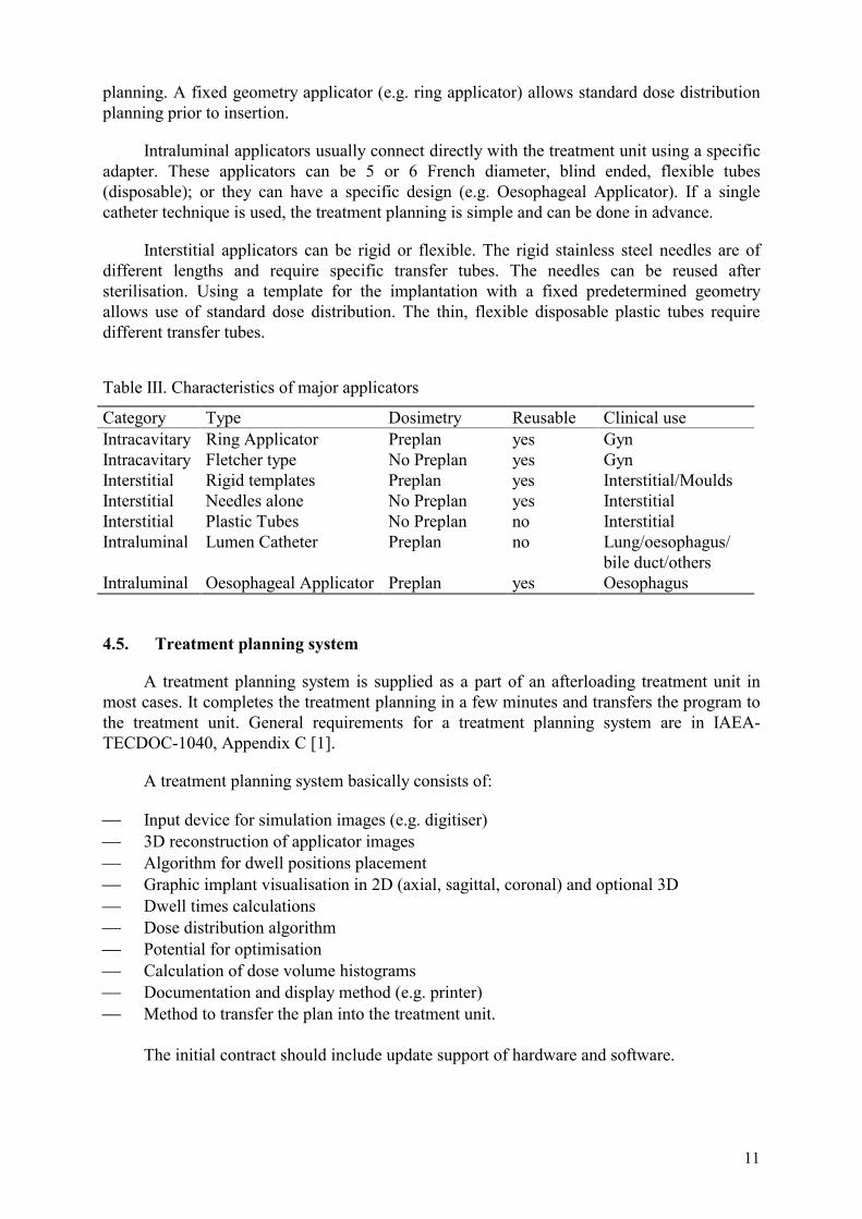
Table III. Characteristics of major applicators
Category Type Dosimetry Reusable Clinical use Intracavitary Ring Applicator Preplan yes Gyn Intracavitary Fletcher type No Preplan yes Gyn Interstitial Rigid templates Preplan yes Interstitial/Moulds Interstitial Needles alone No Preplan yes Interstitial Interstitial Plastic Tubes No Preplan no Interstitial Intraluminal Lumen Catheter Preplan no Lung/oesophagus/
bile duct/others Intraluminal Oesophageal Applicator Preplan yes Oesophagus
4.5. Treatment planning system
A treatment planning system is supplied as a part of an afterloading treatment unit in most cases. It completes the treatment planning in a few minutes and transfers the program to the treatment unit. General requirements for a treatment planning system are in IAEA-TECDOC-1040, Appendix C [1].
A treatment planning system basically consists of:
�� Input device for simulation images (e.g. digitiser) �� 3D reconstruction of applicator images �� Algorithm for dwell positions placement �� Graphic implant visualisation in 2D (axial, sagittal, coronal) and optional 3D �� Dwell times calculations �� Dose distribution algorithm �� Potential for optimisation �� Calculation of dose volume histograms �� Documentation and display method (e.g. printer) �� Method to transfer the plan into the treatment unit.
The initial contract should include update support of hardware and software.
12
5. INFRASTRUCTURE (BUILDING, IMAGING, PLANNING SYSTEM) REQUIRED
Overall requirements for an infrastructure are in the IAEA-TECDOC-1040 [1]. This section focuses on the operational and clinical aspects of an infrastructure.
Setting up an mHDR unit requires an investment of capital and human resources. A new mHDR brachytherapy program should consider the current and future projected patient volume, case mix, the existing infrastructure, and available human resources. The staff should be trained in technical and radiobiological aspects and supported by an experienced radiation oncologist and medical physicist during the initial procedures. Before installing a brachytherapy unit, each step of the treatment procedure must be considered. These include: �� Applicator/catheter placement �� Imaging (simulation and localisation) �� Treatment planning �� Treatment delivery.
Ideally the applicator insertion, radiograph generation, and the mHDR treatment should be done in a dedicated brachytherapy suite so that there is no need to move the patient. If such a facility does not exist, each of these steps can be carried out in a different room. Options include transferring patients either from the operating room or a procedure room in the department to the simulator for radiograph generation. However, it is preferable to minimise movement of the patient by performing the individual procedures within a short distance of the mHDR treatment room. It is especially important that there be minimal movement of the patient after the localisation radiographs have been obtained.
5.1. Building 5.1.1. Infrastructure required for applicator/catheter placement (procedure room)
This room should function like an outpatient surgery room and be suitable for various procedures such as endoscopy, percutaneous insertion of catheters, or gynaecological applicator placement. The factors to be considered includes the availability of: �� Sufficient space for both the brachytherapy team and any other medical or surgical team
that will be involved in the procedure �� Adjustable and mobile table with stirrups, ideally X ray compatible �� Instruments for minor surgery �� Cart with disposable supplies �� Storage cabinet for mHDR applicators and other accessories �� Surgical lights, anaesthesia equipment, and patient telemetry (desirable) �� Clean water supply and sink. 5.1.2. Infrastructure required for localisation radiographs
If the treatment room is separate from the applicator placement room, the size of the shielded treatment room must be adequate to allow localisation radiographs to be obtained on the treatment table in order to minimise the patient movement. Portable X ray equipment can be used or, preferably, dedicated X ray equipment (e.g. C-arm) should be installed. If X ray equipment is not available in the treatment room, there should be sufficient space to allow the patient to be transported on a stretcher from the simulator. In addition to the X ray equipment, there must be a device (simulation box) available if semi-orthogonal films are used for the dosimetry.
13
5.1.3. Infrastructure required for the treatment planning room
The hardware for treatment planning could be placed in a separate room or adjacent to the control console. The only requirements are the space and the power supply. A device for an uninterruptable power supply (UPS) with a voltage regulator should be considered a part of the hardware. It is desirable to have the treatment planning system placed close to the treatment room, as this improves efficiency and communication.
5.1.4. Infrastructure required for the treatment room
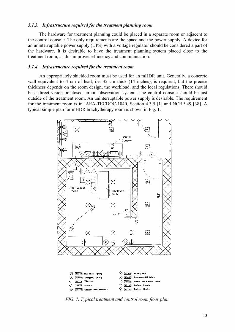
An appropriately shielded room must be used for an mHDR unit. Generally, a concrete wall equivalent to 4 cm of lead, i.e. 35 cm thick (14 inches), is required; but the precise thickness depends on the room design, the workload, and the local regulations. There should be a direct vision or closed circuit observation system. The control console should be just outside of the treatment room. An uninterruptable power supply is desirable. The requirement for the treatment room is in IAEA-TECDOC-1040, Section 4.3.5 [1] and NCRP 49 [38]. A typical simple plan for mHDR brachytherapy room is shown in Fig. 1.
FIG. 1. Typical treatment and control room floor plan.
14
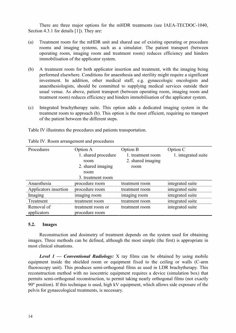
There are three major options for the mHDR treatments (see IAEA-TECDOC-1040, Section 4.3.1 for details [1]). They are: (a) Treatment room for the mHDR unit and shared use of existing operating or procedure
rooms and imaging systems, such as a simulator. The patient transport (between operating room, imaging room and treatment room) reduces efficiency and hinders immobilisation of the applicator system.
(b) A treatment room for both applicator insertion and treatment, with the imaging being performed elsewhere. Conditions for anaesthesia and sterility might require a significant investment. In addition, other medical staff, e.g. gynaecologic oncologists and anaesthesiologists, should be committed to supplying medical services outside their usual venue. As above, patient transport (between operating room, imaging room and treatment room) reduces efficiency and hinders immobilisation of the applicator system.
(c) Integrated brachytherapy suite. This option adds a dedicated imaging system in the treatment room to approach (b). This option is the most efficient, requiring no transport of the patient between the different steps.
Table IV illustrates the procedures and patients transportation. Table IV. Room arrangement and procedures
Procedures Option A 1. shared procedure room 2. shared imaging room 3. treatment room
Option B 1. treatment room 2. shared imaging room
Option C 1. integrated suite
Anaesthesia procedure room treatment room integrated suite Applicators insertion procedure room treatment room integrated suite Imaging imaging room imaging room integrated suite Treatment treatment room treatment room integrated suite Removal of applicators
treatment room or procedure room
treatment room integrated suite
5.2. Images
Reconstruction and dosimetry of treatment depends on the system used for obtaining images. Three methods can be defined, although the most simple (the first) is appropriate in most clinical situations.
Level 1 — Conventional Radiology: X ray films can be obtained by using mobile equipment inside the shielded room or equipment fixed to the ceiling or walls (C-arm fluoroscopy unit). This produces semi-orthogonal films as used in LDR brachytherapy. This reconstruction method with no isocentric equipment requires a device (simulation box) that permits semi-orthogonal reconstruction, to permit taking nearly orthogonal films (not exactly 90� position). If this technique is used, high kV equipment, which allows side exposure of the pelvis for gynaecological treatments, is necessary.
15
Level 2 — Simulator: Having a simulator for external radiotherapy permit taking of not only films as mentioned in the technique of conventional radiology, but also trustworthy orthogonal films. In addition, other (easier) reconstruction techniques such as isocentric and variable angles, which may be required under special circumstances, can be used.
Level 3 — CT and MRI: Axial cuts from a CT Scan or MRI, permit not only the reconstruction of the source position, but also the reconstruction of the anatomic volumes of interest in dosimetry. In the first two methods, it is only possible to get the reconstruction of the applicators, but not their relationship to the anatomical structures.
5.3. Treatment planning procedure
The treatment planning system must be fast, versatile, and specific to control the remote afterloader.
In developing countries, both level 1 and level 2 imaging devices (simulation with conventional radiology or a simulator) can be used for 90% of the cases requiring brachytherapy. These procedures can be done with confidence and good quality assurance without CT-MRI simulation, 3D reconstruction, or sophisticated planning systems.
The hardware and software needed to cope with the different degrees of dosimetric complexity is, of course, directly related to such complexity. Peripheral devices for printing efficiently to show the results (plotters, printers, etc.) and for entering images are needed. The latter can be achieved by means of digitisers or scanners. Ideally, the images can be entered from the diagnostic machine, either through direct connection or through some magnetic or optical device that is able to store information. Once the images are on the worktable, the radiation oncologist should indicate to the physicist the volume to be treated and the dose to be applied.
Treatment planning for fixed geometry applicators or single line catheter can be done before the treatment.
The initial contract should include ongoing support and update of the treatment planning hardware and software.
5.4. Equipment for radiation safety and source handling
Every brachytherapy facility should have the following equipment:
�� A storage container in the treatment room to serve as an emergency source container in case of failure of the afterloader in retracting the source.
�� Long-handled forceps �� A portable radiation monitor instrument and an area radiation monitor (see IAEA-
TECDOC-1040, G.4 [1]).
5.5. Spare parts
All commonly used spare parts should be stored in the department if they are not immediately available from a service centre.
16
5.6. Other requirements
The brachytherapy facility also needs hospital support, such as a laboratory and sterilisation facilities, examination rooms, a pharmacy, air conditioning, etc.
6. PERSONNEL REQUIREMENTS AND TRAINING
6.1. Personnel requirements
The primary prerequisite for the development of an mHDR brachytherapy facility is adequate staff. A multidisciplinary team must be organised. A radiation oncologist, a medical physicist, a technician, and a personnel with nursing skills are the minimum personnel required. Depending on the workload, a dosimetrist, more nurses, radiation oncologists, and technicians may be added. Introduction of mHDR machine leads to wider spectrum diseases and considerable increase in work load. Thus, increase of personnel in proportion to work load should be critical consideration.
6.1.1. Radiation oncologist
The radiation oncologist is responsible for the overall procedure, as brachytherapy is a medical treatment. He/she must be properly accredited according to each country’s regulations.
Specific radiation oncologists responsibilities are [39]: �� Patient evaluation �� Treatment prescription and protocol selection �� Applicator insertion(s) �� Simulation review �� Selecting treatment volumes �� Treatment plan approval �� Applicator(s) removal �� Evaluation of tumour response and side effects �� Patient follow up.
6.1.2. Medical physicist
The medical physicist must be accredited in dosimetry according to each country’s regulations.
Specific medical physicists responsibilities are:
�� Testing equipment for acceptance �� Calibrating sources �� Checking treatment unit �� Verifying source positioning �� Checking patient set-up including applicator positioning �� Supervising simulation

17
�� Treatment planning and calculations �� Supervising treatment administration by the technicians.
The physicist should participate in the preparation of the patient after the applicator has been implanted and prior to getting the aforementioned images, since it is during such preparation that the dummies (X ray marker wires) are to be positioned in the applicators (as specified by the technique used). If catheters are used, it is necessary to measure and identify them. It is also necessary either to select the angles of the radiographic images or to select planes in the event of CT or MRI imaging.
6.1.3. Technician/brachytherapy technician
The technician is in charge of:
�� Checking applicators and specific accessories �� Daily checking of the treatment unit �� Assisting the radiation oncologist during implantation �� Obtaining images for simulation and localisation �� Using treatment planning under the physicists supervision �� Delivering treatment �� Monitoring each treatment from the console �� Recording treatment on appropriate documents.
6.1.4. Nurse
The nurse is in charge of assisting the physician during each procedure.
For an mHDR brachytherapy procedure flow, the reader should refer to the report of AAPM Task Group No. 59 [40].
6.2. Training
The staff needs to be adequately trained on the particular model of mHDR remote afterloading system being used, to prevent possible errors and to promptly identify and correct any errors that may occur.
6.2.1. Radiation oncologist training
If the radiation oncologist has experience in LDR brachytherapy, additional training is required in mHDR specific features such as applicators, insertion techniques, HDR radiobiology, and emergency procedures. mHDR intracavitary, intraluminal, or interstitial applicators are quite similar to those used in LDR, so the radiation oncologist only needs to become familiar with them. The radiation oncologist should be trained to place the applicators quickly and precisely. Some updating in radiobiology knowledge is required to decide on the treatment protocols and fractionation. The linear quadratic model could be used to develop HDR protocols in conjunction with published experience of outcomes and morbidity. The radiation oncologist should be trained in all emergency procedures.
18
A radiation oncologist without experience in LDR brachytherapy requires training in general brachytherapy principles. Subsequently, the radiation oncologist needs to be trained in each site-specific mHDR brachytherapy technique. It is not necessary to have previous LDR brachytherapy experience in order to be trained in mHDR.
6.2.2. Physicist training
The physicist must be trained in the use of the mHDR planning system (a necessary tool in the use of mHDR equipment) and should become thoroughly familiar with applicator image reconstruction. Training in equipment use, security systems, and emergency procedures is mandatory. Physicists must also be trained in the basic principles and procedures of radiation protection.
Preferably, the radiation oncologist and the physicist should be trained at a brachytherapy centre that treats similar types of cancers. Hands-on training is desirable. During the initial phase of working with mHDR brachytherapy, the support of an experienced physician and physicist is very useful for achieving the objectives with confidence and for good quality assurance.
6.2.3. Technician and nurse training
The technicians and nurses can be trained for mHDR brachytherapy procedures by the radiation oncologist and the physicist. Radiation safety instruction and emergency procedures are an essential element to be covered.
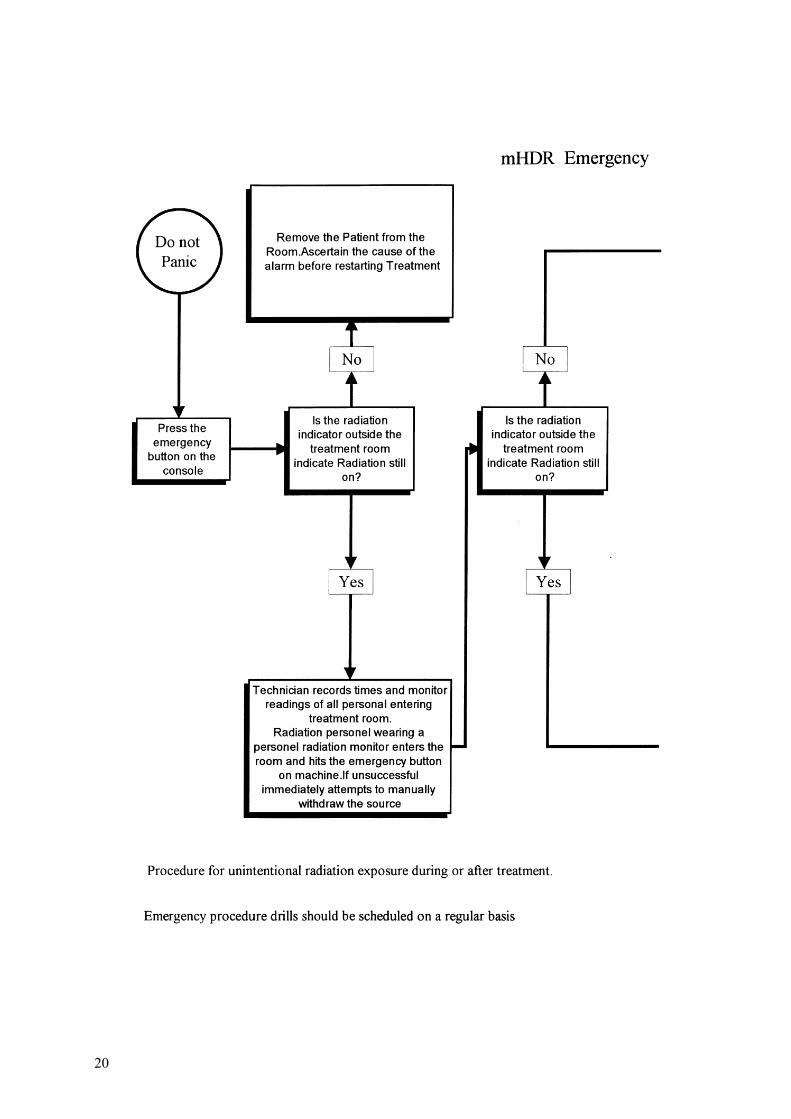
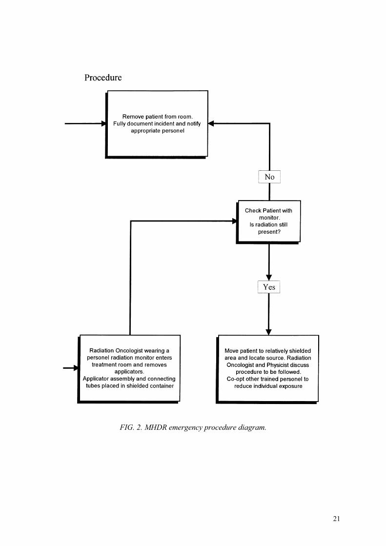
6.2.4. Emergency procedures
The readers are referred to IAEA-TECDOC-1040 Section 7.9 [1], in which emergency procedures are described. These procedures should be practised periodically. An example of a flowchart is given in Fig. 2. This information should be prominently displayed in the treatment room and control room.
7. QUALITY ASSURANCE (QA)
The full scope of the QA programme is beyond the scope of this document and other references are available (e.g. IAEA-TECDOC 989, 1040 and AAPM report of Task Group 59) [1, 40].
QA in radiation therapy is essential for obtaining good results, avoiding unnecessary side effects, and performing mHDR brachytherapy accurately and safely. In mHDR brachytherapy, QA is extremely important because the procedures are carried out quickly with high doses being given in a short time period, with little opportunity for correction.
The QA programme should consider clinical aspects of mHDR brachytherapy, including patient selection criteria, dose determination and specification, fractionation, quality of insertions, tumour volume, treatment volume; and the physical aspects of dosimetry such as checks of the computer information input, sources, strength, and dose at different distances.
19
The imaging should be checked for quality of not only the image but also of the angle of incidence (orthogonal or oblique) and the position of applicators.
The QA for mHDR brachytherapy can be divided into three segments:
�� QA on the treatment unit �� QA on the planning system �� QA on the patient treatment procedure.
7.1. QA on the treatment unit
This consists of a set of tests to be performed periodically to verify the proper function of the treatment unit.
Daily tests — these tests must be done daily, before patient treatment:
�� Emergency systems to withdraw the source into the safe �� Door interlocks of the treatment room �� Interrupt button on the control console �� Emergency stop button �� Interrupting power supply. �� Source positioning �� Room radiation monitors �� Indicator lights.
Daily tests take around 10 minutes and can be performed by a technician.
Monthly tests
The source activity should be checked to show agreement with the predicted radioactive decay. Applicators should be checked in regard to their integrity, internal shields, wells, and joints. Movement of the source to the desired location in the applicator should be confirmed.
Quarterly tests (with each source change):
The source strength must be calibrated using well-type chambers specifically designed for the purpose or with Farmer-style ion chambers. Using the latter, an interpolate technique for deriving a calibrator factor for 192Ir should be used. The calibration of the well-type chamber should be performed at a standard laboratory every two years.
The source positional accuracy should be tested. This means testing the ability of the unit to drive the source to a desired position in the applicator within +/-1 mm precision. This can be done using autoradiography with external markers or a “check ruler” device. [1, 40].
7.2. QA on the planning system
QA on the planning system basically consists of verification of the reconstruction quality and the accuracy of the calculated dose. The quality of the dosimetry is closely linked to the reconstruction used and also to the system of image acquisition of the planning software.
20
21
FIG. 2. MHDR emergency procedure diagram.
22
The quality of the reconstruction can be tested by performing the reconstruction of fixed geometry applicators or by using a phantom to determine the accuracy of X ray marker co-ordinates. This test should be done for each reconstruction method available in the software (orthogonal, isocentric, CT, etc.). The accuracy of dose point calculations can be tested by matching then with manual calculations or independent computer calculations. The accuracy of input and output devices, the accuracy of the device in transferring the plan into the control console, and the consistency of the printed output of plan and other documentation should also be tested. The QA tests on the planning system should be repeated at any significant software change.
7.3. QA on the patient treatment procedure
Medical and physical features are included. The objective is to verify each brachytherapy step during each patient treatment. It has the following components:
�� Consistency and accuracy of the prescription �� Applicator placement or catheter implantation �� Simulation and localisation images �� Treatment planning and calculations �� Treatment delivery. �� Documentation.
For a complete dose specification and documentation of brachytherapy treatments, the following points are necessary, according to the ICRU 38 and the ICRU 58 reports [3, 41]:
�� Description of volume �� Description of method and technique �� Specification of source strength �� Description of source distribution and source pattern �� Reference dose and dose distribution �� Fractionation.
Establishing standardised protocols and policies for common treatments reduces the chances of mistakes. For this reason, the centres should initially perform only simple procedures, using fixed geometry applicators and standard planning. For each treatment, the following must be checked (preferable by a physicist or a dosimetrist not involved in the planning):
�� That the source strength matches the strength used in the calculation and the one indicated at the treatment unit;
�� That the proper source localisation is programmed; �� That programmed dwell times match the plan; �� That positions match the plan; �� That dose per fraction matches the prescription.
The quality control of the treatment itself consists of verifying positioning of the applicator in the patient, the connection of connecting tubes between applicators and the treatment unit, and the presence of staff controlling the treatment at the control console. For
23
each treatment, the completion of quality control tests should be documented by signature, and the personnel responsible for carrying them out should be recorded.
Adequate staff training and quality control checklists are the keys for a successful mHDR brachytherapy.
8. RADIATION SAFETY
8.1. The relevant radiation safety standards and related documents
The use of radiation for brachytherapy is regulated by national regulations. International harmonisation is provided by the IAEA by means of standards of safety. This is recognised in the statutory IAEA’s functions: ‘to establish or adopt…standards of safety for protection of health … and to provide for the application of these standards …’.
For activities related to this TECDOC the relevant radiation safety requirements are the International Basic Safety Standards for Protection against Ionising Radiation and for the Safety of Radiation Sources (the BSS), issued in 1996, co-sponsored by the Food and Agriculture Organization, the International Labor Organization, the Nuclear Energy Agency, the World Health Organization and the Pan American Health Organization [42]. It contains principal requirements covering all practices, including uses of radiation in medicine, agriculture, industry, research and teaching and intervention in the event of accidents and in chronic exposure situations such as that due to residues from past activities. Detailed requirements are given in six Appendices, covering among others, occupational exposure, medical exposure (mainly protection of patients), public exposure, potential exposure and emergencies.
Recommendations on how to comply with the requirements of the BSS are given in Safety Guides. The most relevant to this TECDOC are those related with occupational exposure [43] and with protection in the management of medical exposure (Safety Guide on Radiation Protection in the Management of Medical Exposure [44]), now in the process of publication. This Safety Guide is jointly co-sponsored by PAHO and WHO. It describes strategies to involve organisations, such as professional bodies, whose co-operation is essential to ensure compliance with the requirements of the Basic Safety Standards (BSS) for medical exposure.
The BSS state in the preamble that the Regulatory Authority may need to provide guidance on how certain regulatory requirements are to be fulfilled for various practices. It is not feasible to reproduce detailed safety requirements here. However a few pertinent issues will be highlighted.
8.1.1. Authorizations
In order for a radiotherapy department to operate in compliance with the Standards it is indispensable to obtain an authorisation. The application has to provide evidence of the assessment of safety performed and of the measures taken to work safely. More detailed information is given in the BSS the Safety Guide and the Model Regulations.
24
8.1.2. Responsibilities
The Basic Safety Standards establish that the primary responsibility is with the holder of a license (the licensee) and also assign subsidiary responsibilities to medical practitioners and qualified experts among others.
With regard to medical exposure (mainly the exposure of patients) the responsibilities are given in the following requirements [paragraph II.1]:
(a) no patient be administered a diagnostic or therapeutic medical exposure unless the exposure is prescribed by a medical practitioner;
(b) medical practitioners be assigned the primary task and obligation of ensuring overall patient protection and safety in the prescription of, and during the delivery of, medical exposure;
(c) medical and paramedical personnel be available as needed, and either be health professionals or have appropriate training adequately to discharge assigned tasks in the conduct of the diagnostic or therapeutic procedure that the medical practitioner prescribes;
(d) for therapeutic uses of radiation (including teletherapy and brachytherapy), the calibration, dosimetry and quality assurance requirements of the Standards be conducted by or under the supervision of a qualified expert in radiotherapy physics;”
8.1.3. Radiation protection programme and committee
The licensee (usually the manager of the institution) can delegate functions related to radiation protection and safety, while retaining the overall responsibility. An efficient way of delegating is by establishing a radiation protection programme and a committee to supervise compliance with the programme1. The programme should contain all issues related to radiation protection requirements, including the definition of responsibilities, administrative requirements, and the requirements on occupational exposure, medical exposure, public exposure and emergency exposure situations.
An important part, which is especially important in radiotherapy, is the quality assurance programme, which ensures good practice and radiation protection of the staff, patients and the public. (see also Section 7) Experience has shown that the frequency of accidental exposures is directly related to the absence or inadequacy of an established QA programme in the department.
8.2. Specific remarks with regard to the application of the requirements to high dose rate brachytherapy
This section must be regarded as supplementary to the general instructions in the BSS. Maintenance is a radiation safety related issue. In other areas of radiation therapy, complex 1 It is an extended and growing practice, that hospitals, especially radiotherapy departments, implement a quality assurance system for the entire medical care throughout the treatment, i.e. covering the overall radiotherapy practice. This system involves a quality assurance committee. The radiation protection and the quality assurance committees have many functions in common (namely, quality control of physical and clinical factors in medical exposure, as established in the BSS), and also some members will belong to both committees. Harmonized integration of both committees is needed so that radiation protection issues are given the importance required by regulations and direct reporting to management is ensured.
25
electronic and mechanics has be the cause of severe accident. In the context o f a HDR brachytherapy machine it is necessary to have a strategy for maintenance and preparation with sufficient resources.
8.2.1. Double checks in the quality assurance programme
The quality assurance programme has to incorporate sufficient double and independent checks of all safety-critical parameters, from the commissioning of the machine, source calibration, the treatment plan to the delivery of the doses to the patients. (see also Section 7).
8.2.2. Prevention from accidental exposure
To prevent accidental exposure it is indispensable to identify “what can happen” and what can be done: human mistakes and equipment faults in mHDR brachytherapy has the potential for resulting doses and dose fractions different than prescribed. The most typical identified events are the cases in which the radiation sources were:
(a) Obstructed by a kink in the guide tube or catheter while the source gets stuck in the patient.
(b) Dislodged from the driving mechanism.
Working procedures should be devised to prevent these situations from occurring.
8.2.3. Mitigation of accidental exposure: Emergency plan and response
In spite of the measures to prevent events that may lead to accidental exposure, the probability of occurrence is not zero. It is therefore necessary to be prepared for them. The preparedness involves first and foremost the availability of trained persons responsible for carrying out the response i.e. putting the emergency under control.
The critical emergency arises if the source travel is disrupted while in the applicator or on route to the applicator. Removal of the applicator to permit patient egress from the room is the first action.
Other elements of an emergency plan in HDR brachytherapy suite need to include:
�� list of predictable incidents and accidents and measures to deal with them; �� persons responsible to take actions, with complete relevant information, including
telephone numbers.; �� responsibilities of the individual personnel in emergency procedures (radiation
oncologist, medical physicists, radiation technicians); �� the above persons responsible for carrying out emergency response action shall be on
site; �� set of concise instructions posted in a visible area; �� equipment and tools necessary to carry out the procedures; �� training and periodic rehearsal; and �� recording and reporting system.

26
Emergency procedures (Fig. 2) have an objective: �� to avoid unnecessary radiation doses to patients, staff and public. This involves the
return of sources to the shielded position or waiting receptacle; �� measures to prevent access of persons to the affected area during the time that the
sources are exposed and normal conditions are restored; and
In the case of HDR brachytherapy the time to implement the emergency action is extremely short — of the order of seconds.
After the critical emergency, there is a need to include an estimation of doses from the emergency actions:
�� estimate the time and position of the hands and body of the person removing the applicator with the sources;
�� knowing the time, distance and activity of the source make an estimation of doses to him/her and to the patient.
8.2.4. Investigation of accidental exposure
The requirements of the BSS [42] with regard to investigation of accidental medical exposure are:
(a) Calculate or estimate the doses received and their distribution within the patient; (b) indicate the corrective measures required to prevent recurrence of such an incident; (c) implement all the corrective measures that are under their own responsibility; (d) submit to the Regulatory Authority, as soon as possible after the investigation or as
otherwise specified by the Regulatory Authority, a written report which states the cause of the incident and includes the information specified in (a) to (c), as relevant, and any other information required by the Regulatory Authority; and
(e) inform the patient and his or her refering doctor about the incident.
8.2.5. Identified causes of and contributing factors to accidental exposure in radiotherapy
IAEA Safety Reports Series No. 17 collected these events [45]. The following events were identified:
�� Errors in the calibration of radiotherapy sources; �� errors in the preparation of input parameters from which the treatment; �� errors in acceptance tests and commissioning or lack of tests of both radiation sources
and treatment planning systems; �� maintenance errors; �� communication errors, transmission of information and misunderstanding of
prescription and protocols, or use of obsolete protocols; �� errors in the identification of patient and treatment site; �� dislodging of HDR brachytherapy sources; �� sources left in patient.
27
9. COST–UTILISATION FACTORS
mHDR treatment systems are the most expensive brachytherapy modality, both in terms of capital outlay and ongoing costs. These costs must be offset by the low incremental cost of treating more patients, increased clinical versatility, and cost shifting away from inpatient hospital care. The most appropriate treatment modality depends on the type of clinical practice (case mix) encountered, as well as the likely patient throughput and available infrastructure to undertake such treatments safely. Where quality assurance and safety for both patient and staff are adequate, the choice of treatment modality depends on cost utilisation considerations and the availability of trained staff.
This section gives a framework and methodology for cost-utilisation evaluation but cannot be specific for each set of circumstances. Capital costs as well as ongoing costs are illustrated using US dollars, current for 1999.
9.1. Cost–strategic perspective
Brachytherapy should be placed in the context of establishing health care programs in developing countries. Care must be taken to ensure that any brachytherapy program is sensitive to local cultural and geographic needs and sufficiently adaptable to meet changing needs in the future. Such programs should exist within a framework of resource allocation to preventive health services and to basic health needs at a dispensary, health centre, or primary hospital care level.
A wide disparity in health expenditure per capita and proportion of expenditure for tertiary care exists between countries. Outpatient service, particularly in the curative treatment of cervical cancer, is one of the main advantages of mHDR treatment, since this results in cost shifting from low technology inpatient care to high technology outpatient care. The importance of adequate ongoing funding and commitment for equipment maintenance [42], source replacement, and ongoing training must be acknowledged as an essential part of any mHDR programme development.
9.2. Cost–operational perspective
The cost of providing a brachytherapy service can be examined in different ways. Broadly, three aspects can be defined:
(a) The cost can be divided into direct, indirect, and intangible costs. Thus the cost of a procedure can be examined as the cost of identifiable consumable materials, the cost of providing a bed in a ward to allow the procedure, and the intangible cost of providing alternative services, e.g. palliative care.
(b) Secondly, party based analysis considers the cost to whom. Here, the cost to the state, local health authority, or patient is considered. The costs identified are distinct from the amount claimed for reimbursement.
(c) Process based analysis considers the costs incurred while going through the process: developing infrastructure, treatment procedures quality assurance, and outcome assessment or audit.
28
9.3. Capital cost
The most appropriate operational perspective will depend on the local conditions and model of machines. As a guide, however, example for the main capital costs of mHDR are provided and compared to a LDR remote afterloading unit.
9.3.1. Direct capital costs incurred in setting up a mHDR treatment facility
The following costs should be considered as direct capital costs.
(a) An LDR-afterloader. A 3 channel machine including 18 sources could treat up to 80 cases of cervical cancer per year.
Cost: US$230K
(b) Treatment room(s): The shielding costs for one or two shielded rooms are still likely to be less than a single mHDR treatment room but will depend on local regulations [43] for designated radiation areas and occupancy factors.
Cost: US$5–50K. (More for a new facility)
(c) X ray equipment (C-arm X ray unit or simulator): The X ray facilities must be able to adequately image a lateral view of the pelvis, e.g. 70 kg woman with pelvic separation of 36 cm. They will require a capacity of approximately 89 kV, 200 mA, and 300 mAs.
Cost of C-arm equipment: US$100K.
(d) Applicators: Similar requirements as for mHDR although only gynaecological applicators required.
Cost: US$30K (Minimum).
(e) Patient support table and miscellaneous supplies for the procedures room. Cost: US$10–30K.
(f) Medical physics QA equipment as described in IAEA-TECDOC-1040 Section G5 [1], which are identical to the requirements for mHDR.
Cost US$20K.
The use of a pre-existing facility for mHDR treatment will significantly reduce establishment costs. If a pre-existing shielded treatment or simulator room is used, however, an opportunity cost will be incurred as the room will be unavailable to perform external beam radiotherapy or simulation. Energy and activity of radioactive sources, occupancy factors along with local regulations [43] for designated radiation areas and personnel, are important in determining the cost of room conversion.
9.3.2. LDR brachytherapy remote afterloading units
Brachytherapy remote afterloading units using low dose rate 137Cs sources require a similar capital outlay.
(a) An LDR-afterloader. A 3 channel machine including 18 sources could treat up to 80 cases of cervical cancer per year.
Cost: US$230K
29
(b) Treatment room(s): The shielding costs for one or two shielded rooms are still likely to be less than a single mHDR treatment room but will depend on local regulations [43] for designated radiation areas and occupancy factors.
Cost: US$5–50K. (More for a new facility)
(c) X ray equipment (C-arm X ray unit or simulator): The X ray facilities must be able to adequately image a lateral view of the pelvis, e.g. 70 kg woman with pelvic separation of 36 cm. They will require a capacity of approximately 89 kV, 200 mA, and 300 mAs.
Cost of C-arm equipment: US$100K.
(d) Applicators: Similar requirements as for mHDR although only gynaecological applicators required.
Cost: US$30K (Minimum)
(e) Patient support table and miscellaneous supplies for the procedures room. Cost: US$10–30K.
(f) Medical physics QA equipment as described in IAEA-TECDOC-1040 Section G5 [1], which are identical to the requirements for mHDR.
Cost US$20K.
9.4. Ongoing costs
Source replacement and essential maintenance contracts are the main ongoing cost for mHDR. The cost of trained personnel, both in direct costs (salaries) and indirect cost (opportunity cost), to perform the procedures is more variable. These costs may be off-set by the cost of consumable materials, theatre time, anaesthetics, and ward costs in any comparison with LDR treatment facilities. Marcario [44] estimates in the practice of hospital medicine in the United States that anaesthesia accounts for 5.6% of peri-operative costs, with the largest cost categories being operating room (33%) and ward costs (31%) for a number of defined surgical procedures. The validity of similar comparisons in developing countries will depend on Party based analysis, i.e. the cost to whom. Estimations will also vary with the ability to identify indirect and intangible costs. Thus the generation of revenue and revenue-cost ratio comparisons of LDR and mHDR undertaken in user pay health systems may have little relevance in a centrally funded state where the amount of money available may be relatively fixed.
Similarly, a fixed budget may negate any economy of scale, which is a key advantage of mHDR programs. However, the incremental cost of treating more patients may be relatively small compared to the initial capital outlay.
9.4.1. Source replacement
192Ir sources have a short half-life (74 days) and must be changed 4 times per year. This costs US$15–25K per year.
9.4.2. Maintenance contract
It is preferable that a maintenance contract including spare parts and source replacement (as in 8.4.1) should be included in the initial contract. This amounts to US$30–40k (including sources) per year. Upgrades for treatment planning software supported by the initial hardware should be included in the original contract.

30
9.4.3. Applicator replacement
Applicators used frequently are prone to damage and rapid normal wear. A further US$5–10K should be provided each year for applicator replacement.
9.4.4. Staffing
The time available to plan and treat each patient for multiple fractions is limited. Essential staff should be encouraged to adopt efficient work practices. The use of fixed geometry applicators in gynaecological brachytherapy allows the adoption of standard plans saving considerable work time. Thus planning time for LDR-remote afterloading system and that for mHDR three channel insertions may be similar (approximately 15 minutes). A library of approximately 15–20 plans with orthogonal radiographs and isodose curves of the same scale as the treatment orthogonal radiographs facilitates use of standard plans. These may be adapted for individual patients.
9.4.5. Anaesthesia
As a rule, mHDR intracavitary brachytherapy for cervical cancer requires more fractionation than LDR treatment (e.g. 3 vs. 1 or 5 vs. 2). This increased number of procedures may be offset by simplifying the procedure. The diameter of the mHDR uterine tandem allows applicator insertion into the uterine cavity without the need to dilate the cervical os. Many centres are able to insert the applicators with or even without intravenous sedation [45] and thus avoid using general anaesthesia. Similarly, extensive packing may be replaced by plastic retractors (e.g. Rotterdam Applicators), speeding the procedure. Standby anaesthetic equipment and remote patient telemetry are desirable.
Items for cost consideration are summarized in Table V.
9.5. Comparison of LDR remote after loading with mHDR remote afterloading practice and associated ongoing costs
In this section, the cost for mHDR remote afterloading system is compared with LDR remote afterloading system mainly for the treatment of cervical cancers. Readers should consider the local situation and the local costs. Manual LDR systems are another option to be considered.
9.5.1. Clinical practice
Detailed comparative description of LDR remote afterloading system with mHDR staffing costs for cervical cancer, in conjunction with other costs, is given by Jones et al. [46]. They point out that the most cost-effective treatment modality depends on the number of insertions and the total number of patients treated per year. They acknowledge that mHDR would be recommended for higher patient numbers for practical reasons. A recent literature review by Petreit and Pearcey [23] highlights the optimal fractionation schedule for treating cervical cancer with HDR and concludes that the optimal schedule is unknown.
31
Table V. Items for cost consideration (both for LDR and mHDR)
Category Items Details/Examples Capital cost Room with air conditioning Shielding Treatment equipment
with applicators with treatment planning system
Imaging equipment QA equipment Ongoing cost Source replacement Maintenance contract Applicators Catheters Personnel/staffing
Radiation Oncologist Medical Physicist Radiographer Nurse (Surgical Oncologist) Anaesthesiologist
Training Anaesthesia Consumable materials
IV sedation Patient telemetry
Hospitalisation (bed stay) Operating room cost Operating room supplies
Cleaning
Other consumable materials Sterilisation Autoclave
Gas sterilisation
Imaging Film Developer
Maintenance Electricity Water Heating Air conditioning
9.5.2. Bed stay
An admission of 2–3 days associated with LDR treatment in developed countries may result in higher cost per patient than outpatient treatment with mHDR even when it is fractionated [47].
9.5.3. Case mix
A key consideration is case mix (spectrum of disease), particularly the proportion of potentially curable cervical cancer and the interstitial work likely to be undertaken. This, along with the total number of patients expected to be treated, will largely determine the

32
appropriateness of any equipment purchase. mHDR can be used for a broader range of clinical scenarios and can accommodate a larger patient throughput.
More specific statements rely on variables such as local cancer demographics, referral problems, clinical interest, and training. Moreover, case mix may vary with time, as will clinical demand. mHDR machines are adaptable to such changes, unlike LDR machines.
The appropriateness of other treatment alternatives to treat a similar workload is tabulated in Table VI.
Table VI. Clinical indications Cervix Uterus Nasopharynx Interstitial Bronchus Oesophagus LDR remote after-loading � � � � � �� Manual Afterloading � � � � � �� 60Co HDR � � � X � � mHDR � � � � � � Key: � Appropriate � Acceptable � Unsuitable
9.5.4. Source replacement
Computer-controlled mHDR afterloaders require the radioactive source to be changed 4 times per year because of the relatively short half-life of 192Ir (74 days) and concern over the integrity of the weld connecting the source to the drive cable. 137Cs sources in LDR machines are driven mechanically or by compressed air and have a half-life of 30 years. Manufacturers usually give a 10 year guarantee on the source (18 or 36 sources required at US$3780 each); however, many departments use the sources for longer periods.
9.5.5. Staffing
The staffing requirements will depend very much on the type of procedures performed. The composition and specific responsibilities of key staff are described in Section 6. Additional trained staff are required to monitor sedated or anaesthetised patients and to perform associated procedures, e.g. endoscopy or bronchoscopy.
9.5.6. Special requirements for interstitial brachytherapy
mHDR afterloaders may be used for interstitial work as well as intracavitary treatments. LDR remote afterloading devices using 137Cs are not suitable for interstitial brachytherapy. Iridium wire or ribbon for manual afterloading may be ordered on a patient by patient basis or in bulk by those centres with a high interstitial patient throughput. Cost may be estimated at US$300 per patient. Generally, iridium wire is difficult to reuse. The added radiation exposure to staff, potential for supply/transport disruptions, and concern with safe disposal of used 192Ir should be noted over and above the financial implications. Moreover, such procedures may take up to ten hours of a physicists time if source preparation, calibration, inventory, planning, and loading are considered. This is likely to exceed the time required for mHDR interstitial procedures even when fractionated treatment is considered. Further time savings may be accrued by using mHDR compatible rigid templates and needles, allowing one of a library of standard plans to be used.
33
9.5.7. Patient throughput
Usually, mHDR machines permit larger numbers of patients to be treated, if staffing levels allow. Fixed financial resources may place limitations on the number of patients able to be treated but the potential exists for decreased cost per patient using the extra treatment capacity inherent in mHDR techniques.
9.5.8. Comparison of cost
Table VII shows the costs per year for 80 patients with carcinoma of the cervix. This is assumed to be the maximum capacity of a single LDR remote afterloading system with one shielded room. If a greater number of patients with carcinoma of the cervix were treated, an LDR-afterloader with 6 channels would be required (US$252K plus US$136K for 36 137Cs Sources). This would give the capacity to treat 160 cervical cancer cases per year.
A single mHDR machine would accommodate both a greater number of cervical cancer patients (e.g. 300 as used in this example) and any interstitial work. These procedures could be performed, providing appropriately trained staffs were available. An mHDR machine working at maximum capacity might be expected to treat 750 patients per year.
The figures in Table VII are based on the costs in South Africa and are given as an example in US$. Individual institutes need to modify these figures according to local conditions. Similar exercises using local figures for bed, personnel, and theatre costs are encouraged, along with collection of basic epidemiological data to assess local requirements. [48].
The essential point is that the capacity to treat more patients with mHDR with little incremental cost coupled with the versatility of mHDR could overcome the higher initial cost of mHDR.
Table VII. Cost comparison between LDR and mHDR remote afterloading systems for cervical cancer (US$)
LDR (80 pts/a) mHDR (80 pts/a) mHDR (300 pts/a)
Total/a Per Fraction
Total/a Per Fraction
Total/a Per Fraction
Patients 80 80 300 Fractions/patient 1 3 3 Capital 230,000 275,000 295,000 Depreciation (a) 15 10 10 Depreciation 15,333 27,500 29,500 Sources 8,000 16,000 16,000 Maintenance 10,000 16,000 16,000 Procedure costs 16,000 200 36,000 150 135,000 150 Bed stay 20,000 250 0 0 0 0 Personnel 22,178 277 17,446 73 65,423 73 Theatre 12,000 150 0 0 0 0 Total 103,511 112,946 261,923 Cost/Patient $1,294 $1,411 $873


35
REFERENCES
[1] INTERNATIONAL ATOMIC ENERGY AGENCY, Design and Implementation Of A Radiotherapy Programme: Clinical, Medical Physics, Radiation Protection and Safety Aspects, IAEA-TECDOC-1040,Vienna (1998).
[2] SIEVERT, R.M., Two arrangements for reducing dangers in teleradium treatment, Acta Radiol 18 157–162 (1937).
[3] INTERNATIONAL COMMISSION ON RADIATION UNITS AND MEASUREMENTS, Dose and Volume Specification for Reporting Intracavitary Therapy in Gynecology, ICRU report 38, Bethesda, USA (1985).
[4] WALSTAM, R., Studies on therapeutic short-distance and intracavitary gamma beam techniques, Acta Radiol Suppl 236 1–129 (1965).
[5] HENSCHKE, U.K., HILARIS, B.S., MAHAN, G.D., Remote afterloading with intracavitary applicators, Radiology 83 344–345 (1964).
[6] O'CONNELL, D., HOWARD, N., JOSLIN, C.A., et al., A remotely-controlled unit for the treatment of uterine carcinoma, Lancet 2 570 (1965).
[7] WAKABAYASHI, M, OHSAWA, T, MITSUHASHI, H., et al., High dose rate intracavitary radiotherapy using the RALSTRON. Introduction and Part I. (Treatment of carcinoma of the uterine cervix). Nippon Acta Radiol 31 340–378 (1971).
[8] MUNDINGER, F., SAUERWEIN, K., Gamma Med., ein Gerat zur Bestrahlung von Himgeschwulsten mit Radioisotopen, Acta Radiol 5:48-52, 1966.
[9] VAN'T HOOFT, E., The Selectron. In: Shearer DR ed., Recent Advances in Brachytherapy Physics, PM Monograph No.7, New York, American Institute of Physics, 167–177; 1981.
[10] NAG, S., (Ed.), High Dose Rate Brachytherapy: A Textbook, Futura Publishing Co., Armonk, NY, 1994.
[11] NAG, S, (Ed.). Principles and Practice of Brachytherapy. Armonk, NY: Futura Publishing Company, 1997.
[12] NAG, S, ORTON, C., THOMADSEN, Remote Controlled High Dose Rate Brachytherapy. Crit Rev Oncol Hematol 22 127–150, 1996.
[13] ABITBOL, A., NAG, S., LEWIN, A., HOUDEK, P, MARKOE, A., ORTON, C., “High dose rate brachytherapy”, Textbook of Radiation Oncology (Leibel, S,A., Phillips, T.L., Eds), W B Saunders Co, Philadelphia, 232–252 (1998).
[14] NAG, S, ABITBOL, A., ANDERSON, L.L., et al., Consensus guidelines for High Dose Rate Remote Brachytherapy (HDR) in cervical, endometrial, and endobronchial tumors, Int. J. Radiat Oncol Biol Phys. 27, 1241–1244 (1993).
[15] NAG, S,, ORTON, C., YOUNG, D., ERICKSON, B., American Brachytherapy Society survey of brachytherapy practice for carcinoma of the cervix in the United States. Gynecol Oncol 73, 111–118 (1999).
[16] NAG, S., OWEN, J., FARNAN, N., et al., Survey of brachytherapy practice in the United States: A report of the clinical research committee of The American Endocurietherapy Society, Int. J. Radiat. Oncol. Biol. Phys 31 103–107 (1995).
[17] NAG, S., ORTON, C., PETEREIT, D., ERICKSON, B., DEMANES, J., THOMADSEN, B., The American Brachytherapy Society Recommendations for HDR Brachytherapy of the Cervix, Int. J. Radiat. Oncol. Biol. Phys, in press.
[18] AKINE, Y, TOKITA, N., OGINO, T., KAJIURA, Y., TSUKIYAMA, I., EGAWA, S., Dose equivalence for high-dose rate to low-dose rate intracavitary irradiation in the treatment of cancer of the uterine cervix, Int J. Radiat Oncol Biol Phys. 19, 1511–1514 (1990).
36
[19] ARAI, T, NAKANO, T, MORITA, S, et al., High-dose-rate remote afterloading intracavitary radiation therapy for cancer of the uterine cervix, Cancer, 1992 69 175–180.
[20] FU, K.K., PHILLIPS, T.L., High-dose rate vs. low-dose rate intracavitary brachytherapy for carcinoma of the cervix, Int. J. Radiat Oncol Biol Phys. 1990 19 791–796
[21] ORTON, C,G., SEYEDSADR, M., SOMNAY, A., Comparison of high and low dose rate remote afterloading for cervix cancer and the importance of fractionation, Int. J. Radiat Oncol Biol Phys. 1991 21 1425–1434.
[22] PATEL, F.D., SHARMA, S.C., NEGI, P.S., GHOSHAL, S., GUPTA, B.D., Low dose rate vs. HDR brachytherapy in the treatment of carcinoma of the uterine cervix: a clinical trial, Int. J. Radiat Oncol Biol Phys. 1994 28 335–341.
[23] PETEREIT, D.G., PEARCEY, R., Literature analysis of high dose rate brachytherapy fractionation schedules in the treatment of cervical cancer: is there an optimal fractionation schedule? Int. J. Radiat Oncol Biol Phys 1999 43 358–366.
[24] SHIGEMATSU, Y., et al., Treatment of carcinoma of the uterine cervix by remotely controlled afterloading intracavitary radiotherapy with HDR: a comparative study with a low-dose rate system, Int. J. Radiat Oncol Biol Phys 1983 9 351–356
[25] TESHIMA, T., et al., High-dose rate and low-dose rate intracavitary therapy for carcinoma of the uterine cervix, Cancer 1993 72 2408–2414.
[26] ORTON, C.G., High and Low Dose Rate Brachytherapy for Cervical Cancer, Acta Oncologica 1998 37 117–125.
[27] HISHIKAWA, et al., High-dose-rate intraluminal brachytherapy for espphageal cancer: 10 years experience in Hyogo College of Medicine, Radiother. Oncol. 21 107–14, 1991.
[28] GASPAR L.E., NAG, S., HERSKOVIC, A., MANTRAVADI, P., SPEISER B., American Brachytherapy Society (ABS) consensus guidelines for brachytherapy of esophageal cancer, Int. J. Radiat Oncol Biol Phys. 38 127–132, 1997.
[29] SUR, R.J., et al., Fractionated high dose rate intraluminal brachytherapy in palliation of advanced esophageal cancer, Int. J. Radiation Oncology Biol., Phys., 40(2) 447–453, 1998
[30] ROTH, J.A., LICHTER A.S., PUTNAM, J.B., FORASTIERE, A.A., “Cancer of the esophagus”, DeVitA, V.T., Hellman, S., Rosenberg S.A., (eds), Principles and Practice of Oncology 4th ed. 1993, 776–817
[31] FLORES, A.D., ROWLAND, C.G., YIN, W.-B., “High dose rate brachytherapy of carcinoma of the esophagus”, High Dose Rate Brachytherapy: A Textbook (Nag S., (ed)), Futura Publishing Co., Armonk, NY, 1994, 275–294.
[32] LEVENDAG, P.C., VIKRAM, B., FLORES, A.D., YIN, W.-B., “High dose rate brachytherapy for cancer of the head and neck”, High Dose Rate Brachytherapy: A Textbook (Nag S., (ed)), Futura Publishing Co., Armonk, NY, 1994, 237–273.
[33] JOLLY, D.E., NAG, S., Technique for construction of dental molds for high-dose-rate remote brachytherapy, Special Care in Dentistry 12 218–224, 1992.
[34] MEHTA, M.P., SPEISER, B.L., MACHA, H.N., “High dose rate brachytherapy for lung cancer”, High Dose Rate Brachytherapy: A Textbook (Nag S., (ed)), Futura Publishing Co., Armonk, NY, 1994, 295–319.
[35] NAG, S., MARTINEZ-MONGE, R., GUPTA, N., Intraoperative radiation therapy using electron-beam and high-dose-rate brachytherapy, The Cancer Journal 10 94–101, 1997.
[36] AMERICAN ASSOCIATION OF PHYSICIST IN MEDICINE, Remote Afterloading Technology, AAPM Rep. No. 41, American Institute of Physics, New York (1993).
37
[37] INTERNATIONAL ELECTROTECHNICAL COMMISSION, Medical Electrical Equipment. Part 2, Particular Requirements for the Safety of Remote-Controlled Automatically-Driven Gamma-Ray Afterloading Equipment, IEC, Geneva, General (1989).
[38] NATIONAL COMMITTEE ON RADIATION PROTECTION AND MEASUREMENTS, Structural Shielding Design and Evaluation for Medical Use of X-rays and gamma rays of energies up to 10 MeV, NCRP 49, NCRP, Washington, DC (1976).
[39] KUTCHER, G.J., et al., American Association Of Physicist In Medicine, Comprehensive QA for Radiation Oncology: Report of AAPM Radiation Therapy Committee Task Group-40, Med. Phys. 21 581–618 (1994).
[40] KUBO, H.D., et al., American Association Of Physicists In Medicine. High Dose Rate Brachytherapy Treatment Dose Delivery: Report of AAPM Radiation Therapy Committee Task Group No. 59, Med. Phys. 25 375–403 (1998).
[41] INTERNATIONAL COMMISSION ON RADIATION UNITS AND MEASUREMENTS, Dose and Volume Specification for reporting interstitial therapy, ICRU Rep. 58, Bethesda, MD (1997).
[42] FOOD AND AGRICULTURE ORGANIZATION OF THE UNITED NATIONS, INTERNATIONAL ATOMIC ENERGY AGENCY, INTERNATIONAL LABOUR ORGANIZATION, OECD NUCLEAR ENERGY AGENCY, PAN AMERICAN HEALTH ORGANIZATION, WORLD HEALTH ORGANIZATION, International Basic Safety Standards for Protection against Ionizing Radiation and for the Safety of Radiation Sources, Safety Series No. 115, IAEA, Vienna (1996).
[43] INTERNATIONAL ATOMIC ENERGY AGENCY, Occupational Radiation Protection, Safety Standards Series No. RS-G-1.1, IAEA, Vienna (1999).
[44] The draft Safety Guide on Radiation Protection in the Management of Medical Exposure.
[45] INTERNATIONAL ATOMIC ENERGY AGENCY. Lessons Learned from Accidental Exposures in Radiotherapy, Safety Reports Series No. 17, IAEA, Vienna (2000).
[46] NUNDY, S., Difficulties of surgery in the developing world: a personal view, Lancet 353 (suppl 1) 21–23 (1999).
[47] VAN DEN BROEK, G., WEATHERBURN, H., Re-evaluation of a radiation protection cost benefit analysis study in brachytherapy, The British Journal of Radiology 67 58–62 (1994).
[48] MARCARIO, A., VITEZ, T.S., DUNN, B., MCDONALD, T., What are the costs in peri-operative care? Analysis of hospital costs and charges for inpatient surgical care, Anesthesiology 83 (6) 1138–1144 (1995).
[49] BASTIN, K., et al., Resource utilization. High dose rate versus low dose rate brachytherapy for gynecological cancer, Am J. Clin. Oncol. 16 (3) 256–63 (1993).
[50] JONES, G., LUKKA, H., O’BRIEN, B., High dose rate versus low dose rate brachytherapy for squamous cell carcinoma of the cervix: An economic analysis, Br. J, Radiol. 67 (803) 1113–20 (1994).
[51] PINILLA, J., Cost minimization analysis of high dose rate versus low dose rate brachytherapy in endometrial cancer. Gynecology Tumor Group, Int. J. Radiat. Oncol. Biol. Phys 42 (1) 87–90 (1998).
[52] JOHN, T.J., BALRAJ, V., JOHN, R., Disease surveillance at district level: a model for developing countries, Lancet 352 58-61 (1998).


39
CONTRIBUTORS TO DRAFTING AND REVIEW Andreo, P. International Atomic Energy Agency Chakrabarti-Chakraverty, K. International Atomic Energy Agency De la Torre, M. Buenos Aires University, Argentina Dally, M. William Buckland Radiotherapy Centre, Australia Kizilbash, N. Nuclear Medicine, Oncology and Radiotherapy Institute (NORI), Pakistan Kurusun, S. Khan Kaen University, Thailand Levin, C.V. International Atomic Energy Agency Nag, S. Ohio State University, United States of America Ortiz Lopez, P. International Atomic Energy Agency Pinillos, L. Instituto de Enfermedades Neoplasicas, Peru Pokrajac, B. University Clinics Vienna, Austria Sur, R. University of the Witwatersrand , South Africa Tatsuzaki, H. International Atomic Energy Agency
Advisory Group Meeting Vienna, Austria: April 1999
Consultants Meeting
Vienna, 7–10 September 1999


01-0
2234