Embed Size (px)
Citation preview

dcasip.medicine.duke.edu
IDENTIFICATION OF INFECTIOUS DISEASE PROCESSREBEKAH MOEHRING, MD, MPH; DUKE UNIVERSITY MEDICAL CENTER
MEDICAL DIRECTOR, ANTIMICROBIAL STEWARDSHIP AND EVALUATION TEAM
INFECTION PREVENTION CERTIFICATION COURSE, 2018

DisclosuresGrants to Institution: CDC, AHRQ, CDC FoundationRoyalties: UpToDate, Inc.
Acknowledgements:Andrea (Lynn) Cromer, BSN, MT, CICJohn Juliano, MD

ObjectivesInfectious disease process (aka. Pathogenesis)Clinical signs/symptoms of infection (aka. Immune process)Diagnostics and laboratory reports including specimen handlingInfection vs. colonization vs. contaminationAntimicrobial Use

Disclosures (2)I cannot teach you all of infectious diseases and microbiology in 90 minutes.I will address each core principle broadly and add context.To illustrate the core principles I’ll add a specific scenario.Try to hit key pathogens from each of the (38!) chapters I’m supposed to cover Average 2.4 minutes per chapter
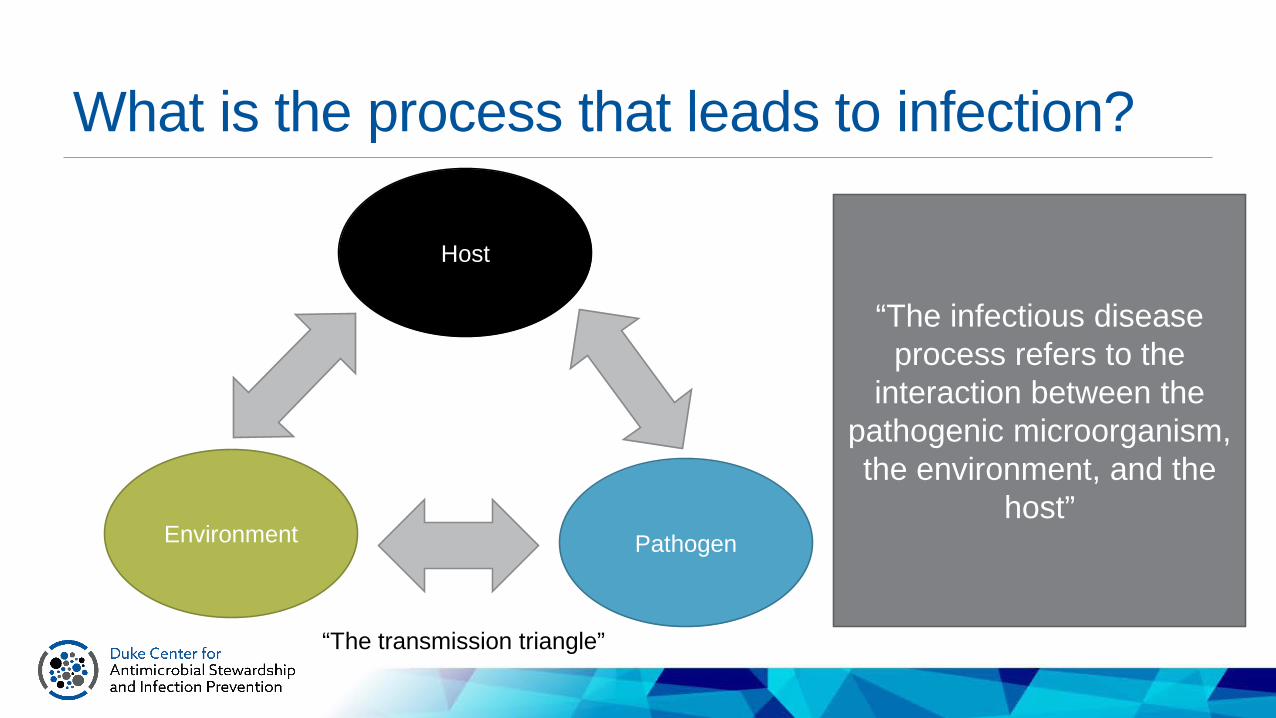
What is the process that leads to infection?
Host
PathogenEnvironment
“The infectious disease process refers to the
interaction between the pathogenic microorganism, the environment, and the
host”
“The transmission triangle”

What is the process that leads to infection?
Host
Pathogen
“The transmission triangle”
Key factors for infection to occur:• Host is susceptible to infection• Pathogen is virulent (can
invade tissues and evade immunity of the host)
• Exposure from the environment is significant enough to allow pathogen entry into the hostEnvironment

Environmental risk factorsPeople, places/physical structure/space, timeHealthcare environments (e.g. Hospitals, OR, Clinic, ED, SNF) or community settingsEach setting and practice/procedure has it’s own unique environmental risks to consider Examples: Likelihood of needle sticks? Likelihood of significant contamination events? Likelihood of an encounter with a returning traveler presenting with fever? What infections are circulating at this time of year?
Environment

Transmission: linear in time, multi-dimensional in space
E. coli lives happily in Patient #1’s gutPatient #1’s feces (E. coli) contaminate the bedNurse touches the bed, but doesn’t effectively clean handsPatient #2 is elderly with impaired immunity and poor functional statusNurse does peri care on Patient #2’s foleycatheterE. coli contaminates the foley catheterPatient #2 develops a CAUTI
TIM
E Patient #1
Patient #2
Nurse
BedHands
Hands Foley
Feces

Reservoirs and intermediariesThe Environmental component may include multiple intermediate steps for transmission to occur:ReservoirPortal of Exit (from the reservoir)Mode of Transmission E.g. surface/skin contact, airborne, fecal-oral,
large droplet, sex, vector-borne
Portal of Entry (to the host)
Environment
https://www.cdc.gov/malaria/about/biology/index.html

TransmissibilityInoculum: number of organisms needed to cause disease during an exposureAbility of the pathogen to survive in the environmentMethod of transmission (move from a reservoir to other hosts)Reproductive number (R0) measure of infectivity: average number of people that one sick person will infect
https://www.quora.com/Whats-the-most-vicious-human-virus-in-the-world
PathogenEnvironment

Practice QuestionWhich of the following statements about influenza is FALSE?A. Influenza is primarily spread between individuals via respiratory secretions (droplets)B. Viral shedding starts 48-72 hours after infection and typically 48 hours before the onset of symptomsC. Viral shedding normally persists for less than 5 days but can be longer in children and in immunocompromised personsD. The typical influenza symptomology is not always predictive of influenza in elderly or immunocompromised persons.

APIC text Figure 21-1. Components of the infectious disease process. Modified source: Centers for Disease Control and Prevention.Principles of epidemiology, 2nd ed. Atlanta: U.S. Department of Health and Human Services, 1992. Available at: http://www.cdc.gov/osels/scientific_edu/ss1978/lesson1/Section10.html.
i
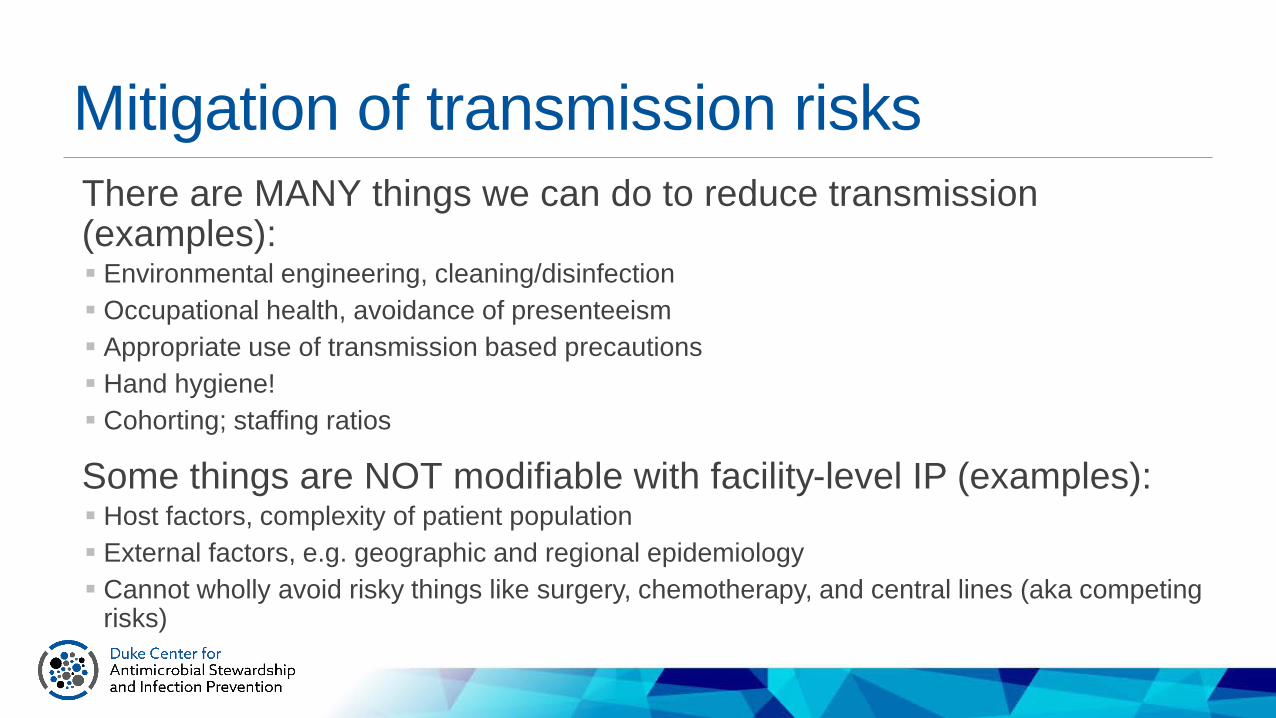
Mitigation of transmission risksThere are MANY things we can do to reduce transmission (examples): Environmental engineering, cleaning/disinfection Occupational health, avoidance of presenteeism Appropriate use of transmission based precautions Hand hygiene! Cohorting; staffing ratios
Some things are NOT modifiable with facility-level IP (examples): Host factors, complexity of patient population External factors, e.g. geographic and regional epidemiology Cannot wholly avoid risky things like surgery, chemotherapy, and central lines (aka competing
risks)

Practice QuestionAn IP is conducting an educational session to help the nursing staff understand infectious disease transmission. She explains that an initial element in transmission is the ability of an organism to survive in the external environment during transit between hosts. What is the second element?A. Secretion of enzymes that enhance spread through tissuesB. A mechanism for transmission to a new hostC. Invasion and dissemination in the hostD. Avoidance of host resistance

Host DefensesNon-specific defenses against invading pathogensPhysiologic barriers: Secretions, Fever, normal floraMechanical barriers: Mucosa or skin
Immune systemNon-specific “Innate” immunity: Phagocytic cells (neutrophils, monocytes),
hormones, fibronectinComplement system: protein pathways that poke holes and ramp up
inflammatory responsePathogen-specific “Adaptive” immunity: Cellular immunity (T cells), Humoral
immunity (B cells, antibodies)
Host
Practice QuestionThe IP is teaching nurses how to assess infection risks in patients. Depletion of what cell type provides the best indication of susceptibility to most bacterial infections?A. monocyteB. EosinophilC. neutrophilD. lymphocyte

ImmunityTerms and numbers to know:Normal WBC : 4,000 – 10,000 cells/mm3
Leukocytosis: WBC >10,000 cells/mm3Leukopenia: WBC <4,000 cells/mm3Neutropenia: PMN or band forms <500 cells/mm3 or absolute neutrophil count less than 1000 cells/mm3Absolute count = total WBC count x % of PMN leukocytesPolys: PMNs: mature or segmented neutrophilsBands: Immature or nonsegmented neutrophils Infection risk is high when absolute neutrophil count is <500 cells/mm

Immunity = DefenseActiveAcquired through prior exposure (+/- resolved infection) or vaccinationMemory of prior antigens and previously produced humoral reaction
(antibodies)
PassiveAntibodies acquired through other means than a patient’s own immune
system Maternal (first 6mo of life) IVIG (all types of immune globulin) Can be specific: e.g. rabies Ig, VZIg
Host

Immunoglobulins/AntibodiesRelevant Facts Where?
IgM Appears FIRST in adaptive response, goes away by ~6mo after exposure, pentamer
Circulating free in blood plasma; too big to go into tissues
IgG MAJOR antibody, 4 subclasses Circulating free in blood plasma; moves easily into tissues
IgA SECRETORY, histamine release, allergic reactions, dimer
Mucous membranes and secretions
IgD On lymphocytes, small amt circulating in plasmaIgE ALLERGY-inducing, “reagin”, increased
with parasitesMucous membranes, incr in seasonal allergies
Host

Practice QuestionThe first immoglobulin response after exposure to a communicable disease pathogen or vaccine is production of:A. Immunoglobulin G (IgG)B. Immunoglobulin M (IgM)C. Immunoglobulin A (IgA)D. Immunoglobulin C (IgC)

IgG = PRIOR exposureIgM = RECENT exposure


Practice questionHigher morbidity rates in chronic hepatitis B virus carriers are associated with a co-infection of which of the following:A. Hepatitis AB. Hepatitis DC. Hepatitis CD. Hepatitis E
Practice QuestionAll of the following are descriptions of patients with immunocompromised status EXCEPT:A. HIV with CD4 count <200B. Leukemia or lymphomaC. Neutropenia (absolute neutrophil count <500/mm3)D. 1 year post bone-marrow transplant
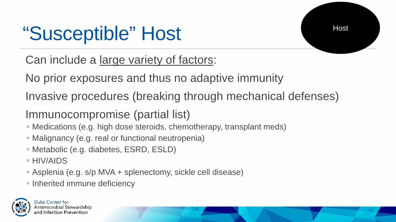
“Susceptible” Host Host
Can include a large variety of factors:No prior exposures and thus no adaptive immunityInvasive procedures (breaking through mechanical defenses) Immunocompromise (partial list) Medications (e.g. high dose steroids, chemotherapy, transplant meds) Malignancy (e.g. real or functional neutropenia) Metabolic (e.g. diabetes, ESRD, ESLD) HIV/AIDS Asplenia (e.g. s/p MVA + splenectomy, sickle cell disease) Inherited immune deficiency

Pathogen VirulenceFactors about the pathogen that can contribute to its ability to invade the host, evade host immunity, or survive:
Pathogen
Advantage Example virulence factor Pathogen/syndrome
Enzymes to increase local tissue damage/spread
Toxin production S. pyogenes and necrotizingfasciitis
Invade, disseminate Motility E. coli swimming up a ureter
Evade host defenses BiofilmsAttach or adhere to surfacesAlter cell wall or membraneCapsule prevents phagocytosis
Coag-neg Staph on IV lineS. aureus on a prosthetic kneeHIVS. pneumoniae
Survive in harsh conditions Spore-formationLipid coat
C. difficile, Bacillus spp.M. tuberculosis
Pathogen-specific features you must knowType of microorganism: bacteria, virus, fungus, parasiteClinical features of infectionLaboratory diagnosis (e.g. culture, serology, PCR)Precautions recommended in healthcare setting (e.g. contact, airborne, droplet, standard)Key transmission data: Mode of transmission Timing: incubation period and “shedding”/contagious period (key for droplet, airborne, and
some contact/viruses), typical duration of symptoms Vectors

List of pathogens/syndromes to knowBordetella pertussis
C. difficile and pseudomembranous colitisCreutzfeldt-Jakob Disease and other prions
Central Nervous System InfectionsEnterobacteriaceae
EnterococciEnvironmental Gram-negative bacilli
FungiDiarrheal diseases – Viral
Diarrheal diseases – BacterialDiarrheal diseases – Parasitic
Herpes VirusesInfluenza
Foodborne IllnessesLegionella pneumophila
HIV/AIDSLyme Disease (Borrelia burgdorferi)
Measles, Mumps, RubellaNeisseria meningitides
ParvovirusRabiesRSVSTDs
Skin and soft tissue infectionsStaphylococciStreptococci
Tuberculosis and other mycobacteriaViral Hemorrhagic Fevers
Viral HepatitisWest Nile Virus
Parasites

Quick and Dirty Micro ClassificationGram positive – skin, lung, guts, devicesGram negative – guts, urine, some lungAtypicals – lung, STIs, ticksAnaerobes –gas- and abscess-forming, bad odors, gutsLess commonly encountered: Mycobacterium (lung), spirochetes (Syphilis and Borrelia) Fungal – guts, devices, really bad in immunosuppressed hosts

Gram stainOften provide clues to etiology (may allow presumptive
diagnosis in some cases) Gram Positive Gram Negative Non-staining
Shape Coccus Rod Spiral Square
DIAGNOSIS: Stains and direct visualization
www.laboratoryinfo.com

Significance of the Gram StainBy knowing the shape and gram staining reaction of the organisms, along with the body site involved; clinicians can make a reasonable guess as to the causative agent.The reasonable guess can guide early empiric antibiotic choices.

Practice QuestionWhen reviewing the Gram stain of a person with a wound infection, the IP sees Gram-positive organisms in clusters. Which organism would this most likely represent?A. StreptococcusB. EnterococcusC. CorynebacteriumD. Staphylococcus

MicrobiologyPhysical requirements for growth of bacteriaNutrition (media)Temperature – 35o for most bacteriaAtmospheric conditionsAerobic (needs oxygen to survive)Anaerobic (needs absence of oxygen to survive)Facultative anaerobes (with or without oxygen)Microaerophilic

Microbiology: Key wordsGrowth mediaBlood agar = Multiple organismsChocolate = Haemophilus, NeisseriaCharcoal = Yeast, legionellaMacConkey = Gram NegativeBile esculin = group D StrepThayer-Martin = N. gonorrhoeaeLowenstein Jensen = Mycobacterium
Biochemical testsCatalase: Strep (-) Staph (+)Coagulase: Staph aureus (+)

Microbiology: Key wordsHemolysis on Blood AgarAlpha = greenBeta = clearGamma = no hemolysis
Lancefield grouping = Strep A to OBile esculin = black pigmentOptochin inhibition: S. pneumoniae (+)

Gram positive rods Bacillus sp. (aerobes) B. antracis Clostridium sp. (anaerobes) Listeria
GRAM POSITIVE ORGANISMSGram positive cocci Staphylococcus aureus Coagulase negative staphylococcus Streptococcus pneumoniae Streptococcus sp. Enterococcus sp.

Gram negative cocci Neisseria meningitidis Neisseria gonorrhoeae
Gram negative rods (non-enteric) Pseudomonas aeruginosa Stenotrophomonas maltophilia Acinetobacter sp.
Gram negative rods (Enterobacteriaceae) E. coli Klebsiella sp. Enterobacter sp. Proteus sp. Serratia sp.
GRAM NEGATIVE ORGANISMS

NON-STAINING/Special stain PATHOGENS Not stained by Gram’s method Legionella sp. Chlamydia Rickettsia Mycobacteria M. tuberculosis Non-tuberculous mycobacteria
Ziehl-Neelsen Stain of TB
Aka. Acid Fast

Organism DiagnosisFungalMorphologyPresence of hyphaeSize of yeastPresence of capsuleVirologyDirect- electron microscopyAntigen detectionVirus isolation from cultureAntibody detection/serology PCR testing
ParasitologyDirect examMicroscopy (oocytes)Antigen detection/serology

Choice of Empiric AntimicrobialsWhat class of pathogen am I likely to be treating? (Bacterial? Viral? Fungal? Other?)
If bacterial, what organisms are most likely? (Gram positive? Gram negative? Anaerobe?)
What information can I get to guide treatment? Microbiology data?
Do I need to order any other diagnostic tests?How sick is my patient? How risky would it be if I miss?

General Indications for AntibioticsProphylaxis: prevent infection EASY! Guidelines and ordersets E.g. surgical prophylaxis, pre-transplant protocol
Empiric: when you suspect infection but don’t exactly know with what Not easy. Local guidelines help (based on local micro data). Clinical syndrome guides choice
Directed: pathogen known Only available for a small portion of folks treated for infection Moderately easy. Follow and interpret patient-specific micro data.
EmpiricBroad-spectrum
TargetedNarrow-spectrum
Tim
e

De-escalationDe-escalation is a core principle of Antimicrobial Stewardship.Target/narrow antibiotic therapies after more clinical data returnsStop therapy when infection has been ruled out
EmpiricBroad-spectrum
TargetedNarrow-spectrum
Tim
e

DIAGNOSIS: Culture“Gold standard” to identify the pathogen Requires sampling of site of infection, best if collected *prior to* therapyAllows determination of antimicrobial susceptibility Can be “banked” for future tests if needed, e.g. outbreak investigations, strain typing, PFGE

DIAGNOSIS: Culture
There are some key limitations to traditional culture methods:Time and resource intensiveTypically have no info other than the stain for 2-3
daysHighly reliant on specimen collection techniquesSometimes positive in absence of infectionSometimes negative when infection is present (e.g. in the setting of antibiotics) or get contaminated/mixed flora result

Practice QuestionGuidelines for transporting specimens include:1) Transport within 2 hours of collecting a specimen2) Transport in leakproof specimen containers and sealable leakproof bags3) Transport specimen in the syringe used to collect it4) Refrigerate all specimens prior to transport
A. 1,4B. 2,3C. 1,2D. 3,4

Culture ContaminationInadequate specimen collection technique can lead to confusing results.Best example: Contaminated blood cultures with skin flora –infection or not? Solutions: Collect blood cultures in pairs. Avoid drawing from existing lines. Hire/educate
phlebotomists.
Other examples: Patients had clinical symptoms, are sick, but culture comes back “mixed flora” and pathogen remains unknown. Patient is treated with broad spectrum therapy. Example: urine cultures from existing foley catheter (doh!) Example: lower respiratory cultures

Specimen Collection: Key pointsDo not contaminate sterile specimens: Aseptic technique + sterile specimen carriers; Appropriate skin prep; Get more than 1Tissue > Fluid >>>>>>>> Swab (avoid!)More volume is better (blood, fluids)Send tissue from the OR to BOTH path and microLabel appropriately and include key clinical clues for the lab, esp for pathogens that are more difficult to cultureDon’t send cultures from drains/foleys that are already in placeDon’t let specimens sit around (to lab within 2h preferred)

Practice QuestionA patient has a nasal swab positive for methicillin-resistant Staphylococcus aureus (MRSA) in the absence of symptoms. This is an example of:A. Normal floraB. ColonizationC. Asymptomatic infectionD. Symptomatic infection

Infection vs. ColonizationHuman beings are not sterile.Clinicians have trouble NOT treating when they see a positive culture.Clinical presentation is very important. Clinical criteria for diagnosis of infection should lead to diagnostic testing (not the other way around).

Infection vs. ColonizationDiagnostic tests used indiscriminately lead to overdiagnosis, overtreatment, and associated negative consequences. Exhibit A: Asymptomatic bacteriuria Exhibit B: Patient colonized with C. difficile had only 1 loose BM after kayexalate = +PCR and
classified as HO-CDI LabID event
Questions to ask before sending diagnostic test: “What is the pre-test probability that this patient has infection?” “What would I do differently if the test comes back positive? Negative?”
?

Establish Criteria for Testing Urine
Diagnoses Urine Culture Clinical Symptoms
Acute, uncomplicatedurinary tract infection
>100,000 bacteria,No more than 2 species of bacteria
• DysuriaOR• Fever AND 1 of the following:-Frequency-Urgency-Suprapubic pain-Incontinence*-Gross Hematuria**
AsymptomaticBacteriuria
>100,000 bacteria,No more than 2 species of bacteria
• No signs or symptomsreferable to the urinary tract
CDC/James GathanyPublic Health Image Library
Stone et al. Infec Control Hosp Epi 2012; *New or worsening of baseline incontinence**I have never known hematuria to a sign of infection in an older adult. Rather, it seems to indicate trauma to the mucosa, which can lead to urinary tract infection or urosepsis.

Diagnoses Urine Culture Clinical Symptoms
Acute, uncomplicatedurinary tract infection
>100,000 bacteria,No more than 2 species of bacteria
AsymptomaticBacteriuria
>100,000 bacteria,No more than 2 species of bacteria
CDC/James GathanyPublic Health Image Library
Stone et al. Infec Control Hosp Epi 2012; *New or worsening of baseline incontinence**I have never known hematuria to a sign of infection in an older adult. Rather, it seems to indicate trauma to the mucosa, which can lead to urinary tract infection or urosepsis.
Establish Criteria for Testing Urine
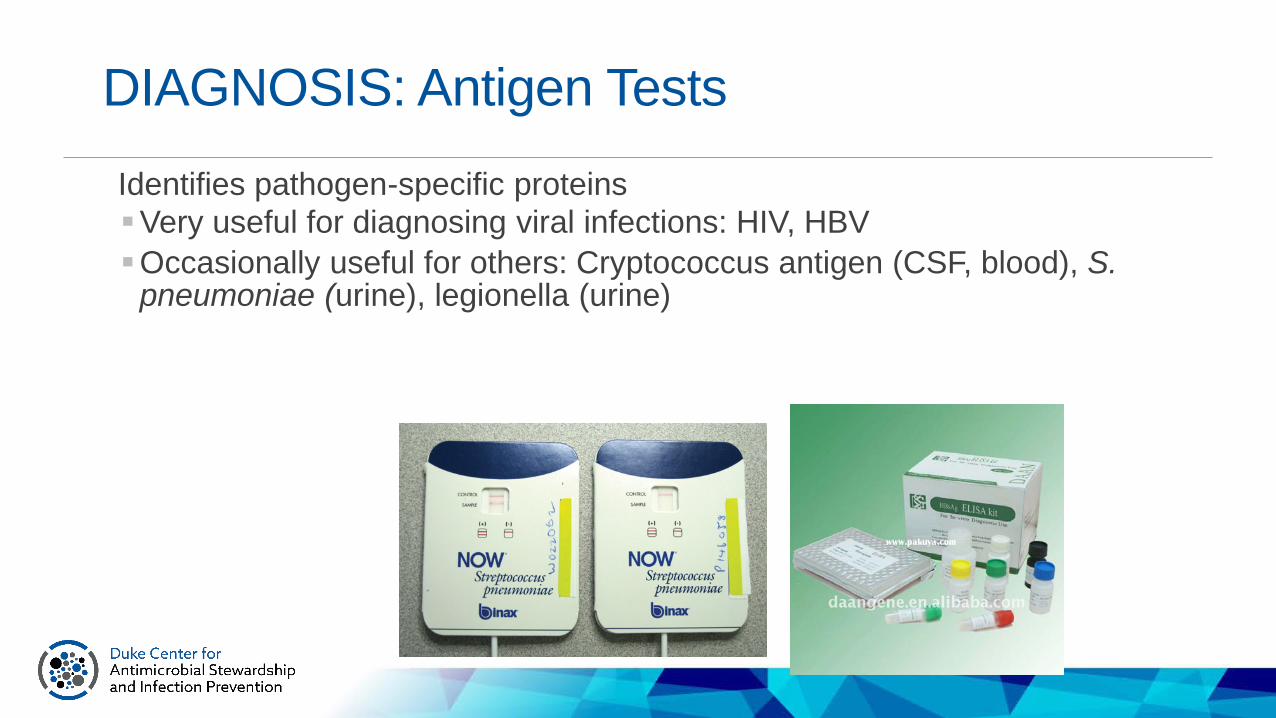
DIAGNOSIS: Antigen Tests
Identifies pathogen-specific proteinsVery useful for diagnosing viral infections: HIV, HBVOccasionally useful for others: Cryptococcus antigen (CSF, blood), S.
pneumoniae (urine), legionella (urine)

DIAGNOSIS: Serologic testing
Detects immune response to a pathogen, or prior exposure to a pathogenFor bacterial infections, generally not useful in
early diagnosis (may require acute and convalescent tests)For viral infections, IgM indicates early diagnosis
or recent exposure (e.g., Hep A) Important for screening for prior exposure,
documenting immunity, and ensuring vaccination e.g. Occupational health titers for varicella, HBVOnce serology is positive, it is typically life-long
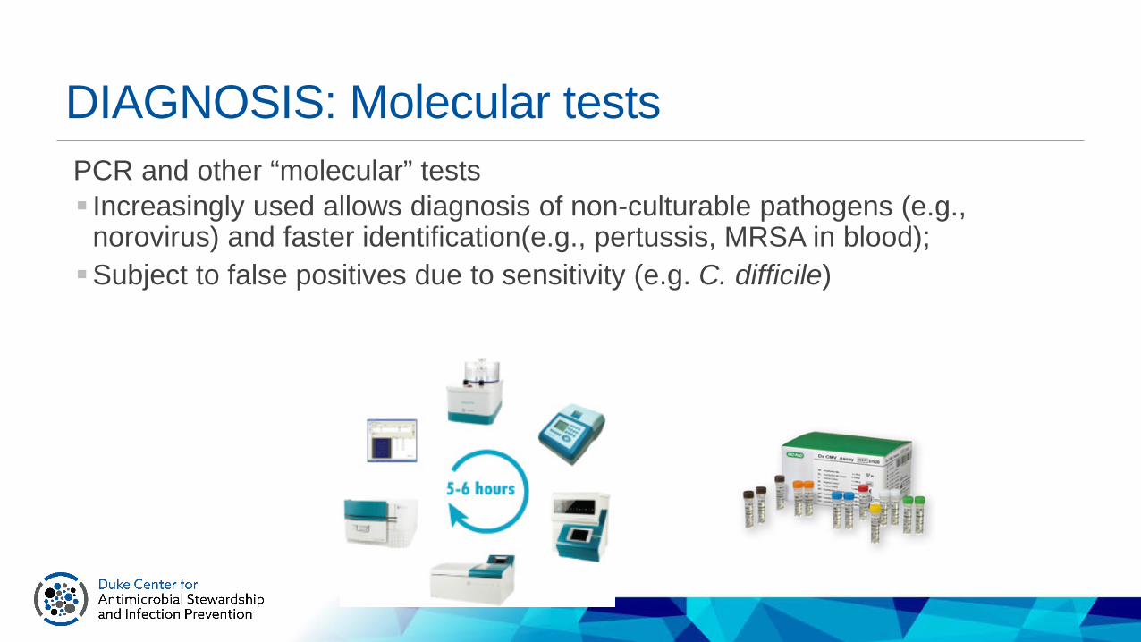
DIAGNOSIS: Molecular testsPCR and other “molecular” tests Increasingly used allows diagnosis of non-culturable pathogens (e.g.,
norovirus) and faster identification(e.g., pertussis, MRSA in blood); Subject to false positives due to sensitivity (e.g. C. difficile)

DIAGNOSIS: Sterile fluid studiesEvidence of infection due to inflammatory, chemical, and cellular changes in body fluidsExamples: synovial fluid, CSF, pleural, and peritoneal fluidTypically combined with GS/culture (which takes a while)

Practice QuestionAn IP is reviewing the cerebrospinal fluid (CSF) result from a patient admitted the previous night. The CSF is cloudy and has an elevated White Blood Cell count (WBC), markedly elevated neutrophils, low glucose, and elevated protein. What type of meningitis should she suspect?A. BacterialB. ViralC. FungalD. Aseptic

CSF studiesOpeningPressure
Glucose (Ratio of CSF to Serum)
WBC count
WBC type Total protein
Stain
Bacteria, “Septic”
Elevated Normal to decreased
≥1,000/mm3
Neutrophils (early or partially treated may have lymphocyte predominance)
Elevated (mild to very)
Gram* stain may show GPC or GNC/GNR
Virus, “Aseptic”
Usually normal
Usually normal <100 per mm3
Lymphocytes Normal to elevated
Gram stain, negative
Fungi Variable Low Variable Lymphocytes Elevated India ink(Crypto), positive
TB Variable Low (can be extremely low)
Variable Lymphocytes Elevated AFB stain, positive
Table 74-1 APIC text

Antibiotic susceptibility testing: Key TermsAntibiotic = A drug that kills or inhibits the growth of microorganisms
Resistant = An antimicrobial will NOT inhibit bacterial growth at clinically achievable concentrations
Susceptible = An antimicrobial WILL inhibit bacterial growth at clinically achievable concentrations
MIC = Minimal inhibitory concentration. Lowest concentration of antimicrobial that inhibits growth of bacteria. Commonly used in clinical lab
MBC = Minimal bactericidal concentration. Concentration of an antimicrobial that kills bacteria. Used clinically only in special circumstances
Breakpoint = The MIC that is used to designate between susceptible and resistant. Set by an expert committee (CLSI).
Key Terms

4.0µg/mL
4.0µg/mL
Methods for Testing Susceptibility: Minimal Inhibitory Concentration
0.25µg/mL
0.5µg/mL
1.0µg/mL
2.0µg/mL
8.0µg/mL
16µg/mL
Known quantity of bacteria placed into each tube
Increasing antibiotic concentration
Lowest concentration of an antimicrobial that results in the inhibition of visible
growth of a microorganism
Sinus and Allergy Health Partnership. Otolaryngol Head Neck Surg. 2000;123(1 Pt 2):S1.

Methods for Testing SusceptibilityBroth dilution = MIC testing (Automated system) = a number
Disc Diffusion = Kirby Bauer (Manual) = a zone size
E test = “Strip” (Manual) = a number

Resistance MechanismsIntrinsic- inherited by the organism species E.g. Serratia is intrinsically resistant to cefazolin
Acquired- results from altered cellular structure and physiology caused by changes in the genetic make-up Efflux pump Point mutation on a penicillin binding protein Beta lactamase production: ESBL K. pneumoniae acquired resistance from a plasmid from E. coli Carbapenemase production

Mechanisms of Action of Antibiotics
TMP-SMX = trimethoprim-sulfamethoxazole.
DNA replication
Nucleotidebiosynthesis Protein
synthesis
Protein
–
Topo-isomerase
mRNA
RNAtranscription
mRNA
–
–
–
–
β-lactamsCephalosporinsCarbapenems
Cell wallsynthesis
–Fluoroquinolones
RifampinCytoplasmicmembrane
integrity
Peptideantibiotics
–
SulfonamidesTMP-SMX
Metronidazole
GlycylcyclinesAminoglycosidesMacrolidesOxazolidinonesStreptograminsLincosamidesTetracyclines
Glycylcyclines
10Adapted from: Chopra I. Curr Opin Pharmacol. 2001;1:464-469.

Practice Question:The ED reports 3 cases of cramping, abdominal pain, and diarrhea within a 24-hour period. All persons are from the same community, and onset of symptoms was within 12 to 36 hours of a picnic they all attended. The IP suspects which of the following foodborne illnesses:A. SalmonellaB. Hepatitis AC. Staphylococcus aureusD. Clostridium perfringens

Foodborne DiseasesFoodborne diseases
Etiology and Incubation periodStaph aureus 30 min to 8 hrs, ave. 2-4 hrs.Bacillus cereus 1-6 hrs vomiting, 6-24h diarrheaSalmonella 6-72 hrs, avg. 12-36 hrsShigella 12-96 hrs, avg. 1-3 daysS. dysenteriae up to 1 weekCampylobacter 1-10 days, avg. 2-5 daysClostridium perfringens 6-24 hrs, avg. 10-12 hrs
Ingestion of toxin

Practice Question A patient is admitted with pruritic lesions on the hands, webs of fingers, wrists, the extensor surfaces of the elbows and knees, and the outer surfaces of the feet, armpits, buttocks, and waist. The most likely diagnosis is:A. Scarlet feverB. Herpes zosterC. ScabiesD. Measles

Practice QuestionA patient who was hospitalized for 2 days calls 3 days after discharge complaining that he has developed healthcare-associated scabies due to his recent inpatient stay. The IP knows that his scabies infestation is not healthcare-associated because:A. Scabies is only transmitted through contaminated linens, and the IP confirmed that all linens the patient came into contact with ahd been properly launderedB. the incubation period for scabies is longer than 5 daysC. the incubation period for scabies is shorter than 3 daysD. Scabies is only transmitted through direct contact and none of the healthcare personnel who cared for the patient are infested

Quick and dirty on parasites “any organism living within or on another living creature and
deriving advantage from doing so while causing disadvantage to the host”
Protozoa
Water or food borneVector borne
AnimalSexually transmitted
Multicellular
LiceScabies
Myiasis (maggots)Bed bugs
WormsRound (Intestine)Round (Tissue)
TapeFluke

Practice QuestionThe causative organism of Creutzfeldt-Jakob disease is a:A. helminthB. diphtheroidC. spirocheteD. prion

Prion Diseases“Transmissible neurodegenerative diseases” or “Transmissible spongiform encephalopathies (TSE)” Infectious protein replicates in the CNS and interrupts neuron functioning Rapidly progressive, ultimately fatal Spongiform appearance of neurologic tissues on path, no inflammatory
response
Ways to acquire TSEs: Sporadic disease; Iatrogenic; Familial transmission (gene mutation); Ingestion of prions (e.g. bovine encephalopathies)Transmission key features: Long incubation period Highly resistant to routine methods of disinfection/sterilization: requires
prolonged cycle/increased temps and/or chemical disinfection for equipment exposed to infectious tissues Early recognition is KEY for the healthcare setting
Humans:Creutzfeldt-Jakob disease (CJD)
KuruGerstmann-Straumlussler-Scheinker
(GSS)Fatal familial insomnia
In animals:Bovine spongiform encephalopathy
(BSE, cattle)Scrapie (sheep)
Chronic wasting disease (deer, elk)
Practice QuestionWhich is TRUE about a tuberculin skin test (TST):A. Positive TST indicates active tuberculosis (TB) infectionB. Negative TST rules out active TB infectionC. Positive TST indications past exposure to TBD. Negative TST indicates past exposure to TB

Practice QuestionThe optimal time to collect a sputum specimen for acid-fast bacilli (AFB) testing to rule out TB would be:A. First thing in the morningB. After a respiratory treatmentC. Prior to the patient going to bedD. Prior to a respiratory treatment

Mycobacterium tuberculosisAcid-fast bacilliClinical features of pulmonary disease: subacute onset, cough/congestion (sputum may be bloody), weakness, fatigue, weight loss, chills, fever, night sweatsDiagnostic testing: acid-fast smear/culture of sputum, PCR DNA probe, chest x-rayTST/PPD or IGRA is a screening test for latent disease or prior exposure
https://radiopaedia.org/cases/pulmonary-tuberculosis-29
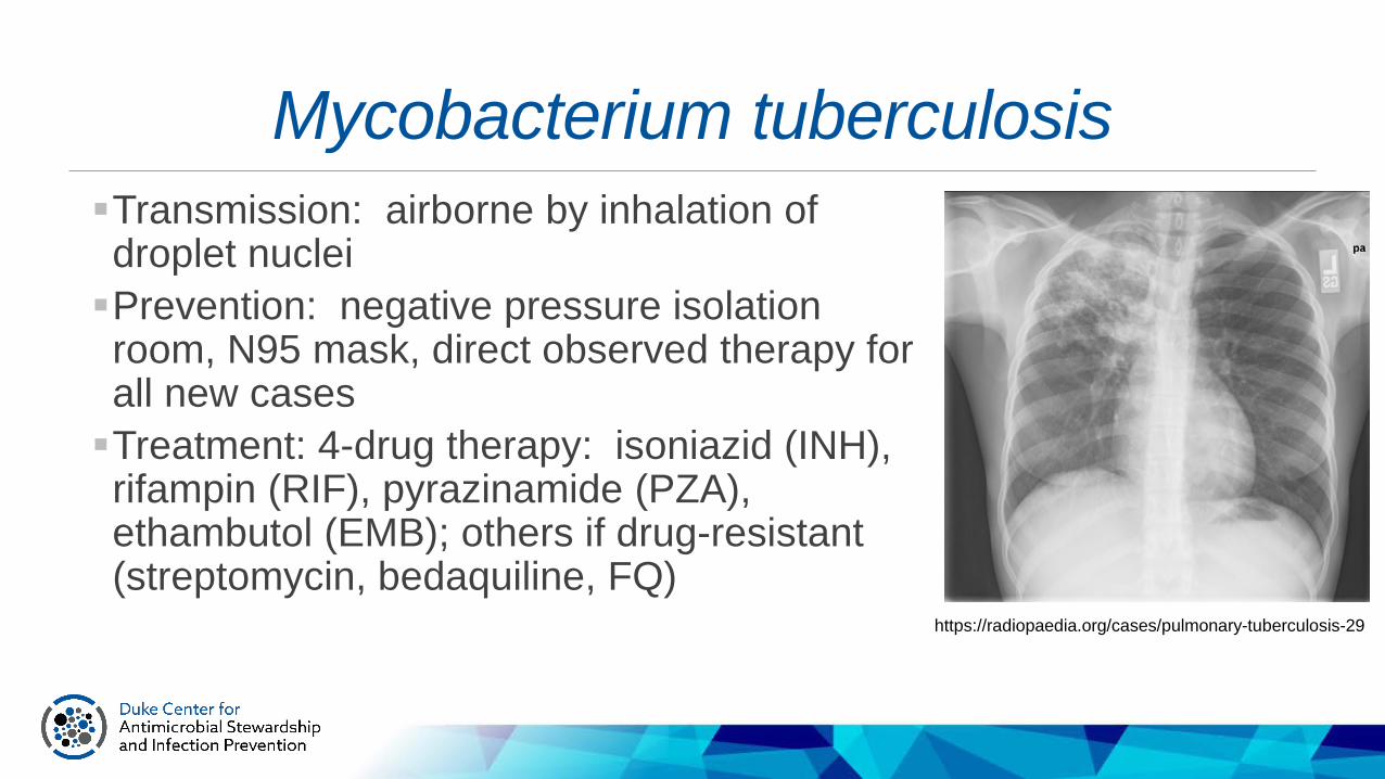
Mycobacterium tuberculosisTransmission: airborne by inhalation of droplet nucleiPrevention: negative pressure isolation room, N95 mask, direct observed therapy for all new casesTreatment: 4-drug therapy: isoniazid (INH), rifampin (RIF), pyrazinamide (PZA), ethambutol (EMB); others if drug-resistant (streptomycin, bedaquiline, FQ)
https://radiopaedia.org/cases/pulmonary-tuberculosis-29
Practice QuestionA 14yo boy from rural Maryland was seen in the emergency department with fever, fatigue, chills, headache, and a large annular lesion on his left thigh. What is the most probable vector of this child’s illness?A. tickB. mosquitoC. fleaD. louse

Spirochetes: spiral shaped bacteriaLyme (Borrelia burgdorfori): tick borne illness in the NE US (but expanding) Target lesions (erythema migrans), fever, headache, arthralgias; can also cause CNS disease
and associated with Bell’s palsy; cardiomyopathy Confusing diagnostics (serology and protein detection) Tick = Ixodes scapularis “black legged” Treatment = doxycycline, ceftriaxone for CNS

Spirochetes: spiral shaped bacteriaSyphilis (Treponema pallidum): “the great imitator” – sexually transmittedMultiple stages of clinical disease: primary, secondary,
early latent, late latent, tabes dorsalis, tertiary, ocular, congenitalDarkfield microscopy is classical diagnostic, but never
used nowSerology IgG plus confirmatory testing for screeningTrending of VDRL or RPR titers for response to
therapy and detection of new exposures Treatment = penicillin www.giantmicrobe.com

Good Luck!
Host
PathogenEnvironment