Embed Size (px)
Citation preview

ICTUS CEREBELOSOSICTUS CEREBELOSOS
Carlos S. Kase, M.D.Carlos S. Kase, M.D.Department of NeurologyDepartment of Neurology
Boston UniversityBoston UniversityBoston, MABoston, MA
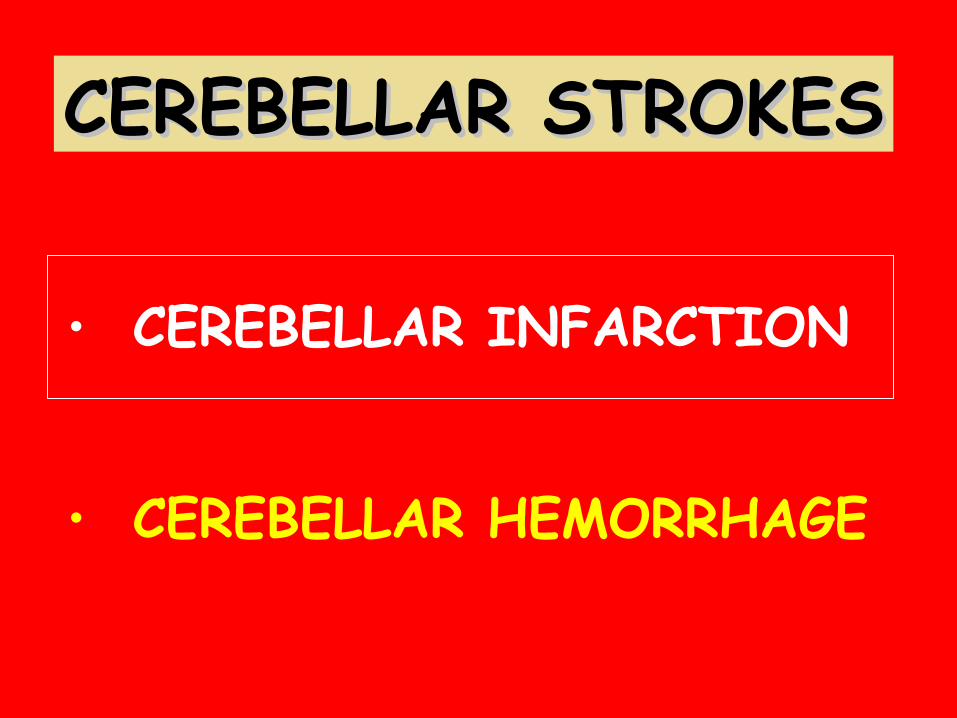
CEREBELLAR STROKESCEREBELLAR STROKES
• CEREBELLAR INFARCTION
• CEREBELLAR HEMORRHAGE

Distribution of Pathologically Distribution of Pathologically Confirmed Cerebellar InfarctionConfirmed Cerebellar Infarction
Vascular territory involved #Posterior inferior cerebellar artery(PICA) 7Superior cerebellar artery 7Multiple random sites 3White matter lacune 1Bilateral watershed (PICA, superior
cerebellar territory) 1
RAL Macdonell et al., Stroke 1987;18:849-855

A.T. – MR # 2449318
PICA-DISTRIBUTION INFARCTPICA-DISTRIBUTION INFARCT

L-N.H. – MR # 2796040
SCA-DISTRIBUTION INFARCTSCA-DISTRIBUTION INFARCT

CEREBELLAR INFARCTIONCEREBELLAR INFARCTIONClinical ImportanceClinical Importance
• Differences in topography, clinical features, vascular pathology, treatment
• Potential for edema, mass effect, brainstem compression
• Associated brainstem infarction

ACUTE CEREBELLAR INFARCTION(Autopsy series, 28 cases)
• Gait-trunk ataxia……………….………71%• Limb dysmetria…………………….…….57%• Facial palsy…………………………….……36%• Facial hypesthesia……………….…….29%• Nystagmus……………………………….….62%• Dysarthria………………………….……….67%• Gaze palsy……………………………….…..32%• 6th nerve palsy……………………….…...12%• Mental changes……………………..……65%

Stroke 1993;24:76-83
• 36 patients with PICA-distribution infarcts
• 30 patients with SCA-distribution infarcts
PURPOSE: To compare clinical features,imaging characteristics, andclinical course in the main two types of cerebellar infarction

CEREBELLAR INFARCTIONCEREBELLAR INFARCTIONSymptoms -Symptoms - PICA v. SCAPICA v. SCA
01020304050607080
PICA SCA
HeadacheVertigoVomitingGait imbal.

CEREBELLAR INFARCTIONCEREBELLAR INFARCTIONSigns -Signs - PICA v. SCAPICA v. SCA
01020304050607080
PICA SCA
Limb ataxiaNystagmusGait ataxia

ACUTE CEREBELLAR INFARCTION ACUTE CEREBELLAR INFARCTION IN THE PICA TERRITORYIN THE PICA TERRITORY
Three pts. with acute onset of: Vertigo ↑‘d by movt. Nausea, vomiting Imbalance Nystagmus No cerebellar ataxia on exam
Initial diagnosis: “Labyrinthitis”
Course: Progressive swelling c/brainstem compression
Duncan GW et al., Arch Neurol 1975;32:364-368

Vestibular syndromeVestibular syndromePICA infarction PICA infarction
75 y/o MD, no significant PMH, while rounding developed severe
vertigo, n/v, gait instability
PE: uncomfortable b/o vertigo; every movement caused severe vertigo, n/v; right-beating, non-
direction-changing nystagmus only abn. in CN examination; no limb
ataxia present

DWI MRI – May 9, 2012
CT – May 13, 2012 CT – May 21, 2012

ISOLATED ACUTE VERTIGO IN THE ISOLATED ACUTE VERTIGO IN THE ELDERLYELDERLY
Norrving et al., Acta Neurol Scand 91:43, 1995Norrving et al., Acta Neurol Scand 91:43, 1995
Prospective study, 24 pts., age 50-75Isolated vertigo/imbalance
Infarct caudal cerebellum in 6 (25%)
Cardioembolic source in 3/6
Normal MRI, but occluded VA by Doppler in 2/18
Infarct caudal cerebellum in 6 (25%)


CEREBELLAR INFARCTIONCEREBELLAR INFARCTIONBrain stem signsBrain stem signs
PICA INFARCTS 12/36 patients (33%)Lateral medullary syndrome
Complete 6 patients Partial 6 patients
SCA INFARCTS 9/30 Patients (30%) Dysarthria 9 patients Horner’s 6 patients Diplopia, vert. gaze
palsy, 6th n. palsy, INO, dyskinesia 1 patient each

CEREBELLAR INFARCTIONCEREBELLAR INFARCTIONCommon presenting signsCommon presenting signs
Vertigo
Dysarthria=
PICA
SCA

CEREBELLAR INFARCTIONCEREBELLAR INFARCTIONMechanisms of infarctionMechanisms of infarction
PICA TERRITORYPICA TERRITORY (N=28)(N=28)
• Cardiac embolism 14 (50%)• VA occlusion 7• VA stenosis 3• PICA occlusion 3• ? Vasculitis 1

CEREBELLAR INFARCTIONCEREBELLAR INFARCTIONMechanisms of infarctionMechanisms of infarction
SCA TERRITORY (N=18)SCA TERRITORY (N=18)
• Cardiac embolism 11 (61%)• Artery-to-artery emb. 3 (17%)• Distal BA stenosis 2• SCA stenosis/occlusion 2

CEREBELLAR INFARCTIONCEREBELLAR INFARCTIONCT FindingsCT Findings
PICA INFARCTS PICA INFARCTS (N = 36)(N = 36)
• Partial territory 27 (75%)
• Full PICA territory 9 (25%)Displacement/obliteration of
4th ventricle 8Obstructive hydrocephalus 7

PICA-DISTRIBUTION CEREBELLARPICA-DISTRIBUTION CEREBELLAR INFARCTSINFARCTS
TOTAL
PARTIAL

PICA–DISTRIBUTION INFARCTS PICA–DISTRIBUTION INFARCTS Clinical CourseClinical Course
In 7/36 (19%) In 7/36 (19%) cc//full territoryfull territory infarct: infarct: ME, brainstem compr., hydrocephalusME, brainstem compr., hydrocephalus
• 3 pts. died without undergoing surgery• 4 pts. operated on
- 1 ventriculostomy- 3 post. fossa decompression

CEREBELLAR INFARCTIONCEREBELLAR INFARCTIONCT findingsCT findings
SCA INFARCTSSCA INFARCTS (N=30)(N=30)
Partial territory (vermian or hemisphere branches) 30
Displacement/obliteration of 4th ventricle 2
Obstructive hydrocephalus 2

SUPERIOR CEREBELLAR ARTERY-DISTRIBUTION INFARCTSSUPERIOR CEREBELLAR ARTERY-DISTRIBUTION INFARCTS
TOTAL
PARTIAL

SCA–DISTRIBUTION INFARCTS SCA–DISTRIBUTION INFARCTS Clinical CourseClinical Course
In 2/30 (7%) c/deep WM infarct: ME, brainstem compr., hydrocephalus
• Both patients had posterior fossa decompression

Pt. SM – MR # 928526Pt. SM – MR # 928526
• 55 y/o ♂, hypertensive• Abrupt onset vertigo,
gait imbalance 9/27/90• PE: A, Ox3; nl. speech;
nl. strength & sensation; mod. ataxia Ⓛ UL > LL; full EOMs, c/horizontalnystag. Ⓛ > Ⓡ gaze; normal pupillary size & reactivity

Pt. SM – MR # 928526Pt. SM – MR # 928526
• 9/28/90, 6 PM:Somnolent, disoriented,dysarthric
• PE: nl. strength & sensation; mod. ataxia Ⓛ UL > LL; full EOMs, c/horizontal nystag. Ⓛ > Ⓡ gaze; normal pupillary size & reactivity; Ⓛ facial palsy

Pt. SM – MR # 928526Pt. SM – MR # 928526
• 9/28/90, 10 PM:VentriculostomyHyperventilation
• 9/29/90: Awake, following commands; full horiz. gaze; toes down
• 9/30/90:Lethargic; full horiz. gaze; paralysis upward gaze; pupils 3 mm, barely reactive; bilat.Babinski
SUB-OCCIPITAL DECOMPRESSIVE CRANIECTOMY

Pt. SM – MR # 928526Pt. SM – MR # 928526
• 10/1/90:More alert, following commands; improved pupillary reactivity; paralysis upward gaze and Babinski signs still present
• 10/2/90: Awake, following commands; improved upward gaze; toes down; normal pupillary reactivity
• 10/3/90:Awake and alert; full upward gaze; toes down; normal pupillary reactivity

Pt. SM – MR # 928526Pt. SM – MR # 928526LessonsLessons
• Large PICA infarct c/progressive neurological decline
• Temporary effect of ventriculostomy
• Potential for upward transtentorial herniation after ventriculostomy
• Value of wide posterior fossa decompressive craniectomy

CEREBELLAR INFARCTIONCEREBELLAR INFARCTIONSwelling and HydrocephalusSwelling and Hydrocephalus
AUTHORS # cases # (%) cases
infarct c/swelling
Scotti et al. 21 6 (29%)
Shenkin, Zavala 55 6 (11%)
Macdonell et al. 30 4 (13%)
Kase et al. 36 PICA 7 (19%)
30 SCA 2 ( 7%)

CEREBELLAR INFARCTIONCEREBELLAR INFARCTIONImaging Features Predictive of Mass Effect Imaging Features Predictive of Mass Effect
and Neurological Deteriorationand Neurological Deterioration
• Fourth ventricle deformity• Fourth ventricle shift• Hydrocephalus• Brain stem deformity• Compression of basal cisterns
MG Koh et al., Stroke 2000;31:2062-2067

CEREBELLAR INFARCTIONCEREBELLAR INFARCTIONSuggested managementSuggested management
• SCA cases more benign than PICA• In PICA cases, close clinical monitoring,
early MRI• If swelling c/brainstem compression,
hydrocephalus → ventriculostomy• If further progession, posterior fossa
decompression


Mean time to neurologicaldecompensation: 50 hrs.


ACUTE ONSET VERTIGO/IMBALANCEACUTE ONSET VERTIGO/IMBALANCEDIFFERENTIAL DIAGNOSISDIFFERENTIAL DIAGNOSIS
Acute unilateral vestibulopathy- Menière’s disease- “Labyrinthitis”- “Vestibular neuronitis”- “Peripheral vestibulopathy”
Posterior fossa vascular disorder- Brainstem infarct/hemorrhage- Cerebellar infarct/hemorrhage

TYPES OF CEREBELLAR HERNIATIONTYPES OF CEREBELLAR HERNIATION
Direction of tissue Direction of tissue displacementdisplacement
Upward Downward
Portion of cerebellum Portion of cerebellum displaceddisplaced
Superior aspect of hemisphere
Tonsils
Site of herniationSite of herniation Free edge of the tentorial incisura
Foramen magnum
Portion of brainstem Portion of brainstem compressedcompressed
Midbrain Medulla
Clinical manifestationsClinical manifestationsLethargy, coma Neck stiffness
AscendingAscendingTranstentorialTranstentorial
DescendingDescendingTonsilarTonsilar

SUPERIOR CEREBELLAR ARTERYPontine territory
• Superior cerebellar peduncle
• Lateral lemniscus
• Spinothalamic tract
• Descending sympathetic tract
• Mesencephalic trigeminal tract
• Locus coeruleus
• Root of contralat. 4th n. (pontine tectum)

CEREBELLAR INFARCTION(Autopsy series, 28 cases)
• Infarct in P-1 aspect of cerebellum (93%)
Vascular occlusion: VA (18 cases) PICA (10 cases)
Mechanism of occlusion: Embolic (25%) Thrombotic (75%)
Assoc. with Wallenberg’s: 18%Death 6-30 hrs. from onset M.S.

CT Findings in Cases of Cerebellar Softening
Location of softening- Left hemisphere 4 9- Right hemisphere 2 6- Deep …a 7- Superficial …a 8
Triventricular hydrocephalus 6 04th ventricle compression 6 0Cisterns obliterated 6 0Contrast enhancement 0 ¼
FINDINGS
Number of CasesGroup 1 Group 2 N=6 N=15

SCA – DISTRIBUTION INFARCT“Classic” Syndrome
Ipsilateral- Limb ataxia- Horner’s syndrome- Choreic dyskinesia
Contralateral- Thermoanalgesia- Fourth n. palsy

CEREBELLAR STROKESCEREBELLAR STROKES
• CEREBELLAR INFARCTION
• CEREBELLAR HEMORRHAGE


CEREBELLAR HEMORRHAGECEREBELLAR HEMORRHAGEClinical PresentationClinical Presentation
• Inability to stand/walk: > 90%
• Vertigo: 50-60%
• Headache: 60-75%
• Vomiting: 70-95%

0
20
40
60
80
100
Inability to Vertigo Headache Vomiting stand/walk
CEREBELLAR HEMORRHAGECEREBELLAR HEMORRHAGEClinical PresentationClinical Presentation

CEREBELLAR HEMORRHAGECEREBELLAR HEMORRHAGECT Features Suggesting Surgical RxCT Features Suggesting Surgical Rx
• Hematoma > 3 cm
• Hydrocephalus


CEREBELLAR HEMORRHAGECEREBELLAR HEMORRHAGECT Aspects and OutcomeCT Aspects and OutcomeLittle et al., J Neurosurg 1978;48:575Little et al., J Neurosurg 1978;48:575
Size
Case # Location >3 cm <3 cm Hydrocephalus Outcome
1 Medial x Moderate
2 Vermis x Marked
3 Medial x Marked
4 Medial x Moderate
5 Vermis x Marked
6 Medial x Moderate
7 Vermis x None
8 Lateral x None
9 Medial x None
10 Medial x None
5 SURGERY2 DEATHS
RECOVERED

CEREBELLAR HEMORRHAGECEREBELLAR HEMORRHAGECT Features Suggesting Surgical RxCT Features Suggesting Surgical Rx
• Hematoma > 3 cm
• Hydrocephalus
• Obliteration quadrigeminal cistern
• Deformation/compression of 4th ventr.?

Neurosurgery 2001;49:1378-1387
50 pts. with cerebellar hemorrhage
Purpose: Assess influence of degree of 4th ventricular mass effect
in clinical and CT features, as well as treatment results
4th ventricular mass effect:
Grade I: no mass effect
Grade II: partial compression, shifted
Grade III: complete obliteration

CEREBELLAR HEMORRHAGECEREBELLAR HEMORRHAGEDegree of 4Degree of 4thth ventricular compression ventricular compression
Neurosurgery 2001;49:1378-1387 ## SizeSize Hydroc. % comaHydroc. % coma % coma% coma
(cm)(cm) (%) (%) onset onset final final
Grade IGrade I 6 3 50 33 33
Grade IIGrade II 26 4.2 61 4 27
Grade IIIGrade III 18 5 100 22 67

CEREBELLAR HEMORRHAGECEREBELLAR HEMORRHAGEDegree of 4Degree of 4thth ventricular compression ventricular compression
Neurosurgery 2001;49:1378-1387 ## Evac.Evac. CSF CSF Conserv.Conserv. % good% good
drain.drain. outcomeoutcome
Grade IGrade I 6 0 2 4 100
Grade IIGrade II 19 4 6 9 58GCS > 8GCS > 8
Grade IIGrade II 7 3 4 0 57GCS < 8GCS < 8
Grade IIIGrade III 8 8 0 0 38GCS > 8GCS > 8
Grade IIIGrade III 10 10 0 0 0GCS < 8GCS < 8


42 pts. with cerebellar hemorrhage,alert on presentation – Deteriorationinto coma after admission
Ott KH, et al., Arch Neurol 1974; 31; 160-167

43 pts. with cerebellar ICH, alert on admissionRate of deterioration into coma in days after onset
Ott KH, Kase CS, Ojemann RG, Mohr JP. Arch Neurol 1974;31:160

Ott KH et al., Arch Neurol 1974; 31; 160-167

CEREBELLAR STROKESCEREBELLAR STROKESConclusionsConclusions
• CEREBELLAR INFARCTIONCEREBELLAR INFARCTION
PICA: Presents with vertigo and headache; limb ataxia may beabsent; severe mass effect/hydrocephalus: 19%
SCA: Presents with gait disturbance, dysarthria; severe masseffect/hydrocephalus: 7%
• CEREBELLAR HEMORRHAGECEREBELLAR HEMORRHAGEPresents with sudden onset of inability to walk; ipsilateral cerebellar ataxia, horizontal gaze palsy and facial palsycommon; notorious tendency to deterioration and brainstemcompression; surgical treatment should be an earlyconsideration

The management of cerebellar hemorrhagic andischemic stroke is controversial. Issues such as the
difference in the treatment algorithm of cerebellar ICH versus infarction, criteria for imaging to exclude an
underlying structural lesion, the value of MRI for patient selection, the role of external ventricular drainage, the
indications for operative management, the timing of surgical intervention, and various options of surgical technique
remainunresolved. Professional society guidelines for these considerations are sparse and based on relatively poor
quality data. Nonetheless, the potential value of neurosurgical intervention remains well established.

CEREBELLAR INFARCTIONConclusions
• PICA: Usually presents with vertigo and headache; limb ataxia
may be absent; severe mass effect/hydrocephalus: 24%
• SCA: Usually presents with gait disturbance; severe mass
effect/hydrocephalus: 7%
• For both groups: Associated with brain stem signs in 1/3 of
cases; cardiac embolism most important mechanism