Embed Size (px)
Citation preview

University of Tennessee Health Science Center University of Tennessee Health Science Center
UTHSC Digital Commons UTHSC Digital Commons
Theses and Dissertations (ETD) College of Graduate Health Sciences
12-2007
Humanized Chimeric Receptors in the Therapy of Multiple Humanized Chimeric Receptors in the Therapy of Multiple
Sclerosis Sclerosis
Ioana Moisini University of Tennessee Health Science Center
Follow this and additional works at: https://dc.uthsc.edu/dissertations
Part of the Immune System Diseases Commons, and the Medical Sciences Commons
Recommended Citation Recommended Citation Moisini, Ioana , "Humanized Chimeric Receptors in the Therapy of Multiple Sclerosis" (2007). Theses and Dissertations (ETD). Paper 184. http://dx.doi.org/10.21007/etd.cghs.2007.0215.
This Dissertation is brought to you for free and open access by the College of Graduate Health Sciences at UTHSC Digital Commons. It has been accepted for inclusion in Theses and Dissertations (ETD) by an authorized administrator of UTHSC Digital Commons. For more information, please contact [email protected].

Humanized Chimeric Receptors in the Therapy of Multiple Sclerosis Humanized Chimeric Receptors in the Therapy of Multiple Sclerosis
Abstract Abstract The role of autoreactive, antigen-specific T-cells in the development of autoimmunity has long been documented. T-cells expressing chimeric receptors are specifically redirected against such cells and have been proven to suppress autoimmune encephalomyelitis, the murine model of multiple sclerosis. We here demonstrate the ability of humanized chimeric receptors to suppress experimental autoimmune encephalomyelitis (EAE) in a humanized mouse model by redirecting T lymphocytes against autoreactive T-cells. The receptors were synthesized by linking the 84-102 epitope of human myelin basic protein (MBP) to the extracellular and transmembrane domains of the beta chain of human major histocompatibility complex (MHC) class II molecule and the cytoplasmic zeta chain of T cell receptor and
pairing it to the alpha chain linked to zeta. CD8+ receptor-modified T-cells (RMTC) were able to recognize the cognate TCR receptor of antigen-specific cells and induce cytokine secretion, proliferation, and cytolysis upon engagement. Most importantly, the RMTC were able to specifically kill antigen-specific cells both in vitro and in vivo and prevent EAE disease. We hypothesize that the humanized chimeric receptors could be used as a therapeutic approach for multiple sclerosis in the future.
Document Type Document Type Dissertation
Degree Name Degree Name Doctor of Philosophy (PhD)
Program Program Pathology
Research Advisor Research Advisor Terrence L. Geiger, MD, Ph.D.
Keywords Keywords multiple sclerosis, autoimmunity, chimeric receptors, receptor-modified T-cells, experimental autoimmune encephalomyelitis, major histocompatibility complex, zeta chain, human myelin basic protein
Subject Categories Subject Categories Diseases | Immune System Diseases | Medical Sciences | Medicine and Health Sciences
This dissertation is available at UTHSC Digital Commons: https://dc.uthsc.edu/dissertations/184

HUMANIZED CHIMERIC RECEPTORS IN THE THERAPY OF MULT IPLE SCLEROSIS
A Dissertation Presented for
The Graduate Studies Council The University of Tennessee
Health Science Center
In Partial Fulfillment Of the Requirements for the Degree
Doctor of Philosophy From The University of Tennessee
By Ioana Moisini
December 2007

ii
Copyright © 2007 by Ioana Moisini
All rights reserved

iii
DEDICATION
This dissertation is dedicated to my mom, Valeria Moisini, who gave me endless love and
support, and was there for me, come rain or come shine.

iv
ACKNOWLEDGEMENTS
I would like to thank my mentor, Dr. Terrence Geiger, for the trust, support,
guidance, and most of all the patience that he has shown during my scientific struggle. He
always knew how to ask things without demanding them and how to soothe my scientific
frustrations. His particular way of approaching people and situations in and out of work
has reshaped my character and my career. He is not only a prodigious contributor in his
field, but also a very caring and considerate person. I appreciate and will always be
grateful for the chance he took when he picked me out of three other colleagues to be his
first graduate student.
I am also extremely thankful to Drs. Lawrence Pfeffer and Edward Schneider.
The first one changed my life when he sent me the acceptance letter and has been like a
father figure to me ever since I came here. He and Dr. Schneider have been the most
“pro-student” figures at the University of Tennessee, supporting me and always being by
my side when I felt that maybe research was not my true calling. Both of them “have
sent” me back to the bench and boosted my spirit when I needed it the most.
Drs. Michael Levin and Elisabeth Fitzpatrick gave me a helpful and well-timed
hand when my committee panel was changed; they did not hesitate for a moment to jump
in and catch up with my research.
I am thankful beyond words to Dr. Richard Cross. My whole project would have
turned to ashes if it had not been for his help with cell sorting. But he did more than
provide scientific help and advice; he was the dearest friend and the strongest shoulder I
could ever ask for and lean on.

v
Last, but definitely not least, I have to acknowledge my colleagues and friends.
Dr. Alina Nico West, Cynthia Lancaster, Dr. Robert Borgon, Yu Fukuda, my closest
friend Dr. Weili Sun, Dr. Jean-Hugues Parmentier, Dr. Noel Lenny, Dr. Rajshekhar Alli,
Dr. Donald Yergeau, Dr. Michelle Hamlet, Dr. Jana Radin, Dr. Kerim Babaoglu and so
many more who have been close to me and supported me throughout these years. Our St.
Jude lunches, parties, “complaints sessions”, lab frustrations and so on are the best
memories that I will carry on with me. I cannot thank you all enough.

vi
ABSTRACT
The role of autoreactive, antigen-specific T-cells in the development of
autoimmunity has long been documented. T-cells expressing chimeric receptors are
specifically redirected against such cells and have been proven to suppress autoimmune
encephalomyelitis, the murine model of multiple sclerosis. We here demonstrate the
ability of humanized chimeric receptors to suppress experimental autoimmune
encephalomyelitis (EAE) in a humanized mouse model by redirecting T lymphocytes
against autoreactive T-cells. The receptors were synthesized by linking the 84-102
epitope of human myelin basic protein (MBP) to the extracellular and transmembrane
domains of the beta chain of human major histocompatibility complex (MHC) class II
molecule and the cytoplasmic zeta chain of T cell receptor and pairing it to the alpha
chain linked to zeta. CD8+ receptor-modified T-cells (RMTC) were able to recognize the
cognate TCR receptor of antigen-specific cells and induce cytokine secretion,
proliferation, and cytolysis upon engagement. Most importantly, the RMTC were able to
specifically kill antigen-specific cells both in vitro and in vivo and prevent EAE disease.
We hypothesize that the humanized chimeric receptors could be used as a therapeutic
approach for multiple sclerosis in the future.
vii
TABLE OF CONTENTS
Chapter 1. General introduction......................................................................................1 1.1 Historical perspective of multiple sclerosis ...................................................................1 1.2 Clinical signs and symptoms .........................................................................................2 1.3 Diagnosis of MS ............................................................................................................3 1.4 Pathobiology of MS .......................................................................................................4 1.5 Animal models of MS....................................................................................................9 1.5.1 Current animal models of MS.............................................................................10 1.5.2 Adoptive transfer EAE........................................................................................12 1.5.3 Transgenic mice as models for MS.....................................................................13 1.6 Therapeutic approaches for MS...................................................................................15 1.6.1 Glucocorticoids ...................................................................................................15 1.6.2 Cytokines ............................................................................................................16 1.6.3 Antigen-derived immunotherapies......................................................................17 1.6.4 Altered peptide ligands .......................................................................................18 1.6.5 Synthetic copolymers..........................................................................................19 1.6.6 Mucosal administration of antigen......................................................................20 1.6.7 T-cell vaccination................................................................................................20 1.6.8 Monoclonal antibodies........................................................................................21 1.6.9 Gene therapy in MS ............................................................................................21 Chapter 2. Development of chimeric receptors.............................................................24 Chapter 3. Significance of a dileucine motif in CD28-zeta (ζ)- containing chimeric receptors.........................................................................................33 3.1 Introduction..................................................................................................................33 3.1.1 [DE]XXXL[LI] signals.......................................................................................34 3.1.2 DXXLL signals...................................................................................................35 3.2 Identification of a murine CD28 dileucine motif that suppresses
single-chain chimeric T-cell receptor expression and function (1)..............................36 3.2.1 Introduction.........................................................................................................36 3.2.2 Materials and methods ........................................................................................38 3.2.3 Results.................................................................................................................42 3.2.4 Conclusions.........................................................................................................48

viii
Chapter 4. Development and function of humanized chimeric receptors............................................................................................................................53 4.1 Introduction to the development of humanized chimeric receptors .............................53 4.2 Materials and methods .................................................................................................55
4.2.1 Design of humanized MBP-DR2-ζ construct .....................................................55 4.2.2 Design of humanized tailless MBP-DR2 construct ............................................65 4.2.3 Generation of retrovirus-producing cell lines.....................................................67 4.2.4 Ob and 6F11 target hybridoma cell lines............................................................71 4.2.5 Stimulation of TCR/DR2 T lymphocytes ...........................................................72 4.2.6 Stimulation of RMTC with HLA-DRB antibody ...............................................72 4.2.7 IL-2 secretion by RMTC in response to recognition of Ob hybridoma ...........................................................................................................73 4.2.8 IFN-γ secretion by RMTC in response to recognition of Ob hybridoma ...........................................................................................................74 4.2.9 Proliferation of RMTC in response to stimulation by Ob hybridoma ...........................................................................................................74
4.2.10 Cytolysis of target cells by RMTC ...................................................................74 4.3 Results..........................................................................................................................77 4.3.1 Design of chimeric receptors ..............................................................................77 4.3.2 Transfection of CRs into transient and permanent viral- producing cell lines .............................................................................................77 4.3.3 Transduction of 4G4 T-cell hybridoma and primary murine T-cells .................................................................................................................77 4.3.4 Specificity of Ob hybridoma...............................................................................82 4.3.5 Specificity of T lymphocytes from TCR/DR2 transgenic mice..........................85 4.3.6 Stimulation of RMTC by HLA-DRB antibody ..................................................88 4.3.7 Specific recognition and stimulation of target cell lines by MBP-DR2-ζ-bearing RMTC...............................................................................91 4.3.8 Cytolysis of target cells by RMTC .....................................................................94 4.4 Conclusions................................................................................................................102
Chapter 5. In vivo activity of RMTC in humanized mice..........................................107 5.1 Introduction................................................................................................................107 5.1.1 Humanized transgenic mice as MS models ......................................................107 5.1.2 Humanized HLA-DR2, TCR, and CD4 mice ...................................................108 5.2 Materials and methods ...............................................................................................109 5.2.1 In vivo cytolysis of CD4+ and CD8+ hMBP-specific cell lines by RMTC ..........................................................................................................109 5.2.2 In vivo cytolysis of naïve antigen-specific T-cells by RMTC..........................110 5.2.3 Prevention of EAE induced by naïve CD4+ T-cells from TCR/DR2/hCD4 using RMTC .........................................................................110 5.2.4 Prevention of hMBP84-102-induced EAE disease in TCR/DR2 double transgenic mice using therapeutic cells ...............................111

ix
5.3 Results........................................................................................................................111 5.3.1 In vivo cytolysis of CD4+ and CD8+ hMBP-specific cell lines by RMTC ..........................................................................................................111 5.3.2 In vivo cytolysis of naïve antigen-specific T-cells by RMTC..........................113 5.3.3 Prevention of EAE induced by naïve CD4+ T-cells from TCR/DR2/hCD4 using RMTC .........................................................................113 5.3.4 Prevention of hMBP84-102-induced EAE disease in TCR/DR2 double transgenic mice using therapeutic cells ...............................115 5.4 Conclusions................................................................................................................120 Chapter 6. Discussion....................................................................................................125 LIST OF REFERENCES ..............................................................................................140 VITA ................................................................................................................................149

x
LIST OF TABLES
Table 1-1. Clinical EAE scores ...................................................................................11 Table 5-1. Clinical manifestations in CD4+ adoptive transfer EAE treated with RMTC ..................................................................................117 Table 5-2. Clinical manifestations in direct induction of EAE with hMBP and treatment with MSCV vector control or ζ-bearing RMTC ......................................................................................119
Table 5-3. Clinical manifestations in direct induction of EAE with hMBP and treatment with MSCV control, ζ-bearing, or tailless RMTC..........................................................................................122

xi
LIST OF FIGURES
Figure 1-1. H&E staining of perivascular infiltrate in active MS plaque (top) and Prussian-blue staining of a MS plaque (bottom)........................................................................................................5 Figure 1-2. Immunopathogenesis of the MS lesion........................................................7 Figure 2-1. Structure of the T-cell receptor and a chimeric receptor ...........................25 Figure 3-1. Chimeric receptor structure and sequence of the dileucine motif ...........................................................................................43 Figure 3-2. Increased surface expression of dileucine-mutated chimeric receptor .......................................................................................44 Figure 3-3. Proliferative response of Kb-CD28- ζ and Kb-CD28 [L G]- RMTC.........................................................................................46 Figure 3-4. IFN- production by Kb-CD28- and Kb-CD28[L G]- RMTC ........................................................................................................47 Figure 3-5. Cytolysis of antigen-specific T-cells by Kb-CD28- and Kb-CD28[L G]- RMTC.......................................................................... 49 Figure 3-6. In vivo killing of antigen-specific T lymphocytes using Kb-CD28- and Kb-CD28[L G]- RMTC................................................50 Figure 4-1. MSCVII-GFP-MBP-DR2-ζ construct........................................................78 Figure 4-2. MSCV II-GFP-MBP-DR2 construct .........................................................79 Figure 4-3. Transient retroviral transfection in 293T cells...........................................80 Figure 4-4. Retroviral transfection in GP+E86 cells....................................................81 Figure 4-5. Expression of HLA-DR2 on 4G4 T-cell hybridoma transduced with chimeric receptors ...........................................................83 Figure 4-6. Expression of HLA-DR2 on primary murine CD8+ T-cells transduced with chimeric receptors ...............................................84 Figure 4-7. Ob cells respond to stimulation by producing IL-2 ...................................86

xii
Figure 4-8. Proliferation of TCR/DR2 cells in response to hMBP peptide........................................................................................................87 Figure 4-9. CD4+ and CD8+ TCR/DR2 cells respond to stimulation by EL4-MBP-DR2.....................................................................................89 Figure 4-10. Stimulation of RMTC by anti-HLA-DR2 antibody...................................90 Figure 4-11. Intracellular IL-2 production by 4G4-MBP-DR2-ζ cells...........................92 Figure 4-12. IFN-γ secretion by MBP-DR2-ζ RMTC stimulated by Ob hybridoma ............................................................................................93 Figure 4-13. Proliferation of CD8+ MBP-DR2-ζ RMTC in response to Ob hybridoma ........................................................................................95 Figure 4-14. Cytolysis of Ob hybridoma by CD8+-MBP-DR2-ζ RMTC ........................................................................................................96 Figure 4-15. CD8+ RMTC show limited cytolysis of naïve TCR/DR2 cells from double transgenic (DTG) mice .................................................98 Figure 4-16. Cytolysis of primary CD4+ DTG target cells by RMTC measured by stimulation of residual MBP-DR2-specific T-cells ......................................................................................................100 Figure 4-17. Cytolysis of primary CD8+ DTG target cells by RMTC measured by stimulation of residual MBP-DR2-specific T-cells ......................................................................................................101 Figure 4-18. Cytolysis of CD4+ hMBP-specific cell lines by RMTC ..........................103 Figure 4-19. Cytolysis of CD8+ hMBP-specific cell lines by RMTC ..........................104 Figure 5-1. In vivo killing of CD4+ hMBP-specific cell line by RMTC ......................................................................................................112 Figure 5-2. Peptide stimulation of T-cells from mice that received RMTC and naïve antigen-specific target cells.........................................114 Figure 5-3. Prevention of CD4+-adoptive transfer EAE with RMTC ........................116 Figure 5-4. Prevention of EAE with ζ-bearing RMTC...............................................118 Figure 5-5. Prevention of EAE with ζ-bearing or tailless RMTC ..............................121
xiii
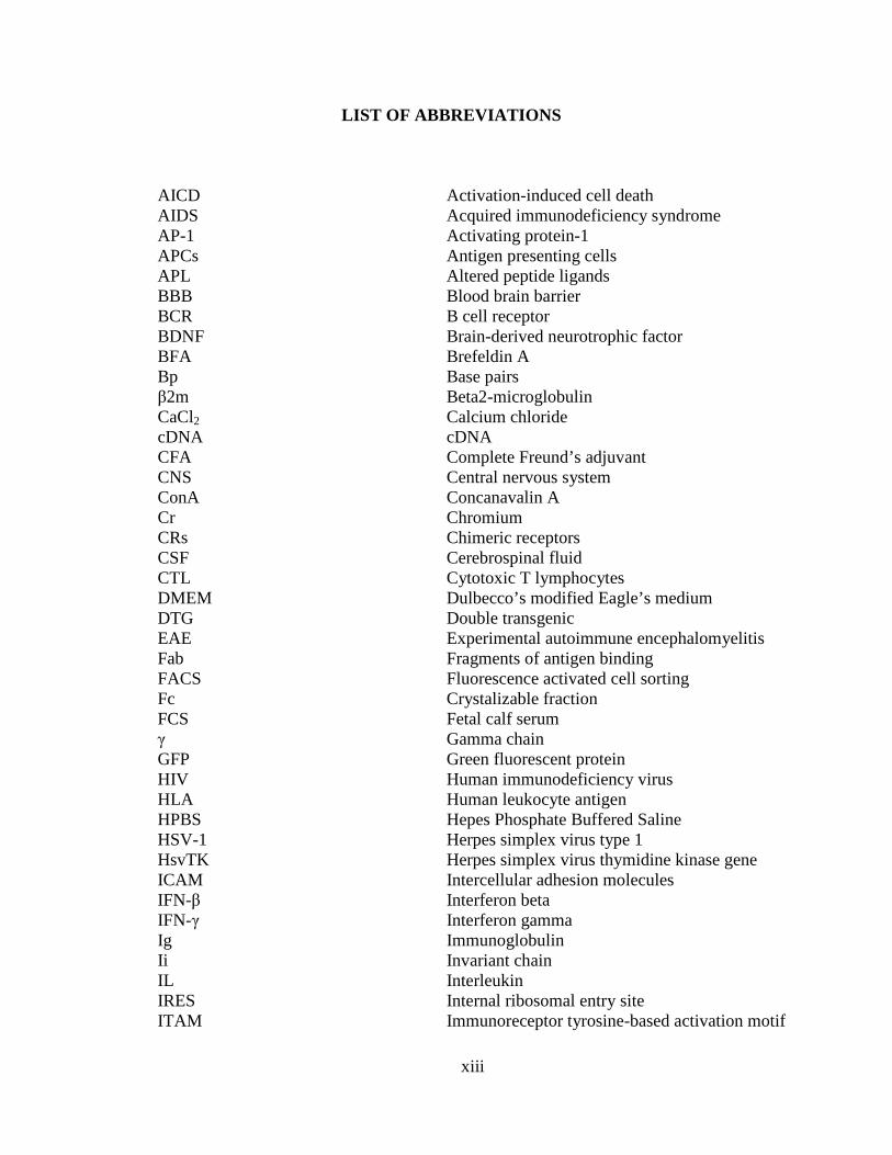
LIST OF ABBREVIATIONS
AICD Activation-induced cell death AIDS Acquired immunodeficiency syndrome AP-1 Activating protein-1 APCs Antigen presenting cells APL Altered peptide ligands BBB Blood brain barrier BCR B cell receptor BDNF Brain-derived neurotrophic factor BFA Brefeldin A Bp Base pairs β2m Beta2-microglobulin CaCl2 Calcium chloride cDNA cDNA CFA Complete Freund’s adjuvant CNS Central nervous system ConA Concanavalin A Cr Chromium CRs Chimeric receptors CSF Cerebrospinal fluid CTL Cytotoxic T lymphocytes DMEM Dulbecco’s modified Eagle’s medium DTG Double transgenic EAE Experimental autoimmune encephalomyelitis Fab Fragments of antigen binding FACS Fluorescence activated cell sorting Fc Crystalizable fraction FCS Fetal calf serum γ Gamma chain GFP Green fluorescent protein HIV Human immunodeficiency virus HLA Human leukocyte antigen HPBS Hepes Phosphate Buffered Saline HSV-1 Herpes simplex virus type 1 HsvTK Herpes simplex virus thymidine kinase gene ICAM Intercellular adhesion molecules IFN-β Interferon beta IFN-γ Interferon gamma Ig Immunoglobulin Ii Invariant chain IL Interleukin IRES Internal ribosomal entry site ITAM Immunoreceptor tyrosine-based activation motif

xiv
KO Knockout LCMV Lymphocytic choriomeningitis virus LN Lymph nodes MBP Myelin basic protein MHC Major histocompatibility complex MOG Myelin oligodendrocyte glycoprotein MRI Magnetic resonance imaging mRNA Messenger RNA MS Multiple sclerosis MSCV Murine stem cell virus NaN3 Sodium azide NFAT Nuclear factor of activated T-cells NF-κB Nuclear factor-kappa OSE Opticospinal EAE PBS Phosphate buffered saline PCR Polymerase chain reaction PE Phycoerythrin-conjugated PLP Proteolipid protein PPMS Primary progressive multiple sclerosis PSG Penicillin-streptomycin-glutamine PTx Bordetella pertussis toxin Rag2-/- Recombination activating gene 2 knockout mice RMTC Receptor-modified T-cells RRMS Relapsing-remitting multiple sclerosis scFV Single chain variable region Spl Spleen SWB Staining wash buffer TAA Tumor associated antigens TaV Thosea asigna virus TCR T-cell receptor TCV T-cell vaccination TGF-β Transforming growth factor-beta Th1 T helper cells type 1 TNF-α Tumor necrosis factor-alpha TTG Triple transgenic VCAM Vascular cell adhesion molecule VH Variable region of the heavy chain VL Variable region of the light chain ZAP-70 ζ-associated protein of 79 kDa ζ Zeta chain

1
Chapter 1. General introduction
1.1 Historical perspective of multiple sclerosis
In 1868, the French neurologist Jean-Martin Charcot examined a young
woman who exhibited a new type of tremor, abnormal eye movements and slurred
speech. On autopsy, she was found to have central nervous system (CNS) “plaques” that
we now associate with multiple sclerosis (MS). Charcot named the disease “sclérose en
plaques”. By the end of the 19th century, the major symptoms of MS were well-
characterized and a new era of neurology arose.
Multiple sclerosis is a chronic progressive demyelinating disorder of the white
matter of CNS characterized by loss of myelin with relative preservation of axons. It is
the most common CNS autoimmune disease, affecting approximately 1 million people
worldwide (250,000 in the United States) (2). The disease disproportionately affects
females at a 2:1 sex ratio, has no single defined cause, and several genetic markers are
associated with susceptibility.
Genes and environment play a major role in the pathology of MS. Among the
primary genetic associations, human leukocyte antigen (HLA) class II genes on
chromosome 6 and particularly HLA-DRB1 (HLA-DRB1*1501 and DQB1*0602) were
found to increase the risk for MS (3). The risk of disease in a monozygotic twin of an
affected individual is far greater than in dizygotic ones (25-30% compared to 2-5%).
Also, the risk that first-degree relatives of patients with MS will get the same disease at
some point in life is seven times higher than for the general population (4). These
findings suggest a very strong genetic component (although not a Mendelian one).

2
Epidemiological studies implicated geography as another factor for MS. Studies
have shown that the disease is more prevalent in the temperate regions and western
hemisphere (Germany, Scandinavia, Canada, northern US) and that migration from a
high-incidence area to a low incidence one before puberty significantly decreases the risk
of acquiring MS later in life (5). Therefore, environmental factors seemingly more
prevalent in temperate regions combine with genetic factors to determine MS
susceptibility.
1.2 Clinical signs and symptoms of MS
MS can exhibit a very large range of signs and symptoms commonly first seen in
the third to the fourth decade of life. 85% of patients have the typical relapsing-remitting
multiple sclerosis (RRMS), with episodes triggered by viral infections followed by
remissions with no residual damage or different cumulative amounts of chronic
impairment. Over time, approximately 30% of these patients will undergo transformation
to a secondary progressive form with less frequent acute attacks, but with gradual
worsening of symptoms and permanent disabilities. A subset of RRF is benign MS, with
few and mild attacks and a limited disease course or total recovery. Ten percent of
patients with MS develop progressive deterioration of neurological functions without
relapses and a more aggressive form of disease, called primary progressive MS (PPMS)
(6).
Symptoms displayed by MS patients vary according to the location of lesions.
Cerebellar and cerebral plaques typically accompany speech and balance problems,
tremors and loss of coordination. Motor and sensory nerve tracts deficits are revealed by
3
spastic paralysis, muscle weakness, diplopia and other visual problems including
blindness, urinary and bowel problems, and tingling, numbness, and loss of touch and
pain. Patients may also show signs of depression, cognitive and emotional problems,
fatigue, and sexual disturbances.
1.3 Diagnosis of MS
MS is not easy to diagnose. There is no single test adequate for diagnosis.
Physicians rely on history, clinical signs and symptoms, and various tests including
magnetic resonance imaging (MRI), cerebrospinal fluid (CSF) analysis, serology, and
sensory evoked potential testing. The classic approach is manifestation of at least two
clinical signs along with MRI lesions localized in the brain or spinal cord which confirm
the diagnosis. MRI with gadolinium contrast shows enhancement of lesions that correlate
with perivascular inflammation. Almost 90% of MS patients will show oligoclonal
immunoglobulin (Ig) G in CSF. Although this finding is not specific for MS and can only
be considered suggestive, it is very useful in ruling out infectious diseases or tumors that
might mimic this autoimmune disease. Optic nerve lesions that might not show up on
MRI can be detected by visual evoked potentials, which will reveal prolonged latencies
consistent with plaques located within optic pathways. Serology is also not specifically a
useful tool in diagnosing MS, but it proves helpful in differential diagnosis with other
entities (7).
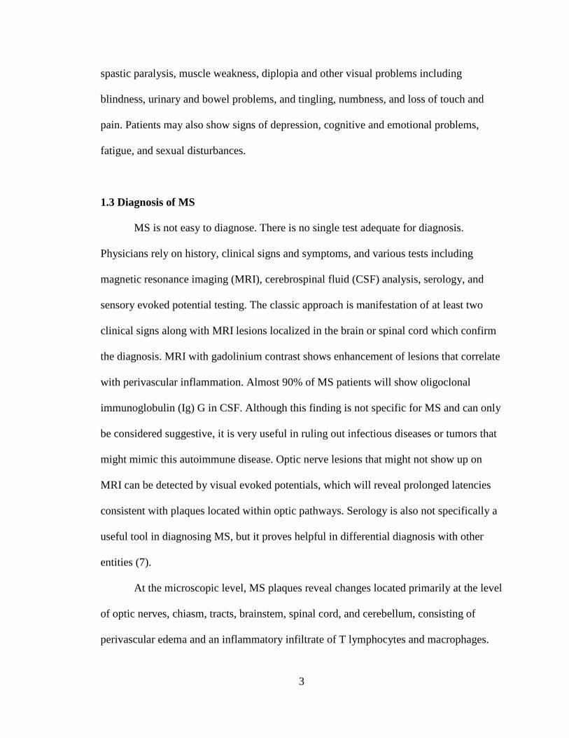
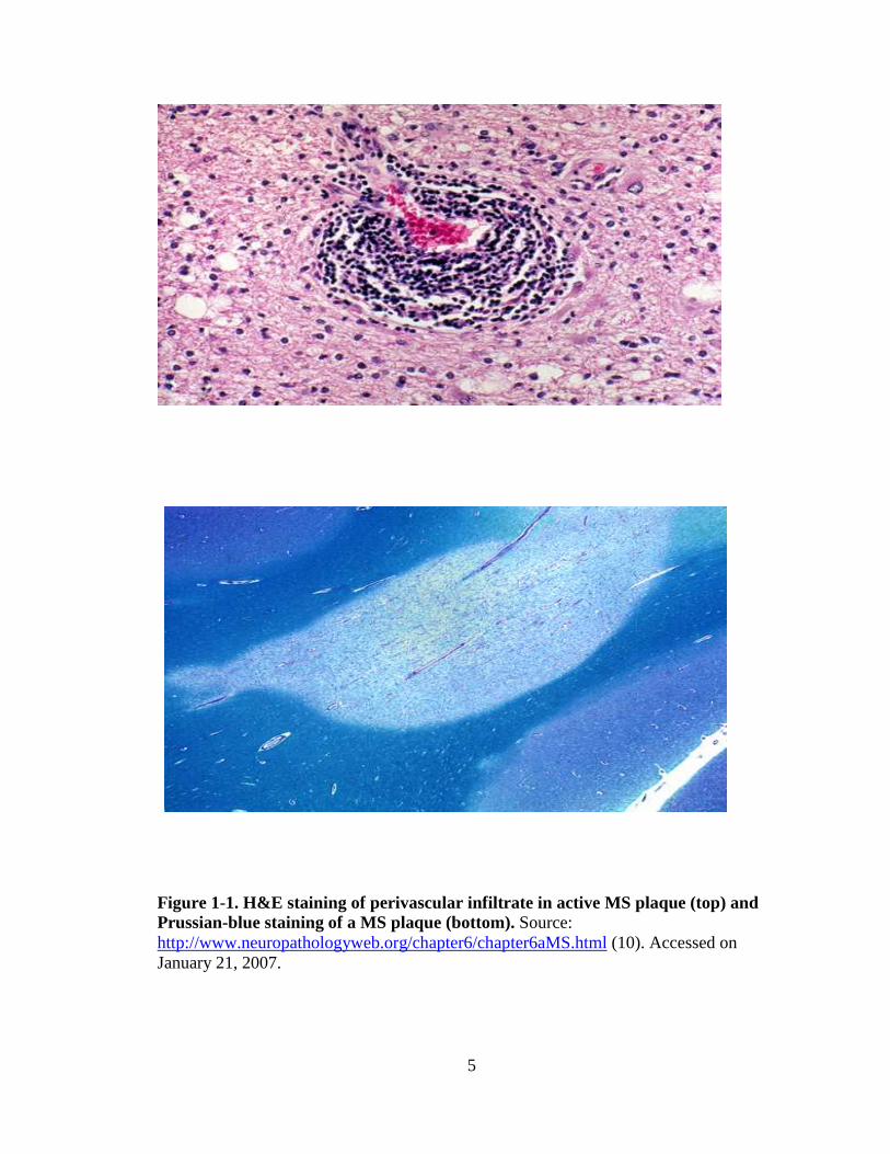
At the microscopic level, MS plaques reveal changes located primarily at the level
of optic nerves, chiasm, tracts, brainstem, spinal cord, and cerebellum, consisting of
perivascular edema and an inflammatory infiltrate of T lymphocytes and macrophages.

4
Myelin is stripped from the axons impairing saltatory conduction and causing conduction
block (Figure 1-1). Axons tend to be spared (8). During periods of remission,
inflammation and edema subside and axons can undergo remyelination and carry out the
normal function again. Repair of damaged areas occurs more completely in early stage
disease when oligodendrocytes are still able to build a new myelin sheath; in time, as the
disease progresses more advanced lesions develop characterized by gliosis. This creates a
boundary between myelin producing cells and axons, therefore rendering remyelination
inefficient.
1.4 Pathobiology of MS
There is no clear proof of the cause of MS. Epidemiological evidence from
genetics, geography, and socio-economic factors has led many to hypothesize that there is
a viral etiology (9). A definite pathogen has not been identified, though some microbes
bear similar structures with self-antigens in the CNS such as myelin basic protein (MBP),
myelin oligodendrocyte glycoprotein (MOG), and proteolipid protein (PLP) and it is
possible that these promote autoimmunity through the mechanism of molecular mimicry.
Molecular mimicry is the process by which a viral or bacterial infection causes activation
of T-cells that are cross-reactive with self antigens. It is still a major mechanism for
triggering autoimmune diseases (8).
There are other hypotheses for the etiology of MS including bystander activation
and superantigenic T cell activation. In the bystander activation hypothesis, T
lymphocytes are activated in the periphery by infectious agents presented on the surface
of antigen presenting cells (APCs) and become capable of crossing the blood brain
5
Figure 1-1. H&E staining of perivascular infiltrate in active MS plaque (top) and Prussian-blue staining of a MS plaque (bottom). Source: http://www.neuropathologyweb.org/chapter6/chapter6aMS.html (10). Accessed on January 21, 2007.

6
barrier (BBB). The cells which develop into activated T-cells are CD4+ or so-called T
helper cells type 1 (Th1). Although both Th1 and Th2 cells are present in MS, Th1 cells
are able to secrete proinflammatory cytokines and also express a high level of adhesion
molecules such as intercellular adhesion molecules (ICAM) or vascular cell adhesion
molecule (VCAM). Inflammation upregulates adhesion molecule expression on the
endothelium of the BBB, thus making it more permeable for penetration. Th1 cells also
secrete matrix metalloproteinases that further compromise the integrity of the BBB (11).
The opening of this natural barricade enables future inflammatory cells to penetrate into
the CNS.
Once within the CNS, activated Th1 cells will die or be eliminated unless
restimulated. An autoantigen or again, a microbe presented on the surface of CNS APCs
(microglia) may restimulate and promote the expansion of T-cells, and induce the release
of proinflammatory cytokines, such as interferon-γ (IFN-γ) and tumor necrosis factor-α
(TNF-α) that lead to macrophage activation. These cells release neurotoxic components
(nitric oxide, reactive oxygen species) that damage the myelin sheath causing the
structural and functional damage that results in MS (Figure 1-2).
In addition to this indirect means of tissue damage, infectious agents can directly
damage tissues through recruitment of T-cells with new specificities, including
autoaggressive T-cells to the CNS. In a process called epitope spread, the immune
response can switch from being initially restricted to a microbial antigen to incorporate
an added self antigen-specific response. Thereby, a secondary autoimmune reaction can
develop (12).
Activation of T-cells by superantigens has also been proposed as a trigger for MS.

7
Figure 1-2. Immunopathogenesis of the MS lesion. APC = antigen presenting cell; IFN = interferon; IL = interleukin; Mic = microglia; MMP = matrix metalloproteinase; MO = monocyte; NAA = nacetylaspartate; NO = nitric oxide; Pl = plasma; VCAM = vascular cell adhesion molecule. Adapted and reprinted with permission – Suhayl Dhib-Jalbut et al. 2006. Neurodegeneration and neuroprotection in multiple sclerosis and other neurodegenerative diseases. Journal of Neuroscience 176:198-215.

8
According to this theory, virus and bacteria superantigen is able to cross-link the T-cell
independent of peptide antigen, thus activating T-cells that can either expand or be
deleted. Since superantigens preferentially recognize particular Vβ families, a large
proportion of T-cells might be activated during this process. As not all self-reactive T-
cells are eliminated in the thymus by negative selection, myelin-specific ones can become
activated and trigger an autoimmune response (13). Similar to the epitope spread theory,
superantigen-mediated stimulation of autoreactive T-cells is detectable in mouse model
but there is no direct evidence for this in MS.
For many years, Th1 CD4+ autoreactive T-cells have been incriminated as the
major T cell offenders in MS. This theory has more recently been challenged by the
recognition of a new subset of autoaggressive T-cells, CD4+Th-17+, whose
differentiation is promoted by APCs in the presence of interleukin (IL)-6 and
transforming growth factor-beta (TGF-β) and whose expansion requires IL-23 (14).
Studies show that Th17 cells secrete proinflammatory cytokines (IL-17, IL-6, TNF-α),
but not IFN-γ and IL-4 and adoptive transfer of these cells can induce severe
experimental allergic encephalomyelitis (EAE) disease (15). In models of MS and other
autoimmune diseases, disease can be suppressed by blockade of IL-23 pathway or the
downstream IL-17 and IL-6 factors (16).
CD8+ T-cells are also incriminated for causing an immune attack by recruitment
and clonal expansion within the CNS. They can recognize peptides presented by major
histocompatibility complex (MHC) class I-expressing brain cells and even outnumber
CD4+ T-cells in the inflammatory infiltrate that characterize MS. Little is known about
the role of CD8 cells in the pathology of MS in regards to the several aspects, such as
9
means of CNS invasion, proliferation, apoptosis, and further clarifications are required
(17). The invading cytotoxic T lymphocytes release pro-inflammatory cytokines, such as
IFN-γ and TNF-α, thus inducing expression of MHC class I molecules in the brain in
vitro (18).
It is clear that the above theories on the cause of MS are demonstrable in the
mouse models but will be difficult to verify in humans. They leave many questions and
lots of alternatives.
1.5 Animal models of MS
Modeling MS is a challenging task. The disease is complex and little is known
about its triggers and mechanisms.
The first attempt to build an animal model of this disease was in the early 1930s
when Rivers and collaborators noticed that certain infections (measles, smallpox) were
followed by a wide range of CNS symptoms. Biopsy of these patients’ brains revealed a
perivascular demyelinating inflammatory infiltrate that characterized the acute
disseminated encephalomyelitis. This group tried to reproduce the disease in monkeys by
repeated intramuscular injections of extracts and emulsions of rabbit brain. Although the
model did not accurately reproduce the human disease, it was still considered to be a
groundbreaking discovery (19). A decade later, Kabat adjusted the disease-induction
procedure by using an adjuvant to increase the immune response (20). This had two
major consequences: it made the immunization protocol more manageable since the
animals, unlike Rivers’ model, only needed one injection, and it incriminated myelin as
the culprit for MS since only animals injected with adult rabbit or monkey brain plus

10
adjuvant got sick but not those injected with rabbit lung or fetal rabbit brain.
In 1960s and 1970s the concept of EAE developed as adoptive transfer of
splenocytes from rats immunized with spinal cord extract into normal recipients was
shown to induce disease (21).
1.5.1 Current animal models of MS
EAE is considered to be primarily mediated by MHC II-restricted CD4+ Th1 cells
that secrete proinflammatory cytokines such as tumor TNF-α and IFN-γ (22). EAE can be
induced in susceptible strains of small, easy-to-handle animals such as mice, guinea pigs,
and rats by active immunization with myelin antigens including MBP, PLP, MOG, and
others, plus complete Freund’s adjuvant (CFA). The administration of Bordetella
pertussis toxin (PTx) increases the permeability of the BBB, thus creating a “breach” for
activated T-cells that enter the CNS and cause the local inflammation (23). The first signs
of neurological disease can be detected as early as ten days post immunization and
depending on the model system may resemble the human relapsing-remitting or
progressive disease forms. Inbred mice are most commonly used as the animal model for
EAE due to their small size and well-defined genetics. Typically, disease is scored on a 1
to 5 scale (24) and the animals are euthanized at a score of 4 or 5 according to the ethical
guidelines (Table 1-1).
Not all strains of mice exhibit the same disease course and symptoms when
immunized with myelin antigens. While a certain strain of mice might be susceptible to a
peptide antigen, another one could be resistant to the same antigen. There are now clear
“recipes” for EAE induction in different mouse strains (25). Some of the standard

11
Table 1-1. Clinical EAE scores.
Disease score Clinical signs of disease
0 No signs of disease
1 Limp tail
2 Partial hind leg paralysis
3 Complete hind leg paralysis
4 Hind and front leg paralysis
5 Moribund or dead

12
protocols include relapsing-remitting EAE in SJL mice immunized with PLP139-151, MBP-
induced EAE in PL/J or B10.PL mice, or MOG35-55-induced EAE in C57BL/6 mice. The
disease is mostly T cell-mediated. However, MOG not only induces a T cell response, but
also production of demyelinating autoantibodies (26). MOG-specific T-cells and
autoantibodies were also found in circulation in patients with MS (27), but the cause that
triggers their activation is still unknown.
1.5.2 Adoptive transfer EAE
The discovery that adoptive transfer of myelin-specific T-cells can induce EAE in
naïve syngeneic recipients validated the autoimmune nature of EAE in mice (28).
Susceptible mice are immunized with a particular antigenic peptide followed by isolation
of T-cells from draining lymph nodes and spleens, and in vitro stimulation of the T-cells
with the myelin peptide. The T-cells are then injected into naïve recipients who develop
disease. This approach showed that the CNS can be invaded by activated
encephalitogenic T cell clones that are capable of crossing the BBB and emphasized the
autoimmune nature of the phenomenon. The encephalitogenic cells were thought to bear
a CD4+Th1 cell phenotype and recognize self peptides presented in the context of MHC
class II molecules. Later data showed that the MOG35-55 epitope is able to activate CD8+
MOG-specific αβ T cell receptor (TCR) positive cells that can be adoptively transferred
into naïve C57BL/6 recipients, causing a much more severe clinical disease sustained by
more destructive histopathologic CNS lesions. Furthermore, these antigen-specific cells
were capable of surviving in vivo as shown by their ability to be retrieved from recipient
mice (29). A role for CD8+ T-cells in murine EAE has been controversial and not

13
uniformly supported.
Although EAE induction seems fairly straightforward if following the rule “right
strain-right antigenic peptide”, one must not overlook the association between the antigen
and its corresponding MHC. T-cells cannot recognize an antigenic peptide properly
unless it is presented on the right MHC. Beta2-microglobulin knockout (β2m-/-) mice
lacking MHC class I are resistant to EAE induced by adoptive transfer of CD8+ MOG-
specific TCR+ cells (29). Likewise, CIITA mice lacking MHC class II, as well as mice
deficient for invariant chain (Ii) and H-2M (DM) are resistant to both direct priming with
peptide and adoptive transfer of CD4+ peptide-specific T-cells. Interestingly, APCs from
these knockout (KO) mice can present MOG peptide to CD4+ T-cells, but they are not
capable of presenting and processing the myelin protein, thus rendering the mice resistant
to EAE induction. The fact that MOG EAE cannot be induced in class II deficient mice
but can in β2m-/- mice argues against a significant role for MOG-specific CD8+ T-cells
(30).
1.5.3 Transgenic mice as models for MS
Key to understanding human autoimmune diseases is to dissect the mechanisms
of tolerance induction. The challenge for MS is to discover how seemingly tolerant self-
reactive cells in the periphery become activated, are able to penetrate the CNS, and
mediate disease. B10.PL mice were engineered to express a transgenic α2+ and Vβ8.2+
TCR specific for MBP epitope 1-11. These mice were highly susceptible to MBP-
induced EAE and some even developed spontaneous disease. T-cells removed from
spleens or lymph nodes of these animals proliferated and secreted cytokines in response
14
to in vitro MBP stimulation (31). This MBP-specific transgenic mouse model has
provided not only a unique model to study MS but also important information concerning
the failure of the thymus to delete the antigen-specific T-cells, the mechanisms of
peripheral tolerance, and the circumstances in which autoreactive, antigen-specific cells
can cross the BBB and cause inflammation and myelin destruction (32).
Recently, another animal model has been developed that has proved extremely
useful in the research of demyelinating diseases: the opticospinal EAE (OSE) mouse. A
transgenic mouse expressing a TCR specific for MOG 35-55 (denoted TCRMOG) was
crossed with a MOG-specific Ig heavy-chain knock-in mouse (denoted IgHMOG) both on
a C57BL/6 background. The latter mouse had B cells producing antibodies against MOG.
Single-transgenic mice did not undergo spontaneous EAE disease, but the double-
transgenic animals exhibited signs of an EAE-like disease, closer to the human Devic
disease than to MS. Devic disease differs from MS only in regards to the site of the
primary attack (spinal cord and optic tracts instead of the brain) and more frequent and
severe attacks compared to MS. Nevertheless, mice exhibited a pathologic finding similar
to what is seen in MS: inflammatory infiltrate with prevalence of CD4+T-cells and
macrophage, demyelination, and sometimes axonal loss. Since single-transgenic mice did
not develop spontaneous disease, one can infer that interaction between MOG-specific T
and B cells present in the double-transgenic mice is responsible for development of
disease. Although OSE mice do not reproduce the classical MS, they still are extremely
valuable for the understanding of the role of B cells in the pathogenesis of EAE since
MOG-specific B lymphocytes do not only act as APCs but also proficiently capture even
smallest amounts of peptide, process it, and present it to T-cells (33).
15
1.6 Therapeutic approaches for MS
The concept that MS is a non treatable disease has changed dramatically over
time. There is currently no definitive cure, but long-term survival has increased in recent
years due to new discoveries about the pathology of the disease, better and earlier
diagnostics including MRI, and better clinical trial designs. While some therapies have
been successfully introduced and have shown to improve symptoms, others, having been
proven to work in animal models, have not shown any benefit or even worsened the
disease course in humans. Nonetheless, efforts are increasingly centered towards making
MS a much more manageable disease than in the past.
There are two goals to therapy: the first one is to treat disease symptoms, such as
spasticity, vertigo, depression, bladder and bowel dysfunctions. The second one targets
the pathogenic cascade: peripheral activation of antigen-specific T-cells, penetration of
BBB and activation and proliferation in the CNS, demyelination, and interaction of TCR
with peptides loaded on MHC complexes.
1.6.1 Glucocorticoids
Corticosteroids have long been considered a panacea for autoimmune conditions,
and MS is no exception to the rule. Studies with intravenous (iv) methylprednisolone
showed improvement of symptoms in patients with chronic progressive disease as well as
acute relapses (34). Steroids have also proved beneficial for optic neuritis which is often
the first clinical manifestation of MS (35). Therapeutic use of corticosteroids nevertheless
has more recently decreased, partly due to their side effects (osteoporosis, glaucoma,
worsened diabetes, suppression of adrenal glands, etc.), and partly due to the advent of

16
new therapies. However, they are still important tools though in treating RRF of MS and
secondary progressive MS.
1.6.2 Cytokines
Interferon beta (IFN-β) 1a and 1b acts through a mechanism that is not fully
understood, but it is proposed to involve a drop in IFN-γ levels, blockade of myelin
attack, inhibition of metalloproteinases, and various modulatory effects on chemokine
and adhesion molecule production (36). Several drugs currently on the market (Avonex,
Rebif, Betaseron) were shown to reduce the annualized relapse rate by approximately
one-third. A new study revealed a decreased concentration of monocyte-derived non-
classical MHC molecule (class Ib) called HLA-G in patients with MS. This molecule is
important since it inhibits both Th1 and Th2 cytokine production (IFN-γ, IL-2, and IL-10
respectively) by CD4+ T-cells. HLA-G levels were increased to almost normal levels
after treatment with IFN-β (37).
TNF-α or cachectin has already been shown to be an important mediator in the
pathology of MS due to its ability to mediate inflammatory responses. However, its
therapeutic blockade in MS has been very controversial. Studies have shown that TNF-α
is increased in the CSF of patients with MS in direct correlation to the severity of the
disease and neurological impairment (38). However, results of anti-TNF-α therapy with
TNF-receptor fusion proteins such as etanercept (Enbrel) or monoclonal antibodies such
as infliximab (Remicade) have been disappointing. Disease course was worsened and
there was actually a new case of MS reported (39). Data of TNF blockade in EAE has
yielded confusing results. Treatment of adoptive transfer EAE with soluble TNF
17
receptors reversed the disease and protected against recurrent episodes (40). In contrast,
complete deletion of the gene in TNF knockout mice led to high mortality and severe
neurological defects. Moreover, treatment with recombinant TNF reduced the disease
course and even prevented progression of EAE (41).
IL-10 and TGF-β2 are suppressive cytokines in EAE. IL-10 is produced by
regulatory CD4+ cells and selectively upregulated during recovery in EAE model. Just
like TNF-α, studies of IL-10 treatment of EAE were mixed, particularly because the route
of administration seemed to play an important role. Intravenous injections exacerbated
the disease (42), whereas intranasal immunizations partially inhibited EAE (43). TGF-β2
has not been approved for the therapy of MS due to the nephrotoxic effects seen in mice.
1.6.3 Antigen-derived immunotherapies
As corticosteroids and other drugs impair the general immune defense by
eliminating or suppressing not only the disease-causing cells, but other T-cells, it has
become imperative that therapeutic approaches specifically target antigen-specific cells.
A major limitation in the therapy of MS is the phenomenon of epitope spread. This
develops after the initiating event when CNS provides the proper environment for
reactivation of T-cells (self-antigens and MHC complex and co-stimulatory signals
necessary for reactivation), thus recruiting and stimulating cells reactive not only against
the initial causative antigen, but also against other neighboring self-peptides. This leads
to a more extensive repertoire of immune responses that may amplify tissue destruction.
A major goal of antigen-derived therapies is to circumvent this problem by mediating
bystander suppression. For example, self-reactive antigen-specific Th2 or Th3 cells that
18
were initially generated by oral tolerization of mice and are capable of secreting anti-
inflammatory cytokines (IL-4, IL-10) may globally downregulate the immune response
after activation (44). One method to generate these regulatory T-cells is through peptide
tolerization. Two approaches to tolerization with peptides have been extensively studied:
altered peptide ligands (APL) and mucosal administration of antigen.
1.6.4 Altered peptide ligands
It is well known that activation of CD4+ Th1 cells depend on the interaction of
immunogenic peptide bound to MHC class II with the TCR along with a costimulatory
signal from APCs. Activation leads to proliferation, cytokine production, and cytolysis.
Lack of costimulation renders the cells anergic to subsequent antigenic stimulation. Past
studies have shown that an immunogenic peptide that has some of its residues mutated
can stimulate Th1 and Th2 cells to fulfill some functions, but not proliferation. This
phenomenon is called partial activation. The basic principle is that the surface expression
of important molecules, such as CD3 did not change, nor did the MHC binding residues
in the peptide. The only residues that were changed were the TCR binding moieties.
When the cells were cultured with the original peptide and APCs, the T-cells proliferated
normally. When the cells were first stimulated with the mutated peptide, upon subsequent
stimulation with the original peptide, T-cells were rendered unresponsive to the
immunogenic peptide (45).
APLs were also approached in the therapy of EAE induced by adoptive transfer of
a pathogenic T cell clone specific for MBP epitope p87-99. This clone caused a
heterogenous inflammatory infiltrate of the CNS that disappeared when the clone was

19
tolerized in vivo with an analogue of the immunogenic peptide that carried a
phenylalanine to alanine substitution at position 96. Direct transfer of the same clone
treated with the APL led to prevention of EAE and even reversion of paralysis. The
mechanism of action is still unclear since the therapeutic APL has no influence on
proliferation of pathogenic T cell clone to the immunogenic peptide MBP 87-99, so there
is no MHC competition or TCR antagonism. It has though, been noticed that deletion of
the inflammatory infiltrate by APL depends on the availability of IL-4. Treatment with
APL promoted a shift in the ratio between IL-4 and TNF-α to an increase in the former
which downregulates the latter (46). Even though treatment of EAE with APL showed
encouraging results, this approach in human MS has led to a controversial outcome, as
some trials ending in exacerbations of disease (47).
1.6.5 Synthetic copolymers
Glatiramer acetate (Copaxone) consists of a mixture of alanine, glutamic acid,
lysine, and tyrosine that acts like a universal antigen, “luring” autoreactive T-cells (48). It
efficiently binds to MHC class II molecules (DR, but not DQ or class I), thus hindering
self-peptide from binding in the same groove. It is not fully understood how the drug
works: it was initially thought to cross-react with MBP peptide and then compete for
MHC binding, but has also been found to induce regulatory T-cells (49). Since
Glatiramer-activated, Th2-like cells can cross the BBB, they enter the CNS and secrete
anti-inflammatory cytokines, such as IL-4, IL-10, IL-6, thus promoting a non-
inflammatory environment. These Glatiramer-activated cells also exert a neurotrophic
effect by producing brain-derived neurotrophic factor (BDNF) (50). Copaxone can
20
successfully prevent EAE induced by several peptides (MBP, PLP, or MOG) and phase
III clinical trials have also shown it to be beneficial in RRMS (51).
1.6.6 Mucosal administration of antigen
Oral or nasal administration of antigens responsible for MS and EAE has yielded
controversial results. Self-specific T-cells can be activated within six hours of oral
administration of peptide and consecutive administration of the same peptide will
decrease the number of effector T-cells. The route of administration is important, with the
intranasal one seemingly more efficient than oral (“nasal” versus “oral tolerance”).
Peptide administered intranasally seems to be able to reach the thymus where it can
mediate apoptosis of high affinity thymocytes as opposed to the oral route of
administration where the antigenic peptide might be destroyed by the acid in the
gastrointestinal tract. The mechanisms governing mucosal tolerance - anergy or deletion
of antigen-specific T-cells - are not clear. Although feeding the inducing peptide
at time of disease induction prevented EAE in mice and generated enthusiasm regarding
the therapeutic outcome of the human disease (37), this approach failed in MS clinical
trials (52).
1.6.7 T-cell vaccination
Antigen-stimulated T-cells can induce EAE equally as well as the antigenic
peptide itself in adjuvant. T-cell vaccination (TCV) uses irradiated, activated antigen-
specific CD4+ T-cells to “vaccinate” mice without causing EAE similar to microbial
vaccination against infectious agents. These cells induce CD8+ T-cells capable of killing

21
the autoreactive CD4+ T-cells as well as preventing antigen-induced proliferation of the
vaccine T-cells themselves. Pilot trials in MS patients have used TCV with MBP-reactive
T-cells isolated from their blood, activated in vitro and irradiated to abrogate subsequent
proliferation. These clones were then injected back into the patients, resulting in the
specific deletion of circulatory MBP-specific T-cells (53). Although this procedure has
not been used to treat patients on a large scale, it is still considered for future evaluation.
1.6.8 Monoclonal antibodies
Activated T-cells express high levels of surface adhesion molecules like VCAM
or ICAM and upregulate their receptors on the endothelium of the BBB, thus enabling T-
cells to cross the BBB and cause inflammation in the CNS. Antibodies directed against
the ligand-receptor pair could potentially block this first step in the CNS homing of T-
cells. VCAM-1 expression is low on blood vessels under homeostatic conditions;
however, expression is increased under conditions of inflammation, such as found in
brain tissue of EAE-induced animals and human MS. Administration of antibody against
α4β1-integrin prevents accumulation of leukocytes in the brain and subsequent
development of EAE (54). The monoclonal antibody anti-α4β1-integrin was named
Natalizumab and has been licensed for clinical use (55).
1.6.9 Gene therapy in MS
Gene therapy for autoimmune disease has emerged as a result of progress in
deciphering in greater detail the pathologic mechanisms by which self-antigens mediate
autoimmune diseases. This approach can be very specific and aims to deliver a gene or
22
gene product that can specifically block disease. Gene therapies may be antigen-specific,
while hopefully avoiding the general suppression of the immune system associated with
conventional treatments. The goal of gene therapy in MS is generally to deliver immune-
modulating molecules (blocking antibodies, anti-inflammatory cytokines, etc.) by
different means such that antigen-specific immune tolerance can be achieved.
One new tactic is the delivery of anti-inflammatory cytokine genes (IL-4, IL-10,
or IL-12 p40 subunit) by viral vectors that are administered intrathecally in order to
directly concentrate the gene product in the CNS, slowly releasing the cytokine of
interest. Different vectors may be used: non-replicative herpes simplex virus type 1(HSV-
1), retroviruses or adenoviruses are able to accommodate the genes and infect cells.
Potentially, EAE could be both prevented and treated using an HSV-IL-4 system by
downregulating proinflammatory cytokines and therefore, macrophage activation and
CNS invasion (56).
An even more practical way of delivering anti-inflammatory cytokines by means
of viruses is to retrovirally transduce antigen-specific CD4+ T-cells. The autoreactive T-
cells will migrate to the CNS and therefore, provide a “home delivery” of Th2 regulatory
cytokines to the autoimmune lesions (57).
Receptor-modified T-cells (RMTC) have emerged in the past five years as a
means to redirect T-cells against antigen-specific T-cells and have already proven useful
in infectious diseases and cancer. In this case new, often chimeric, signaling receptors are
expressed on T lymphocytes. The benefit of using T-cells is their effector and regulatory
functions, their ability to grow well in vitro and traffic to most sites of the body. Chimeric
receptors containing extracellular domains from MHC class I linked to a signaling
23
domain from TCR can be retrovirally transduced in T-cells or T-cell hybridoma. Upon
encounter and recognition of their cognate TCRs, these chimeric receptors exhibit
effector functions such as cytokine secretion, proliferation, or cytolysis depending on the
type of T cell in which they are transduced (CD4+ or CD8+) (58).
A more specific surrogate receptor able to target autoreactive, encephalitogenic
Th1 cells has recently been designed. This chimeric receptor contains the extracellular
and transmembrane domains of mouse MHC class II I-A β and α, the zeta cytoplasmic
signaling domain, and also an antigenic peptide, MBP89-101, linked on its surface. This
receptor not only recognizes the cognate TCR but can also be stimulated as a result of
this interaction. Among outcomes of this TCR-TCR interaction, CD8+ T-cells transduced
with retrovirus containing this construct can specifically kill CD4+Th1 antigen-specific
T-cells. Although the chimeric receptor was designed to only carry one peptide epitope,
experimental results show that this approach could also address the main problem of
EAE, epitope spread, even when RMTC are administered one month after disease
induction (59). One benefit of this approach is that it does not interfere with the whole
immune system, but selectively targets encephalitogenic, antigen-specific cells.
24
Chapter 2. Development of chimeric receptors
Chimeric receptors (CRs) are hybrid combinations of a recognition domain
(variable regions of an Ig or a MHC molecule) and a signaling domain (TCR moieties
responsible for signal transduction). The name “chimeric” resides in their mixed
structure: CRs carry a recognition domain containing variable regions in charge of
antigen recognition and an intracytoplasmic domain responsible for signal transduction
(60) (Figure 2-1).
For a better understading of how CRs were engineered, I will briefly discuss the
structure of IgG and TCR.
IgG is composed of two identical heavy and two light chains (kappa or lambda).
The light chain has a variable region and a constant one and the heavy chain has
additionally two or three constant domains. The chains are kept together by disulfide
bonds. When treated with papain, the Ig breaks into two equal fragments of 45-50 KDa
called fragments of antigen binding (Fab) and a third fragment of 50 KDa called
crystalizable fraction (Fc). The antigen binding site consists of the variable domains of
the light and heavy chains.
TCR is also composed of two chains, alpha and beta, each of which has a variable
and a constant region. Its role is to recognize antigen-MHC complexes. The signals
triggered by antigen recognition are not transduced by the TCR but by two proteins, CD3
and zeta (ζ), that are noncovalently linked to TCR, forming the TCR complex. Upon
recognition of peptide, a cascade of signals including tyrosine phosphorylation and
activation of nuclear factor-kappa (NF-κB), nuclear factor of activated T-cells (NFAT),

25
Figure 2-1. Structure of the T-cell receptor and a chimeric receptor. Reprinted with permission – Claudia Rössig, Malcolm K. Brenner. Chimeric T-Cell Receptors for the Targeting of Cancer Cells. Acta Haematol 2003;110:154-159.

26
and activating protein-1 (AP-1), is triggered, eventually leading to cell proliferation and
differentiation. The cytoplasmic domain of CD3 and ζ contain a conserved sequence
called immunoreceptor tyrosine-based activation motif (ITAM) that plays a crucial role
in signaling. Upon phosphorylation, ITAMs become docking sites for a tyrosine kinase
called ζ-associated protein of 79 kDa (ZAP-70), ultimately leading to changes in gene
expression in the T-cells.
CRs represent a smart combination between the ability of TCR and B cell
receptor (BCR) to recognize different antigens and elicit intracytoplasmic signal
transduction events leading to different effects. Some of the earlier CRs were constructed
by using both variable region of the heavy chain (VH) and variable region of the light
chain (VL) combined with the α or β constant domain of the TCR (61). Eshhar engineered
a chimeric TCR composed of the variable region domain Fv of an antibody and the
constant region of TCR. This receptor can be expressed as a transgene in T-cells via
retrovirus, but due to the fact that two genes (VH and VL) have to be transfected into the
same cell by two separate retroviral vectors, the efficiency of transduction was low.
Consequently, this problem was overcome by joining together VH and VL into a single
chain variable region (scFV) connected by a linker peptide (62, 63). The scFv is part of
the extramembrane portion of the construct and is responsible for antigen recognition.
This is linked to a region consisting of the gamma (γ) or zeta cytoplasmic tail of the TCR.
The two domains are linked by a hinge region that works as a spacer, increasing the
distance between scFV and the plasma membrane. The hinge region belongs to IgG and
accounts for the differences in the amino acid composition of the four classes of IgGs. It
is placed between the Fab fragment and the constant CH2 and CH3 domains of the heavy

27
chains and determines the flexibility of the IgG molecule. Flexibility is important for
further effector functions of the IgG such as C1q binding and complement activation
(64); this feature and also the number of inter-heavy chain disulfide bonds in the hinge
region is strictly characteristic and different for each IgG subclass. Different hinges were
used, such as the hinge region of human IgG1 or CD8 or part of the extracellular region
of CD28 some of which showing better expression in T-cells than others (65). The
advantage of using the variable domain from Ig resides in the non-MHC restricted,
antibody-type specificity that leads to a more ubiquitous array of specificities that can be
transferred to T-cells through CRs.
Similar to the classic TCR, the mere contact between T-cells bearing chimeric
receptors and target cells does not lead to cytolysis of latter. The CR can only guarantee
specific recognition of target, but does not confer effector function to the T-cells unless
they are activated upon this recognition. This may require the presence of a co-
stimulatory signal and although a definite role has been established for induction of
effector T-cells from naïve T-cells, it is not clear whether the costimulation is also
required for the induction of effector cells from memory T-cells. The two signals theory
states that T-cell activation requires recognition of antigen-MHC complex and
costimulation; T-cells stimulated in the absence of costimulation are rendered tolerant
rather than memory T-cells (66).
CD28 is a membrane protein with well-defined ability to promote T-cell
proliferation and differentiation and induction of cytokine secretion upon binding of B7
molecules on APCs. The addition of the cytoplasmic domain of the co-stimulatory CD28
molecule to the engineered zeta signaling tail improved the efficacy of CR-transduced T-
28
cells (67). Another co-stimulatory pathway is mediated by CD137 that belongs to the
TNF family. Stimulation of CD 137 inhibits activation-induced cell death (AICD) (68).
CRs engineered as described can be transferred into human or murine T-cells and
redirected against microbial antigens or tumor antigens in a manner independent of MHC
restriction. Replication-defective viral vectors are used for transduction of CRs into T-
cells. Several different vectors have been tried for this strategy, each with advantages and
disadvantages. Adeno-associated vectors can be easily delivered into dividing and non-
dividing cells and have high transduction efficiency. Unfortunately, they integrate at low
frequency and gene expression is, therefore, temporally limited (69). Retroviral vectors
are better at integrating into the target genome but the target cell must be activated to
incorporate the retrovirus. Future concerns have been raised as to whether viral
integration may be oncogenic.
Pre-clinical trials using CRs for the therapy of infectious diseases have yielded
controversial results. Human immunodeficiency virus (HIV) constitutes an example.
Infusion of CD4+ and CD8+ T lymphocytes transduced with a chimeric receptor
containing the extracellular domain of human CD4 linked to the zeta chain of TCRs were
followed by a decrease in the viral load and an increase in CD4+ T-cell counts. Upon
recognition of TCR on the surface of HIV-infected T-cells, retrovirally-transduced T-
cells get activated and exhibit effector function such as cytokine production, antigen-
specific proliferation, and cytolysis of target cells (70). Other studies contradict these
results and did not show any change in the HIV p24 or RNA plasma levels in patients
that received cytotoxic T lymphocytes (CTL) transduced with CD4-ζ chimeric receptor,
which indicates a lack of correlation between their in vitro and in vivo cytolytic
29
capacity (71).
Human cancer cells possess tumor associated antigens that can be recognized and
bound by monoclonal antibodies. These antibodies can recognize the extracellular
domain of these genes; an example is ERBB2 oncogene present in breast ovarian, gastric,
and colon cancers. CR bearing scFv derived from the ERBB2 antibody linked to a hinge
region and the zeta cytoplasmic domain were engineered, followed by transduction into
CTL. The newly modified CTL were capable of efficient in vitro lysis of fibroblasts and
epithelial cells transfected with human ERBB2 oncogene. Adoptive transfer of both
target cells and CTL into nude mice slowed ERBB2 tumor growth for ten days (72).
Based on the ability of monoclonal antibodies to recognize tumor associated
antigens (TAA), cytotoxic T lymphocytes can be redirected using these antibodies in a
clinical trial for metastatic ovarian cancer. One-third of the patients showed objective
clinical responses, but the approach failed in most of them due to the limited accessibility
of the solid tumors by antibodies, dissociation of antibodies from CTL, and the limited
ability of re-directed T-cells to kill more than one cell (73). Another clinical trial using
chimeric receptors was directed against renal cell cancer, an immunogenic tumor, with a
specific monoclonal antibody, G250, that recognizes a carboxy-anhydrase expressed on
the cell membrane in both primary tumors and metastases. A CR was engineered, bearing
the scFv domains of G250 linked to the ζ-chain from Fc receptor of IgE and was
administered to G250 positive patients whose metastatic lesions were not amenable for
resection in a phase I clinical trial protocol. Although infusions of T-cells retrovirally
transduced with CR were clinically well tolerated, the patients developed liver toxicity
and hyperbilirubinemia, but these laboratory abnormalities were reversible. This

30
phenomenon occurred due to the interaction of G250 antibody on the surface of gene-
modified T-cells with G250L target antigen also expressed on the cells lining the bile
ducts. The CR-transduced T-cells were detected in the peripheral blood and showed
increased specific cytolysis against G250L target cells and increased secretion of IFN-γ
upon chimeric receptor stimulation (74). Another report has shown that a chimeric
receptor designed to specifically recognize and target EBV was detected even 18 months
after injection of EBV-specific CTL into patients (75). The long-term persistence and
effects of therapeutic T-cells brings further hope for therapy with CRs.
Another caveat of immunotherapy with chimeric receptors is their functional
limitations. There’s certainly an advantage of CRs designed to carry the variable region
of an antibody as recognition domain and the zeta cytoplasmic moiety for signal
transduction in the fact that they can activate T-cells to trigger antigens regardless of their
MHC restriction. Still, there are differences between the interaction of CR-bearing T-
cells with their targets and a “classic” TCR interaction with a peptide held on a MHC
molecule. These differences can lead to important functional issues. For the most part,
upon recognition of a MHC-peptide complex, the CD4 or CD8 coreceptors are also
recruited and interact with the nonpolymorphic regions of MHC class I or II, thus
bringing lck (a tyrosine kinase from the Src family located in their cytoplasmic domain)
in close association with ITAMs on the CD3 or ζ chains, leading to augmented activation
of transcription factors and ultimately a more potent T-cell response. We can therefore
assume that incorporation of CD4 or CD8 coreceptors might enhance the strength of CRs.
Therefore, new CRs have been designed comprising multiple tandem linked signaling
domains. These include zeta, CD4, CD28 with or without lck in different combinations

31
(zeta only, CD4-zeta, CD28-zeta). The presence of lck promotes signaling by CRs
because it enhances receptor phosphorylation. CD4 cytoplasmic tail does not possess
intrinsic kinase activity, but in the form of CR-CD4-zeta, it was shown to be able to
enhance the phosphorylation of CR by recruitment of p56lck to the CR similar to the way
lck on CD4 coreceptor is recruited by TCR. Overall, the novelty of this particular model
of CRs is the improved signaling ability upon recognition of antigen when either CD4 or
CD28 signaling regions are incorporated. T-cells transduced with these type of CRs show
better proliferation and cytokine production than the ones having ζ only as signaling
domain. Among all the combinations tried, the chimeric receptor with a CD28-ζ-lck
intracellular signaling domain revealed to be the most efficient regarding IL-2 production
and sensitivity to stimulation. However, its low surface expression level limited the
practicality of using this receptor (76).
An issue to be considered in the evaluation of therapeutic T-cells bearing CRs is
the difference between their excellent in vitro effects and the poor in vivo survival and
expansion of CR-expressing T-cells. For in vivo survival of transduced T-cells, proper
stimulation and expansion is vital since prolonged culture of these cells might diminish
their functional effects. Thus, the essence is fulfillment of the right conditions for T-cell
stimulation since insufficient amounts of cytokines lead to passive cell death, whereas
inappropriate stimulation leads to AICD. Another problem might be the need for CD4+ T-
cells presence. CD8+ T-cells can exercise their role in antigen clearance in the absence of
any help in short-term acute infections. Chronic infections last longer and take more time
to clear and CD4+ lymphocytes are required to sustain virus-specific CD8+ CTL (77). For
example, CD8+ lysis ability seen in the late stages of acquired immunodeficiency

32
syndrome (AIDS) is lost in correspondence to a dramatic drop in the number of CD4+ T-
cells.
In conclusion, adoptive immunotherapy with receptor-modified T-cells bearing
chimeric receptors on their surface comprises a potential novel and specific therapy for
malignancies and infectious diseases.
33
Chapter 3. Significance of a dileucine motif in CD28-zeta (ζ)-containing chimeric
receptors
3.1 Introduction
Lysosomes are the ultimate destination of macromolecules transported from the
extracellular space or cell membrane by endocytosis. These organelles can be accessed
via internalization of carrier proteins into endosomes and then transportation to the
lysosomes or via the biosynthetic pathway that involves an intermediate organelle, the
trans-Golgi network, followed by intracellular delivery to endosomes and then
lysosomes. Sorting of transmembrane proteins to endosomes and lysosomes is mediated
by signals present in the cytosolic domain of the proteins. These signals include short
amino acid sequences that can be tyrosine-based or dileucine motifs. There are two
consensus dileucine motifs, [DE]XXXL[LI] or DXXLL. Dileucine (LL) motifs only have
four to seven amino acid residues, but only two or three of them are critical for their
function. These are recognized by proteins that play an important role in the endosomal-
lysosomal system. Clathrin coats forming around plasma membrane contain
heterotetrameric adaptor protein (AP) complex AP-2 and other accessory factors.
Endosomal clathrin coats and the trans-Golgi network contain AP-1 and ADP-
ribosylation factor-binding proteins (GGA1, 2, and 3) and monomeric adaptors.
[DE]XXXL[LI] sorting signals are recognized by the µ and β subunits of AP-1, AP-2,
AP-3, and AP-4, leading to internalization, lysosomal, and basolateral targeting. DXXLL
are recognized by the VHS domain GGAs, leading to sorting from the trans-Golgi to
endosomes (78).

34
The dileucine motifs have been identified in multiple proteins in a quest to
characterize protein motifs responsible for lysosomal targeting. For example, to eliminate
multiple targeting signals, Letourneur engineered chimeras containing the extracellular
and transmembrane domain of IL-2 receptor antigen Tac (the alpha chain of the IL-2
receptor) linked to the cytoplasmic domain of each CD3 chain. Using these chimeras, a
new dileucine-based targeting sequence in the cytoplasmic domain of CD3 γ and δ was
revealed, responsible for both rapid internalization and delivery to lysosomes (79). This
sequence shown to be important in lysosomal targeting contains six amino acids,
DKQTLL; site-directed mutagenesis of either of the leucines L130 or 131 established
their ranking. The first leucine is invariant since replacement with any other amino acid
lead to decreased signal, whereas the second leucine could be replaced by isoleucine
without affecting the function.
3.1.1 [DE]XXXL[LI] signals
The [DE]XXXL[LI] signals are important in the sorting of many transmembrane
proteins, such as: CD3-γ (human Tm-8-SDKQTLLPN-26), tyrosinase (human Tm-8-
EEKQPLLME-12), CD4 (human tm-12-SQIKRLLSE-17), etc. In CD3-γ chain,
SDKQTLL sequence plays a part in serine phosphorylation-dependent downregulation of
the TCR from the surface that involves rapid internalization and lysosomal degradation.
CD4 has a very similar LL-containing sequence.
As previously mentioned, [DE]XXXL[LI] signals are recognized by the µ and β
subunits of various AP complexes. Each [DE]XXXL[LI] signal has a preference for a
specific AP complex but not others. Although they bind to the µ2 subunit of AP (similar

35
to tyrosine-based motifs), these determinants do not compete with each other.
3.1.2 DXXLL signals
DXXLL signals constitute a particular type of sorting determinants present in
proteins or transmembrane receptors that cycle between the trans-Golgi and endosomes,
such as cation-independent (CI-MPRs) or cation-dependent mannose-6-phosphate
receptors (CD-MPRs). These signals mediate incorporation into clathrin-coated vesicles
that move from the Golgi apparatus to the endosomal system. Similar to [DE]XXXL[LI]
signal, this has a very strict requirement for the LL and also D residues, because
mutations of any of these amino acids inactivate signaling and increase the protein
expression at the cell surface (80). This signal does not bind to AP complexes but instead
is recognized by the amino terminal VHS domain of GGAs, which are ADP-ribosylation
factor-dependent clathrin adaptors within the trans Golgi and endosomes (81). This
recognition is very specific since the VHS domain cannot bind the other dileucine motif,
[DE]XXXL[LI], or the tyrosine motif YXXø. Regulation of the recognition of DXXLL
signals also involves serine residues in a consensus sequence placed two or three amino
acids upstream of DXXLL signals that are phosphorylated by casein kinase II (CK II).
Upon phosphorylation of serine, the negatively charged oxygens in the phosphate group
interact with the positively charged residues within the VHS domains of GGAs. Upon
this interaction, MPRs are incorporated into clathrin-coated vesicles emerging from the
trans-Golgi network. These vesicles will then deliver the receptors to endosomes and
eventually, to lysosomes (82). Another interaction involves GGA 1 and 3 that also bear
DXXLL motifs able to bind their own VHS domain (83), followed by auto-inhibition.
36
Therefore, GAs should be dephosphorylated in order to displace from their own VHS
domain, making it available for interaction with domains in the cytosolic part of other
proteins.
3.2 Identification of a murine CD28 dileucine motif that suppresses single-chain
chimeric T-cell receptor expression and function (1)
3.2.1 Introduction
RMTC are T-cells modified to express surrogate chimeric receptors that can
target antigens not normally recognized by the immune system. These chimeric receptors
that redirect therapeutic RMTC against their targets substitute for the classic T cell
receptor. They recognize target antigen through an extracellular antigen-recognition
domain, such as a single-chain Fv fragment, and signal through a TCR-derived signal
transduction domain, such as the TCR ζ chain (84). RMTC have shown therapeutic
properties in cancer of infectious diseases systems, selectively targeting malignant or
infected cells in model systems. No significant toxicity has been observed in phase I
clinical trials (85).
A possible problem in redirecting therapeutic cells with chimeric receptors is the
limited signal the receptors are able to transduce. The physiologic coreceptor and
costimulatory signals that are normally delivered to T-cells when they interact with an
APC are not necessarily available in the case of RMTC engaging with the ligand on a
target cell. These signals are important, for they promote T cell survival, proliferation,
and effector function. To avoid this drawback, we have developed single-chain chimeric

37
receptors that incorporate moieties from both the TCR and costimulatory and/or
coreceptor molecules. We decided to genetically link the signaling domain of the CD28
costimulatory molecule to the cytoplasmic tail of the TCR ζ chain. Several studies have
shown that RMTC that express chimeric receptors composed of a CD28-ζ signaling
region showed improved functional responses compared to those that only bear the ζ
signaling tail.
In our study, we used RMTC to specifically target T lymphocytes. Some RMTC
may be useful in transplantation or other settings. The specificity of the TCR is the
defining feature of a pathologic T cell. In transplantation, these TCR are generally
directed against allogeneic major histocompatibility complex (MHC) or syngeneic MHC
linked to minor histocompatibility antigens. We designed surrogate CRs to specifically
redirect RMTC against class I MHC-restricted T-cells. These CRs include the
extracellular and transmembrane domains of the MHC class I Kb molecule linked to either
a murine ζ or CD28-ζ signaling tail. The Kb extracellular region serves as bait for Kb-
restricted T-cells; the signaling domain promotes the RMTC’s effector functions.
Biochemical analysis of CR-mediated signal transduction in Kb-CD28-ζ or Kb-ζ-
transduced T cell hybridoma showed that the presence of CD28 enhanced receptor
phosphorylation and calcium flux. Furthermore, the CD28 domain allowed direct receptor
association with the src kinase p56lck, critically involved in initiating and sustaining
receptor-mediated signal transduction. Kb-CD28-ζ T cell hybridoma also showed
increased IL-2 production and signaling sensitivity.
Unlike these data with hybridoma, when we transduced primary murine T
lymphocytes with the CD28-ζ or the ζ-CR, we did not observe significant differences in
38
chimeric receptor-mediated functional responses (82). We further observed a 2-fold to 4-
fold decrease in the surface expression level of the Kb-CD28-ζ CR in primary T-cells
when compared with the Kb-ζ receptor. Therefore, addition of the CD28 costimulatory
molecule in chimeric receptor leads to conflicting effects: it provides an enhanced signal
into RMTC but at the same time diminishes the receptor surface expression and thus
limits the extent and/or duration of this signal.
In order to find a sensible explanation for the decreased expression of the Kb-
CD28-ζ CR, we analyzed the sequence of the murine CD28 cytoplasmic tail and we
noticed a noncanonical dileucine internalization motif. Dileucine motifs have been well
characterized in other proteins, but not CD28. To clarify the role of this motif in the
CD28-ζ CR function, we inactivated it by mutating leucine to glycine, [L G]. We found
that this mutation increased surface expression of the Kb-CD28-ζ receptor 2-fold to 5-fold
compared with the wild type receptor. Moreover, Kb-CD28[L G]-ζ-modified T-cells
showed increased sensitivity in cytokine production, proliferation, and cytolysis of target
cells when compared with Kb-CD28-ζ RMTC. Therefore, this study identifies a
previously undescribed dileucine motif within the murine CD28 tail and demonstrates its
specific role in the restriction of CR function in RMTC.
3.2.2 Materials and methods
3.2.2.1 Construct
cDNA clones or splenic cDNA were used to isolate cDNA fragments encoding
the extracellular and transmembrane domain of the H-2Kb molecule and the cytoplasmic

39
tails of murine CD28 and ζ by PCR. Flanking restriction sites were added to the
fragments by PCR and the dileucine mutation was introduced by PCR mutagenesis. The
constructs were assembled and subcloned into the MSCV retroviral vector that contains
an internal ribosome entry site (IRES) linked to the green fluorescence protein (GFP) (gift
from E. Vanin, St Jude Children's Research Hospital). Prior to ligation of all genes into
the final construct, all DNA sequences were confirmed by sequencing at the St Jude
Hartwell Center for Biotechnology.
3.2.2.2 Antibodies, cells, mice
TG-B transgenic mice were used as a source of CD8+ T-cells for transducing the
two constructs. These mice are transgenic for a rearranged SV40-T/H-2Kk-restricted TCR
that were bred more than 20 generations with B10.BR mice. As a source of target cells
we used OT-1 mice (Jackson Laboratories, Bar Harbor, ME), transgenic for a rearranged
ovalbumin 257-264/H-2Kb-restricted TCR. Antibodies included clone B20.1 anti-mouse
V 2 (Pharmingen, San Diego, CA), clone 2C11 anti-mouse CD3 (gift from M.
Blackman, Trudeau Institute, Saranac, NY), clone AF6-88.5 anti-mouse H-2Kb
(Pharmingen and gift from M. Blackman), goat anti-mouse IgG (Jackson Laboratories),
and goat anti-rat IgG (Jackson Laboratories).
3.2.2.3 Retroviral transduction and T cell culture
10 µg of CR constructs and 10 µg of the retrovirus helper DNA PEQPAM (gift
from J. Cleveland) were cotransfected into 293 T cells using calcium phosphate
precipitation. After 16 hours, the cells were washed and cultured in Dulbecco modified

40
Eagle medium/10% fetal calf serum (DMEM/10% FCS) for 48 hours. Supernatant was
collected twice daily and used to infect GP+E86 retroviral producer cells in the presence
of 8 µg/mL polybrene, for five days. Transduced GP+E86 cells were sorted for GPF by
fluorescence-activated cell sorting (FACS). To transduce T lymphocytes, we isolated
lymph nodes, processed them in a single-cell suspension, and stimulated them in vitro
with soluble anti-CD3- and CD28 antibodies in the presence of 2 ng/mL recombinant
murine IL-2 (rmIL-2) (R&D Systems, Minneapolis, MN) for 48 hours. At that point, the
medium was replaced with retroviral supernatant and 8 µg/mL polybrene, and the cells
were spun at 1800 rpm for 90 minutes in a Jouan CR422 tabletop centrifuge (Winchester,
VA). Transduced T-cells were sorted for expression of GFP and CD8 and expanded in
EHAA medium (Biosource International, Camarillo, CA) in the presence of rmIL-2 for
up to 5 days. The cells were restimulated every 10 days using 2 µg/mL Concanavalin A
(ConA; Sigma, St Louis, MO), 2 x 10⁶/mL irradiated syngeneic splenocytes (3000 rad),
and 2 ng/mL rmIL-2. The assays were performed on day five or six after stimulation, in
the absence of exogenously added IL-2.
3.2.2.4 Proliferation
96-well plates were coated with goat anti-mouse IgG followed by loading of
purified AF6-88.5 antibody. 5 x 10⁴ CR-transduced T-cells and 2.5 x 10⁵ 25 irradiated
syngeneic B10.BR splenocytes were added. After 48 hours, the cells were pulsed with 1
µCi 3[H]-thymidine for 16 hours and harvested onto filtermats. Liquid scintillation
counting of incorporated thymidine was used to assess proliferation. All samples were
analyzed in triplicate and plotted as means.

41
3.2.2.5 Cytotoxicity assay
RMTC were incubated overnight in medium with different concentrations of
ovalbumin (OVA) 257-264 peptide (St Jude Hartwell Center for Biotechnology) in PBS
or control PBS, then washed 3 times and resuspended in medium. Effector RMTC were
incubated with 105 OT-1 target T-cells in various ratios. The target cells were isolated
from OT-1 TCR transgenic lymph node cells or splenocytes. After 5 to 6 hours of
coincubation, 5000 to 10 000 6-µm fluorescent TruCount beads (Becton Dickinson,
Franklin Lakes, NJ) were added. Samples were stained for V 2, washed once, and
analyzed by flow cytometry. The target cells stained positive for V 2 and GFP negative,
and were readily distinguished from the GFP+ effector cells. The TruCount beads serve as
a tool for normalization of cellular events and provide a quantitative assessment of the
absolute number of target cells. Percent specific cytotoxicity was determined as 100 x (1 -
viable target cell count after incubation with peptide-pulsed effectors/viable target cell
count after incubation with unpulsed effectors). Parallel cultures of target cells in the
absence of effector cells were performed simultaneously in all experiments as negative
controls. Essentially identical results were obtained when cytotoxicity was alternatively
calculated as 100 x (1 - viable target cell count after incubation with peptide-pulsed
effectors/viable target cell count after incubation without effectors). All samples were
analyzed in quintuplicate.
3.2.2.6 Cytokine analysis
IFN-γ was analyzed using a Bioplex assay (Bio-Rad, Hercules, CA). 3000
analytical beads per well were added to a prewet 96-well filter plate. The samples were

42
then added to the beads and incubated for 1 hour at room temperature (RT), followed by
aspiration of supernatant, beads washing, and incubation for 1 hour with biotinylated anti-
IFN-γ detection antibody. Detection was performed by staining with streptavidin-
phycoerythrin (PE) and fluorescence analysis with a Bioplex plate reader (Bio-Rad).
3.2.3 Results
3.2.3.1 Design and expression of chimeric receptors
H-2Kb extracellular and transmembrane domains were linked to the cytoplasmic
domains of CD28 and ζ in order to engineer the wild-type Kb-CD28-ζ and dileucine-
mutated Kb-CD28[L G]-ζ receptors (Figure 3-1). Both constructs were subcloned into
the MSCV retroviral vector, which includes an IRES and GFP gene. Upon transduction of
primary CD8+ T lymphocytes with retroviral supernatant, we observed efficiencies
ranging from 15% to 50%.
To determine the role of the dileucine motif in CR expression, CD8+GFP+ T-cells
transduced with either the Kb-CD28-ζ or Kb-CD28[L G]-ζ receptor were sorted and
stained with a Kb-specific antibody. We noticed a 2-fold to 5-fold increase in the surface
expression level in the cells bearing the mutated receptor compared to the wild-type one,
regardless the amount of GFP cotranscribed in the cells (Figure 3-2). These results
demonstrate that the dileucine motif in the CD28 molecule significantly restricts the level
of surface chimeric receptor, while its disruption enhances surface expression, but this
finding does not necessarily ensure a more robust functional response.

43
Figure 3-1. Chimeric receptor structure and sequence of the dileucine motif. Chimeric constructs were created by linking components in a cassette fashion. Extracellular and transmembrane domains are derived from the MHC class I H-2Kb molecule. The murine CD28 and TCR-ζ cytoplasmic tails were attached as described.10 PCR mutagenesis was used to introduce the leucine to glycine change in the CD28 tail. This corresponds to an L184G and L185G conversion in the CD28 sequence (GenBank accession NP_031668).

44
Figure 3-2. Increased surface expression of dileucine-mutated chimeric receptor. Chimeric receptor (anti-Kb) expression is plotted as a function of cotranscribed GFP level. Transduced cell populations were analyzed for GFP (FL1) expression level and gated into regions comprising approximately 0.2 to 0.3 log fluorescence using Cellquest software (BD Biosciences, San Jose, CA). For cells within each of these gated regions, FL1 MFI (GFP) and FL2 MFI (anti-Kb staining or control unstained) was calculated and plotted.

45
3.2.3.2 Functional response of RMTC
Previous data has shown that the T cell response to stimulation depends on the
intensity and duration of the stimulus. This implies that the increased expression of [L
G] chimeric receptors should theoretically result in improved signaling compared with the
wild type receptors. However, since the role of the dileucine motif in CD28 signaling has
not been established yet, it is also possible that this disruption would hinder signal
transduction. To determine whether this mutation proves beneficial on the functional
response or not, we first measured T cell proliferation after stimulation through both
receptors using a non-specific mitogen. Kb-CD28-ζ, Kb-CD28[L G]-ζ, and MSCV
retroviral control cells responded equivalently to Concanavalin A. This result
demonstrates that the expression of CR does not affect the proliferative ability of
therapeutic, CR-transduced T-cells in response to non-specific stimuli (Figure 3-3). In
contrast, differences were observed after stimulation through the CR. T-cells transduced
with the [L G]-mutated CR proliferated better than wild type CR-transduced T-cells in
response to CR-specific stimulation. Therefore, we can conclude that the dileucine motif
in CD28 functionally restricts chimeric receptor activity, and the L G mutation alleviates
this restriction.
To determine whether the enhanced function of mutated CR-transduced RMTC
also affected the amount of cytokines produced by T-cells upon stimulation, we analyzed
IFN-γ secretion. A more than 3.5-fold IFN-γ production was noted upon stimulation of
Kb-CD28[L G]-ζ RMTC with CR-specific antibody (Figure 3-4). Therefore, disabling
the dileucine motif also improves RMTC cytokine response.
It was already proven that Kb-CD28-ζ RMTC can kill Kb-restricted target cells
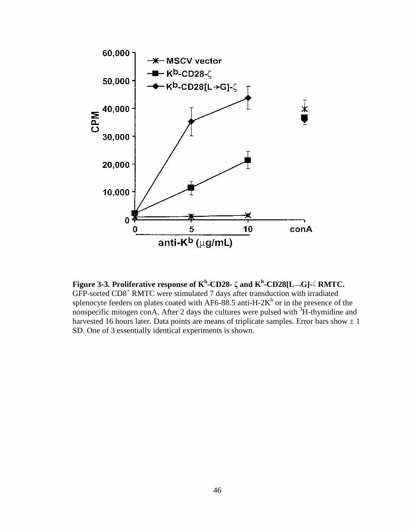
46
Figure 3-3. Proliferative response of Kb-CD28- ζ and Kb-CD28[L G]- RMTC. GFP-sorted CD8+ RMTC were stimulated 7 days after transduction with irradiated splenocyte feeders on plates coated with AF6-88.5 anti-H-2Kb or in the presence of the nonspecific mitogen conA. After 2 days the cultures were pulsed with 3H-thymidine and harvested 16 hours later. Data points are means of triplicate samples. Error bars show ± 1 SD. One of 3 essentially identical experiments is shown.

47
Figure 3-4. IFN- production by Kb-CD28- and Kb-CD28[L G]- RMTC. GFP-sorted CD8+ RMTC were stimulated 7 days after transduction in the presence of splenocyte feeders on plates coated with 5 µg/mL AF6-88.5 anti-Kb, with conA, or cultured in the absence of stimulation. Stimulation-induced IFN- production was measured by Bioplex assay using anti-IFN--coated beads. Data points are means of triplicate samples. Error bars show ± 1 SD; *, less than 1 ng/mL.

48
(58). We tested if the new mutated CR is more efficient in this aspect. We used OT-1
cells from the lymph nodes or spleen of mice transgenic for a TCR specific for Kb/Ova
257-264 as target cells. Ova peptide-pulsed Kb-CD28[L G]-ζ RMTC are able to
specifically kill target OT-1 T-cells more efficiently than the Kb-CD28-ζ RMTC (Figure
3-5). This is a confirmation that the dileucine to glycine mutation enhances the effector
function of RMTC, namely the ability to proliferate, secrete cytokines, and lyse Ova-
specific target cells.
The data above show that mutations in the dileucine motif of CD28 enhance the
effector function of RMTC in vitro. We next wanted to find out if therapeutic cells were
also effective in vivo. To do this, we coinjected Ova peptide-pulsed RMTC transduced
with the wild type or mutated CR along with OT-1 transgenic cells labeled with
carboxyfluorescein succinnimidyl ester (CFSE) into severe combined immunodeficiency
(SCID) mice. Both Kb-CD28[L G]-ζ and Kb-CD28-ζ peptide-pulsed RMTC efficiently
eliminated OT-1 T-cells. This means that the mutated CR-transduced therapeutic cells are
functional in vivo. These cells also show a slight – though not statistically significant –
increase in the cytolytic activity compared to the unmutated CR-transduced RMTC,
which might be an indication for their preferential use in the future (Figure 3-6).
3.2.4 Conclusions
T-cells redirected against pathologic lymphocytes are potential tools in cellular
immunotherapy. Therapeutic cells can migrate to different sites in the body, are long-
lasting, and exhibit different effector functions. Chimeric receptors composed of antigen-
recognition and signaling domains from the TCR, such as the cytoplasmic ζ tail, were
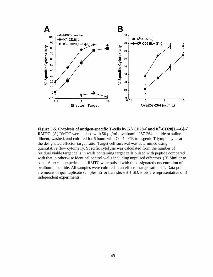
49
Figure 3-5. Cytolysis of antigen-specific T-cells by Kb-CD28- and Kb-CD28[L G]-RMTC. (A) RMTC were pulsed with 50 µg/mL ovalbumin 257-264 peptide or saline diluent, washed, and cultured for 6 hours with OT-1 TCR transgenic T lymphocytes at the designated effector-target ratio. Target cell survival was determined using quantitative flow cytometry. Specific cytolysis was calculated from the number of residual viable target cells in wells containing target cells pulsed with peptide compared with that in otherwise identical control wells including unpulsed effectors. (B) Similar to panel A, except experimental RMTC were pulsed with the designated concentration of ovalbumin peptide. All samples were cultured at an effector-target ratio of 1. Data points are means of quintuplicate samples. Error bars show ± 1 SD. Plots are representative of 3 independent experiments.

50
Figure 3-6. In vivo killing of antigen-specific T lymphocytes using Kb-CD28- and Kb-CD28[L G]- RMTC. A total of 107 CFSE-labeled OT-1 lymph node cells were adoptively transferred intravenously into SCID mice; 107 Kb-CD28-ζ or Kb-CD28[LG]-ζ peptide-pulsed or unpulsed RMTC were then adoptively transferred intravenously at an anatomically separate location. Twenty-four hours after transfer, spleen and mixed lymph nodes (mesenteric, cervical, axillary, inguinal) were isolated and single-cell suspensions prepared, stained with V2-specific antibody, and analyzed by flow cytometry. (C) Normalized numbers of target cells in the spleens of treated animals. The ratio of residual transferred (CFSE-positive) RMTC targets (Vα2-positive) to nontargets (Vα2-negative) was calculated to control for the efficiency of adoptive transfer in mice treated with peptide-pulsed or control unpulsed effectors. (D) Analysis of LN cells. Error bars show ± 1 SD. Results are representative of 3 independent experiments.
51
proven effective in redirecting T-cells against target cells. Since their effector abilities
depend upon the efficiency of signal transduction and the presence of costimulatory
molecules, new generation of CRs include both the CD28 molecule and the ζ tail.
Although we and others have observed enhanced function of the CR bearing the
extracellular and transmembrane part of the Kb molecule linked to CD28-ζ in T cell
hybridoma, there was no noticeable difference between the functional effectiveness of
this construct compared to the one lacking CD28 when transduced into primary murine
T-cells. Also, the surface expression of this CR was diminished. We hypothesized that
the poor expression resulted in the lack of improvement of their effector abilities. We
here identify a novel dileucine motif in the murine CD28 molecule that hampers CR
expression and function.
Up to now, two classes of dileucine motifs have been described, containing
[DE]XXXL[LI] or DXXLL. They play critical roles in the sorting of transmembrane
proteins. DXXLL signals are distinct dileucine-based sorting signals that cycle between
the trans-Golgi network and endosomes and mediate incorporation into clathrin-coated
vesicles that bud from the trans-Golgi network for transport to the endosomes. They bind
to the GGA family of ARF-dependent clathrin adaptors localized in the trans-Golgi
network. Mutations of either D or LL upstream inactivate the signals and result in
increased surface expression of the proteins. [DE]XXXL[LI] motifs bind to AP-1, 2, or 3
and are less intolerant to mutations of the second isoleucine that can be mutated without
impairing activity (74). The SRRNRLL that we discovered in the CD28 molecule is not
identical to either [DE]XXXL[LI] or DXXLL motifs; rather, it is more similar to a
[DE]XXXL[LI] motif that can also be found in other molecules, such as human CD4
52
(SQIKRLLSE), mouse GLUT4 (RRTPSLLEQ) or human VAMP4 (SERRNLLED). This
motif has a positively charged arginine residue upstream the dileucine motif instead of
the canonical negatively charged amino acid residues.
Our experimental results disentangle the functional properties of the dileucine
motif in CD28 molecule. Although disruption of this motif leads to enhanced surface
expression and effector function, internalization might not be directly related to this fact.
CD3γ molecule also has a SDKQTLL sequence responsible for internalization of the
TCR but only after phosphorylation of serine (80). It is therefore, possible that
internalization requires a conformational shift that makes the motif accessible to sorting-
associated proteins. In our CR, the CD28 tail was taken out of its normal
environment/structure, possibly making the dileucine motif more exposed to the protein-
sorting apparatus, and leading to its constitutive activation.
The dileucine motif in CD28 limits the CR expression and signaling when
transduced into primary murine T-cells. Mutation of leucine residues to glycine improves
surface expression and upregulates proliferation, cytokine secretion, and cytolysis of
target cells.
In addition to the in vitro evidence, we also provide evidence that effector T-cells
bearing CR with mutated dileucine motifs are active in vivo, leading to selective killing
of specific target cells. This constitutes a possible future therapeutic approach in
transplant tolerance and autoimmunity.

53
Chapter 4. Development and function of humanized chimeric receptors
4.1 Introduction to the development of humanized chimeric receptors
Surrogate chimeric receptors designed to bear an antigenic peptide linked to parts
of MHC class I or II molecule and signaling parts of the TCR may be used as therapeutic
tools (57). These CRs can be transduced into T lymphocytes giving rise to receptor-
modified T-cells designed to specifically target only pathogenic, peptide-specific T-cells.
In initial studies our laboratory used a T-lineage specific promoter to express in
transgenic mice a chimeric receptor that included the immunodominant epitope of myelin
basic protein (MBP) in SJL mice linked to its restricting I-As MHC and the signaling
domain of TCR-ζ. MBP-specific T-lymphocytes stimulated these transgenic RMTC,
inducing effector functions, such as cytokine secretion, proliferation, and/or cytolysis of
target cells. CD8+, T helper 2, or CD4+CD25+ transgenic RMTC showed therapeutic
activity in EAE, even after the dissemination of T cell responses through epitope spread
(55). These data supported the application of RMTC immunotherapy in autoimmune
diseases.
I have examined whether RMTC carrying humanized CRs can be similarly
redirected against autoantigen-specific T lymphocytes. These humanized CRs were
engineered by genetically linking autoantigenic peptide, MHC, and TCR-ζ or other
signaling domains. The MHC-Ag serves as bait that specifically recognizes the TCR of
autoantigen-specific T lymphocytes. Cognate TCR recognition of the extracellular MHC-
Ag crosslinks the chimeric receptor, activating the RMTC through the receptor’s
signaling domains.

54
Linkage analyses have associated HLA-DR2 (DRA*01/DRB1*1501) in patients
of European descent with susceptibility to MS. Additionally, reactivity to a specific
epitope, MBP84-102, restricted to this HLA has been well characterized in patients with
MS (86). In this study we took advantage of this established association to design two
chimeric receptors specific for DR2-restricted, MBP84-102-specific T-cells. Each
heterodimeric receptor includes an extracellular and transmembrane domain comprised of
HLA DRA*0101/DRB1*1501 with genetically linked MBP84-102 peptide. One receptor
pair incorporates the cytoplasmic ITAM-rich domain of TCR-ζ on both the α and β
chains (MBP-DR2-ζ). The second receptor pair lacks these signaling chains (MBP-
DR2), allowing us to define the role of receptor signaling in therapeutic cell function,
which we were unable to do with the transgenic cells in our previous mouse model. The
receptors were placed in an MSCV-based retroviral vector to allow us to determine
whether adoptively transferred T-lymphocytes retrovirally transduced with the tri-
cistronic (α, β receptor, GFP) chimeric constructs are functional, able to re-direct effector
activity, and capable of modulating autoimmune disease in a humanized model system.
In this chapter, I describe our efforts to create humanized chimeric receptors and
validate them through in vitro studies of the therapeutic cells’ effector abilities. The
receptors were transduced into two types of effector cells, immortalized TCR-CD4-CD8-
4G4 T-cell hybridoma and primary CD8+ T lymphocytes. The activity of the receptors
against cognate MBP84-102/DR2-specific T-cells was tested using either a hMBP84-
102-specific Ob1A12 T-cell hybridoma (or Ob hybridoma), a T-cell clone derived from a
patient with relapsing-remitting multiple sclerosis, or T-cells from mice made transgenic
with rearranged TCR derived from the Ob hybridoma, Ob Tg mice (87). These Ob Tg

55
mice were bred with another strain that constitutively expresses human DR2 gene using
the human DR2 promoter (Fug 5 mice) (88). As a negative control for our in vitro studies
we used 6F11 T cell cell hybridoma specific for MBP89-101 epitope (same as hMBP 84-
102) restricted by I-As.
4.2 Materials and methods
4.2.1 Design of humanized MBP-DR2-ζ construct
The MBP-DR2-ζ construct contains two polypeptides. The first one includes the
extracellular and transmembrane domains of the human MHC class II beta chain
DRB1*1501 linked to the human MBP peptide epitope 84-102 and the ζ cytoplasmic
domain of the TCR. This was then linked to the human MHC class II alpha chain
DRA*0101 paired with the ζ cytoplasmic tail of TCR. The alpha and beta chains were
connected by a small amino acid sequence extracted from the Thosea asigna virus (TaV).
We cloned the two genes in a single open reading frame by eliminating the stop codon at
the end of the first ζ tail. The 2A sequence allows for polycistronic message formation.
As the mRNA is translated, the ribosome pauses at the 2A sequence and cleaves the N-
terminal peptide from the nascent protein product. The ribosome does not release the
mRNA however, and continues to synthesize the C-terminal polypeptide. Following
cleavage, the 2A peptide remains attached to the C-terminus of the first gene. The
advantage of this bicistronic system using 2A sequences is the same amount of two
distinct proteins, α and β chains of the chimeric construct, is produced (89). Internal
ribosomal entry sites (IRES) can also be added to mRNA to permit for polycistronic

56
message production, however, variable translational efficiencies are observed from the 5’
mRNA cap and the IRES, leading to non-stoichiometric protein production. This
synthetic heterodimeric gene was subcloned into the murine stem cells virus (MSCV)
retroviral expression vector that contains an IRES-linked green fluorescent protein
(GFP) for an easy identification of the gene by fluorescence activated cell sorting (FACS)
analysis.
4.2.1.1 Generation of leader sequence
We started by extracting total RNA from MGAR cell lines using RNeasy
procedure (Qiagen, Valencia, CA). MGAR cells are Epstein-Barr virus (EBV)-
transformed B cells that express DRA*0101/DRB1*1501. We used the following
protocol:
10x106 MGAR cells were counted and spun for 5 minutes in a centrifuge tube.
The supernatant was discarded and 600 µl lysis buffer RTL (Quiagen) containing 10
µl/ml 2-mercaptoethanol was added to the pellet which was then resuspended by
vortexing. The disrupted cells were then homogenized by pipetting the lysate directly
onto a QIAshredder column followed by a 2 minute-centrifugation in a microcentrifuge at
maximum speed and collection of the homogenized lysate. One volume (600 µl) of 70%
ethanol was added to the homogenized lysate, mixed, applied to an RNeasy minispin
column carried by a 2-ml collection tube, and centrifuged for 15 seconds at 8000 x g.
This step was repeated until the whole volume was used and the flow-through was
discarded every time. 700 µl buffer RW1 (Quiagen) was added to the column and
centrifuged again like above, followed by transfer of the column onto a new collection
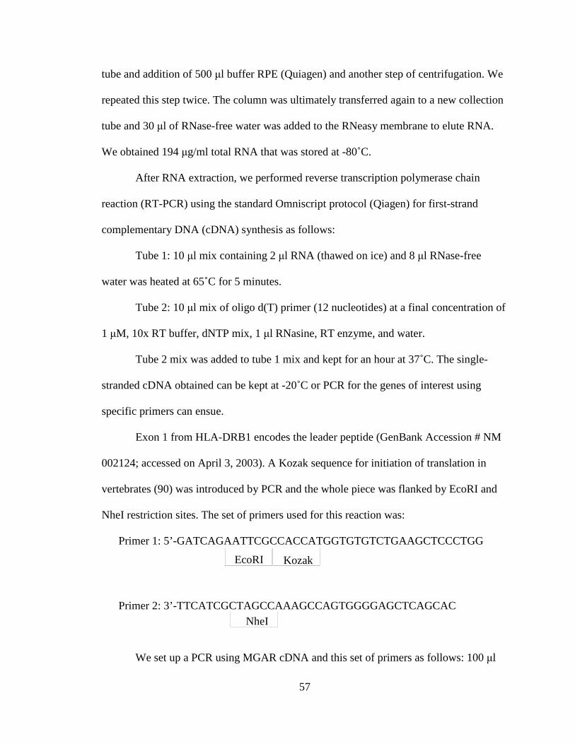
57
tube and addition of 500 µl buffer RPE (Quiagen) and another step of centrifugation. We
repeated this step twice. The column was ultimately transferred again to a new collection
tube and 30 µl of RNase-free water was added to the RNeasy membrane to elute RNA.
We obtained 194 µg/ml total RNA that was stored at -80˚C.
After RNA extraction, we performed reverse transcription polymerase chain
reaction (RT-PCR) using the standard Omniscript protocol (Qiagen) for first-strand
complementary DNA (cDNA) synthesis as follows:
Tube 1: 10 µl mix containing 2 µl RNA (thawed on ice) and 8 µl RNase-free
water was heated at 65˚C for 5 minutes.
Tube 2: 10 µl mix of oligo d(T) primer (12 nucleotides) at a final concentration of
1 µM, 10x RT buffer, dNTP mix, 1 µl RNasine, RT enzyme, and water.
Tube 2 mix was added to tube 1 mix and kept for an hour at 37˚C. The single-
stranded cDNA obtained can be kept at -20˚C or PCR for the genes of interest using
specific primers can ensue.
Exon 1 from HLA-DRB1 encodes the leader peptide (GenBank Accession # NM
002124; accessed on April 3, 2003). A Kozak sequence for initiation of translation in
vertebrates (90) was introduced by PCR and the whole piece was flanked by EcoRI and
NheI restriction sites. The set of primers used for this reaction was:
Primer 1: 5’-GATCAGAATTCGCCACCATGGTGTGTCTGAAGCTCCCTGG
Primer 2: 3’-TTCATCGCTAGCCAAAGCCAGTGGGGAGCTCAGCAC
We set up a PCR using MGAR cDNA and this set of primers as follows: 100 µl
NheI
EcoRI Kozak

58
reaction mix containing 2 µl cDNA, 10 µl buffer and magnesium chloride (MgCl2), 2 µl
dNTPs, 2 µl primer 1, 2 µl primer 2 (at a concentration of 25 pmol/µl), 0.5 µl high fidelity
Taq polymerase and 81.5 µl water was submitted to 35 cycles of amplification using the
following set-up:
First cycle of amplification: denaturation 5 minutes at 94˚C, annealing 2 minutes
at 55˚C, extension 3 minutes at 72˚C. 34 subsequent cycles followed using a gradient for
the annealing step, starting at 65˚C and subtracting 0.2˚C each cycle until it reached
55˚C. We checked the size of the leader sequence on a 1% agarose gel and we obtained
the expected size of 101 base pairs (bp). The PCR product was submitted for sequencing
in a tube of 12 µl total mix containing 1 µl of DNA, 1 µl of each primer, forward (primer
1) and reverse (primer 2) at 3.2 pmol, and water at the Hartwell Center, St. Jude
Children's Research Hospital.
Once the product was proven correct, we used the two enzymes, EcoRI and NheI,
to digest the leader sequence, gel purify it, and subclone it into pBluescript (pBS) II KS
(+) vector (Stratagene, La Jolla, CA) digested with the same enzymes.
4.2.1.2 Generation of MBP peptide sequence
NCBI nucleotide search found the sequence for Homo sapiens MBP (GenBank
Accession # NM 002385; accessed on April 30, 2003).The MBP epitope 84-102
(DENPVVHFFKNIVTPRTPP) was synthesized as oligonucleotides by the Hartwell
Center at St. Jude Children's Research Hospital, Memphis, TN. The oligonucleotides
used have the following sequence:
Forward primer: 5’-CTAGCGATGAAAACCCCGTAGTCCACTTCTTCAAGA
NheI

59
Reverse primer: 3’-GATCCGCCTGACCCTCCGCCACCGGACCCGCCACCTCCG
GGTGGTGTGCGAGGCGTCACAATGTTCTTGAAGAAGTGGACTACGGGTTTTC
ATCG
These two oligos were mixed in a 1:1 ratio, annealed at 94˚C, cooled down at
room temperature, and ligated it together with EcoRI/NheI-digested leader sequence into
pBS II KS vector using T4 DNA ligase (New England BioLabs, Ipswich, MA). This
time, the vector was digested with EcoRI (that the leader sequence has a site for) and
BamHI (present at the end of the peptide sequence or beginning of the beta chain). We
ended up with the following piece of 185 bp that we sequenced and confirmed to be
correct:
4.2.1.3 Generation of HLA-DRB1*1501
NCBI nucleotide search found the sequence for Homo sapiens HLA-DRB1
(GenBank Accession NM_002124 on May 5, 2003) mRNA with exons 2 and 3 coding
for the extracellular domains. We amplified the cDNA obtained as previously described
(4.2.1.1) with the following set of primers, adding two restriction sites, BamHI and AatII:
Forward primer: 5’-TCAGGCGGATCCGGGGACACCCGACCACGTTTCTTG
BamHI
BamHI
leader hMBP84-102EcoRI BamHI
NheI

60
Reverse primer: 3’-TCTTGACGTCTGATTCCTGAAGTAGATGAACAGCCC
PCR amplification yielded a 682 bp product. We subcloned this gene into pBS II
vector digested with BamHI/AatII, sequenced this, and confirmed the correct sequence.
4.2.1.4 Generation of the ζ chain
NCBI nucleotide search found the sequence for Homo sapiens CD3ζ (GenBank
Accession NM_000734 on May 1, 2003)) mRNA. cDNA was provided by courtesy of
Dr. Dario Campana, St. Jude Children's Research Hospital, Memphis, TN and amplified
by PCR to obtain the cytoplasmic tail flanked by AatII and ApaI restriction sites using
the following set of primers:
Forward primer: 5’-AATCAGACGTCAAGAGTGAAGTTCAGCAGGAGCGCA
Reverse primer: 3’-GCCATGGGCCCGGGATTTTCCTCCACGTCCCCGCATGTT
AGAAGACTTCCCCTGCCCTCGGCTCTGCGAGGGGGCAGGGCCTGCATGTGAA
G
The reverse primer also included the reversed 2A sequence (discussed below),
which was synthesized as part of the oligonucleotide. Similar to the other genes, the zeta-
2A gene was also sequenced, confirmed correct, and subcloned into pBS II vector. The
product was 396 bp.
AatII
Aat II
Apa I

61
4.2.1.5 The 2A sequence
Thosea asigna virus (TaV) is a member of the Tetraviridae family comprised of
single-stranded RNA picornavirus whose polyproteins undergo co-translational cleavage
to produce several proteins (P1, P2, P3). The 2A sequence is a proteinase that cleaves at
its own amino terminus generating two protein products, P1 and P2. The 2A sequence
was previously used in retroviral vectors as an alternative system to IRES to generate a
polycistronic message (91). In the MBP-DR2-ζ and MBP-DR2 constructs the 2A
sequence is flanked by α and β chimeric receptor chains forming a unique open reading
frame. The cleavage of the polyprotein product occurs at the C-terminus end of the 2A
sequence, leaving it fused to the upstream β chain protein which is otherwise released
intact. The amino acid sequence of the 2A was:
5’-RAEGRGSLLTCGDVEENPGP-3’
We ligated the β chain gene to zeta-2A and subcloned them into pBS II vector
digested with BamHI and Apa I. We ended up with the following piece of 1078 bp that
was confirmed correct by sequencing:
4.2.1.6 Generation of HLA-DRA*0101
NCBI nucleotide search found the sequence for Homo sapiens HLA-DRA
(GenBank Accession NM_019111 on May 5, 2003) mRNA with exons 2 and 3 coding
Beta chain Zeta 2ABamHI ApaI
AatII
62
for the extracellular domains and exon 4 coding for the transmembrane domain and the
cytoplasmic tail. cDNA produced from MGAR cells as described at 4.2.1.1 was used to
isolated the DRA fragment. The extracellular and transmembrane domains of interest
were flanked by ApaI and AatII restriction sites. The following set of primers was used
for amplification of the two domains:
Forward primer: 5’-AATCCCGGGCCCATGGCCATAAGTGGAGTCCCTGTG
Reverse primer: 3’-CTCTTGACGTCAATCCCTTGATGATGAAGATGGTCCCAA
PCR amplification yielded a 736 bp product. This gene was subcloned into pBS II
vector digested with the two enzymes after sequencing and checking for the correct
sequence.
4.2.1.7 Generation of synthetic ζ tail
Two zeta tails were required for the MBP-DR2-ζ construct, one linked to the α
chain, the second linked to the β. For the β chain, native ζ sequence was used. For the α
chain, we chose to redesign the ζ tail by keeping the same amino acid sequence but
changing the codons that determine each amino acid. This was to ensure that
rearrangement between the two ζ chains would not occur during the production of
retrovirus. Synthetic ζ was engineered as two sets of large oligos, one for the first half of
the construct and the second one for the rest of the construct. The 3’ end of the first set
and the 5’ end of the second set, respectively, had a PstI restriction site. The two sets are
as follows:
ApaI
AatII

63
Set 1 - Forward primer: 5’-GGAGCGGATCCGACGTCCGCGTAAAATTTTCTCG
TTCAGCTGATGCACCAGCTTATCAACAAGGACAAAATCAACTATACAATGAA
CTGAACCTTGGTAGGCGTGAAGAATATGAC
Reverse primer: 3’-TTTGTCCTTCTGCAGCTCGTTATAAAGTCCCTCTTGTGGA
TTTTTTGCGACGTGGTTTACCTCCCATTTCTGGATCTCTTCCTCTACGTTTAAT
CTAG TACGTCATATTCTTCACGCCT
Set 2 – Forward primer: 5’-AACGAGCTGCAGAAGGACAAAATGGCAGAAGCA
TATTCAGAAATCGGAATGAAGGGAGAAAGAAGACGAGGTAAAGGCCATGAC
GGATTATAT
Reverse primer: 3’-GACTACTCGAGTTATTACCTTGGAGGTAATGCTTGCATA
TGTAAAGCATCATAAGTATCTTTTGTAGCCGTTGATAATCCTTGATATAACCG
TCATGGCC
The two sets of primers were annealed and made double stranded using one PCR
cycle (4 minutes denaturation at 94˚C, 1 minute annealing at 55˚C, 3 minutes extension at
72˚C) and high fidelity Taq polymerase, followed by purification and digestion with the
two sets of enzymes, BamHI – PstI and PstI – XhoI for the second one. The two pieces of
the synthetic ζ gene were then subcloned into pBS II vector digested with BamHI and
XhoI. The product was 354 bp long.
This synthetic gene was then ligated together with the α chain and subcloned
AatII BamHI
PstI
PstI
XhoI

64
again into pBS II vector digested with ApaI and XhoI. We ended up with a gene of 1090
bp shown below that was confirmed correct by sequencing:
4.2.1.8 Ligation into the final construct
We have avoided the inconvenience of cloning six genes of different sizes into a
considerably large vector such as MSCV (6.825 Kb) by subcloning two genes at a time
into a smaller vector, pBS II KS (+) of only 3 Kb. We ended up with three pieces of the
final product: leader-MBP, beta-ζ-2A, and alpha-synthetic ζ of 185, 1078, and 1090 bp,
respectively. We then subcloned the leader-MBP and the beta-zeta-2A into pBS II. This
step yielded the following clone of 1263 bp, shown below:
We digested this new gene with EcoRI/ApaI and inserted it along with the alpha-
synthetic ζ one digested with ApaI/XhoI into MSCV retroviral vector cut with
EcoRI/XhoI that includes an IRES linked to GFP. The whole leader-MBP-β-ζ-2A-α-
synthetic ζ construct was 2.352 Kb.
Alpha chainSynthetic zetaApaI XhoI
AatII
leader hMBP84-102EcoRI
NheI
Beta chain Zeta 2A ApaI
AatIIBamHI

65
4.2.2 Design of humanized tailless MBP-DR2 construct
This signaling deficient construct was produced in order to clarify the role of CR
signaling for RMTC function.
4.2.2.1 Generation of leader-MBP peptide
We used the same leader-MBP peptide gene that we previously used for the tail-
bearing construct restricted by the same sites, EcoRI and BamHI (see 4.2.1.1 and
4.2.1.2).
4.2.2.2 Generation of HLA-DRB1*1501
We amplified the cDNA with the following set of primers, adding the same
BamHI as for the previous construct, but changing the second site to an ApaI. The
reverse primer was a large oligonucleotide that also contained the 2A sequence in a
manner analogous to how we added 2A to the ζ chain in the MBP-DR2-ζ construct:
Forward primer: 5’-TCAGGCGGATCCGGGGACACCCGACCACGTTTCTTG
Reverse primer: 3’-GCCATGGGCCCGGGATTTTCCTCCACGTCCCCGCATGTT
AGAAGACTTCCCCTGCCCTCGGCTCTGCGAGGGGGCAGGGCCTGCATGTGAA
G
Similar to the other genes, the beta 2A gene was also sequenced and subcloned
into pBS II vector.
BamHI
Apa I
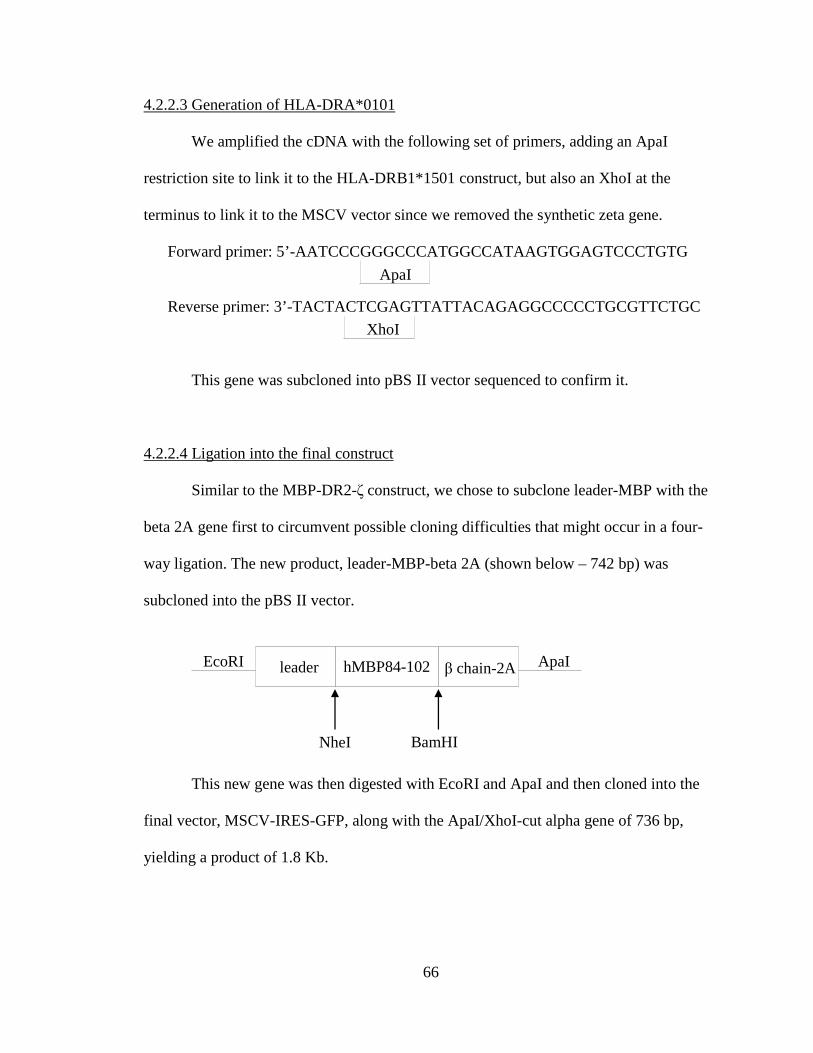
66
4.2.2.3 Generation of HLA-DRA*0101
We amplified the cDNA with the following set of primers, adding an ApaI
restriction site to link it to the HLA-DRB1*1501 construct, but also an XhoI at the
terminus to link it to the MSCV vector since we removed the synthetic zeta gene.
Forward primer: 5’-AATCCCGGGCCCATGGCCATAAGTGGAGTCCCTGTG
Reverse primer: 3’-TACTACTCGAGTTATTACAGAGGCCCCCTGCGTTCTGC
This gene was subcloned into pBS II vector sequenced to confirm it.
4.2.2.4 Ligation into the final construct
Similar to the MBP-DR2-ζ construct, we chose to subclone leader-MBP with the
beta 2A gene first to circumvent possible cloning difficulties that might occur in a four-
way ligation. The new product, leader-MBP-beta 2A (shown below – 742 bp) was
subcloned into the pBS II vector.
This new gene was then digested with EcoRI and ApaI and then cloned into the
final vector, MSCV-IRES-GFP, along with the ApaI/XhoI-cut alpha gene of 736 bp,
yielding a product of 1.8 Kb.
XhoI
ApaI
leader hMBP84-102EcoRI
NheI
β chain-2A ApaI
BamHI
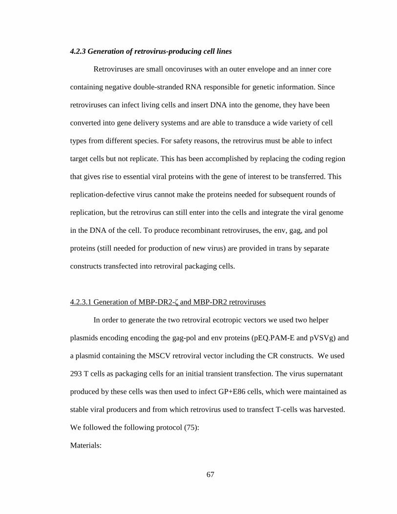
67
4.2.3 Generation of retrovirus-producing cell lines
Retroviruses are small oncoviruses with an outer envelope and an inner core
containing negative double-stranded RNA responsible for genetic information. Since
retroviruses can infect living cells and insert DNA into the genome, they have been
converted into gene delivery systems and are able to transduce a wide variety of cell
types from different species. For safety reasons, the retrovirus must be able to infect
target cells but not replicate. This has been accomplished by replacing the coding region
that gives rise to essential viral proteins with the gene of interest to be transferred. This
replication-defective virus cannot make the proteins needed for subsequent rounds of
replication, but the retrovirus can still enter into the cells and integrate the viral genome
in the DNA of the cell. To produce recombinant retroviruses, the env, gag, and pol
proteins (still needed for production of new virus) are provided in trans by separate
constructs transfected into retroviral packaging cells.
4.2.3.1 Generation of MBP-DR2-ζ and MBP-DR2 retroviruses
In order to generate the two retroviral ecotropic vectors we used two helper
plasmids encoding encoding the gag-pol and env proteins (pEQ.PAM-E and pVSVg) and
a plasmid containing the MSCV retroviral vector including the CR constructs. We used
293 T cells as packaging cells for an initial transient transfection. The virus supernatant
produced by these cells was then used to infect GP+E86 cells, which were maintained as
stable viral producers and from which retrovirus used to transfect T-cells was harvested.
We followed the following protocol (75):
Materials:

68
293 T cells and GP+E86 cells
Dulbecco’s modified Eagle’s medium (DMEM) supplemented with 10% fetal calf serum
(FCS) and penicillin-streptomycin-glutamine (PSG) 1%
pEQ.PAM-E plasmid
pVSVg plasmid
MBP-DR2-ζ/MBP-DR2 plasmid
Calcium chloride CaCl2 2.5M
2X Hepes Phosphate Buffered Saline (HPBS) pH 7.12
Method:
24-hours prior to transfection, 2x10⁶ (or 4x10⁶ the day of transfection) 293 T cells
were plated on a 10 ml tissue-culture treated Petri dish. On the day of transfection, four
hours prior to the procedure, the medium was changed with fresh complete DMEM. At
the time of transfection, cells were approximately 70% confluent. DNA mix was prepared
using 4 µg pEQ.PAM-E and 2 µg pVSVg and 4µg MBP-DR2-ζ/MBP-DR2 plasmid, 50
µl 2.5M CaCl2 and adjusted to 500 µl with distilled water at room temperature (RT). This
mix was then added to 500 µl HPBS dropwise while vortexing and incubated 1 minute at
RT. The whole mixture was added onto 293 T cells and incubated for 16 hours at 37˚C.
The next day the plates were washed with prewarmed phosphate buffered saline (PBS)
twice and new complete DMEM was added. 3x10⁵ GP+E86 cells were plated in a 10 ml
dish. 24-hours later, the medium from 293T cells was replaced, spun at 3000 rotations per
minute (RPM) for 5 minutes in a centifuge with rotor and then the supernatant was used
to infect GP+E86 cells twice a day, adding 8 µg/ml polybrene to facilitate binding and
entry of the retrovirus. The GP+E86 cell line was used to produce retroviral supernatant
69
which was then used to transduce 4G4 T cell hybridoma and activated primary murine T-
cells. It is recommendable that the GP+E86 cells be frozen in freezing mix (EHAA
medium with 20% heat inactivated FCS and 10% dimethyl sulfoxide (DMSO) and kept at
-80˚C as soon as the transfection has been finished. Since they are stably transfected with
retrovirus, they can be thawed out and frozen again multiple times. After thawing,
GP+E86 cells were washed once in HBSS medium and plated onto large or medium
tissue culture-treated flasks. Retroviral supernatant was collected daily and the medium
was replaced until the cells were completely confluent.
4.2.3.2 Retroviral transduction of 4G4 T-cell hybridoma
GP+E86 cells previously transfected with either MBP-DR2-ζ CR (GP+E86-MBP-
DR2-ζ) or MBP-DR2 CR (GP+E86-MBP-DR2) were thawed out from the liquid nitrogen
tank, washed once with 1x Hanks’ balanced salt solution (HBSS, Gibco, Carlsbad, CA),
and cultured in 75 or 150 ml culture-treated flanks. Viral supernatant was collected at 24
hours and spun at 3000 rpm for 5 minutes. 4x10⁶ 4G4 T-cell hybridoma were added per
well in a 6-well plate and 2 mls of retroviral supernatant and 8 µg/ml polybrene were
added, and the cells were spun at1800 rpm for 90 minutes in a centrifuge with rotor. The
procedure was repeated twice for a better yield of GFP+ cells.
4.2.3.3. Flow cytometry of retrovirally-transduced cells
To check for HLA class II expression, transduced 4G4 cells were resuspended in
FACS buffer (PBS with 1% BSA and 0.1% sodium azide (NaN3, Sigma) and incubated
30 min on ice with primary phycoerythrin-conjugated (PE) anti-human HLA-DR

70
antibody (Becton Dickinson, Franklin Lakes, NJ) at the concentration recommended by
the supplier. The cells were then washed and resuspended in FACS buffer and analyzed
using a FACSCalibur (BD Biosciences) and CellQuest software. CR-transduced
hybridoma were then flow cytometric sorted for GFP+ on a MoFlo high-speed cell sorter
(DakoCytomation) (courtesy of Dr. Richard Cross, PhD, St. Jude Children's Research
Hospital).
4.2.3.4 Retroviral transduction of primary murine T-cells
Lymph nodes and spleen from C57BL/6 mice or OT-1 mice (transgenic for CD8+
cells) were harvested and processed into a single cell suspension by crushing through a
40-µm nylon filter. Red blood cells were lysed with Gey’s solution (7.0 g/l NH4Cl, 0.37
g/l KCl , 0.3 g/l Na2HPO4.12H2O, 0.024 g/l KH2PO4, 1.0 g/l glucose, 10.0 mg/l phenol
red, 8.4 mg/l MgCl2.6H2O, 7.0 mg/l MgSO4.7H2O, 6.8 mg/l CaCl2 and 45 mg/l NaHCO3)
and the lymphocytes were cultured in 6-well plates and stimulated with 2 µg/ml purified
mouse anti-CD3ε and 2 µg/ml anti-CD28 antibodies (Pharmingen, San Diego, CA) or 2
µg/ml Concanavalin A (ConA) overnight. After 24 and 48 hours, the media was carefully
removed and 2 mls/well of viral supernatant was added along with 8 µg/ml polybrene and
sorted for GFP expression by flow cytometry. GFP+ cells were re-fed and split into 6-
well plates when they reached a high density. They were also sorted for GFP+ and on day
10-12, the cells were restimulated with 2 µg/ml ConA and irradiated (4,000 rad)
syngeneic splenocytes.
71
4.2.4 Ob and 6F11 target hybridoma cell lines
4.2.4.1 Stimulation of Ob hybridoma with hMBP84-102 peptide
105 Ob and 6F11 cells were incubated overnight with four different
concentrations of purified hMBP84-102 peptide on a 96-well round-bottom plate in
complete Bruff medium (EHAA or Click’s medium enriched with 10% FCS, 1%
Penicillin-Streptomycin-Glutamine (PSG), and 4.2 µl 2-mercaptoethanol per 1,000 ml
medium). Irradiated (20, 000 rad) MGAR cells were used as antigen presenting cells. 24-
hours later, 60 µl of supernatant was removed and submitted for IL-2 secretion analysis
by Multiplex (Bio-Rad, Hercules, CA).
4.2.4.2 Stimulation of Ob hybridoma with mouse anti-humanVβ2 antibody
A 96-well flat bottom plate was coated with 10 µg/ml purified rat anti-mouse IgG
(Jackson Immunoresearch Laboratories, Inc., West Grove, PA) and incubated at 4˚C
overnight. 24-hours later, the plate was washed three times with HBSS solution and
coated with 50 µg/ml mouse anti-human TCR Vβ2 monoclonal antibody (Immunotech,
Warrenale, PA) either overnight at 4˚C or for two hours at 37˚C. The plate was washed
again and 105 Ob or 6F11 cells were added. 24-hours later 60 µl of supernatant was
removed and submitted for IL-2 secretion.

72
4.2.5 Stimulation of TCR/DR2 T lymphocytes
4.2.5.1 Stimulation of TCR/DR2 T lymphocytes with hMBP84-102 peptide
5x10⁵ cells from lymph nodes or splenocytes of double transgenic mice were
cultured in 96-well flat-bottom plates without antigenic peptide or with peptide in
different concentrations and irradiated splenocytes from humanized DR2 transgenic mice
(denoted as Fug5). After 72 hours of culture, the cells were pulsed with 1 µCi of
[3H]thymidine and harvested on filtermats16 hours later. All samples were analyzed in
triplicates.
4.2.5.2 Stimulation of TCR/DR2 T lymphocytes with EL4-MBP-DR2 cells
2x10⁶ T-cells from lymph nodes or splenocytes of transgenic mice were
stimulated with 4x10⁶ irradiated (20,000 rad) EL4-MBP-DR2 antigen-presenting cells
(EL4 thymoma cells transduced with the tailless MBP-DR2 construct) and the same
number of irradiated feeders. The cells were grown in complete Bruff medium with
10 U/ml recombinant IL-2 and split every other day. We have also used this procedure to
build an antigen-specific cell line by re-stimulating them in the same conditions on day
ten, using Con A as a mitogen.
4.2.6 Stimulation of RMTC with HLA-DRB antibody
The plate-bound antibody stimulation was set up as described at 4.2.4.2. Briefly,
transduced 4G4 RMTC were stimulated with three different concentrations of plate-
bound mouse anti-human HLA-DR2 antibody or control goat anti-mouse IgG. 24-hours
73
later, 60 µl of supernatant was removed and submitted for IL-2 secretion analysis by
Multiplex.
4.2.7 IL-2 secretion by RMTC in response to recognition of Ob hybridoma
5x10⁵ 4G4 hybridoma transduced with MSCV and MBP-DR2-ζ were cocultured
in three different ratios with Ob or 6F11 target hybridoma in a 96-well round bottom
plate for 8 hours. The cells were spun, the medium was replaced, and Brefeldin A (BFA,
Epicentre Technologies) was added for 6 hours at 37˚C and 5-10% CO2. The cells were
spun again at 4˚C and then washed with staining wash buffer (SWB; 1x PBS, 2% FBS,
0.1% NaN3) and BFA 10µg/ml. The primary surface antibody (mouse anti-human Vβ2
PE-conjugated) diluted in SWB+BFA was added and the plate was then incubated on ice
for 20 minutes, then washed with PBS+BFA twice and resuspended in the same mix. The
cells were then fixed with 2% formaldehyde in PBS for 20 minutes at RT, washed and
incubated in permeabilization buffer (SWB + 0.5% saponin) for 10 minutes at RT. The
cells were then incubated with the APC (Allophycocyanin)-conjugated rat anti-mouse IL-
2 monoclonal antibody (Becton Dickinson) for 30 minutes on ice, followed by a wash
with PBS, and resuspension in SWB. Flow cytometric analysis was performed after
gating on the double positive, APC+ (on FL4 axis)/GFP+ (on FL1 axis) cells or
alternately, on APC+ (on FL4 axis)/Vβ2+ (on FL2 axis) cells. This way we can identify
the cells that secrete IL-2 (featured by APC staining) by gating on either FL1/FL4 (GFP+
cells secreting IL-2) or FL2/FL4 (Ob hybridoma secreting IL-2).
74
4.2.8 IFN-γ secretion by RMTC in response to recognition of Ob hybridoma
5x10⁵ Ob hMBP84-102-specific hybridoma or 6F11 control cells were cocultured
with RMTC transduced with MSCV, MBP-DR2-ζ, and MBP-DR2 chimeric receptors for
24 hours. 60 µl of supernatant was then removed and submitted for cytokine secretion
analysis by Multiplex.
4.2.9 Proliferation of RMTC in response to stimulation by Ob hybridoma
5x10⁴ GFP+-sorted CD8+-transduced RMTC cells were incubated with different
ratios of irradiated Ob or 6F11 hybridoma and syngeneic feeders on a 96-flat bottom
plate. After 72 hours, the cells were pulsed with 1µCi [3H] thymidine and harvested after
16 hours. Proliferation was measured by liquid scintillation counting of incorporated [3H]
thymidine. The samples were analyzed in duplicates and plotted as mean ± 1 stdev.
4.2.10 Cytolysis of target cells by RMTC
4.2.10.1 Cytolysis of Ob target hybridoma by RMTC assayed by radioactive chromium
(Cr) release
4x10⁶ Ob hybridoma specific for human MBP84-102 peptide or 6F11 control
hybridoma specific for MBP89-101 were incubated with 200 µCi 51Cr for 30 minutes at
37˚C with intermittent shaking. After half an hour, the cells were washed three times with
PBS saline solution and cocultured in different effector to target ratios with CD8+-RMTC
transduced with MBP-DR2-ζ, MBP-DR2, or MSCV vector for 4 hours on a 96-well
round bottom plate. All cell combinations were set up as triplicates. Target cells in

75
medium only were also set for controls. Labeled antigenic targets are recognized and
lysed by RMTC, releasing radioactivity into the supernatant. 20 µl TritonX (Sigma-
Aldrich, St Louis, MO) was added to 51Cr-labeled Ob hybridoma (control wells) and
served as a positive control for maximal release of radioactive Cr. Spontaneous lysis
(control 51Cr) was estimated by the release of radioactive Cr by Ob hybridoma alone.
After incubation at 37˚C, the plate was centrifuged and 100 µL supernatant was
harvested and counted. We used the formula below to calculate the percent of specific
cytolysis from the release of 51Cr:
(Experimental 51Cr – control 51Cr)/(maximal 51Cr – control 51Cr) x100
4.2.10.2 In vitro cytolysis of primary MBP-DR2-specific cells by RMTC
105 T-cells/well from TCRxDR2 double transgenic (DTG) or TCRxDR2xhCD4
triple transgenic (TTG) mice were cocultured with CD8+ RMTC at different ratios
overnight in a 96-well round bottom plate. The next day, the plate was centrifuged and
cells were double stained with the mouse anti-human Vβ2.1 antibody (PE) and rat anti-
mouse CD4+ or CD8+ antibody (APC). After 30 minutes incubation on ice, the cells were
washed and resuspended in SWB. In order to find out the absolute number of target cells
left after overnight cytolysis, we used TruCOUNT assay and added TruCount high
control fluorescent beads (BD Bioscience, San Jose, CA) at a final concentration of 2000
beads/sample. These beads were used as markers to guarantee that equal fractions of each
sample were enumerated, thus allowing quantitative comparison of the number of events
and eliminating the variability between samples. The absolute number of cells in the
sample can be determined by normalizing to the number of acquired beads.

76
FACSCaliburTM was used with forward (FCS) and side scatter (SSC) set in a logarithmic
mode. TruCount beads were gated in a FCS/SSC plot and analysis was stopped when 500
beads were counted. We then ran the sample tubes and analyzed the double positive
hVβ2.1+CD4+ or hVβ2.1+CD8+ cells after gating and counting 500 beads.
4.2.10.3 In vitro cytolysis of primary MBP-DR2-specific T-cells by RMTC detected by
stimulation with EL4-MBP-DR2 cells
This experiment was set up similarly to 4.2.10.2. Unlike the previous experiment,
we chose not to directly analyze the residual cells after the overnight coculture of the two
cell types, but instead we added 2x10⁵ EL4-MBP-DR2 antigen-presenting cells or control
EL4 cells at the time of the culture, for five days. On day five we stained each well with
mouse anti-human Vβ2.1 and rat anti-mouse CD4+ and CD8+ antibodies and analyzed the
residual double positive cell counts by quantitative flow cytometry.
4.2.10.4 Cytolysis of target DTG cell lines by RMTC
105 double or triple transgenic cell lines (obtained as described in 4.2.5.2) were
cocultured with MSCV retroviral vector control, MBP-DR2-ζ, or MBP-DR2 RMTC in a
96-well round bottom plate for six hours at four different ratios. After 6 hours, we stained
the cells as described in 4.2.10.2 and evaluated the number of residual hVβ2.1+CD4+ and
hVβ2.1+CD8+ cells by quantitative FACS.

77
4.3 Results
4.3.1 Design of chimeric receptors
In order to specifically target pathologic, hMBP-specific T lymphocytes, we
engineered two chimeric receptors: MPB-DR2-ζ and MBP-DR2. The first CR included
the extracellular and transmembrane domains of the human MHC class II beta chain
DRB1*1501 linked to the human MBP peptide epitope 84-102 and the ζ cytoplasmic
domain of the TCR. A 2A sequence from TaV connected this part of the construct with
the human MHC class II alpha chain DRA*0101 paired with the ζ cytoplasmic tail of
TCR. The whole gene was subcloned in MSCV retroviral expression vector that contains
an IRES site and the GFP gene (Figure 4-1). In parallel we designed a tail-deficient CR
that lacked any signaling moiety from the TCR (Figure 4-2).
4.3.2. Transfection of CRs into transient and permanent viral-producing cell lines
Transfection of both retroviruses had a good yield in both 293T and GP+E86 cells
(Figures 4-3 and 4-4). The latter cell line was used to produce retroviral supernatant
which was then used to transduce 4G4 T-cell hybridoma and activated murine T-cells.
4.3.3 Transduction of 4G4 T-cell hybridoma and primary murine T-cells
Transduction of both 4G4 T-cell hybridoma and primary murine T-cells had a
good yield, although we noticed a difference between the efficiency of transduction in
4G4 hybridoma and primary T lymphocytes. 4G4 cells transduced with the ζ-bearing C
showed an equivalent surface expression of the receptor compared to the ζ-deficient CR,

78
Figure 4-1. MSCVII-GFP-MBP-DR2-ζ construct. The immunodominant epitope of the human myelin basic protein was genetically linked to the extracellular and transmembrane domains of MHC class II β chain chain and human cytoplasmic TCR ζ chain. This was then paired with the extracellular and transmembrane domains of MHC class II α chain and another cytoplasmic signaling tail. The 2A sequence from FMDV was inserted between the DRB β and α chain to provide equal amounts of the bicistronic message.
GFPDRB1*1501
leaderMBP
84-102DRB1*1501
EC+TMCytoplasmicZeta tail+2A
DRA*0101EC+TM
Syntheticcytoplasmic
Zeta tail
MSCV
(8905 bp)
IRES
EcoRI
(1406)
NheI
(1502)
BamHI
(1598)
AatII
(2279)
ApaI
(2676)
AatII
(3407)
XhoI
(3752)

79
Figure 4-2. MSCV II-GFP-MBP-DR2 construct. This construct is similar to MSCV II-GFP-MBP-DR2-ζ construct (in Figure 4-1) with the exception of the ζ cytoplasmic signaling tail.
GFPDRB1*1501leader
MBP84-102
DRB1*1501 EC+TM
2A DRA*0101EC+TM
MSCV(8257)
IRES
EcoRI
(1406)
NheI
(1502)
BamHI
(1598)
ApaI
(2334)
XhoI
(3104)
GFPDRB1*1501leader
MBP84-102
DRB1*1501 EC+TM
2A DRA*0101EC+TM
MSCV(8257)
IRES
EcoRI
(1406)
NheI
(1502)
BamHI
(1598)
ApaI
(2334)
XhoI
(3104)
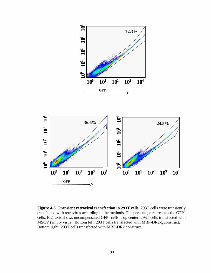
80
Figure 4-3. Transient retroviral transfection in 293T cells. 293T cells were transiently transfected with retrovirus according to the methods. The percentage represents the GFP+ cells. FL1 axis shows uncompensated GFP+ cells. Top center: 293T cells transfected with MSCV (empty virus). Bottom left: 293T cells transfected with MBP-DR2-ζ construct. Bottom right: 293T cells transfected with MBP-DR2 construct.
102100 101 103 104100 101 103 10410
010
110
210
310
410
010
110
210
310
4
72.3%
GFP
100
101
102
103
104
100
101
102
103
104
36.6% 24.5%10
010
110
210
310
410
010
110
210
310
4
GFP
102100 101 103 104100 101 103 104102100 101 103 104100 101 103 104
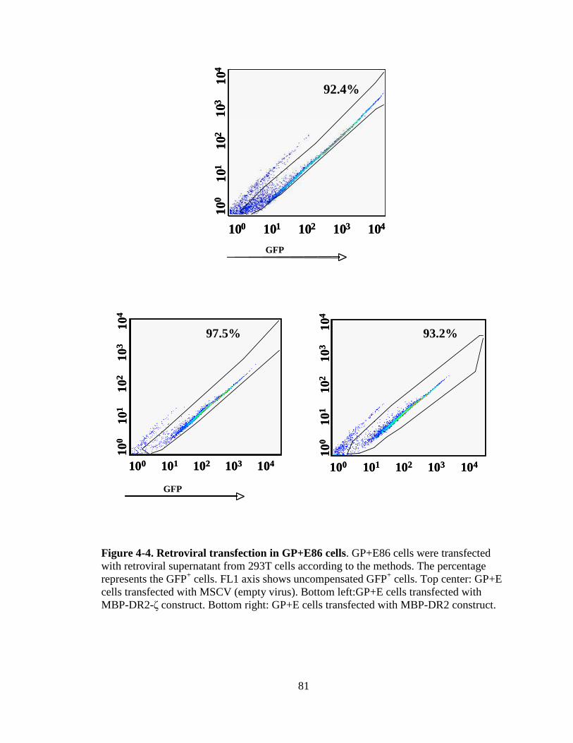
81
Figure 4-4. Retroviral transfection in GP+E86 cells. GP+E86 cells were transfected with retroviral supernatant from 293T cells according to the methods. The percentage represents the GFP+ cells. FL1 axis shows uncompensated GFP+ cells. Top center: GP+E cells transfected with MSCV (empty virus). Bottom left:GP+E cells transfected with MBP-DR2-ζ construct. Bottom right: GP+E cells transfected with MBP-DR2 construct.
100 101 102 103 104100 101 102 103 10410
010
110
210
310
410
010
110
210
310
4
92.4%
GFP
GFP
100 101 102 103 104100 101 102 103 104
100
101
102
103
104
100
101
102
103
104
97.5%
100 101 102 103 104100 101 102 103 104
100
101
102
103
104
100
101
102
103
10493.2%

82
whereas transduction in murine T lymphocytes showed an approximately 30% increase in
the surface expression levels of MBP-DR2 CR compared with MBP-DR2-ζ (Figures 4-5
and 4-6).
In conclusion, there’s a clear difference between the transduction efficiency in
hybridoma compared to activated T-cells. Hybridoma are very uniform, clonally
expanded cells that would exhibit the same ability to harbor a retrovirally-transduced
gene. The intracytoplasmic signaling tail renders the CR capable of signal transduction
and therefore, primary activated T-cells transduced with the tail-bearing CR will
internalize the surrogate receptor, leading to a decreased surface expression of the CR.
Removal of the signaling tail restores the surface expression of the CR to almost the same
levels as MSCV control.
4.3.4 Specificity of Ob hybridoma
Before we proceeded to use Ob hybridoma as target cell lines, we first needed to
ensure their antigenic-specificity by testing their response to stimulation with peptide and
the mouse anti-human Vβ2 antibody.
4.3.4.1 Stimulation of Ob hybridoma with hMBP84-102 peptide
To verify the specificity of Ob hybridoma for hMBP, we stimulated the cells with
different concentrations of antigenic peptide. The Ob hybridoma secreted IL-2 in
response to peptide stimulation. The amount of IL-2 secreted by the cells was directly
proportional to the amount of antigenic peptide used to stimulate the cells. Control 6F11
hybridoma did not show any response to stimulation with hMBP restricted by HLA-DR2

83
Figure 4-5. Expression of HLA-DR2 on 4G4 T-cell hybridomas transduced with chimeric receptors. MSCV retroviral vector control, ζ-bearing, and ζ-deficient CRs were transduced into 4G4 TCR-/- hybridomas. The efficiency of transduction is shown by the amount of GFP which represents the surface expression level of the chimeric receptors. The cells were stained with mouse anti-human HLA-DR2 antibody and then analyzed by FACS. Left panel: MSCV-transduced 4G4 T cells. Middle panel: MBP-DR2-ζ-transduced 4G4 T cells. Right panel: MBP-DR2-transduced 4G4 T cells.
58.1%
100 101 102 103 104100 101 102 103 104
100
101
102
103
104
100
101
102
103
104
GFP
DR
259.1%
100
101
102
103
104
100
101
102
103
104
100 101 102 103 104100 101 102 103 104
69.2%
100
101
102
103
104
100
101
102
103
104
100 101 102 103 104100 101 102 103 104

84
Figure 4-6. Expression of HLA-DR2 on primary murine CD8 + T-cells transduced with chimeric receptors. MSCV retroviral vector control, ζ-bearing, and ζ-deficient CRs were transduced into primary CD8+ T-cells. The efficiency of transduction is shown by the amount of GFP which represents the surface expression level of the chimeric receptors. The cells were stained with mouse anti-human HLA-DR2 antibody and then analyzed by FACS. Left panel: MSCV-transduced T-cells. Middle panel: MBP-DR2-ζ-transduced T-cells. Right panel: MBP-DR2-transduced T-cells.
40.4%
52.7%
71.1%
78%
GFP
DR
2 100
101
102
103
104
100
101
102
103
104
100
101
102
103
104
100
101
102
103
104
100
101
102
103
104
100
101
102
103
104
100 101 102 103 104100 101 102 103 104 100 101 102 103 104100 101 102 103 104 100 101 102 103 104100 101 102 103 104

85
(Figure 4-7 upper panel).
4.3.4.2 Stimulation of Ob hybridoma with mouse anti-humanVβ2 antibody
Ob hybridoma are specific for hMBP84-102 restricted by HLA-DR2 and they
stain for hVβ2 antibody. Upon stimulation with plate-bound mouse anti-human Vβ2, Ob-
specific, but not 6F11 control cells responded to this stimulation by IL-2 secretion
(Figure 4-7 lower panel).
In conclusion, we hereby proved that Ob hybridoma constitutes a clonal
population of cells specific for hMBP84-102 peptide and they respond stimulation with
either the peptide or the specific Vbeta antibody by proliferation.
4.3.5 Specificity of T lymphocytes from TCR/DR2 transgenic mice
4.3.5.1 Stimulation of TCR/DR2 T lymphocytes with hMBP84-102 peptide
To reproduce previously published data, we stimulated T lymphocytes from
double transgenic TCRxDR2 mice with hMBP84-102. We did not obtain consistent
results, and the proliferative response of T-cells proved extremely variable with different
mice and experiments. T-cells removed from the lymph nodes of some of the mice
responded very well to peptide stimulation, while others did not (Figure 4-8).
4.3.5.2 Stimulation of TCR/DR2 T lymphocytes with EL4-MBP-DR2 cells
Considering the results from the previous experiment, we searched for alternative
ways of stimulating antigen-specific cells from DTG mice. Specifically, because we

86
Figure 4-7. Ob cells respond to stimulation by producing IL-2. Ob and control 6F11 hybridoma were stimulated with different dilutions of hMBP peptide (upper panel) or mouse anti-human TCR Vβ2 (lower panel) antibody and secretion of IL-2 was assayed after 24 hours. Only Ob hMBP-specific cells, but not 6F11 control cells responded to stimulation by the secretion of IL-2.
0100200300400500600700800900
1000
0 25 50 100
Ob 6F11
IL-2
sec
retio
n (p
g/m
l)
hMBP 84-102 (µg/ml)
0100200300400500600700800900
1000
0 25 50 100
Ob 6F11
IL-2
sec
retio
n (p
g/m
l)
hMBP 84-102 (µg/ml)
050
100150200250300350400450
no antibody Vβ2 IgG IgG+Vβ2
Ob 6F11
IL-2
sec
retio
n (p
g/m
l)

87
Figure 4-8. Proliferation of TCR/DR2 cells in response to hMBP peptide. T-cells from lymph nodes of TCR/DR2 mice were stimulated with different concentrations of peptide and 72 hours later 1µCi [3H] thymidine was added to each well. Proliferation was measured by liquid scintillation counting. Top figure: in this particular experiment, the transgenic T-cells proliferated robustly in response to peptide stimulation. Bottom figure: in this experiment, the transgenic T-cells did not respond to peptide stimulation.
CP
M
hMBP84-102(µg/ml)
0
50000
100000
150000
200000
250000
300000
350000
35 25 15 5 0
CP
M
hMBP84-102(µg/ml)
0
50000
100000
150000
200000
250000
300000
350000
35 25 15 5 0
0
50000
100000
150000
200000
250000
300000
350000
40 30 20 10 0
CP
M
0
50000
100000
150000
200000
250000
300000
350000
40 30 20 10 0
hMBP84-102 (µg/ml)

88
suspected that the low expression levels of DR2 gene on B cells from humanized class II
transgenic mice (Fug5) were impeding the proper stimulation of hMPB-specific cells, we
we tested the use for EL4 thymoma cells transduced with the MBP-DR2 tail-deficient
construct as a stimulus. This way the antigen-specific cells would undergo a proper
stimulation, but will not suffer the effects that the tail-bearing CR would provide. We
showed that EL4-MBP-DR2 cells, though not EL4 cells alone, stimulate lymph node or
spleen-derived T-cells from DTG or TTG mice due to expression of hMBP-DR2 (Figure
4-9).
4.3.6 Stimulation of RMTC by HLA-DRB antibody
Since RMTC are transduced with CRs containing the DR2 gene, we wanted to
first test the ability of our CRs to be stimulated by the specific MHC class II antibody.
We used 4G4 T cell hybridoma expressing the CRs. Upon stimulation with the plate-
bound mouse anti-human DR2 antibody, only 4G4-MBP-DR2-ζ cells responded by IL-2
secretion. This demonstrates that the chimeric receptor bearing the signal transduction
domain is the only one able to transmit a functional signal and that deletion of the ζ
cytoplasmic domain impairs this ability (Figure 4-10).

89
Figure 4-9. CD4+ and CD8+ TCR/DR2 cells respond to stimulation by EL4-MBP- DR2. CD4+ (top panel) and CD8+ (bottom panel) cells from lymph nodes and spleens of TCR/DR2 mice were sorted and stimulated with hMBP peptide, EL4 control, or EL4-DR2 antigen-presenting cell lines (EL4-MBP-DR2) for 72 hours. Proliferation was assayed by [3H]thymidine incorporation.
0
50000
100000
150000
200000
250000
APC APC+hMBP84-102
EL4
CP
M
CD4+ObFug5
EL4-MBP-DR2
0
50000
100000
150000
200000
250000
300000
350000
400000
APC APC+hMBP84-102
EL4 EL4-MBP-DR2
CP
M
CD8+ObFug5
0
50000
100000
150000
200000
250000
300000
350000
400000
APC APC+hMBP84-102
EL4 EL4-MBP-DR2
CP
M
CD8+ObFug5

90
Figure 4-10. Stimulation of RMTC by anti-HLA-DR2 antibody. 4G4 T-cells were transduced with MBP-DR2-ζ, MBP-DR2, and MSCV control retrovirus and cultured overnight on an anti-DR2 antibody-coated plate. 24-hours later, 60 µl supernatant was removed and checked for secretion of IL-2. 4G4-MBP-DR2-ζ cells showed exclusive secretion of IL-2.
0
100
200
300
400
500
600
700
no antigen IgG IgG+DRB1ug/ml
IgG+DRB2.5ug/ml
IgG+DRB5ug/ml
IL-2
(pg
/ml)
4G4 4G4-MSCV 4G4-MBP-DR2-ζ 4G4-MBP-DR2

91
4.3.7 Specific recognition and stimulation of target cell lines by MBP-DR2-ζ-bearing
RMTC
4.3.7.1 Recognition of cognate TCR by RMTC
Having proven that the MBP-DR2-ζ-RMTC can be specifically stimulated by the
anti-DR2 antibody, we next wanted to test the ability of the RMTC to specifically
recognize cognate TCR on the surface of Ob target hybridoma and transmit a signal in
response to this recognition. Upon coculture with irradiated hMBP84-102/DR2-specific
Ob hybridoma but not control MBP89-101/I-As-specific 6F11 hybridoma, MBP-DR2-
ζ-transduced 4G4 T-cells secreted IL-2 detectable by flow cytometry after
intracytoplasmic staining. Although this approach does not provide information about the
amount of IL-2 secreted, we were nevertheless able to identify the specific interaction
that yielded this cytokine (Ob hybridoma and 4G4-MBP-DR2-ζ cells) and the specific
cell type that produced it (4G4-MBP-DR2-ζ cells). This was an important piece of
information since both the Ob and RMTC cells can produce IL-2 and it proved that the
RMTC were specifically responding to cognate TCR stimulation (Figure 4-11).
To analyze similar recognition by CR-transduced primary CD8+ T-cells, we
stimulated the MBP-DR2-ζ, MBP-DR2, and MSCV control CTL RMTC with Ob or
6F11 control and quantitated the secretion of IFN-γ. Hybridoma do not produce this
cytokine, whereas primary T-cells do. This shows that the only cell type producing INF-γ
would be the tail-bearing RMTC (Figure 4-12).
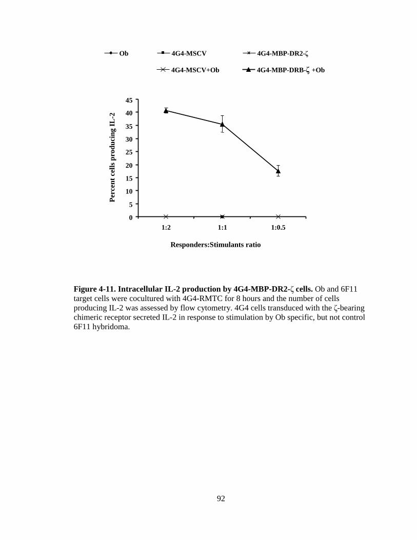
92
Figure 4-11. Intracellular IL-2 production by 4G4-MBP-DR2-ζ cells. Ob and 6F11 target cells were cocultured with 4G4-RMTC for 8 hours and the number of cells producing IL-2 was assessed by flow cytometry. 4G4 cells transduced with the ζ-bearing chimeric receptor secreted IL-2 in response to stimulation by Ob specific, but not control 6F11 hybridoma.
0
5
10
15
20
25
30
35
40
45
1:2 1:1 1:0.5
Per
cent
cel
ls p
rodu
cing
IL-2
Ob 4G4-MSCV 4G4-MBP-DR2-ζ
4G4-MSCV+Ob 4G4-MBP-DRB-ζ +Ob
Responders:Stimulants ratio
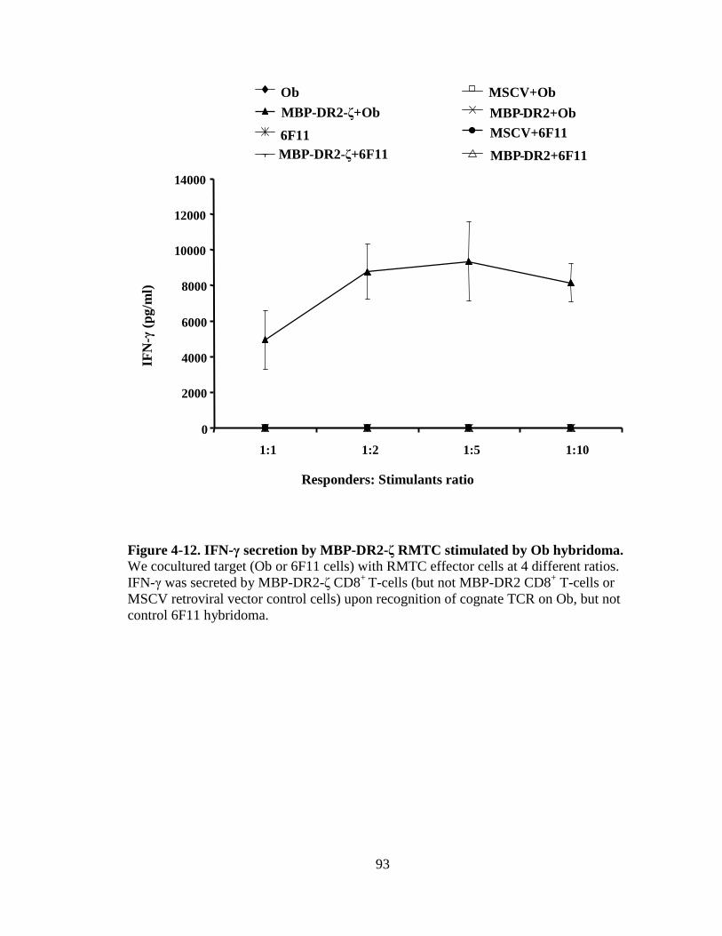
93
Figure 4-12. IFN-γ secretion by MBP-DR2-ζ RMTC stimulated by Ob hybridoma. We cocultured target (Ob or 6F11 cells) with RMTC effector cells at 4 different ratios. IFN-γ was secreted by MBP-DR2-ζ CD8+ T-cells (but not MBP-DR2 CD8+ T-cells or MSCV retroviral vector control cells) upon recognition of cognate TCR on Ob, but not control 6F11 hybridoma.
0
2000
4000
6000
8000
10000
12000
14000
1:1 1:2 1:5 1:10
Responders: Stimulants ratio
IFN
-γ(p
g/m
l)
Ob MSCV+Ob
MBP-DR2-ζ+Ob MBP-DR2+Ob
6F11 MSCV+6F11
MBP-DR2+6F11MBP-DR2-ζ+6F11
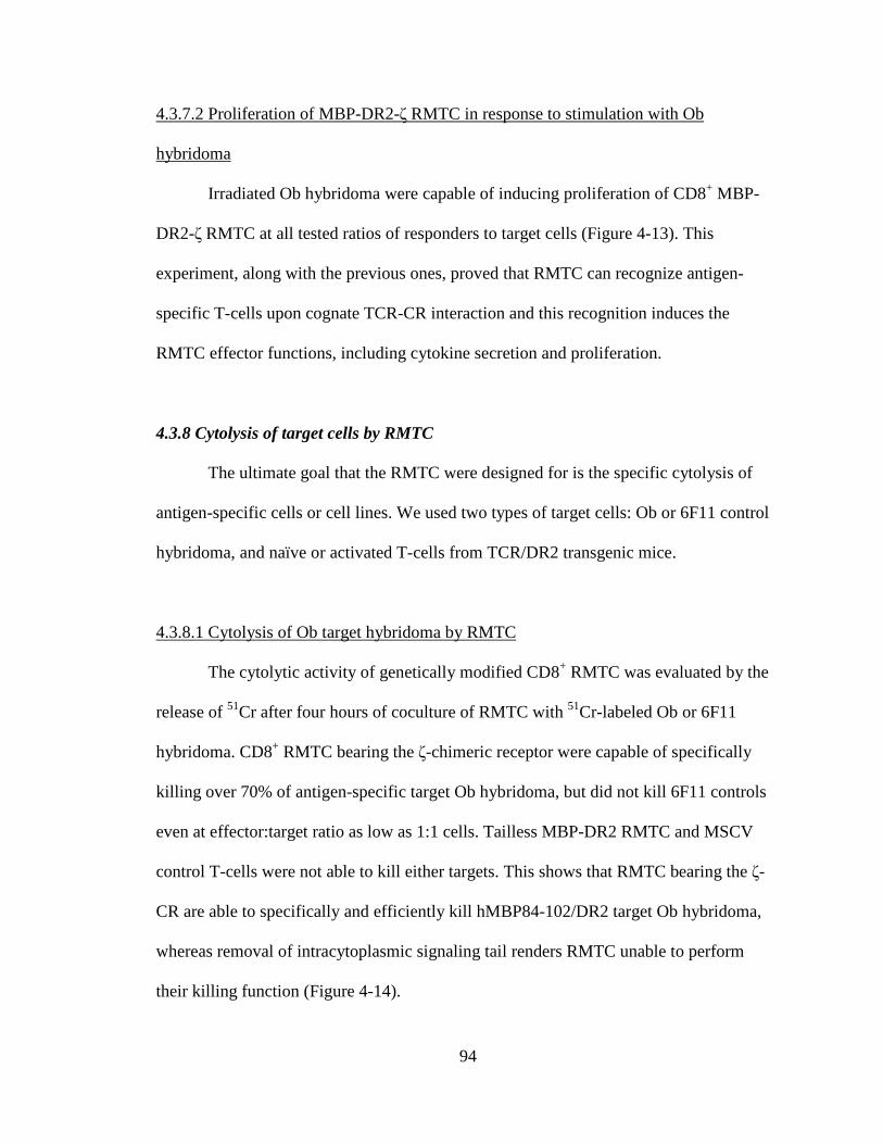
94
4.3.7.2 Proliferation of MBP-DR2-ζ RMTC in response to stimulation with Ob
hybridoma
Irradiated Ob hybridoma were capable of inducing proliferation of CD8+ MBP-
DR2-ζ RMTC at all tested ratios of responders to target cells (Figure 4-13). This
experiment, along with the previous ones, proved that RMTC can recognize antigen-
specific T-cells upon cognate TCR-CR interaction and this recognition induces the
RMTC effector functions, including cytokine secretion and proliferation.
4.3.8 Cytolysis of target cells by RMTC
The ultimate goal that the RMTC were designed for is the specific cytolysis of
antigen-specific cells or cell lines. We used two types of target cells: Ob or 6F11 control
hybridoma, and naïve or activated T-cells from TCR/DR2 transgenic mice.
4.3.8.1 Cytolysis of Ob target hybridoma by RMTC
The cytolytic activity of genetically modified CD8+ RMTC was evaluated by the
release of 51Cr after four hours of coculture of RMTC with 51Cr-labeled Ob or 6F11
hybridoma. CD8+ RMTC bearing the ζ-chimeric receptor were capable of specifically
killing over 70% of antigen-specific target Ob hybridoma, but did not kill 6F11 controls
even at effector:target ratio as low as 1:1 cells. Tailless MBP-DR2 RMTC and MSCV
control T-cells were not able to kill either targets. This shows that RMTC bearing the ζ-
CR are able to specifically and efficiently kill hMBP84-102/DR2 target Ob hybridoma,
whereas removal of intracytoplasmic signaling tail renders RMTC unable to perform
their killing function (Figure 4-14).

95
Figure 4-13. Proliferation of CD8+ MBP-DR2-ζ RMTC in response to Ob hybridoma. Irradiated Ob or control 6F11 hybridoma were cocultured in different ratios with CD8+ RMTC and on day 3, [3H]thymidine was added and liquid scintillation counting was performed 24 hours later. At any given ratio, Ob hybridoma could only stimulate the proliferation of MBP-DR2-ζ-RMTC, but not the tailless CR-transduced T-cells or MSCV retroviral vector-transduced cells.
0
5000
10000
15000
20000
25000
1:1 1:2 1:5
Responders:Stimulants ratio
CP
M
Ob 6F11 MSCVMBP-DR2-ζ MBP-DR2 MSCV+ObMBP-DR2-ζ+Ob MBP-DR2+Ob
MSCV+6F11MBP-DR2-ζ+6F11 MBP-DR2+6F11
0
5000
10000
15000
20000
25000
1:1 1:2 1:5
Responders:Stimulants ratio
CP
M
Ob 6F11 MSCVMBP-DR2-ζ MBP-DR2 MSCV+ObMBP-DR2-ζ+Ob MBP-DR2+Ob
MSCV+6F11MBP-DR2-ζ+6F11 MBP-DR2+6F11

96
Figure 4-14. Cytolysis of Ob hybridoma by CD8+-MBP-DR2-ζ RMTC. Ob hMBP-specific or control 6F11 hybridoma were labeled with 51Cr and cocultured with RMTC for four hours. Specific cytolysis was calculated based on chromium release from the dead target cells. ζ-bearing CR-transduced cells were shown to have an exclusive and potent cytolytic activity against MBP-DR2-specific Ob hybridoma, but not control 6F11 cells.
0
10
20
30
40
50
60
70
80
90
100
1:1 2:1 5:1 10:1 20:1
Effector:Target ratio
Per
cent
Cyt
otox
icity
MBP-DR2-ζ+Ob MBP-DR2+Ob
MBP-DR2-ζ+6F11 MBP-DR2+6F11
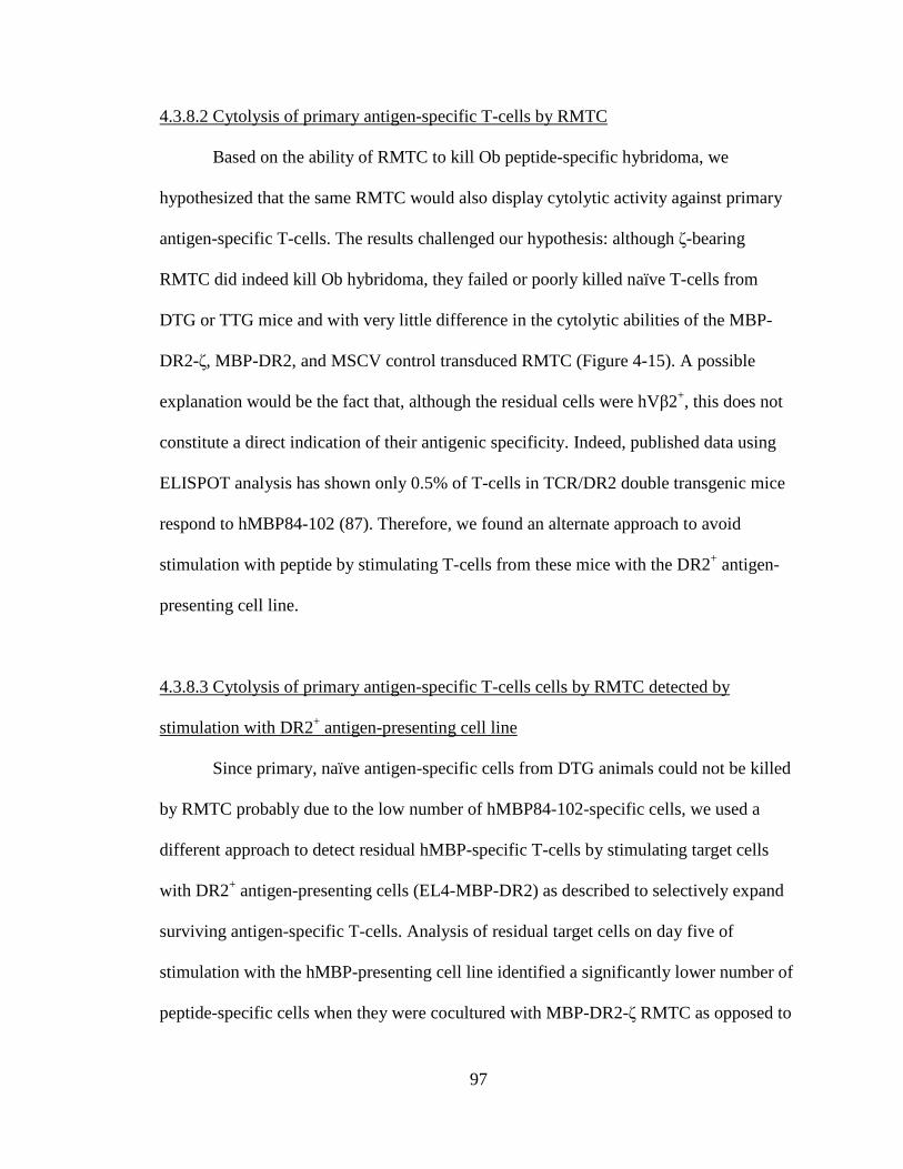
97
4.3.8.2 Cytolysis of primary antigen-specific T-cells by RMTC
Based on the ability of RMTC to kill Ob peptide-specific hybridoma, we
hypothesized that the same RMTC would also display cytolytic activity against primary
antigen-specific T-cells. The results challenged our hypothesis: although ζ-bearing
RMTC did indeed kill Ob hybridoma, they failed or poorly killed naïve T-cells from
DTG or TTG mice and with very little difference in the cytolytic abilities of the MBP-
DR2-ζ, MBP-DR2, and MSCV control transduced RMTC (Figure 4-15). A possible
explanation would be the fact that, although the residual cells were hVβ2+, this does not
constitute a direct indication of their antigenic specificity. Indeed, published data using
ELISPOT analysis has shown only 0.5% of T-cells in TCR/DR2 double transgenic mice
respond to hMBP84-102 (87). Therefore, we found an alternate approach to avoid
stimulation with peptide by stimulating T-cells from these mice with the DR2+ antigen-
presenting cell line.
4.3.8.3 Cytolysis of primary antigen-specific T-cells cells by RMTC detected by
stimulation with DR2+ antigen-presenting cell line
Since primary, naïve antigen-specific cells from DTG animals could not be killed
by RMTC probably due to the low number of hMBP84-102-specific cells, we used a
different approach to detect residual hMBP-specific T-cells by stimulating target cells
with DR2+ antigen-presenting cells (EL4-MBP-DR2) as described to selectively expand
surviving antigen-specific T-cells. Analysis of residual target cells on day five of
stimulation with the hMBP-presenting cell line identified a significantly lower number of
peptide-specific cells when they were cocultured with MBP-DR2-ζ RMTC as opposed to
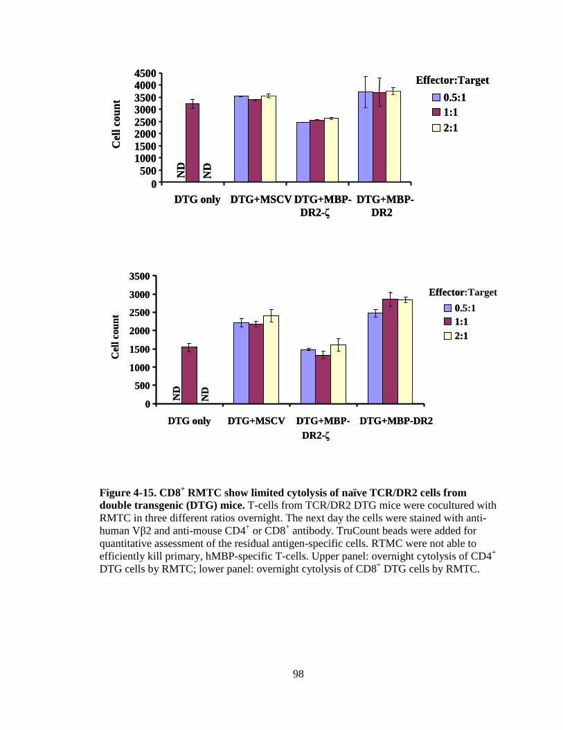
98
Figure 4-15. CD8+ RMTC show limited cytolysis of naïve TCR/DR2 cells from double transgenic (DTG) mice. T-cells from TCR/DR2 DTG mice were cocultured with RMTC in three different ratios overnight. The next day the cells were stained with anti-human Vβ2 and anti-mouse CD4+ or CD8+ antibody. TruCount beads were added for quantitative assessment of the residual antigen-specific cells. RTMC were not able to efficiently kill primary, hMBP-specific T-cells. Upper panel: overnight cytolysis of CD4+ DTG cells by RMTC; lower panel: overnight cytolysis of CD8+ DTG cells by RMTC.
0500
10001500200025003000350040004500
DTG only DTG+MSCV DTG+MBP-DR2-ζ
DTG+MBP-DR2
Cel
l cou
nt
ND
ND
0.5:11:12:1
Effector:Target
0500
10001500200025003000350040004500
DTG only DTG+MSCV DTG+MBP-DR2-ζ
DTG+MBP-DR2
Cel
l cou
nt
ND
ND
0.5:11:12:1
Effector:Target
DTG+MBP-DR2
0
500
1000
1500
2000
2500
3000
3500
DTG only DTG+MSCV DTG+MBP-DR2-ζ
Cel
l cou
nt
ND
ND
0.5:11:12:1
Effector:Target
DTG+MBP-DR2
0
500
1000
1500
2000
2500
3000
3500
DTG only DTG+MSCV DTG+MBP-DR2-ζ
Cel
l cou
nt
ND
ND
0.5:11:12:1
Effector:Target

99
the number of target cells cocultured with MSCV control or tailless RMTC (Figures 4-16
and 4-17). This leads to two conclusions. The first one is that, by day five after
stimulation, the antigen-specific T-cells would undergo clonal expansion, whereas the
non-antigen-specific cell would die. The second conclusion is that the significantly lower
number of residual CD4 and CD8 antigen-specific cells when naïve target cells were
cocultured with MBP-DR2-ζ CTL is due to killing by CTL as opposed to expansion of
target cells were cocultured with MBP-DR2 or MSCV control CTL.
Surprisingly, both CD4+ and CD8+ target cells from TCR/DR2 transgenic animals
showed sustained proliferation in response to antigenic stimulation. Human MBP peptide
is presented by HLA class II, DR2 gene. The fact that not only CD4 T-cells, but also CD8
T-cells respond to this peptide by proliferation contradicts the dogma of immunology that
CD4 T-cells would recognize antigens presented on an MHC class II molecule, whereas
CD8 T-cells would recognize antigens complexed with MHC class I (92). This accounts
for the coreceptor independence of CD8+ antigen-specific cells from TCR/DR2
transgenic mice and explains their proliferation in response to peptide stimulation.
4.3.8.4 Cytolysis of target DTG cell lines by RMTC
Knowing that peptide-specific cells from DTG mice can be expanded with the
DR2+ antigen-presenting cell line, we were interested in determining whether we could
produce hMBP-specific T-cell lines and whether the RMTC could similarly kill these cell
lines. CD4+ and CD8+ hMBP-specific target cell lines were grown as described in
Materials and methods (paragraph 4.2.5.2) and cocultured with RMTC. Only the CD4+
MBP-specific cell line was efficiently killed by MBP-DR2-ζ RMTC, whereas none of the

100
Figure 4-16. Cytolysis of primary CD4+ DTG target cells by RMTC measured by stimulation of residual MBP-DR2-specific T-cells. T lymphocytes from DTG mice TCR/DR2 were cocultured with therapeutic cells or MSCV retroviral control in three different ratios in the absence or presence of DR2+ antigen-presenting cell line (EL4-MBP-DR2) for five days. At the end of the fifth day, the residual cells were stained with hVβ2 and mCD4 antibodies and quantitative FACS analysis was performed. We found a considerably lower number of residual CD4+ peptide-specific cells when these were cocultured with MBP-DR2-ζ RMTC as opposed to the MSCV control cells or MBP-DR2 RTMC.

101
Figure 4-17. Cytolysis of primary CD8+ DTG target cells by RMTC measured by stimulation of residual MBP-DR2-specific T-cells. T lymphocytes from DTG mice TCR/DR2 were cocultured with therapeutic cells or MSCV retroviral control in three different ratios in the absence or presence of DR2+ antigen-presenting cell line (EL4-MBP-DR2) for five days. At the end of the fifth day, the residual cells were stained with hVβ2 and mCD4 antibodies and quantitative FACS analysis was performed. We found a considerably lower number of residual CD8+ peptide-specific cells when these were cocultured with MBP-DR2-ζ RMTC as opposed to the MSCV control cells or MBP-DR2 RTMC.

102
other RTMC (MBP-DR2 and MSCV control) showed this ability. Moreover, both CD4
and CD8 antigen-specific cell lines survived and expanded after six hour-coculture with
tailless RMTC or MSCV control cells. Prior data show that RMTC are only able to kill
primary CD8+ antigen-specific T-cells and not stimulated CTL and that activated CD8+
antigen-specific T-cells are resistant to fratricidal lysis (56). Our experience with the
antigen-specific T-cell lines from humanized transgenic mice yielded the same results:
CD4+ antigen-specific T-cells can be killed by the tail-bearing RMTC, whereas CD8+
antigen-specific T-cells survived after coculture with MBP-DR2-ζ RMTC (Figures 4-18
and 4-19).
4.4 Conclusions
We developed receptor-modified T-cells that can specifically recognize and target
pathologic, antigen-specific T lymphocytes by transducing chimeric receptors into 4G4 T
cell hybridoma or primary murine T-cells. The MBP-DR2-ζ CR heterodimer bears an
antigen-MHC extracellular domain linked to the functional ζ signaling moiety from the
TCR. The MBP-DR2 CR lacked the cytoplasmic signaling domain. Both constructs were
subcloned in MSCV retroviral vector with an IRES site and GFP gene. Their surface
expression was proportional to level of expression of the GPF protein. Transduction of
4G4 T cell hybridoma with the ζ-bearing and ζ-deficient retrovirus showed similar
surface levels of expression whereas transduction of activated, primary T-cells showed a
discrepancy between the two CRs, with a 30% increase in surface expression of the
tailless CR. Upon encounter of cognate TCR on the surface of Ob target hybridoma
(specific for hMBP84-102 restricted by HLA-DR2), therapeutic RMTC recognized the
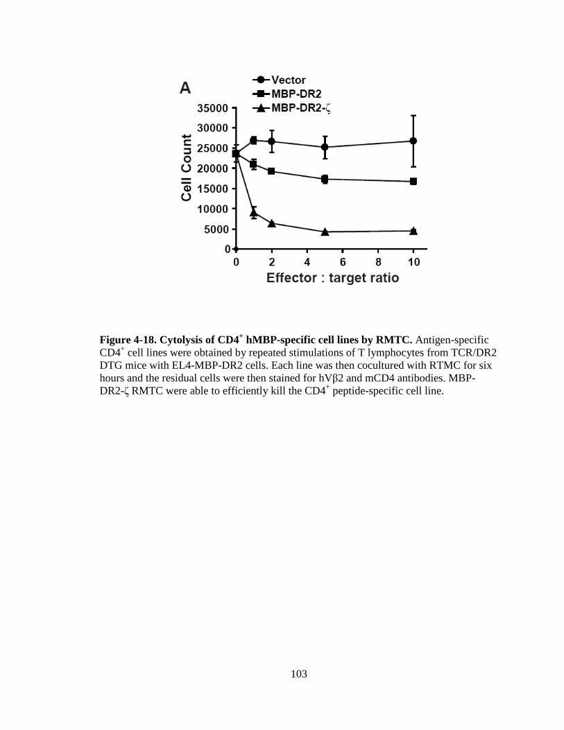
103
Figure 4-18. Cytolysis of CD4+ hMBP-specific cell lines by RMTC. Antigen-specific CD4+ cell lines were obtained by repeated stimulations of T lymphocytes from TCR/DR2 DTG mice with EL4-MBP-DR2 cells. Each line was then cocultured with RTMC for six hours and the residual cells were then stained for hVβ2 and mCD4 antibodies. MBP-DR2-ζ RMTC were able to efficiently kill the CD4+ peptide-specific cell line.
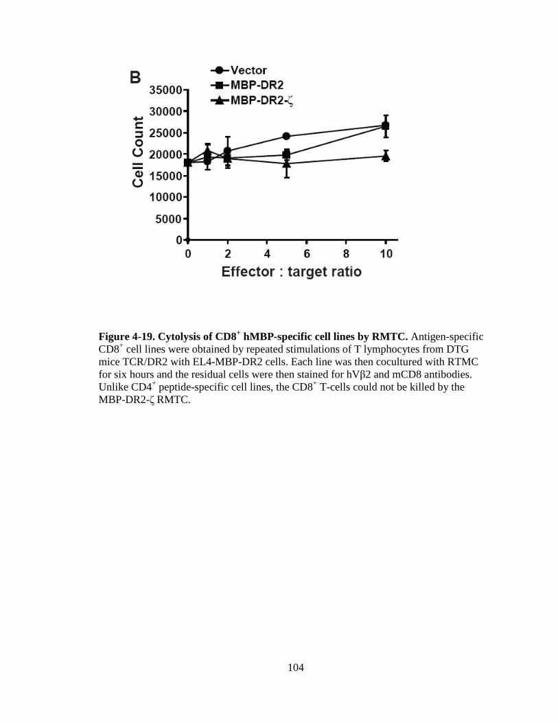
104
Figure 4-19. Cytolysis of CD8+ hMBP-specific cell lines by RMTC. Antigen-specific CD8+ cell lines were obtained by repeated stimulations of T lymphocytes from DTG mice TCR/DR2 with EL4-MBP-DR2 cells. Each line was then cocultured with RTMC for six hours and the residual cells were then stained for hVβ2 and mCD8 antibodies. Unlike CD4+ peptide-specific cell lines, the CD8+ T-cells could not be killed by the MBP-DR2-ζ RMTC.
105
target cells, responded by proliferation, secretion of IL-2 and IFN-γ, and were able to
specifically kill target cells. The in vitro cytolytic ability of CTL RMTC was significant:
effector cells killed over 70% of target cells when cocultured in a 1:1 effector to target
ratio and even up to 90% when five times more effectors were added to the target cells.
Cytolysis was only detected in RMTC transduced with the ζ-bearing CRs, but not MSCV
control or the ζ-deficient RMTC.
Several attempts to direct RMTC against naïve, antigen-specific T-cells from
lymph nodes of TCR/DR2 humanized transgenic mice failed repeatedly in a six hour-
killing assay or overnight killing. Analysis of these double transgenic mice revealed that
only 0.5% for their T-cells were peptide-specific cells in an ELISPOT analysis (85).
Therefore, the reduced ability of MBP-DR2-ζ RMTC to specifically kill target cells and
the lack of difference between the activity of ζ-bearing and ζ-deficient RMTC and MSCV
control was rather due to the very low number of antigen-specific cells that could not be
detected and isolated from the non-specific. The latter cells would not be subjected to
RMTC lysis, but would interfere in the assay, making identification of specific cytolysis
of hMBP-specific T-cell impossible to detect. We adopted an alternate way to stimulate
antigen-specific cells using a DR2+ antigen-specific cell line (EL4-MBP-DR2) to
selectively expand only residual antigen-specific cells. Five days later, we noticed a
decreased number of both CD4+ and CD8+ hMBP-specific T-cells when cocultured with
MBP-DR2-ζ RMTC in the presence of DR2+ cell line, but an expansion of these cells
when cocultured with MBP-DR2 or MSCV control RMTC. These data show that the ζ-
CR-transduced RMTC can also kill naïve antigen-specific T-cells from TCR/DR2 double
transgenic mice in the presence of antigen, whereas removal of the signaling domain

106
from the CR impeded this ability. Surprisingly, although the humanized transgenic mice
have T-cells specific for hMBP presented in the context of DR2, an MHC class II
molecule, upon antigenic stimulation, both CD4+ and CD8+ T-cells from TCR/DR2 mice
responded by proliferation. Although fairly rare, this is not an isolate finding and
accounts for the coreceptor independence of CD8+ TCR/DR2 T-cells.
The ability of MBP-DR2-ζ RMTC to kill naïve, antigen-specific T-cells from
TCr/DR2 mice did not apply in the case of peptide-specific cell lines. Stimulated T
lymphocytes behave differently from naïve T lymphocytes. We built a CD4 and a CD8-
specific cell line by several rounds of antigenic stimulation using DR2+ antigen-
presenting cells. Just like naïve CD4 T-cells, peptide-specific CD4+ cell lines were killed
upon in vitro coculture with the tail-bearing RMTC, but not MSCV control or ζ-deficient
therapeutic cells. CD8+ hMBP-specific cell line proved resistant to lysis. This fact was
demonstrated in the early 1990s and we have also proven that CD8 T-cells acquire
resistance to lysis in between day three and seven post stimulation (56). EAE is
classically considered a CD4 disease and therefore, the ability of our MBP-DR2-ζ RMTC
to specifically kill both naïve and activated CD4+ hMBP-specific cell lines are grounds to
hypothesize that they might also work in the in vivo mouse model.

107
Chapter 5. In vivo activity of RMTC in humanized mice
5.1 Introduction
5.1.1 Humanized transgenic mice as MS models
Similar to other autoimmune diseases (rheumatoid arthritis, celiac disease,
insulin-dependent diabetes), MS is associated with certain MHC class II genes. The
disease is triggered by association of autoantigenic peptides with certain MHC class II;
MHC-peptide is recognized by CD4+ T-cells that, upon activation, mediate inflammation
and destruction of the myelin sheath, ultimately leading to impaired saltatory conduction
(93).
In the previous chapter, we described the development of RMTC as therapeutic T
lymphocytes bearing humanized chimeric receptors, which can be redirected against
antigen-specific T-cells. Upon encounter of target, hMBP-specific T lymphocytes,
therapeutic cells will engage the target cells’ TCR through their surrogate chimeric
receptor, leading to various effector functions, such as proliferation, cytokine secretion,
and specific cytolysis of peptide-specific cells.
Once we have shown that RMTC can recognize cognate TCR and exhibit effector
functions in response to this recognition, the next step was to test whether this interaction
would lead to the same effects in vivo.
In an effort to better understand autoimmune diseases and MS in particular, mice
that express disease-relevant MHC II molecules have been developed. These mice were
then crossed with mice transgenic for other genes encoding TCRs, human CD4, and

108
candidate autoantigens. Since they bear human genes, these mice present similar disease
features either spontaneously or after immunization with peptides. Although they are not
a perfect representation of the human immunopathology, these animals are, nevertheless,
the closest mouse model to study MS-causing mechanisms and symptoms (87).
5.1.2 Humanized HLA-DR2, TCR, and CD4 mice
After analyzing autoantigens and MHC molecules associated with MS, studies
have shown that one of the immunodominant MBP peptide epitope in DR2+ patients was
84-102. This epitope was identical in humans and mice (94).
Serologic typing of patients serum has identified the common MHC class II
molecule as HLA-DRB1*1501 and DRA*0101 that recognized the MBP peptide (86).
Based on these data, Fugger and collaborators engineered three humanized transgenic
mice. A first mouse bore the human DR2 gene (HLA-DRB1*1501 and DRA*0101)
expressed under the control of its native promoters. A second mouse bore a TCR derived
from a patient with MS, denoted Ob (Ob.1A12). Because human TCR is difficult to
express in mouse cells, the TCR construct comprised human variable domains and mouse
constant and regulatory domain. This set-up ensures that the MBP peptide will be able to
be recognized and bound by the human MHC class II and, since the constant and
signaling domains of the TCR are murine, they could interact with the mouse signaling
machinery. These two strains of transgenic mice were crossbred and then also crossed
with another transgenic mouse carrying the human CD4 coreceptor. Caveats of this
double and triple transgenic mouse model are that only 10-30% of B-cells were DR2+ and
only 80% of T-cells were Vβ2+ (87). T-cells from these mice showed proliferation and
109
IL-2 secretion in response to hMBP84-102 stimulation. Upon immunization with peptide,
the mice developed relapsing-remitting or primary progressive EAE similar to MS
patients and with similar histopathologic aspect. Single transgenic TCR or HLA-DR2
mice did not develop signs and symptoms of EAE following MBP immunization.
Breeding of the double transgenic TCR/DR2 mice with Rag2-/- mice leads to
development of spontaneous EAE due to the complete absence of T and B lymphocytes
(including regulatory T-cells) in these knockout mice and repopulation of their immune
system with hMBP-specific cells.
5.2 Materials and methods
5.2.1 In vivo cytolysis of CD4+ and CD8+ hMBP-specific cell lines by RMTC
Recombination activating gene 2 knockout mice (Rag2-/-) were kept in
Helicobacter pylori-free facility of the Animal Research Center at St. Jude Children's
Research Hospital. These mice were coinjected with 5x10⁶ mixed CD4+ and CD8+ triple
transgenic cells from T-cell lines and 20x106 receptor-modified CTLs. CD4+ and CD8+
peptide-specific cell lines were obtained by three consecutive stimulations of T-cells
from TCR/DR2 transgenic mice with irradiated antigen-presenting cell line (EL4-MBP-
DR2) every ten days, according to the protocol described in chapter 4.2.5.2. After three
days, recipient mice were sacrificed and spleens were processed in a single cell
suspension, followed by double staining of splenocytes human Vβ2/mouse CD4 and CD8
antibodies and flow cytometry analysis.

110
5.2.2 In vivo cytolysis of naïve antigen-specific T-cells by RMTC
Irradiated (450 rad) transgenic humanized DR2 mice (strain Fug5) received
10x106 freshly isolated T-cells from TCR/DR2 transgenic mice and 30x106 therapeutic
RMTC by retroorbital injection. Two irradiated recipient Fug5 mice were used for each
group (MSCV control, MBP-DR2-ζ, and MBP-DR2 RMTC).The mice were sacrificed
after nine days or the indicated time. Lymph nodes (LN) were collected and processed to
obtain a single cell suspension. 105 cells/well were stimulated for three days in the
presence or absence of antigen (2x10⁵ EL4-MBP-DR2 cells or EL4 cells, respectively,
irradiated at 20,000 rad). 72-hours later, the cells were pulsed with 1µCi [3H] thymidine
and harvested on filtermats after 16 hours. Proliferation was measured by liquid
scintillation counting of incorporated [3H]thymidine. The samples were analyzed in
duplicates.
5.2.3 Prevention of EAE induced by naïve CD4+ T-cells from TCR/DR2/hCD4 using
RMTC
Lymph nodes and spleen from ten TCR/DR2/hCD4 transgenic mice were
processed in a single cell suspension and stained for PE anti-mouse CD4 microbeads.
Cells were then separated by magnetic cell sorting (Miltenyi Biotec Inc., Auburn CA).
13x106 pure CD4+ T-cells were then transferred into 8 irradiated Fug5 recipients along
with 35x106 RMTC. Two days later, the mice were immunized with 300 µg hMBP84-
102 and 400 ng PTx on day 0 and 2. The animals were scored daily for signs of EAE
disease and euthanized at score 4 to 5 (complete paralysis or moribund).
111
5.2.4 Prevention of hMBP84-102-induced EAE disease in TCR/DR2 double transgenic
mice using therapeutic cells
TCR/DR2 transgenic mice were immunized with 300 µg hMBP84-102 and at the
same time, received the indicated numbers of MBP-DR2-ζ, MBP-DR2 or MSCV control
RMTC. 400 ng PTx was injected retroorbitally at the time of peptide injection and on day
2. Mice were followed and scored daily for signs of EAE.
5.3 Results
5.3.1 In vivo cytolysis of CD4+ and CD8+ hMBP-specific cell lines by RMTC
This experiment was designed to investigate the in vivo efficacy of therapeutic
RMTC in vivo when coinjected with hMBP-specific cell lines into Rag-/- mice for three
days. Upon interaction with the antigen-specific cell lines, RMTC transduced with
chimeric receptor MBP-DR2-ζ and the MBP-DR2 recognized the specific target cells and
exhibited their effector function, namely cytolysis of CD4+ antigen-specific cells. As
expected from previous data, CD8+ MBP-specific cell line did not undergo cytolysis.
Surprisingly, even though MBP-DR2-transduced RMTC failed to show any effector
function in vitro due to the absence of the ζ cytoplasmic signaling tail, in vivo co-
adoptive transfer of these cells along with MBP-specific cell lines showed almost
the same efficacy of killing as the MBP-DR2-ζ RMTC (Figure 5-1).

112
Figure 5-1. In vivo killing of CD4+ hMBP-specific cell line by RMTC. CD4+ and CD8+ hMBP-specific cell lines and RMTC were coinjected into Rag2-/- recipients. Three days later, mice were sacrificed, splenocytes were stained with mouse anti-human Vβ2/rat anti-mouse CD4 or CD8 antibodies, and analyzed by FACS. The plot represents the number of cells found in spleens after three days treatment with RMTC.
0
500
1000
1500
2000
2500
3000
3500
4000
4500
Mouse 1 Mouse 2 Mouse 1 Mouse 2 Mouse 1 Mouse 2
MSCV MBP-DR2-ζ MBP-DR2
Cel
l Cou
nt
CD4+ TTG cell line CD8+ TTG cell line
0
500
1000
1500
2000
2500
3000
3500
4000
4500
Mouse 1 Mouse 2 Mouse 1 Mouse 2 Mouse 1 Mouse 2
MSCV MBP-DR2-ζ MBP-DR2
Cel
l Cou
nt
CD4+ TTG cell line CD8+ TTG cell line

113
5.3.2 In vivo cytolysis of naïve antigen-specific T-cells by RMTC
The previous experiment confirmed that CD4+ antigen-specific cell lines could be
killed by MBP-DR2-ζ RMTC, but these effector cells cannot kill CD8+ hMBP-specific
cell lines. The next step was to find out if whether this also applies to naïve peptide-
specific T-cells. We coinjected freshly isolated cells from TCR/DR2 transgenic mice
along with RMTC into irradiated recipients, sacrificed them after nine days, and looked
for residual peptide-specific cells after stimulation with antigen (irradiated EL4-MBP-
DR2 cells) or in the absence of antigenic stimulation (irradiated EL4 cells). T-cells from
lymph nodes of MBP-DR2-ζ RMTC-injected recipients showed little proliferation in
response to antigenic stimulation and decreased number of residual cells compared to the
mice that received MBP-DR2 RTMC or MSCV control virus. These results demonstrate
that the signaling-competent therapeutic cells effectively eliminate hMBP-specific T-cells
in vivo and further demonstrate that signaling through the ζ cytoplasmic tail is essential
for this activity (Figure 5-2).
5.3.3 Prevention of EAE induced by naïve CD4+ T-cells from TCR/DR2/hCD4 using
RMTC
To find out whether EAE induced by adoptive transfer of CD4+ T-cells from
transgenic mice (as described in 5.2.3) could be prevented by MBP-DR2-ζ RMTC, we
followed all three groups of mice daily for signs of disease. The first signs of EAE in the
group that received MSCV CTLs became visible after only 7 days and followed a
progressive course with very little remission. The mice that received ζ-CR RMTC did not
show any signs of disease. This pilot experiment, which requires repetition, shows that

114
Figure 5-2. Peptide stimulation of T-cells from mice that received RMTC and naïve antigen-specific target cells. Cells from mice that received TCR/DR2 cells and RMTC for nine days were stimulated in the presence/absence of antigen for 72-hours and [3H]thymidine was added. 16-hours later, the cells were harvested onto filtermats and proliferation was measured by liquid scintillation counting. hMBP-specific cells from mice that received retroviral vector control and the MBP-DR2 RMTC proliferated in response to antigen as opposed to the ones that received the MBP-DR2-ζ RMTC.
0
5000
10000
15000
20000
25000
30000
35000
40000
45000
Mouse 1 Mouse 2 Mouse 1 Mouse 2 Mouse 1 Mouse 2
MSCV MBP-DR2-ζ MBP-DR2
CP
M
EL4 EL4-MBP-DR2
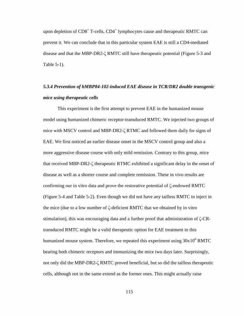
115
upon depletion of CD8+ T-cells, CD4+ lymphocytes cause and therapeutic RMTC can
prevent it. We can conclude that in this particular system EAE is still a CD4-mediated
disease and that the MBP-DR2-ζ RMTC still have therapeutic potential (Figure 5-3 and
Table 5-1).
5.3.4 Prevention of hMBP84-102-induced EAE disease in TCR/DR2 double transgenic
mice using therapeutic cells
This experiment is the first attempt to prevent EAE in the humanized mouse
model using humanized chimeric receptor-transduced RMTC. We injected two groups of
mice with MSCV control and MBP-DR2-ζ RTMC and followed them daily for signs of
EAE. We first noticed an earlier disease onset in the MSCV control group and also a
more aggressive disease course with only mild remission. Contrary to this group, mice
that received MBP-DR2-ζ therapeutic RTMC exhibited a significant delay in the onset of
disease as well as a shorter course and complete remission. These in vivo results are
confirming our in vitro data and prove the restorative potential of ζ-endowed RMTC
(Figure 5-4 and Table 5-2). Even though we did not have any tailless RMTC to inject in
the mice (due to a low number of ζ-deficient RMTC that we obtained by in vitro
stimulation), this was encouraging data and a further proof that administration of ζ-CR-
transduced RMTC might be a valid therapeutic option for EAE treatment in this
humanized mouse system. Therefore, we repeated this experiment using 30x106 RMTC
bearing both chimeric receptors and immunizing the mice two days later. Surprisingly,
not only did the MBP-DR2-ζ RMTC proved beneficial, but so did the tailless therapeutic
cells, although not in the same extend as the former ones. This might actually raise

116
Figure 5-3. Prevention of CD4+-adoptive transfer EAE with RMTC. Irradiated Fug5 recipients received concomitantly sorted CD4+ target cells from TCR/DR2/hCD4 transgenic mice and therapeutic RMTC followed by immunization with hMBP84-102 peptide and PTx. Recipients of MSCV control cells showed early disease onset and severe course with little remission as opposed to the ones that received therapeutic MBP-DR2-ζ RMTC that were resistant to EAE induction.
0
0.5
1
1.5
2
2.5
3
3.5
day1
day3
day5
day7
day9
day1
1
day1
3
day1
5
day1
7
day1
9
day2
1
day2
3
day2
5
day2
7
day2
9
day3
1
day3
3
day3
5
day3
7
day3
9
day4
1
day4
3
day4
5
Days after induction
Dis
ease
sco
re
MSCV MBP-DR2-ζ
0
0.5
1
1.5
2
2.5
3
3.5
day1
day3
day5
day7
day9
day1
1
day1
3
day1
5
day1
7
day1
9
day2
1
day2
3
day2
5
day2
7
day2
9
day3
1
day3
3
day3
5
day3
7
day3
9
day4
1
day4
3
day4
5
Days after induction
Dis
ease
sco
re
MSCV MBP-DR2-ζ

117
Table 5-1. Clinical manifestations in CD4+ adoptive transfer EAE treated with RMTC . Therapeutic cells Disease frequency Mean onset Mean maximal
(diseased/total) day x±s.d. (range) score x±s.d.
MSCV 4/4 9.5±7 3±2.3
MBP-DR2-ζ 0/0 0±0 0±0

118
Figure 5-4. Prevention of EAE with ζ-bearing RMTC. TCR/DR2 mice received MBP-DR2-ζ RMTC or MSCV control cells and were immunized with peptide and PTx. Control group showed early onset and severe disease course, whereas treated group had a milder form of disease followed by complete remission.
0
0.5
1
1.5
2
2.5
3
3.5
4
1 3 5 7 9 11 13 15 17 19 21 23 25 2729 31 33 35 37 39 41 43 4547 49 51 53
Days after induction
Dis
ease
sco
re
MSCV MBP-DR2-ζ
0
0.5
1
1.5
2
2.5
3
3.5
4
1 3 5 7 9 11 13 15 17 19 21 23 25 2729 31 33 35 37 39 41 43 4547 49 51 53
Days after induction
Dis
ease
sco
re
MSCV MBP-DR2-ζ

119
Table 5-2. Clinical manifestations in direct induction of EAE with hMBP and treatment with MSCV vector control or ζ-bearing RMTC. Therapeutic cells Disease frequency Mean onset Mean maximal
(diseased/total) day x±s.d. (range) score x±s.d.
MSCV 4/4 8.75±3.5 3.5±1.7
MBP-DR2-ζ 4/5 11.8±6.6 1.6±1.1

120
question about the absolute requirement for the cytoplasmic signaling tail in the chimeric
receptor for the in vivo treatment of EAE (Figure 5-5 and Table 5-3).
5.4 Conclusions
The results obtained from in vitro testing of RMTC encouraged us to move
forward to the in vivo system. We coinjected therapeutic RMTC along with either
peptide-specific cell lines or naïve antigen-specific cells obtained from the transgenic
mice into recipient Rag-/- or irradiated DR2 (Fug5) mice. We sacrificed the mice at
various intervals, starting from day 3 to 9, and estimated the number of residual CD4 and
CD8 antigen-specific T-cells. According to our in vitro data, therapeutic RMTC bearing
the ζ-signaling tail should be able to specifically recognize and kill naive antigen-specific
T-cells. When we sacrificed recipient mice nine days after coinjection of MBP-DR2-ζ
and naïve target cells, we found a decreased number of hVβ2+CD4+/CD8+ T-cells. Since
we have already established that very few of the target cells provided by TCR/DR2 mice
are antigen-specific, we stimulated cells from lymph nodes and spleen of recipient mice
with antigen as previously described. T-cells from irradiated recipients that received
MBP-DR2 and MSCV-control RMTC along with naïve antigen-specific cells showed
good proliferation upon peptide stimulation unlike mice that received MBP-DR2-ζ
RMTC. These data resemble the ones we obtained from in vitro experiments and prove
that recognition through surrogate chimeric receptor also takes place in the mouse
system. Surprisingly, when Rag-/- recipients received both therapeutic cells and hMBP-
specific cell lines, both ζ-bearing and ζ-deficient RMTC were effective in eliminating the
CD4+ peptide-specific cell line. We did not pursue this observation, but the fact that

121
Figure 5-5. Prevention of EAE with ζ-bearing or tailless RMTC. Three groups of TCR/DR2 transgenicmice received MBP-DR2-ζ, MBP-DR2, and MSCV control RTMC and 48 hours later peptide and PTx. Control group showed early onset and severe disease course, whereas ζ-competent therapeutic cells showed a later onset and milder disease followed by complete remission. ζ-deficient therapeutic cells showed an intermediate effect, but still managed to induce remission although not in the same extent as MBP-DR2-ζ RMTC.
0
0.5
1
1.5
2
2.5
3
3.5
4
4.5
5
1 2 3 4 5 6 7 8 9 1011 1213 141516 1718 192021 2223 2425 262728 29
Days after induction
Dis
ease
sco
re
MSCV MBP-DR2-ζ MBP-DR2
0
0.5
1
1.5
2
2.5
3
3.5
4
4.5
5
1 2 3 4 5 6 7 8 9 1011 1213 141516 1718 192021 2223 2425 262728 29
Days after induction
Dis
ease
sco
re
MSCV MBP-DR2-ζ MBP-DR2

122
Table 5-3. Clinical manifestations in direct induction of EAE with hMBP and treatment with MSCV control, ζ-bearing, or tailless RMTC. Therapeutic cells Disease frequency Mean onset Mean maximal
(diseased/total) day x±s.d. (range) score x±s.d.
MSCV 4/4 8.5±1 5±0
MBP-DR2-ζ 3/3 14±0 1±0
MBP-DR2 4/4 13±0 3±1.4
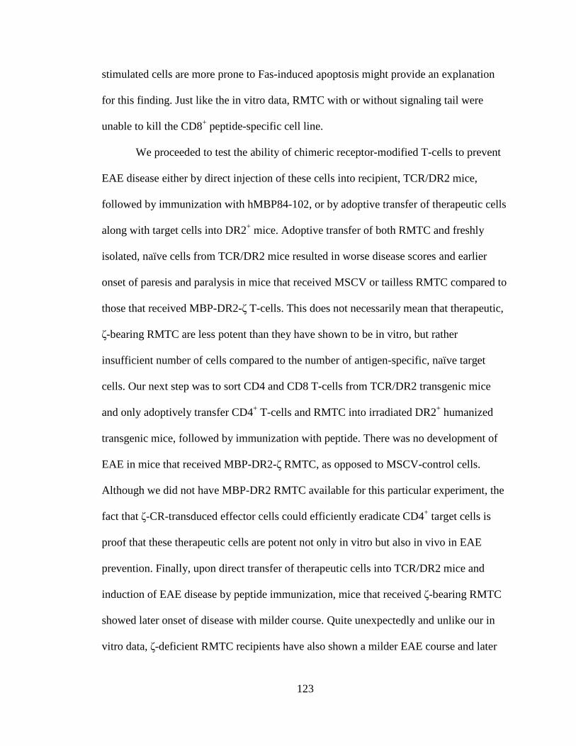
123
stimulated cells are more prone to Fas-induced apoptosis might provide an explanation
for this finding. Just like the in vitro data, RMTC with or without signaling tail were
unable to kill the CD8+ peptide-specific cell line.
We proceeded to test the ability of chimeric receptor-modified T-cells to prevent
EAE disease either by direct injection of these cells into recipient, TCR/DR2 mice,
followed by immunization with hMBP84-102, or by adoptive transfer of therapeutic cells
along with target cells into DR2+ mice. Adoptive transfer of both RMTC and freshly
isolated, naïve cells from TCR/DR2 mice resulted in worse disease scores and earlier
onset of paresis and paralysis in mice that received MSCV or tailless RMTC compared to
those that received MBP-DR2-ζ T-cells. This does not necessarily mean that therapeutic,
ζ-bearing RMTC are less potent than they have shown to be in vitro, but rather
insufficient number of cells compared to the number of antigen-specific, naïve target
cells. Our next step was to sort CD4 and CD8 T-cells from TCR/DR2 transgenic mice
and only adoptively transfer CD4+ T-cells and RMTC into irradiated DR2+ humanized
transgenic mice, followed by immunization with peptide. There was no development of
EAE in mice that received MBP-DR2-ζ RMTC, as opposed to MSCV-control cells.
Although we did not have MBP-DR2 RMTC available for this particular experiment, the
fact that ζ-CR-transduced effector cells could efficiently eradicate CD4+ target cells is
proof that these therapeutic cells are potent not only in vitro but also in vivo in EAE
prevention. Finally, upon direct transfer of therapeutic cells into TCR/DR2 mice and
induction of EAE disease by peptide immunization, mice that received ζ-bearing RMTC
showed later onset of disease with milder course. Quite unexpectedly and unlike our in
vitro data, ζ-deficient RMTC recipients have also shown a milder EAE course and later

124
onset compared to MSCV control mice, though their potency was lower than the one
exhibited by the ζ-bearing therapeutic cells.
All these data lead us to believe that surrogate ζ-chimeric receptor-transduced
RMTC as just as effective in killing antigen-specific T-cells from TCR/DR2 mice in
vivo as they were in vitro, thus preventing EAE disease. A question that rises naturally is
whether these therapeutic cells would also work after the first signs of EAE disease in
forestalling the fatal outcome and improving the clinical signs and symptoms, and this
may be a topic for future studies.

125
Chapter 6. Discussion
Multiple sclerosis is the most common autoimmune disease involving the white
matter of the CNS. It is reported to affect 300,000 Americans with 200 people diagnosed
weekly and 2.5 million people worldwide (2). The neurologic signs and symptoms of MS
are varied and can also present in other diseases. Double vision, paralysis, tremor,
numbness, and gait difficulties are caused by demyelination resulting in impaired nerve
conduction; axons and cell bodies are spared early in the disease. The initial disease
target is the white matter of the brain and spinal cord, but after a while the gray matter
also becomes involved due to secondary degeneration resulting from the loss of the white
matter. Axons will eventually be destroyed and replaced by gliosis that gives the sclerotic
aspect on MRI.
There are currently several therapies approved for MS. Regardless of the
approach, treatment should target both the symptoms and the pathologic cascade.
Gene therapy is the newest approach in the treatment of MS. It has emerged as a
direct result of increased knowledge concerning pathologic mechanisms in autoimmune
diseases and aims to more specifically target pathologic T-cells than current therapies.
Anti-inflammatory cytokine genes can be delivered into the CNS by viral vectors. More
recently, our laboratory has developed receptor-modified T-cells bearing chimeric
receptors that specifically target autoreactive T-cells. A similar approach has already
been used in infectious diseases and cancer.
Chimeric receptors are hybrid molecules that encompass a recognition domain –
the variable region involved in antigen recognition from MHC or Ig – and a signaling

126
domain – the intracytoplasmic signaling moieties of the TCR. This new therapeutic
approach emerged in the 1980s-1990s when it was found that T-cells endowed with
antibody-like abilities and no MHC restriction can be redirected against infected and
neoplastic cells. The necessity of having a signaling domain resides in the fact that
recognition of antigen by the variable region of the chimeric receptor is not sufficient to
activate T-cells. Similar to the cascade of events triggered in a regular T lymphocyte, T-
cells bearing chimeric receptors undergo phosphorylation of ITAMs on the receptor’s
signaling moiety and turn on the transcription of nuclear factors responsible for activation
of these cells, thus inducing effector function. Engagement of the TCR constitutes the
first signal necessary for activation of T lymphocytes but for a full activation a second
signal is mandatory. This can be through either of several molecules including the CD28
molecule that stimulates the expansion of CD4+ cells and CD137 (or 4-1BB) that
prevents activation-induced cell death of activated T-cells and stimulates the expansion
of CD8+ cells.
The choice of the cytoplasmic signaling domain has long been a critical topic in
the design of chimeric receptors. Increasing the number of ITAMs did not necessarily
enhance signaling of the CR (95). In an attempt to test the effect of different signaling
domains on the potency of signal transduction, several intracytoplasmic domains have
been used in a CR engineered to bear the recognition domain of MHC class I (Kb)
molecule. The combinations of CD28-zeta, zeta-lck, CD4-zeta, and CD28-zeta-lck
signaling domains increased receptor function measured by IL-2 secretion after
stimulation with the anti-Kb specific antibody.
Although CRs bearing the intracytoplasmic CD28 moiety showed very high
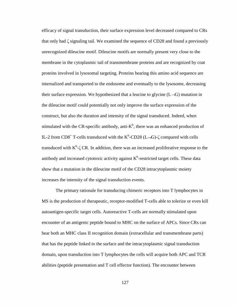
127
efficacy of signal transduction, their surface expression level decreased compared to CRs
that only had ζ signaling tail. We examined the sequence of CD28 and found a previously
unrecognized dileucine motif. Dileucine motifs are normally present very close to the
membrane in the cytoplasmic tail of transmembrane proteins and are recognized by coat
proteins involved in lysosomal targeting. Proteins bearing this amino acid sequence are
internalized and transported to the endosome and eventually to the lysosome, decreasing
their surface expression. We hypothesized that a leucine to glycine (LG) mutation in
the dileucine motif could potentially not only improve the surface expression of the
construct, but also the duration and intensity of the signal transduced. Indeed, when
stimulated with the CR-specific antibody, anti-Kb, there was an enhanced production of
IL-2 from CD8+ T-cells transduced with the Kb-CD28 (L G)-ζ compared with cells
transduced with Kb-ζ CR. In addition, there was an increased proliferative response to the
antibody and increased cytotoxic activity against Kb-restricted target cells. These data
show that a mutation in the dileucine motif of the CD28 intracytoplasmic moiety
increases the intensity of the signal transduction events.
The primary rationale for transducing chimeric receptors into T lymphocytes in
MS is the production of therapeutic, receptor-modified T-cells able to tolerize or even kill
autoantigen-specific target cells. Autoreactive T-cells are normally stimulated upon
encounter of an antigenic peptide bound to MHC on the surface of APCs. Since CRs can
bear both an MHC class II recognition domain (extracellular and transmembrane parts)
that has the peptide linked to the surface and the intracytoplasmic signal transduction
domain, upon transduction into T lymphocytes the cells will acquire both APC and TCR
abilities (peptide presentation and T cell effector function). The encounter between
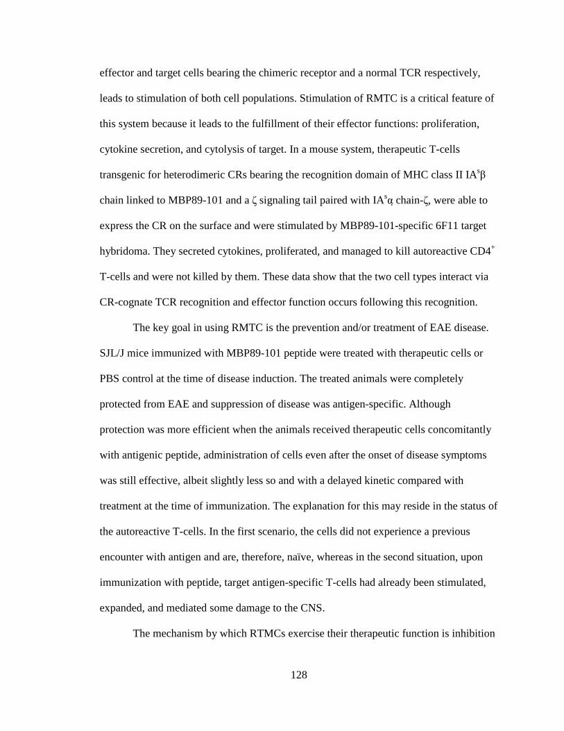
128
effector and target cells bearing the chimeric receptor and a normal TCR respectively,
leads to stimulation of both cell populations. Stimulation of RMTC is a critical feature of
this system because it leads to the fulfillment of their effector functions: proliferation,
cytokine secretion, and cytolysis of target. In a mouse system, therapeutic T-cells
transgenic for heterodimeric CRs bearing the recognition domain of MHC class II IAsβ
chain linked to MBP89-101 and a ζ signaling tail paired with IAsα chain-ζ, were able to
express the CR on the surface and were stimulated by MBP89-101-specific 6F11 target
hybridoma. They secreted cytokines, proliferated, and managed to kill autoreactive CD4+
T-cells and were not killed by them. These data show that the two cell types interact via
CR-cognate TCR recognition and effector function occurs following this recognition.
The key goal in using RMTC is the prevention and/or treatment of EAE disease.
SJL/J mice immunized with MBP89-101 peptide were treated with therapeutic cells or
PBS control at the time of disease induction. The treated animals were completely
protected from EAE and suppression of disease was antigen-specific. Although
protection was more efficient when the animals received therapeutic cells concomitantly
with antigenic peptide, administration of cells even after the onset of disease symptoms
was still effective, albeit slightly less so and with a delayed kinetic compared with
treatment at the time of immunization. The explanation for this may reside in the status of
the autoreactive T-cells. In the first scenario, the cells did not experience a previous
encounter with antigen and are, therefore, naïve, whereas in the second situation, upon
immunization with peptide, target antigen-specific T-cells had already been stimulated,
expanded, and mediated some damage to the CNS.
The mechanism by which RTMCs exercise their therapeutic function is inhibition
129
of antigen-specific T cell response of target cells which may occur through the killing of
the target cells. In addition to this, pathologic cells are skewed from Th1 to Th2 type and
secrete anti-inflammatory cytokines, such as IL-4, instead of IFN-γ.
In the work described here, we proposed extending our studies in murine EAE by
developing a new model for the therapy of MS using humanized chimeric receptors. We
designed two constructs based on the same principle that has already been described for
CRs. The first construct, MBP-DR2-ζ, had a recognition domain composed of an
immunodominant MBP epitope (84-102) linked to the β chain of a HLA class II molecule
(DR2) and a signaling domain composed of the ζ cytoplasmic signaling tail.The α chain
of DR2 was also linked to ζ. The DR2 HLA molecule is highly associated with MS
disease, and T-cells specific for the MBP84-102 epitope can be detected in DR2 patients
with MS. We then engineered a second CR, MBP-DR2 that lacked the signaling tails to
identify the role of signal transduction for the in vitro and in vivo efficacy of this
particular CR. The constructs were each tricistronic, with the β and α chains separated by
a 17-mer 2A sequence from T. asigna and an IRES linked to GFP. Both constructs were
subcloned into MSCV retroviral vector. CRs were transduced in either 4G4 TCR-
hybridoma or primary murine T lymphocytes from C57Bl/6 mice, sorted for GFP
expression and expanded in complete Bruff medium and IL-2.
The tail-bearing and tailless CRs showed good surface expression on flow
cytometry in both 4G4 and primary T-cells when stained with anti-CR specific antibody
mouse anti-human DR2. Transduced hybridoma showed similar levels of expression of
CR; transduced T-cells showed better expression of the tailless CR compared to the ζ tail-
bearing one (71.7% versus 40.4%).
130
We first wanted to see if receptor-modified T-cells bearing TCR-ζ, tailless CRs,
or MSCV retroviral vector control could be stimulated by plate-bound DR2-specific
antibody. Only MBP-DR2-ζ responded to stimulation by secreting IL-2.
We next looked for the ability of CR-transduced T-cells to recognize the cognate
TCR on the surface of target cells. The latter were either Ob1A12 hybridoma specific for
hMBP84-102/DR2 T-cells or T cell lines from the double or triple transgenic mice,
TCR/DR2 and TCR/DR2/hCD4, respectively, with similar specificity. These humanized
mice have develop a disease that strongly resembles human MS model; they carry one of
the most frequent immunodominant epitope of myelin basic protein, 84-102, presented by
one of the most frequently used class II MHC in MS patients, DRB1*1501. Even though
very few of the T-cells in the transgenic mice were MBP-specific in Tg mice bred on a
Rag+/+ background, on a Rag-/- background all of the T-cells will express exclusively the
Tg receptor and these mice develop spontaneous EAE. We further demonstrated that both
CD4 cells and CD8 cells from the double Tg mice responded to MBP peptide restricted
to the DR2 (DRB1*1501) HLA molecule. Thus the T cell response in these mice is not
coreceptor dependent. As controls we used other targets, specifically 6F11 hybridoma
reactive against MBP89-101/IAs and T-cells from non-Tg mice.
Upon recognition of cognate TCR on the surface of Ob1A12 specific hybridoma
but not 6F11 controls, RMTC bearing the ζ signaling tail were stimulated and secreted
IL-2 and IFN-γ. Also, due to the interaction between the two cells, RMTC and target
Ob1A12 cells, RMTC proliferated and killed the target cells. The cytolytic effect was
specific and very effective as even at a 1:1 ratio effector to target ratio, the RMTC
managed to eliminate over 70% of the targets. It is noteworthy that only the cells bearing

131
the cytoplasmic signaling tail were capable of both proliferation and killing upon
recognition of cognate TCRs.
Our attempt to demonstrate similar results in cytolysis assays using primary
murine antigen-specific cells from lymph nodes or spleens of DTG (TCR/DR2) mice did
not yield the same results. In vitro overnight killing assays in which RMTC targeted T-
cells from transgenic mice showed very little difference between control and
experimental cultures. This seemed be due to the fact that although 80% of T-cells in the
humanized transgenic mice were hVβ2+, only ~ 0.5% of them are hMBP84-102-specific
in ELISPOT assays. Therefore a preponderance of non-antigen-specific cells that cannot
be recognized and killed by RMTC were present therefore preventing detection of any
target cell lysis.
We approached this problem in two ways. First, we developed a functional assay
to overcome it in which we stimulated T-cells with antigen in vitro for five days. The
MBP-specific cells survive, get activated, and proliferate in response to peptide, whereas
the non-specific cells die. After 5 days we could demonstrate that virtually all surviving
T-cells are antigen specific. We treated cultures at the time of antigen-stimulation either
with control MSCV-vector transduced RMTC, MBP-DR2 RMTC or MBP-DR2-ζ
RMTC. After 5 days we used quantitative flow cytometry to determine the number of
surviving T-cells. This demonstrated that the MBP-specific cells were killed by ζ-
bearing RMTC, whereas the target cells cocultured with MSCV control or tailless RMTC
proliferated due to peptide stimulation. Surprisingly, these results were true not only for
CD4+ antigen-specific cells, but also for the CD8+ MBP-specific cells.
As a second approach, we established T cell lines using T-cells from DTG

132
(TCR/DR2) or TTG (TCR/DR2/hCD4) mice stimulated repeatedly with exogenous
antigen (irradiated EL4-MBP-DR2 cells) and used them to test the cytolytic ability of
RMTC. Results showed that, unlike the previous experiment where therapeutic cells
bearing the signaling tail were capable of killing naïve, CD4 and CD8 antigen-specific T-
cells, the RMTC could not achieve the same outcome with CD8+ hMBP-specific cell line.
CD8+ T-cells develop resistance to lysis between days 3 and 7 after activation and this
might explain the lack of potency of the RMTC in killing the CD8+ cell line (57). It may
also impair the therapeutic usefulness of RTMCs in EAE models, such as potentially the
TCR/DR2 model of ours, where class II restricted CD8 T-cells may have a pathologic
role.
We went on to test our RMTC in vivo. These studies highlighted the in vitro
results and provided evidence for the therapeutic applicability of humanized RMTC.
Adoptive transfer of freshly isolated lymphocytes from humanized transgenic mice
TCR/DR2 along with therapeutic cells into irradiated humanized DR2 recipients showed
decreased proliferation upon antigenic stimulation in mice treated with the ζ- bearing
RMTC as opposed to a vigorous proliferative response generated in mice that received
MSCV control or tailless RMTC.
Adoptive transfer of antigen-specific cell lines and therapeutic cells into Rag-/-
mice followed by treatment with RMTC yielded somewhat different results. As we saw
in vitro, the number of CD8+ peptide-specific cell line cells was not diminished by either
ζ- or tailless RMTC when compared with the control treatment. Therefore these cells
seemed to be resistant to lysis in vivo, much as they were in vitro. Results with the CD4
cell line were more surprising. Not unexpectedly, the number of residual CD4 peptide-

133
specific cells was decreased in mice injected with MBP-DR2-ζ RMTC. However, mice
injected with MBP-DR2 RMTC showed a similar and nearly complete loss of these cells.
This in vivo effect of the tailless CR-transduced cells could not be predicted based on any
of the in vitro experiments. One potential explanation for the difference is that
stimulation by the MBP-DR2 RMTC in vivo induces the MBP-reactive cell line but not
naive T-cells to undergo activation induced cell death. However, at the current time
experimental evidence to support this does not exist.
The ultimate goal of this project is to prevent or treat EAE in a humanized mouse
model. We adopted two approaches. The first one was to adoptively transfer cells from
triple transgenic TCR/DR2/hCD4 mice along with therapeutic cells followed by
immunization with hMBP and PTx. The mice were observed and scored for EAE disease
for 50 days. Recipients of the zeta-bearing therapeutic cells showed later onset of disease
symptoms and milder course compared to the ones receiving MSCV control or zeta-
deficient RMTC. We have also tried to induce EAE by adoptive transfer of CD4+ cells
from TTG mice and therapeutic cells followed by peptide immunization. MBP-DR2-ζ
CTL completely blocked development of EAE disease, whereas mice that received
MSCV CTL developed EAE within a week from injection. Since EAE is classically
considered a CD4+ Th1-induced disease, the efficient elimination of these cells by the
zeta-bearing therapeutic cells is an indication of their potency in blocking the disease. We
also noticed that the mice that received MSCV control cells along with CD4+ target cells
developed less severe signs of EAE than the mice that received unsorted target cells
which might account for the role of CD8+ MBP-specific cells as “inducers” of EAE in
this particular mouse model. Due to the lack of humanized MHC class II mice (Fug5) we

134
could not repeat these experiments, but this is a task we intend to pursue in the future. We
also intend to test the ability of therapeutic cells to block disease induced by adoptive
transfer of CD8+ cells from TCR/DR2/hCD4 transgenic mice.
The second approach for in vivo prevention of EAE disease involved the use of
TCR/DR2 double transgenic mice as recipients of therapeutic cells. These mice received
active peptide immunization and PTx two days after the transfer of RTMCs. Although
these experiments need to be repeated, they yielded very encouraging results: mice that
received zeta-bearing RMTC showed a delayed onset of disease, a milder course, and
remission compared to the mice that received control CTL. The tailless chimeric
receptor-transduced T-cells seemed to be efficient as well, though not in the same extent
as the ones bearing the zeta signaling tail. Two more experiments validating these data
are ongoing.
These studies are important for several reasons. The use of RMTC expressing
humanized CR in humanized transgenic mice brings us one step closer to their use in
treating multiple sclerosis in humans. Experimental therapies in humans are limited by
both technical and ethical considerations whereas mice have similarities but are simpler
to use as disease models. Conversely, although genetically similar, mice are not humans,
and some therapeutic approaches that proved beneficial for them have not worked in
humans or could even be harmful. HLA-transgenic mice are particularly useful in
modeling autoimmune diseases associated with specific HLA alleles, such as MS.
Although it is rather difficult to identify exactly the genes responsible for MS, due to the
strong linkage disequilibrium, HLA class II has a clear contribution in predisposing to the
development of disease (85). HLA is crucial as it governs T cell selection in the thymus

135
and antigen presentation in periphery. Upon binding of a TCR to a peptide antigen in the
groove of MHC II, a cascade of signal transduction events ensues.
CD4 costimulation may be required as a TCR interacts with MHC. The
requirement in the humanized mouse model we have studied is less clear. The class II
MHC used in this model is entirely human in sequence. Data from literature are
extremely controversial on whether murine CD4 can bind human class II MHC. Some
studies demonstrate that an entirely human MHC class II mouse cannot interact with
mouse CD4 (96, 97). Therefore, in some humanized systems, a mixed, chimeric
human/mouse class II molecule was engineered so that murine CD4 coreceptor could
bind the β2 domain (also murine) of the transgenic HLA II (98). Another set of data
seemingly contradicted this requirement. Altman et al. engineered humanized HLA-DR1
transgenic mice and crossbred them with human CD4 transgenic mice (Hu-CD4) and
tested the response of Hu-CD4 positive versus Hu-CD4 negative T-cells to influenza
haemagglutinin (HA). T-cells proliferated strongly to this peptide regardless the CD4
status (99). This suggests the possibility of interaction between human HLA class II and
mouse CD4; however this was not formally proven. In our experience, using antigen-
specific T-cells from double transgenic or triple transgenic mice (including or lacking
Hu-CD4) did not make a difference in the experimental results.
Another issue regarding our system is the proliferative response of CD8+ antigen-
specific T-cells to peptide stimulation. EAE is classically considered a CD4 Th1 disease
(100) and until recently the research was predominantly focused on this T cell phenotype.
One study that used MOG35-55 as the immunodominant epitope showed equal
proliferative potential of both CD4 and CD8 cells to this antigen and EAE induction by
136
adoptive transfer of CD8 antigen-specific cells (28). On the same note, the inflammatory
infiltrate in the brain of the patients with MS incriminated both CD4 and CD8 T cell
types along with B cells, and depletion of CD4 cells in MS patients only minimally
decreased relapse rates (101). Invasion of the CNS after intravenous delivery of MBP-
activated, cytotoxic CD8 cells result in very severe ascending flaccid paralysis with high
mortality (102). The aggressive and somewhat different manifestation of disease is a
critical aspect for therapy as CD8-induced disease can be dampened by anti-IFN-γ
antibodies (unlike the CD4-induced disease), while anti-TNF-α does not show any effect
on the disease course. It is also noteworthy that perivascular inflammation leading to
further demyelination in MS is composed mainly of CD8+ T-cells as these cells recognize
endogenous antigens processed via the MHC class I pathway.
CD8+ T-cells have been incriminated as culprits for MS. As a logical
consequence, the humanized mice that we used for this project might actually be closer to
the human disease than EAE mediated exclusively by CD4 T-cells, as the disease in this
model might be caused by both CD4 and CD8 autoreactive T-cells. Since HLA-DR2
mice bear human MHC class II, but not class I, a CD4 response to hMBP-stimulation is
to be expected. All our experiments showed a proliferative ability of both CD4+ and
CD8+ MBP-specific cells. Under the circumstances, we hypothesized that there are MHC
class II-restricted CD8+ T-cells able to respond to peptide stimulation.
If CD8+ T-cells play a prominent pathologic role in disease, they might become
problematic for our therapeutic approach since we transduced the chimeric receptors into
CD8+ T-cells that can only target and kill CD4+ antigen-specific cell lines, but not CD8+
antigen-specific cell lines. On the other hand, our RMTC are able to kill both cell types as

137
long as they are still naïve. Chimeric receptor-transduced CTL can still efficiently kill
long-term activated antigen-specific CD4+ T-cells, whereas activated CD8+ MBP-specific
cells become resistant to lysis. Under these circumstances, we attempted to prevent rather
than treat disease by injecting therapeutic cells before EAE induction. This way we
ensured very early cytolysis of the majority CD4 and CD8 naïve, antigen-specific cells.
Indeed, when treated with zeta-bearing CR-transduced RMTC the mice developed EAE
disease later than controls, had less severe disease, and developed a complete remission.
A sensible explanation would be that the RMTC inhibited both CD4 and CD8 naïve,
antigen-specific cells.
Both in vitro and in vivo experiments using RMTC as a therapeutic tool did not
show complete cytolysis or abrogate proliferation of target cells from TCR/DR2 mice. A
rational question would be why residual MBP-specific cells could not get activated,
proliferate, and induce disease. There are several possible explanations for this issue. One
would be that the therapeutic cells are very potent in killing autoreactive T-cells and
therefore, the low number of residual target cells would not be able to cause disease. This
finding is true for both CD4+ and CD8+ population. A second reason refers to the
relationship between CD4+ and CD8+ T-cells in the development of EAE disease. If CD4
T-cells are effectively eliminated, CD8 T-cells would not be able to survive without the
signals delivered by CD4 T helper cells. Therefore, although the chimeric receptor was
transduced into CD8+ CTL that can only target and kill CD4 hMBP-specific cells, their
disappearance would also impede survival of CD8+ hMBP-specific cells.
The in vivo efficacy of the ζ-deficient RMTC is still an open question. Removal
of the cytoplasmic signaling tail impeded signal transduction and recognition of target T-
138
cells by MBP-DR2-CTL. MBP-DR2 therapeutic cells cannot kill hMBP-specific cells.
Likewise, they cannot inhibit proliferation of antigen-specific cells after peptide
stimulation. The only option left that might explain is the conversion of autoreactive, Th1
cells intoTh2 ones and secretion of anti-inflammatory cytokines (IL-4, 5, 13). This is one
of the hypotheses that remain to be proven in the future.
A major limitation in the therapy of EAE is the phenomenon of epitope spread.
The disease in primarily induced by a particular antigenic peptide that activates T-cells
but due to local inflammation and damage, the T-cell response will extend to other
epitopes within the same antigenic protein or different proteins (103). Naïve T-cells can
penetrate the inflamed CNS without the need of peripheral activation (104). Dendritic
cells in the CNS will then activate these naïve T-cells, thus initiating epitope spread
(105). This phenomenon occurs after the peak of acute disease, around day 16 after
immunization with a specific peptide and it is inhibited by treatment with IFN-β (106).
HLA-DR15/MBP85-99 humanized transgenic mice were shown to undergo epitope
spread to other HLA-DR15-restricted MBP epitopes. We do not know if this would also
happen in our humanized system. Previous reports from our lab using RMTC specific for
MBP89-101 reactive T-cells after epitope spread showed symptomatic improvement of
EAE-induced mice. In addition lymphocytes from the mice showed decreased
proliferation not only to MBP89-101, but also to two PLP epitopes, 139-151 and 178-
191, that are important in epitope spread (58). Although these constitute encouraging
data, we can not assume that humanized chimeric receptor-transduced therapeutic cells
would also block epitope spread in our humanized system and this is an important
question to be addressed in the future.
139
The ultimate goal of this entire project using T-cells transduced with chimeric
receptors for the therapy of MS in a humanized mouse model is to eventually use this
approach in human clinical trials. Several adjustments should be made for the chimeric
receptors to meet the safety requirements of such trials. The GFP gene is suitable for
experiments in mice but it induces an immune response in humans (107). Therefore, this
gene should be removed as the RMTC can be easily detected and quantified by staining
with the DR2 antibody. Moreover, since there is very little data about the side-effects of
RMTC presenting an autoantigen, a suicide mechanism would be beneficial so that their
prompt elimination becomes readily possible. One way to do this is insertion of herpes
simplex virus thymidine kinase gene (hsvTK) into the transduced construct. This
converts the dideoxynucleoside prodrug gancyclovir into its phosphorylated form, thus
terminating the DNA chain and leading to cell death (108).
In this project we only tested the ability of CD8+ T-cells transduced with CRs to
prevent or treat EAE in the humanized system. There are several other variations to this
theme. One of them is transduction of CRs into Th2 lymphocytes or regulatory
CD4+CD25+ T-cells. These therapeutic cells will not directly kill autoreactive, peptide-
specific cells, but rather act through different mechanisms, such as anti-inflammatory
cytokines. In this manner they may be more potent than the CD8+ RMTC cells in settings
where significant epitope spreading has already taken place. The field is still wide open
for the use of CRs and other, more “classical”, non antigen-specific approach for the fight
for the cure of this disease.

140
LIST OF REFERENCES
1. Nguyen, P., I. Moisini, and T. L. Geiger. 2003. Identification of a murine CD28 dileucine motif that suppresses single-chain chimeric T-cell receptor expression and function. Blood 102:4320-4325.
2. Pugliatti, M., S. Sotgiu, and G. Rosati. 2002. The worldwide prevalence of multiple sclerosis. Clinical Neurology and Neurosurgery 104:182-191.
3. Barcellos, L. F., S. Sawcer, P. P. Ramsay, S. E. Baranzini, G. Thomson, F. Briggs, B. C. A. Cree, A. B. Begovich, P. Villoslada, X. Montalban, A. Uccelli, G. Savettieri, R. R. Lincoln, C. DeLoa, J. L. Haines, M. A. Pericak-Vance, A. Compston, S. L. Hauser, and J. R. Oksenberg. 2006. Heterogeneity at the HLA-DRB1 locus and risk for multiple sclerosis. Human Molecular Genetics 15:2813-2824.
4. A. D. Sadovnick, H. Armstrong, G. P. A. Rice, D. Bulman, L. Hashimoto, D. W. Paty, S. A. Hashimoto, S. Warren, W. Hader. T. J. Murray. 1993. A population-based study of multiple sclerosis in twins: Update. Annals of Neurology 33:281-285.
5. Detels, R., B. R. Visscher, R. W. Haile, R. M. Malmgren, J. P. Dudley, and A. H. Coulson. 1978. Multiple sclerosis and age at migration. American Journal of Epidemiology 108:386-393.
6. Confavreux, C., S. Vukusic, T. Moreau, and P. Adeleine. 2000. Relapses and progression of disability in multiple sclerosis. New England Journal of Medicine 343:1430-1438.
7. Calabresi, P. A. 2004. Diagnosis and management of multiple sclerosis. American Family Physician 70:1935-1944.
8. Smith, K. J., and W. I. McDonald. 1999. The pathophysiology of multiple sclerosis: the mechanisms underlying the production of symptoms and the natural history of the disease. Philosophical Transactions of the Royal Society of London 354:1649-1673.
9. Olson, J. K., J. L. Croxford, M. A. Calenoff, M. C. Dal Canto, and S. D. Miller. 2001. A virus-induced molecular mimicry model of multiple sclerosis. Journal of Clinical Investigation 108:311-318.
10. Neuropathology:http://www.neuropathologyweb.org/chapter6/chapter6aMS.html, accessed on January, 2007.
11. Dhib-Jalbut, S., D. L. Arnold, D. W. Cleveland, M. Fisher, R. M. Friedlander, M. M. Mouradian, S. Przedborski, B. D. Trapp, T. Wyss-Coray, and V. Wee Yong. 2006. Neurodegeneration and neuroprotection in multiple sclerosis and other neurodegenerative diseases. Journal of Neuroimmunology 176:198-215.
12. McRae, B. L., C. L. Vanderlugt, M. C. Dal Canto, and S. D. Miller. 1995. Functional evidence for epitope spreading in the relapsing pathology of experimental autoimmune encephalomyelitis. Journal of Experimental Medicine 182:75-85.

141
13. Herman, A., J. W. Kappler, P. Marrack, and A. M. Pullen. 1991. Superantigens: mechanism of T-cell stimulation and role in immune responses. Annual Review of Immunology 9:745-772.
14. Langrish, C. L., Y. Chen, W. M. Blumenschein, J. Mattson, B. Basham, J. D. Sedgwick, T. McClanahan, R. A. Kastelein, and D. J. Cua. 2005. IL-23 drives a pathogenic T-cell population that induces autoimmune inflammation. Journal of Experimental Medicine. 201:233-240.
15. Gocke, A. R., P. D. Cravens, L. H. Ben, R. Z. Hussain, S. C. Northrop, M. K. Racke, and A. E. Lovett-Racke. 2007. T-bet regulates the fate of Th1 and Th17 lymphocytes in autoimmunity. Journal of Immunology 178:1341-1348.
16. Chen, Y., C. L. Langrish, B. McKenzie, B. Joyce-Shaikh, J. S. Stumhofer, T. McClanahan, W. Blumenschein, T. Churakovsa, J. Low, L. Presta, C. A. Hunter, R. A. Kastelein, and D. J. Cua. 2006. Anti-IL-23 therapy inhibits multiple inflammatory pathways and ameliorates autoimmune encephalomyelitis. Journal of Clinical Investigation. 116:1317-1326.
17. Neumann, H., I. M. Medana, J. Bauer, and H. Lassmann. 2002. Cytotoxic T lymphocytes in autoimmune and degenerative CNS diseases. Trends in Neurosciences 25:313-319.
18. Vass, K., and H. Lassmann. 1990. Intrathecal application of interferon gamma. Progressive appearance of MHC antigens within the rat nervous system. American Journal of Pathology 137:789-800.
19. Rivers, T. M., D. H. Sprunt, and G. P. Berry. 1933. Observation on attempts to produce acute dissmeinated encephalomyelitis in monkeys. Journal of Experimental Medicine 58:39-53.
20. Kabat, E. A., A. Wolf, and A. E. Bezer. 1947. The rapid production of acute disseminated encephalomyelitis in Rhesus monkeys by injection of heterologous and homologous brain tissue with adjuvants. Journal of Experimental Medicine 85:117-130.
21. Paterson, P. Y. 1960. Transfer of allergic encephalomyelitis in rats by means of lymph node cells. Journal of Experimental Medicine 111:119-136.
22. Zamvil, S. S., and L. Steinman. 1990. The T lymphocyte in experimental allergic encephalomyelitis. Annual Review of Immunology 8:579-621.
23. Bergman, R. K., J. J. Munoz, and J. L. Portis. 1978. Vascular permeability changes in the central nervous system of rats with hyperacute experimental allergic encephalomyelitis induced with the aid of a substance from Bordetella pertussis. Infection & Immunity. 21:627-637.
24. Oliveira-dos-Santos, A. J., A. Ho, Y. Tada, J. J. Lafaille, S. Tonegawa, T. W. Mak, and J. M. Penninger. 1999. CD28 costimulation is crucial for the development of spontaneous autoimmune encephalomyelitis. Journal of Immunology 162:4490-4495.
25. Gold, R., C. Linington, and H. Lassmann. 2006. Understanding pathogenesis and therapy of multiple sclerosis via animal models: 70 years of merits and culprits in experimental autoimmune encephalomyelitis research. Brain 129:1953-1971.
26. Schluesener, H. J., R. A. Sobel, C. Linington, and H. L. Weiner. 1987. A monoclonal antibody against a myelin oligodendrocyte glycoprotein induces

142
relapses and demyelination in central nervous system autoimmune disease. Journal of Immunology 139:4016-4021.
27. Erik Wallström, M. Khademi, M. Anderosson, R. Weissert, C. Linington, T. Olsson. 1998. Increased reactivity to myelin oligodendrocyte glycoprotein peptides and epitope mapping in HLA DR2(15)+ multiple sclerosis. European Journal of Immunology 28:3329-3335.
28. Ben-Nun, A., H. Wekerle, and I. R. Cohen. 1981. The rapid isolation of clonable antigen-specific T lymphocyte lines capable of mediating autoimmune encephalomyelitis. European Journal of Immunology 11:195-199.
29. Sun, D., J. N. Whitaker, Z. Huang, D. Liu, C. Coleclough, H. Wekerle, and C. S. Raine. 2001. Myelin antigen-specific CD8+ T-Cells are encephalitogenic and produce severe disease in C57BL/6 mice. Journal of Immunology 166:7579-7587.
30. Tompkins, S. M., J. Padilla, M. C. Dal Canto, J. P. Ting, L. Van Kaer, and S. D. Miller. 2002. De novo central nervous system processing of myelin antigen is required for the initiation of experimental autoimmune encephalomyelitis. Journal of Immunology 168:4173-4183.
31. Goverman, J., A. Woods, L. Larson, L. P. Weiner, L. Hood, and D. M. Zaller. 1993. Transgenic mice that express a myelin basic protein-specific T cell receptor develop spontaneous autoimmunity. Cell 72:551-560.
32. Brabb, T., A. W. Goldrath, P. von Dassow, A. Paez, H. D. Liggitt, and J. Goverman. 1997. Triggers of autoimmune disease in a murine TCR transgenic model for multiple sclerosis. Journal of Immunology 159:497-507.
33. Krishnamoorthy, G., H. Lassmann, H. Wekerle, and A. Holz. 2006. Spontaneous opticospinal encephalomyelitis in a double-transgenic mouse model of autoimmune T cell/B cell cooperation. Journal of Clinical Investigation 116:2385-2392.
34. Milligan, N. M., R. Newcombe, and D. A. Compston. 1987. A double-blind controlled trial of high dose methylprednisolone in patients with multiple sclerosis: 1. Clinical effects. Journal of Neurology, Neurosurgery & Psychiatry 50:511-516.
35. Beck, R. W., P. A. Cleary, J. D. Trobe, D. I. Kaufman, M. J. Kupersmith, D. W. Paty, C. H. Brown, and G. The optic neuritis study. 1993. The effect of corticosteroids for acute optic neuritis on the subsequent development of multiple sclerosis. New England Journal of Medicine 329:1764-1769.
36. Alfons, B., C. K. Bernd, and H. Hans-Peter. 2004. Biologic role of interferon beta in multiple sclerosis. Journal of Neurology V251:ii10-ii14.
37. Mitsdoerffer, M., B. Schreiner, B. C. Kieseier, O. Neuhaus, J. Dichgans, H.-P. Hartung, M. Weller, and H. Wiendl. 2005. Monocyte-derived HLA-G acts as a strong inhibitor of autologous CD4 T cell activation and is upregulated by interferon-[beta] in vitro and in vivo: rationale for the therapy of multiple sclerosis. Journal of Neuroimmunology 159:155-164.
38. Sharief, M. K., and R. Hentges. 1991. Association between tumor necrosis factor-alpha and disease progression in patients with multiple sclerosis. New England Journal of Medicine 325:467-472.
143
39. Titelbaum, D. S., A. Degenhardt, and R. P. Kinkel. 2005. Anti-tumor necrosis factor alpha-associated multiple sclerosis. American Journal of Neuroradiology 26:1548-1550.
40. Selmaj, K., W. Papierz, A. Glabinski, and T. Kohno. 1995. Prevention of chronic relapsing experimental autoimmune encephalomyelitis by soluble tumor necrosis factor receptor I. Journal of Neuroimmunology 56:135-141.
41. Liu, J., M. W. Marino, G. Wong, D. Grail, A. Dunn, J. Bettadapura, A. J. Slavin, L. Old, and C. C. Bernard. 1998. TNF is a potent anti-inflammatory cytokine in autoimmune-mediated demyelination. Nature Medicine 4:78-83.
42. Cannella, B., Y. L. Gao, C. Brosnan, and C. S. Raine. 1996. IL-10 fails to abrogate experimental autoimmune encephalomyelitis. Journal of Neuroscience Research 45:735-746.
43. Cua, D. J., B. Hutchins, D. M. LaFace, S. A. Stohlman, and R. L. Coffman. 2001. Central nervous system expression of IL-10 inhibits autoimmune encephalomyelitis. Journal of Immunology 166:602-608.
44. Chen, Y., V. K. Kuchroo, J. Inobe, D. A. Hafler, and H. L. Weiner. 1994. Regulatory T-cell clones induced by oral tolerance: suppression of autoimmune encephalomyelitis. Science 265:1237-1240.
45. Sloan-Lancaster, J., B. D. Evavold, and P. M. Allen. 1993. Induction of T-cell anergy by altered T-cell receptor ligand on live antigen presenting cells. Nature 363:156-159.
46. Brocke, S., K. Gijbels, M. Allegretta, I. Ferber, C. Piercy, T. Blankenstein, R. Martin, U. Utz, N. Karin, D. Mitchell, T. Veromaa, A. Waisman, A. Gaur, P. Conlon, N. Ling, P. J. Fairchild, D. C. Wraith, A. O'Garra, C. G. Fathman, and L. Steinman. 1996. Treatment of experimental encephalomyelitis with a peptide analogue of myelin basic protein. Nature 379:343-346.
47. Bielekova, B., B. Goodwin, N. Richert, I. Cortese, T. Kondo, G. Afshar, B. Gran, J. Eaton, J. Antel, J. A. Frank, H. F. McFarland, and R. Martin. 2000. Encephalitogenic potential of the myelin basic protein peptide (amino acids 83-99) in multiple sclerosis: results of a phase II clinical trial with an altered peptide ligand. Nature Medicine 6:1167-1175.
48. Duda, P. W., M. C. Schmied, S. L. Cook, J. I. Krieger, and D. A. Hafler. 2000. Glatiramer acetate (Copaxone) induces degenerate, Th2-polarized immune responses in patients with multiple sclerosis. Journal of Clinical Investigation 105:967-976.
49. Lando, Z., D. Teitelbaum, and R. Arnon. 1979. Effect of cyclophosphamide on suppressor cell activity in mice unresponsive to EAE. Journal of Immunology 123:2156-2160.
50. Aharoni, R., B. Kayhan, R. Eilam, M. Sela, and R. Arnon. 2003. Glatiramer acetate-specific T-cells in the brain express T helper 2/3 cytokines and brain-derived neurotrophic factor in situ. Proceedings of the National Academy of Sciences of the United States of America 100:14157-14162.
51. Johnson, K. P., B. R. Brooks, J. A. Cohen, C. C. Ford, J. Goldstein, R. P. Lisak, L. W. Myers, H. S. Panitch, J. W. Rose, and R. B. Schiffer. 1995. Copolymer 1 reduces relapse rate and improves disability in relapsing-remitting multiple

144
sclerosis: results of a phase III multicenter, double-blind placebo-controlled trial. The Copolymer 1 multiple sclerosis study group. Neurology 45:1268-1276.
52. Weiner, H. L. 1997. Oral tolerance: immune mechanisms and treatment of autoimmune diseases. Immunology Today 18:335-343.
53. Zhang, J., R. Medaer, P. Stinissen, D. Hafler, and J. Raus. 1993. MHC-restricted depletion of human myelin basic protein-reactive T-cells by T-cell vaccination. Science 261:1451-1454.
54. Yednock, T. A., C. Cannon, L. C. Fritz, F. Sanchez-Madrid, L. Steinman, and N. Karin. 1992. Prevention of experimental autoimmune encephalomyelitis by antibodies against [alpha]4[beta]l integrin. Nature 356:63-66.
55. Hauser, S. L., and H. L. Weiner. 2006. Natalizumab: immune effects and implications for therapy. Annals of Neurology 59:731-732.
56. Martino, G., P. L. Poliani, R. Furlan, P. Marconi, J. C. Glorioso, L. Adorini, and G. Comi. 2000. Cytokine therapy in immune-mediated demyelinating diseases of the central nervous system: a novel gene therapy approach. Journal of Neuroimmunology 107:184-190.
57. Costa, G. L., J. M. Benson, C. M. Seroogy, P. Achacoso, C. G. Fathman, and G. P. Nolan. 2000. Targeting rare populations of murine antigen-specific T lymphocytes by retroviral transduction for potential application in gene therapy for autoimmune disease. Journal of Immunology 164:3581-3590.
58. Nguyen, P., and T. L. Geiger. 2003. Antigen-specific targeting of CD8+ T-cells with receptor-modified T lymphocytes. GeneTherapy 10:594-604.
59. Jyothi, M. D., R. A. Flavell, and T. L. Geiger. 2002. Targeting autoantigen-specific T-cells and suppression of autoimmune encephalomyelitis with receptor-modified T lymphocytes. Nature Biotechnology 20:1215-1220.
60. Rossig, C., and M. K. Brenner. 2003. Chimeric T-cell receptors for the targeting of cancer cells. Acta Haematologica 110:154-159.
61. Goverman, J., S. M. Gomez, K. D. Segesman, T. Hunkapiller, W. E. Laug, and L. Hood. 1990. Chimeric immunoglobulin T-cell receptor proteins form functional receptors: implications for T cell receptor complex formation and activation. Cell 60:929-939.
62. Bird, R. E., K. D. Hardman, J. W. Jacobson, S. Johnson, B. M. Kaufman, S. M. Lee, T. Lee, S. H. Pope, G. S. Riordan, and M. Whitlow. 1988. Single-chain antigen-binding proteins. Science 242:423-426.
63. Eshhar, Z., T. Waks, G. Gross, and D. G. Schindler. 1993. Specific activation and targeting of cytotoxic lymphocytes through chimeric single chains consisting of antibody-binding domains and the gamma or zeta subunits of the immunoglobulin and T-cell receptors. Proceedings of the National Academy of Sciences of the United States of America 90:720-724.
64. Tan, L. K., R. J. Shopes, V. T. Oi, and S. L. Morrison. 1990. Influence of the hinge region on complement activation, C1q binding, and segmental flexibility in chimeric human immunoglobulins. Proceedings of the National Academy of Sciences of the United States of America 87:162-166.
65. Moritz, D., and B. Groner. 1995. A spacer region between the single chain antibody and the CD3 zeta-chain domain of chimeric T-cell receptor components

145
is required for efficient ligand binding and signaling activity. Gene Therapy 2:539-546.
66. Mueller, D. L., M. K. Jenkins, and R. H. Schwartz. 1989. Clonal expansion versus functional clonal inactivation: a costimulatory signalling pathway determines the outcome of T-cell antigen receptor occupancy. Annual Review of Immunology 7:445-480.
67. Krause, A., H. F. Guo, J. B. Latouche, C. Tan, N. K. Cheung, and M. Sadelain. 1998. Antigen-dependent CD28 signaling selectively enhances survival and proliferation in genetically modified activated human primary T lymphocytes. Journal of Experimental Medicine 188:619-626.
68. Hurtado, J. C., Y. J. Kim, and B. S. Kwon. 1997. Signals through 4-1BB are costimulatory to previously activated splenic T-cells and inhibit activation-induced cell death. Journal of Immunology 158:2600-2609.
69. Kootstra, N. A., and I. M. Verma. 2003. Gene therapy with viral vectors. Annual Review of Pharmacology and Toxicology 43:413-439.
70. Mitsuyasu, R. T., P. A. Anton, S. G. Deeks, D. T. Scadden, E. Connick, M. T. Downs, A. Bakker, M. R. Roberts, C. H. June, S. Jalali, A. A. Lin, R. Pennathur-Das, and K. M. Hege. 2000. Prolonged survival and tissue trafficking following adoptive transfer of CD4zeta gene-modified autologous CD4+ and CD8+ T-cells in human immunodeficiency virus-infected subjects. Blood 96:785-793.
71. Walker, R. E., C. M. Bechtel, V. Natarajan, M. Baseler, K. M. Hege, J. A. Metcalf, R. Stevens, A. Hazen, R. M. Blaese, C. C. Chen, S. F. Leitman, J. Palensky, J. Wittes, R. T. Davey, Jr., J. Falloon, M. A. Polis, J. A. Kovacs, D. F. Broad, B. L. Levine, M. R. Roberts, H. Masur, and H. C. Lane. 2000. Long-term in vivo survival of receptor-modified syngeneic T-cells in patients with human immunodeficiency virus infection. Blood 96:467-474.
72. Moritz, D., W. Wels, J. Mattern, and B. Groner. 1994. Cytotoxic T lymphocytes with a grafted recognition specificity for ERBB2-expressing tumor cells. Proceedings of the National Academy of Sciences of the United States of America 91:4318-4322.
73. Canevari, S., G. Stoter, F. Arienti, G. Bolis, M. I. Colnaghi, E. M. Di Re, A. M. Eggermont, S. H. Goey, J. W. Gratama, C. H. Lamers, and et al. 1995. Regression of advanced ovarian carcinoma by intraperitoneal treatment with autologous T lymphocytes retargeted by a bispecific monoclonal antibody. Journal of the National Cancer Institute 87:1463-1469.
74. Bleumer, I., A. Knuth, E. Oosterwijk, R. Hofmann, Z. Varga, C. Lamers, W. Kruit, S. Melchior, C. Mala, S. Ullrich, P. De Mulder, P. F. Mulders, and J. Beck. 2004. A phase II trial of chimeric monoclonal antibody G250 for advanced renal cell carcinoma patients. British Journal of Cancer 90:985-990.
75. Heslop, H. E., C. Y. Ng, C. Li, C. A. Smith, S. K. Loftin, R. A. Krance, M. K. Brenner, and C. M. Rooney. 1996. Long-term restoration of immunity against Epstein-Barr virus infection by adoptive transfer of gene-modified virus-specific T lymphocytes. Nature Medicine 2:551-555.
76. Geiger, T. L., P. Nguyen, D. Leitenberg, and R. A. Flavell. 2001. Integrated src kinase and costimulatory activity enhances signal transduction through single-chain chimeric receptors in T lymphocytes. Blood 98:2364-2371.

146
77. Matloubian, M., R. J. Concepcion, and R. Ahmed. 1994. CD4+ T-cells are required to sustain CD8+ cytotoxic T-cell responses during chronic viral infection. Journal of Virology 68:8056-8063.
78. Bonifacino, J. S., and L. M. Traub. 2003. Signals for sorting of transmembrane proteins to endosomes and lysosomes. Annual Review of Biochemistry 72:395-447.
79. Letourneur, F., and R. D. Klausner. 1992. A novel di-leucine motif and a tyrosine-based motif independently mediate lysosomal targeting and endocytosis of CD3 chains. Cell 69:1143-1157.
80. Johnson, K. F., and S. Kornfeld. 1992. A His-Leu-Leu sequence near the carboxyl terminus of the cytoplasmic domain of the cation-dependent mannose 6-phosphate receptor is necessary for the lysosomal enzyme sorting function. The Journal of Biological Chemistry 267:17110-17115.
81. Dell'Angelica, E. C., R. Puertollano, C. Mullins, R. C. Aguilar, J. D. Vargas, L. M. Hartnell, and J. S. Bonifacino. 2000. GGAs: A family of ADP ribosylation factor-binding proteins related to adaptors and associated with the Golgi complex. Journal of Cell Biology 149:81-94.
82. Kato, Y., S. Misra, R. Puertollano, J. H. Hurley, and J. S. Bonifacino. 2002. Phosphoregulation of sorting signal-VHS domain interactions by a direct electrostatic mechanism. Nature Structural Biology 9:532-536.
83. Doray, B., K. Bruns, P. Ghosh, and S. A. Kornfeld. 2002. Autoinhibition of the ligand-binding site of GGA1/3 VHS domains by an internal acidic cluster-dileucine motif. Proceedings of the National Academy of Sciences of the United States of America 99:8072-8077.
84. Finney, H. M., A. D. G. Lawson, C. R. Bebbington, and A. N. C. Weir. 1998. Chimeric receptors providing both primary and costimulatory signaling in T-cells from a single gene product. Journal of Immunology 161:2791-2797.
85. Kreitman, R. J., W. H. Wilson, J. D. White, M. Stetler-Stevenson, E. S. Jaffe, S. Giardina, T. A. Waldmann, and I. Pastan. 2000. Phase I trial of recombinant immunotoxin anti-Tac(Fv)-PE38 (LMB-2) in patients with hematologic malignancies. Journal of Clinical Oncology 18:1622-1636.
86. Wucherpfennig, K. W., A. Sette, S. Southwood, C. Oseroff, M. Matsui, J. L. Strominger, and D. A. Hafler. 1994. Structural requirements for binding of an immunodominant myelin basic protein peptide to DR2 isotypes and for its recognition by human T-cell clones. Journal of Experimental Medicine 179:279-290.
87. Mycko, M. P., H. Waldner, D. E. Anderson, K. D. Bourcier, K. W. Wucherpfennig, V. K. Kuchroo, and D. A. Hafler. 2004. Cross-reactive TCR responses to self antigens presented by different MHC class II molecules. Journal of Immunology 173:1689-1698.
88. Madsen, L. S., E. C. Andersson, L. Jansson, M. Krogsgaard, C. B. Andersen, J. Engberg, J. L. Strominger, A. Svejgaard, J. P. Hjorth, R. Holmdahl, K. W. Wucherpfennig, and L. Fugger. 1999. A humanized model for multiple sclerosis using HLA-DR2 and a human T-cell receptor. Nature Genetics 23:343-347.

147
89. de Felipe, P., V. Martin, M. L. Cortes, M. Ryan, and M. Izquierdo. 1999. Use of the 2A sequence from foot-and-mouth disease virus in the generation of retroviral vectors for gene therapy. Gene Therapy 6:198-208.
90. Kozak, M. 1986. Point mutations define a sequence flanking the AUG initiator codon that modulates translation by eukaryotic ribosomes. Cell 44:283-292.
91. de Felipe, P., G. A. Luke, L. E. Hughes, D. Gani, C. Halpin, and M. D. Ryan. 2006. E unum pluribus: multiple proteins from a self-processing polyprotein. Trends in Biotechnology 24:68-75.
92. Heemskerk, M. H., M. W. Schilham, H. M. Schoemaker, G. Spierenburg, W. J. Spaan, and C. J. Boog. 1995. Activation of virus-specific major histocompatibility complex class II-restricted CD8+ cytotoxic T-cells in CD4-deficient mice. European Journal of Immunology 1109-1112.
93. Steinman, L., R. Martin, C. Bernard, P. Conlon, and J. R. Oksenberg. 2002. Multiple sclerosis: deeper understanding of its pathogenesis reveals new targets for therapy. Annual Review of Neuroscience 25:491-505.
94. Ota, K., M. Matsui, E. L. Milford, G. A. Mackin, H. L. Weiner, and D. A. Hafler. 1990. T-cell recognition of an immunodominant myelin basic protein epitope in multiple sclerosis. Nature 346:183-187.
95. van Oers, N. S. C., P. E. Love, E. W. Shores, and A. Weiss. 1998. Regulation of TCR signal transduction in murine thymocytes by multiple TCR {zeta}-chain signaling motifs. Journal of Immunology 160:163-170.
96. Vignali, D. A., J. Moreno, D. Schiller, and G. J. Hammerling. 1992. Species-specific binding of CD4 to the beta 2 domain of major histocompatibility complex class II molecules. Journal of Experimental Medicine 175:925-932.
97. Lombardi, G., L. Barber, G. Aichinger, T. Heaton, S. Sidhu, J. R. Batchelor, and R. I. Lechler. 1991. Structural analysis of anti-DR1 allorecognition by using DR1/H-2Ek hybrid molecules. Influence of the beta 2-domain correlates with CD4 dependence. Journal of Immunology 147:2034-2040.
98. Woods, A., H. Y. Chen, M. E. Trumbauer, A. Sirotina, R. Cummings, and D. M. Zaller. 1994. Human major histocompatibility complex class II-restricted T-cell responses in transgenic mice. Journal of Experimental Medicine 180:173-181.
99. Altmann, D. M., D. C. Douek, A. J. Frater, C. M. Hetherington, H. Inoko, and J. I. Elliott. 1995. The T-cell response of HLA-DR transgenic mice to human myelin basic protein and other antigens in the presence and absence of human CD4. Journal of Experimental Medicine 181:867-875.
100. Zamvil, S. S., P. A. Nelson, D. J. Mitchell, R. L. Knobler, R. B. Fritz, and L. Steinman. 1985. Encephalitogenic T-cell clones specific for myelin basic protein. An unusual bias in antigen recognition. Journal of Experimental Medicine 162:2107-2124.
101. Lindsey, J. W., S. Hodgkinson, R. Mehta, D. Mitchell, D. Enzmann, and L. Steinman. 1994. Repeated treatment with chimeric anti-CD4 antibody in multiple sclerosis. Annals of Neurology 36:183-189.
102. Huseby, E. S., D. Liggitt, T. Brabb, B. Schnabel, C. Ohlen, and J. Goverman. 2001. A pathogenic role for myelin-specific CD8+ T-cells in a model for multiple sclerosis. Journal of Experimental Medicine 194:669-676.

148
103. Vanderlugt, C. L., and S. D. Miller. 2002. Epitope spreading in immune-mediated diseases: implications for immunotherapy. Nature Reviews 2:85-95.
104. Greter, M., F. L. Heppner, M. P. Lemos, B. M. Odermatt, N. Goebels, T. Laufer, R. J. Noelle, and B. Becher. 2005. Dendritic cells permit immune invasion of the CNS in an animal model of multiple sclerosis. Nature Medicine 11:328-334.
105. McMahon, E. J., S. L. Bailey, C. V. Castenada, H. Waldner, and S. D. Miller. 2005. Epitope spreading initiates in the CNS in two mouse models of multiple sclerosis. Nature Medicine 11:335-339.
106. Tuohy, V. K., M. Yu, L. Yin, P. M. Mathisen, J. M. Johnson, and J. A. Kawczak. 2000. Modulation of the IL-10/IL-12 cytokine circuit by interferon-beta inhibits the development of epitope spreading and disease progression in murine autoimmune encephalomyelitis. Journal of Neuroimmunology 111:55-63.
107. Imai, C., and D. Campana. 2004. Genetic modification of T-cells for cancer therapy. Journal of Biological Regulators and Homeostatic Agents 18:62-71.
108. Marktel, S., Z. Magnani, F. Ciceri, S. Cazzaniga, S. R. Riddell, C. Traversari, C. Bordignon, and C. Bonini. 2003. Immunologic potential of donor lymphocytes expressing a suicide gene for early immune reconstitution after hematopoietic T-cell-depleted stem cell transplantation. Blood 101:1290-1298.

149
VITA
Ioana Moisini was born in Iasi, Romania. She attended “Emil Racovita” high
school and from September 1988 to July 1994 she was a medical student at the
University of Medicine and Pharmacy “Gr. T. Popa” in Iasi, Romania. She graduated
with a 9.57 average (on a scale of 1 to 10) and pursued a clinical residency in Pathology
from 1995 to 1999. She passed the Pathology Board exam in November 1999 and worked
as an attending at “Socola Psychiatric Hospital” until 2001. In August 2001 Ioana was
enrolled as a graduate student in the department of Pathology at University of Tennessee
Health Science Center and began working on autoimmune conditions in the laboratory of
Dr. Terrence Geiger at St. Jude Children's Research Hospital. She defended her doctoral
dissertation in June 2007 and will graduate as Doctor of Philosophy in December 2007.
Ioana Moisini has received a postdoctoral fellowship at Albert Einstein University in
New York and will continue working on autoimmune conditions at North Shore Jewish
Long Island Medical Center/Feinstein Institute, New York.