Embed Size (px)
Citation preview

Hodgkin Lymphomas:
An Update
November 10th, 2018
Roberto N. Miranda, M.D.
Professor
UT MD Anderson Cancer Center

Disclosures
• Scientific Advisory Board, Allergan Inc,
2018
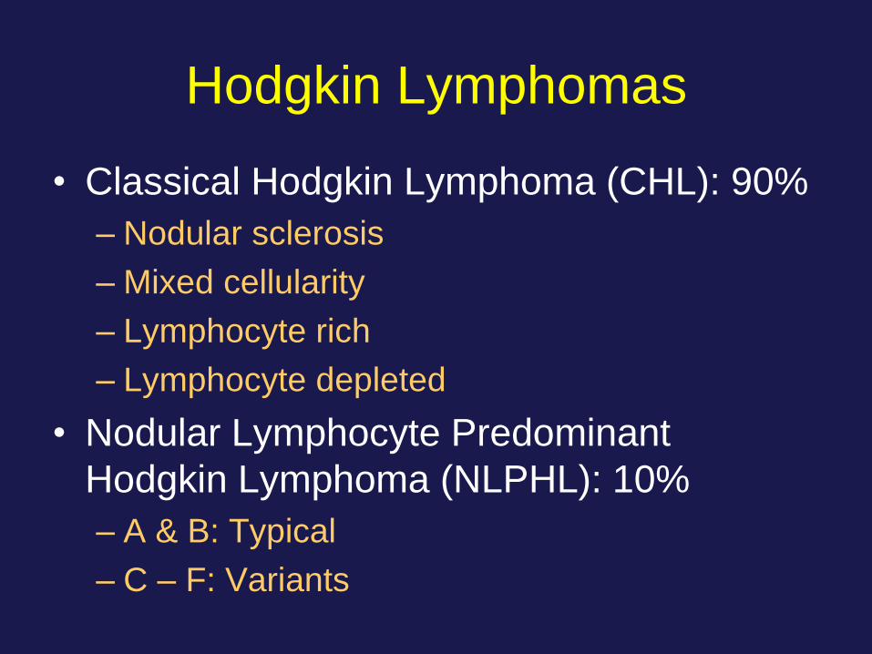
Hodgkin Lymphomas
• Classical Hodgkin Lymphoma (CHL): 90%
– Nodular sclerosis
– Mixed cellularity
– Lymphocyte rich
– Lymphocyte depleted
• Nodular Lymphocyte Predominant
Hodgkin Lymphoma (NLPHL): 10%
– A & B: Typical
– C – F: Variants
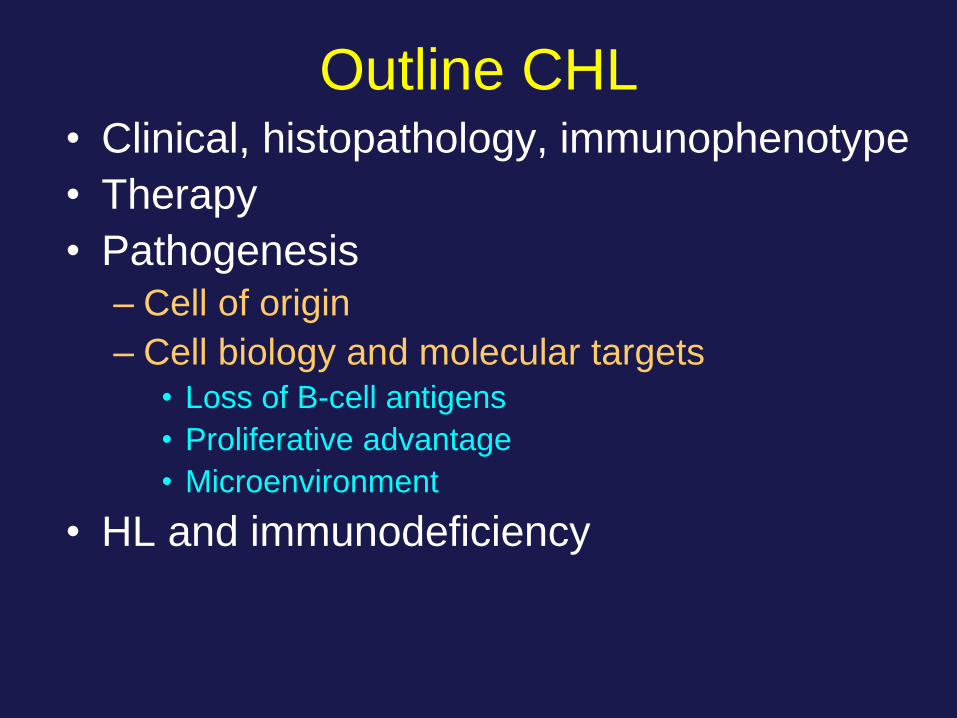
Outline CHL • Clinical, histopathology, immunophenotype
• Therapy
• Pathogenesis
– Cell of origin
– Cell biology and molecular targets • Loss of B-cell antigens
• Proliferative advantage
• Microenvironment
• HL and immunodeficiency
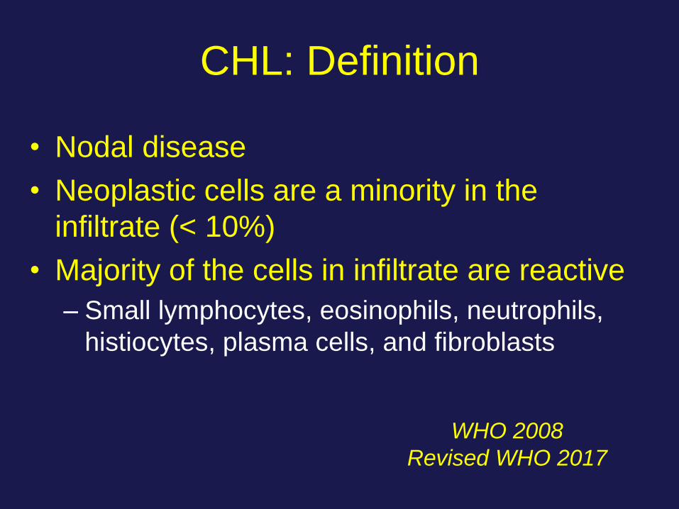
CHL: Definition
• Nodal disease
• Neoplastic cells are a minority in the
infiltrate (< 10%)
• Majority of the cells in infiltrate are reactive
– Small lymphocytes, eosinophils, neutrophils,
histiocytes, plasma cells, and fibroblasts
WHO 2008
Revised WHO 2017
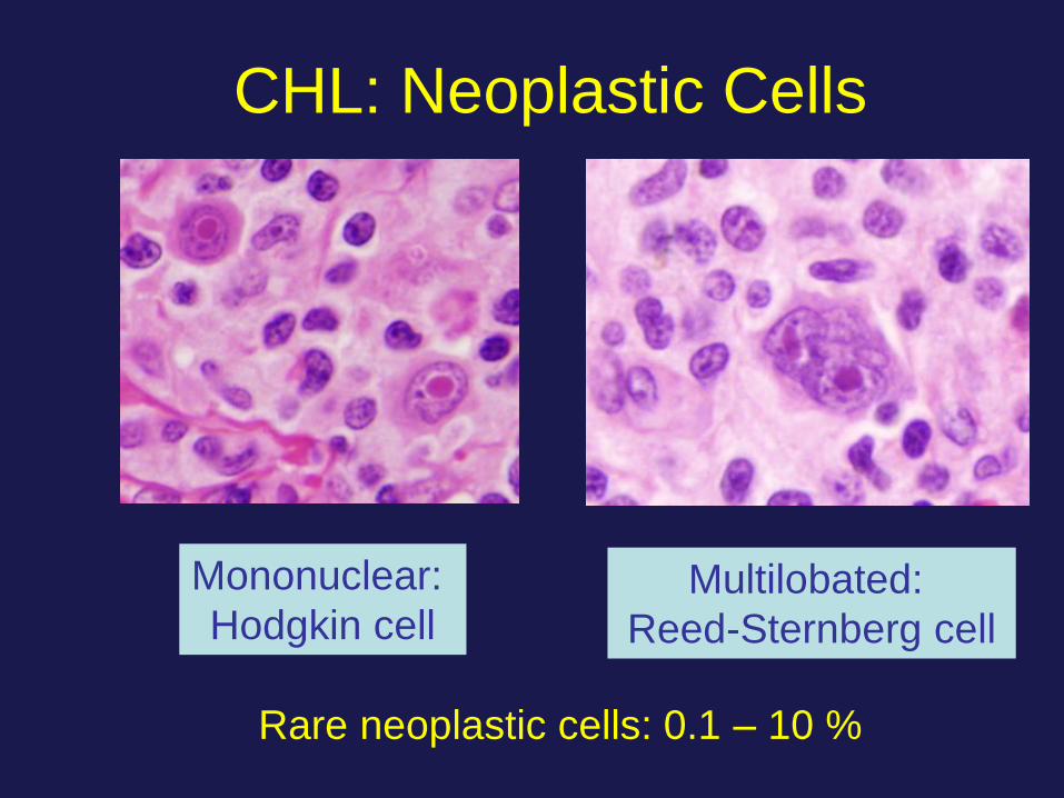
Mononuclear:
Hodgkin cell
Rare neoplastic cells: 0.1 – 10 %
Multilobated:
Reed-Sternberg cell
CHL: Neoplastic Cells
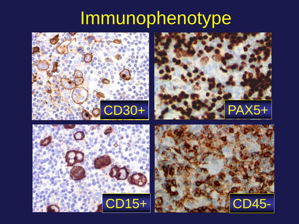
Immunophenotype
CD30+
CD15+ CD45-
PAX5+
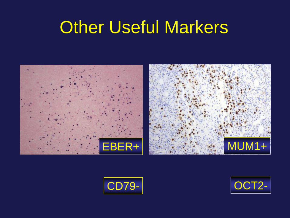
Other Useful Markers
EBER+ MUM1+
CD79- OCT2-
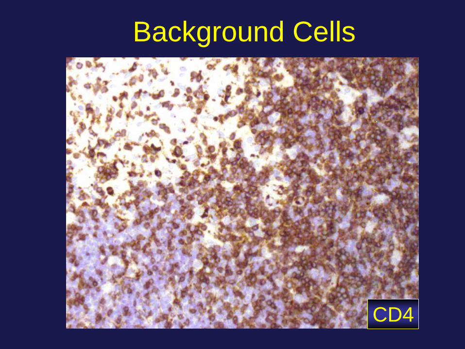
Background Cells
CD4
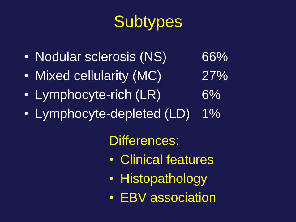
Subtypes
• Nodular sclerosis (NS) 66%
• Mixed cellularity (MC) 27%
• Lymphocyte-rich (LR) 6%
• Lymphocyte-depleted (LD) 1%
Differences:
• Clinical features
• Histopathology
• EBV association
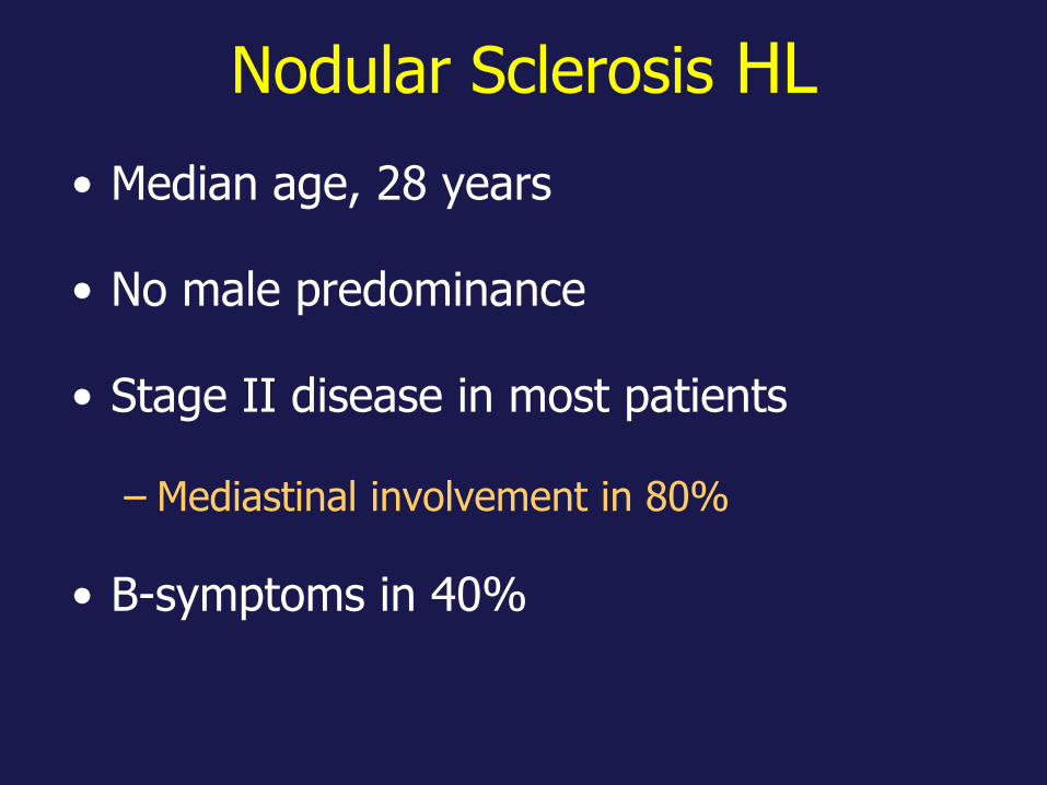
Nodular Sclerosis HL
• Median age, 28 years
• No male predominance
• Stage II disease in most patients
– Mediastinal involvement in 80%
• B-symptoms in 40%

Nodular Sclerosis HL
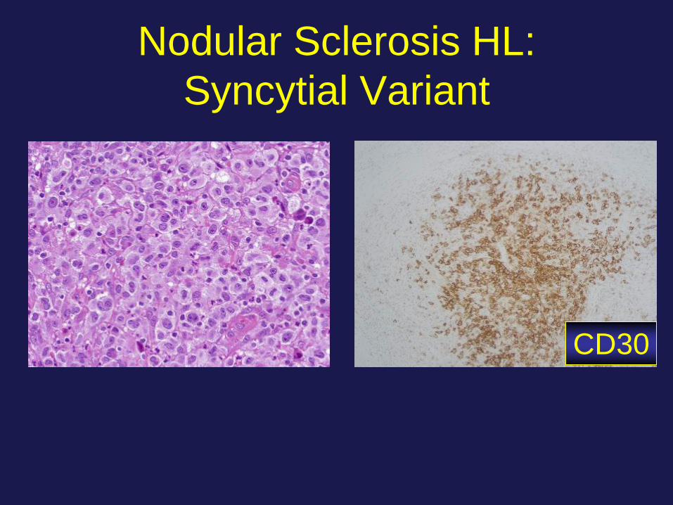
Nodular Sclerosis HL:
Syncytial Variant
CD30
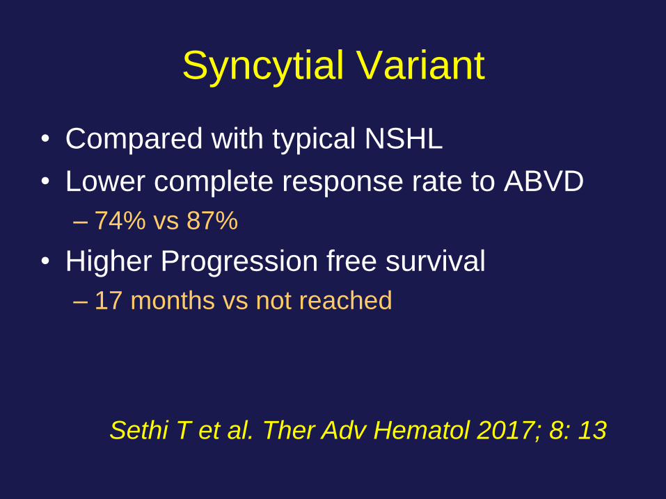
Syncytial Variant
• Compared with typical NSHL
• Lower complete response rate to ABVD
– 74% vs 87%
• Higher Progression free survival
– 17 months vs not reached
Sethi T et al. Ther Adv Hematol 2017; 8: 13
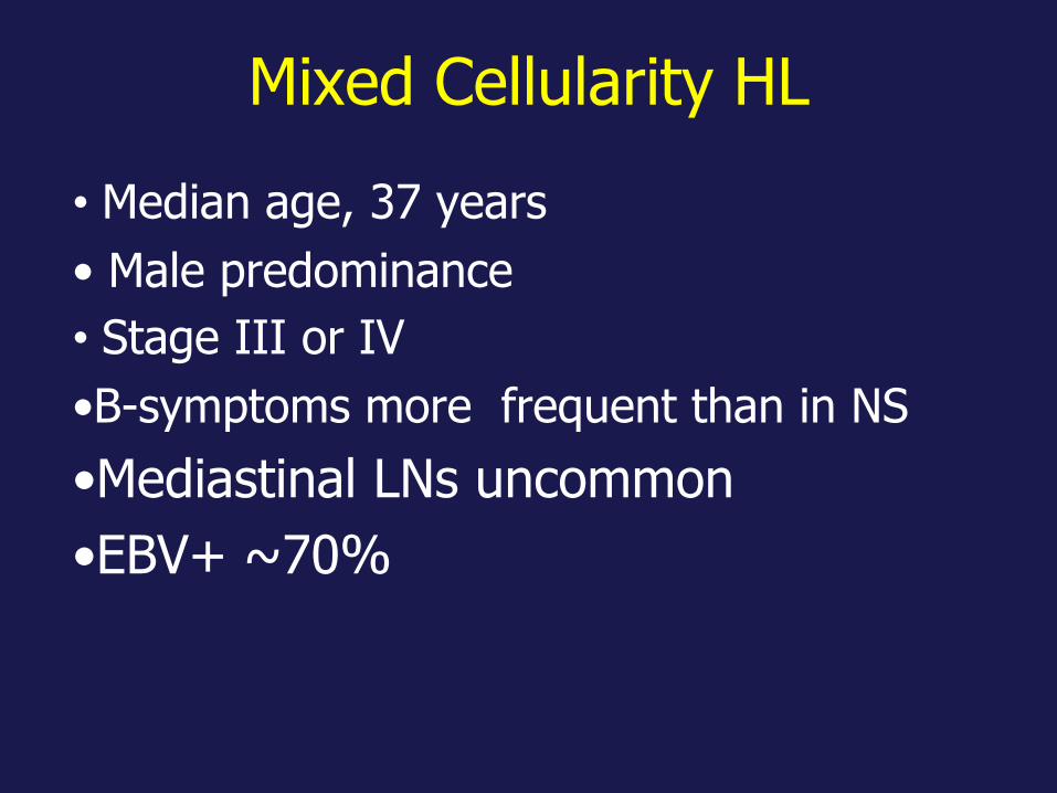
Mixed Cellularity HL
• Median age, 37 years
• Male predominance
• Stage III or IV
•B-symptoms more frequent than in NS
•Mediastinal LNs uncommon
•EBV+ ~70%
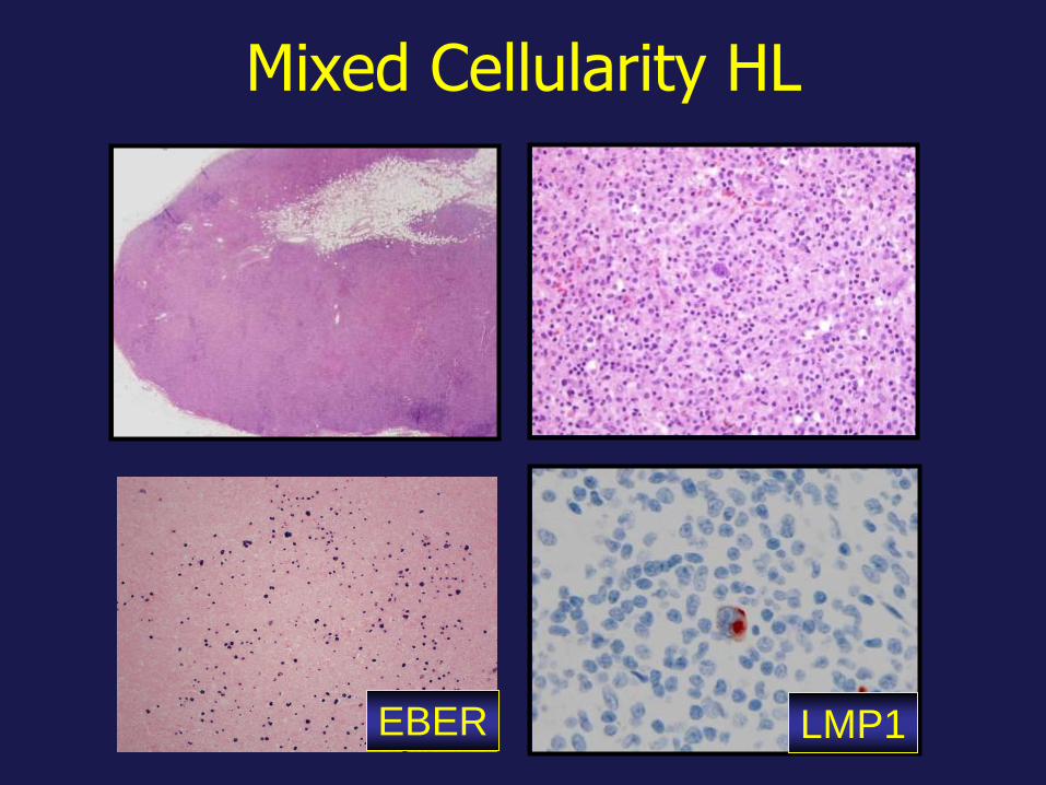
Mixed Cellularity HL
LMP1 EBER
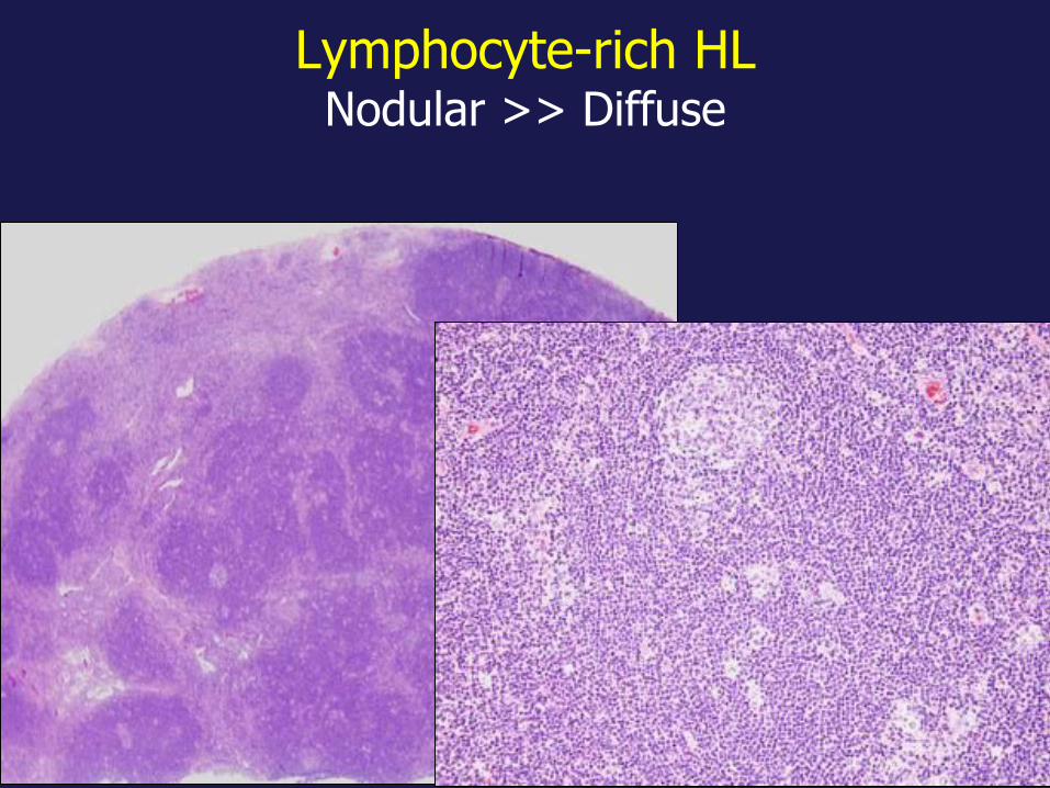
Lymphocyte-rich HL Nodular >> Diffuse

Lymphocyte-Rich HL Nodular pattern
CD30 CD20
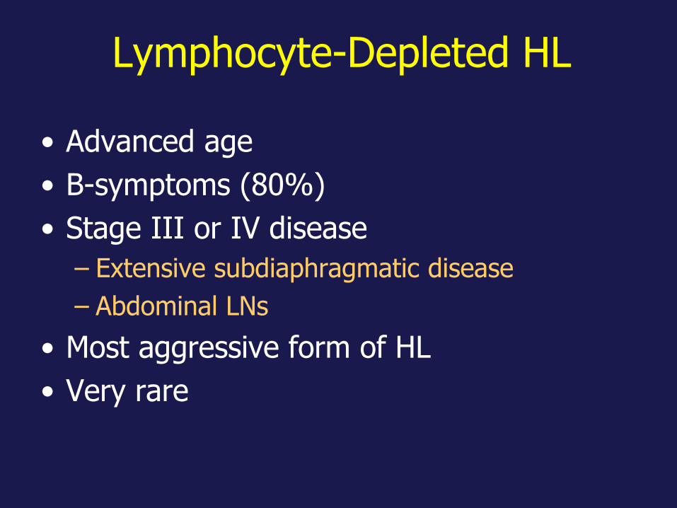
Lymphocyte-Depleted HL
• Advanced age
• B-symptoms (80%)
• Stage III or IV disease
– Extensive subdiaphragmatic disease
– Abdominal LNs
• Most aggressive form of HL
• Very rare

• Diffuse fibrosis: Fibroblastic proliferation
• Reticular: Abundant HRS cells
Lymphocyte Depleted HL

HL: Therapy
• Chemotherapy (ABVD or BEACOPP) +
Radiation: Standard of care in USA
– 90% 5-y OS
– 60% 5-y FFS
• Chemotherapy alone for early stage HL
Nat Oncol 2008; 5:543
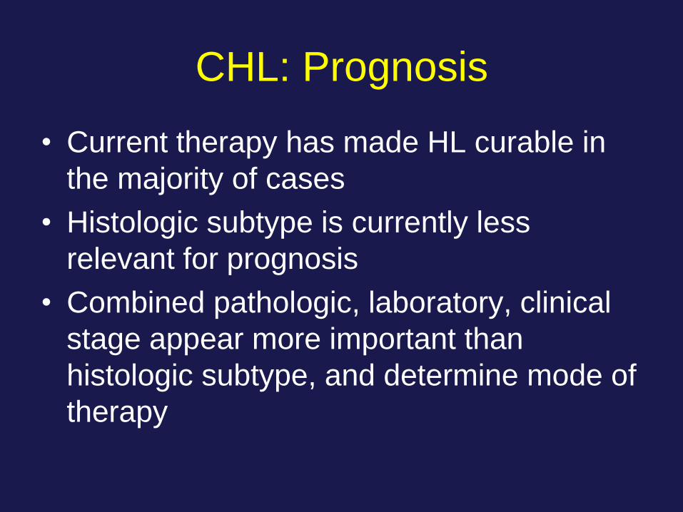
CHL: Prognosis
• Current therapy has made HL curable in
the majority of cases
• Histologic subtype is currently less
relevant for prognosis
• Combined pathologic, laboratory, clinical
stage appear more important than
histologic subtype, and determine mode of
therapy

Secondary MDS/AML in German
GHSG Trials
• N= 11,805
• 86 (0.72% developed MDS/AML)
– Early stages: 6
• < 4 cycles
– Intermediate stage: 18
– Advanced stage: 62
• > 4 cycles
Eichenauer, Blood 2010
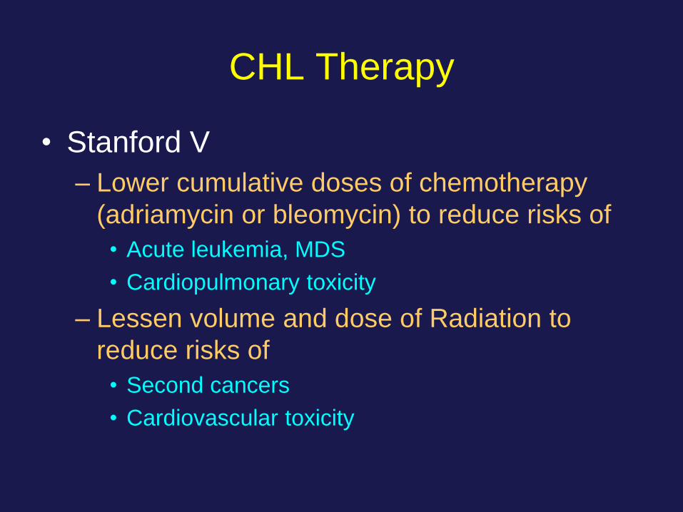
CHL Therapy
• Stanford V
– Lower cumulative doses of chemotherapy
(adriamycin or bleomycin) to reduce risks of
• Acute leukemia, MDS
• Cardiopulmonary toxicity
– Lessen volume and dose of Radiation to
reduce risks of
• Second cancers
• Cardiovascular toxicity
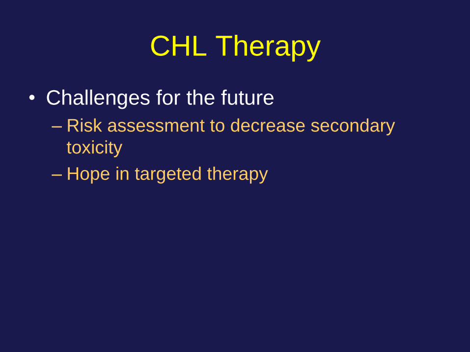
CHL Therapy
• Challenges for the future
– Risk assessment to decrease secondary
toxicity
– Hope in targeted therapy
Targeted Therapy:
Vedotin Brentuximab (Anti-CD30)
• Auristatin is bound to anti-CD30 • Potent anti-tubulin (vincristine like) arrests G2-M
phase and triggers apoptosis
• FDA-approved for relapsed and refractory CHL
• Studies underway for other CD30+ lymphomas of B- or T-cell lineage
Exp Op Inv Drugs 2011; 20: 141
Berger et al. Crit Rev Oncol/Hem 2017
Viviani et al. Tumori 2017; 103: 101
Pathogenesis of HL
• Nature of the malignant cell
– B-cell, very abnormal, pre-apoptotic
• Many reactive cells
– HRS cells secrete cytokines that attract
inflammatory cells: IL-4, IL-5, TNF-α,
GM-CSF
Cell Biology of HRS Cells
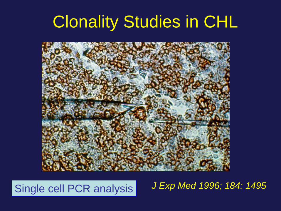
Clonality Studies in CHL
J Exp Med 1996; 184: 1495 Single cell PCR analysis

Single Cell Analysis in CHL
Antigen receptor-genes
• HRS cells show clonal Ig gene
rearrangements
Somatic mutations of VH immunoglobulin
genes
• The rearranged Ig genes harbor a high
load of somatic mutations
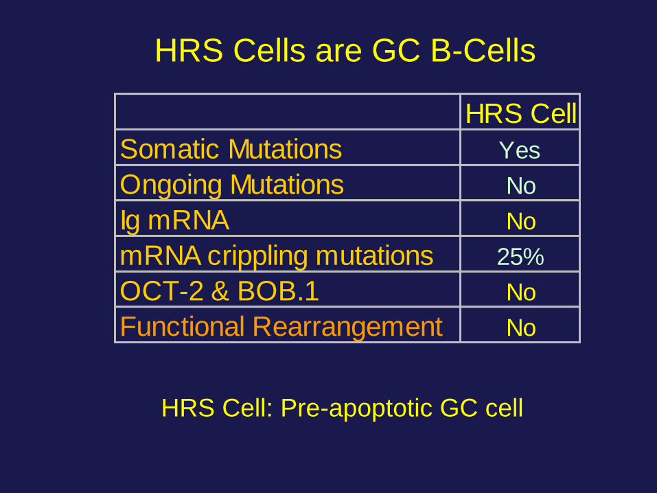
HRS Cells are GC B-Cells
HRS Cell
Somatic Mutations Yes
Ongoing Mutations No
Ig mRNA No
mRNA crippling mutations 25%
OCT-2 & BOB.1 No
Functional Rearrangement No
HRS Cell: Pre-apoptotic GC cell
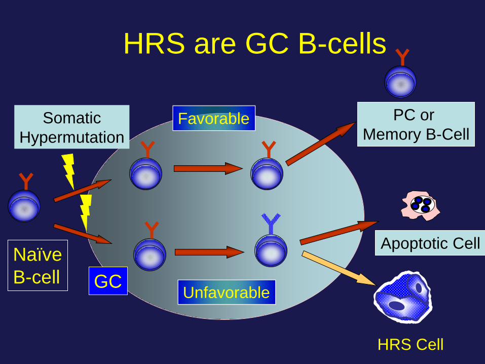
HRS are GC B-cells
Naïve
B-cell
Somatic
Hypermutation Favorable
Unfavorable
PC or
Memory B-Cell
Apoptotic Cell
HRS Cell
GC
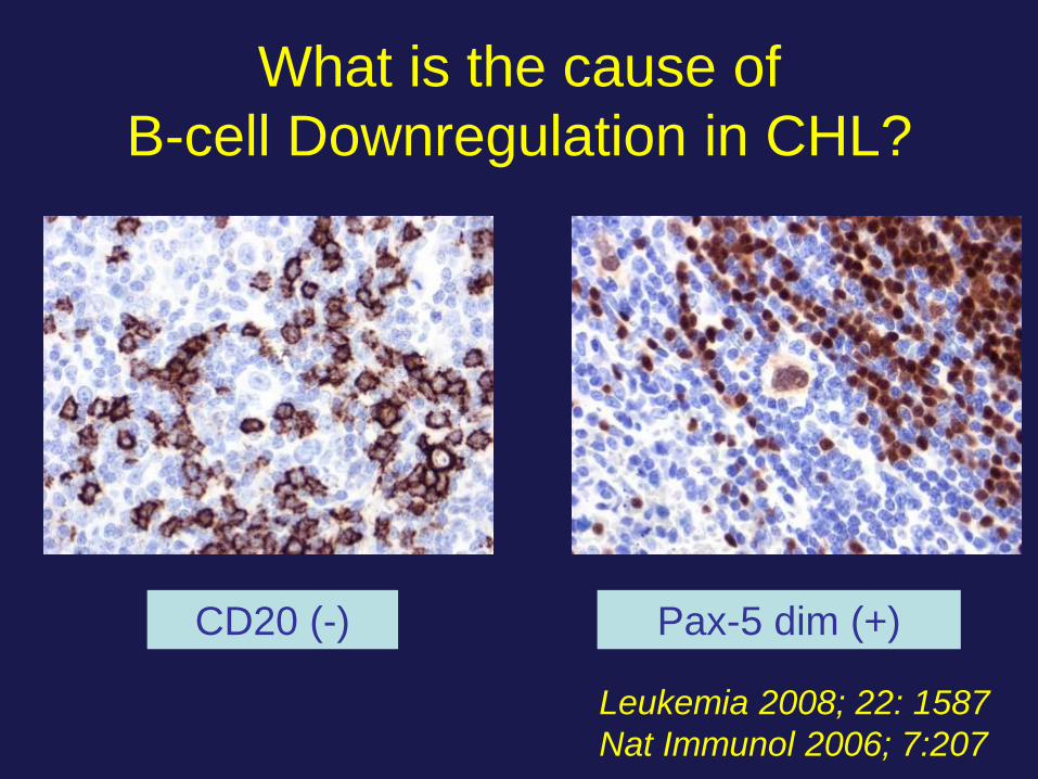
What is the cause of
B-cell Downregulation in CHL?
Leukemia 2008; 22: 1587
Nat Immunol 2006; 7:207
CD20 (-) Pax-5 dim (+)

Downregulation of B-cell genes
CD19, CD20 and CD79a
• Aberrant expression of Id2 and ABF1
– Inactivate E2A (Early B factor)
• Notch-1 antagonizes B-cell
transcription factors E2A and EBF
(early B-cell factor)
Leukemia 2008; 22: 1587
Nat Immunol 2006; 7:207
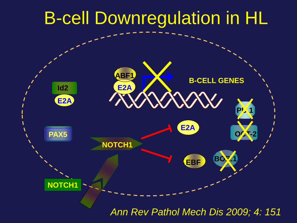
B-cell Downregulation in HL
Ann Rev Pathol Mech Dis 2009; 4: 151
PAX5
BOB.1
OCT-2
PU.1
NOTCH1
E2A
E2A
E2A
ABF1
EBF
Id2
NOTCH1
B-CELL GENES
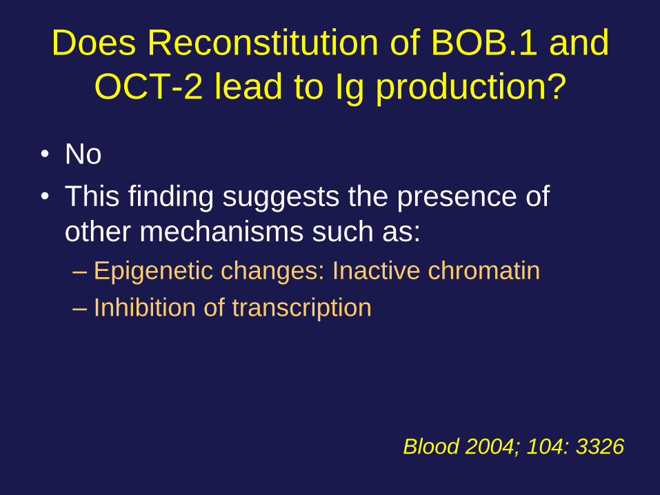
Does Reconstitution of BOB.1 and
OCT-2 lead to Ig production?
• No
• This finding suggests the presence of
other mechanisms such as:
– Epigenetic changes: Inactive chromatin
– Inhibition of transcription
Blood 2004; 104: 3326
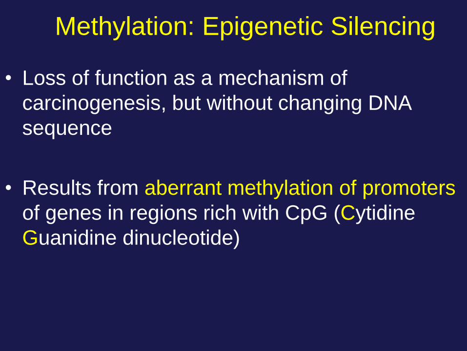
Methylation: Epigenetic Silencing
• Loss of function as a mechanism of
carcinogenesis, but without changing DNA
sequence
• Results from aberrant methylation of promoters
of genes in regions rich with CpG (Cytidine
Guanidine dinucleotide)

Epigenetics in HL
• Downregulation of B-cell transcription factors
– BCMA
– LCK
– SYK
– TCL1
• Downregulation of B-cell genes
– CD19
– CD79a
– Ig
Leukemia 2008; 22: 835
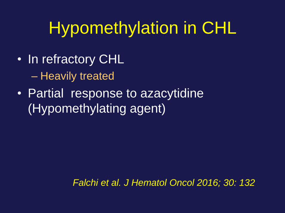
Hypomethylation in CHL
• In refractory CHL
– Heavily treated
• Partial response to azacytidine
(Hypomethylating agent)
Falchi et al. J Hematol Oncol 2016; 30: 132
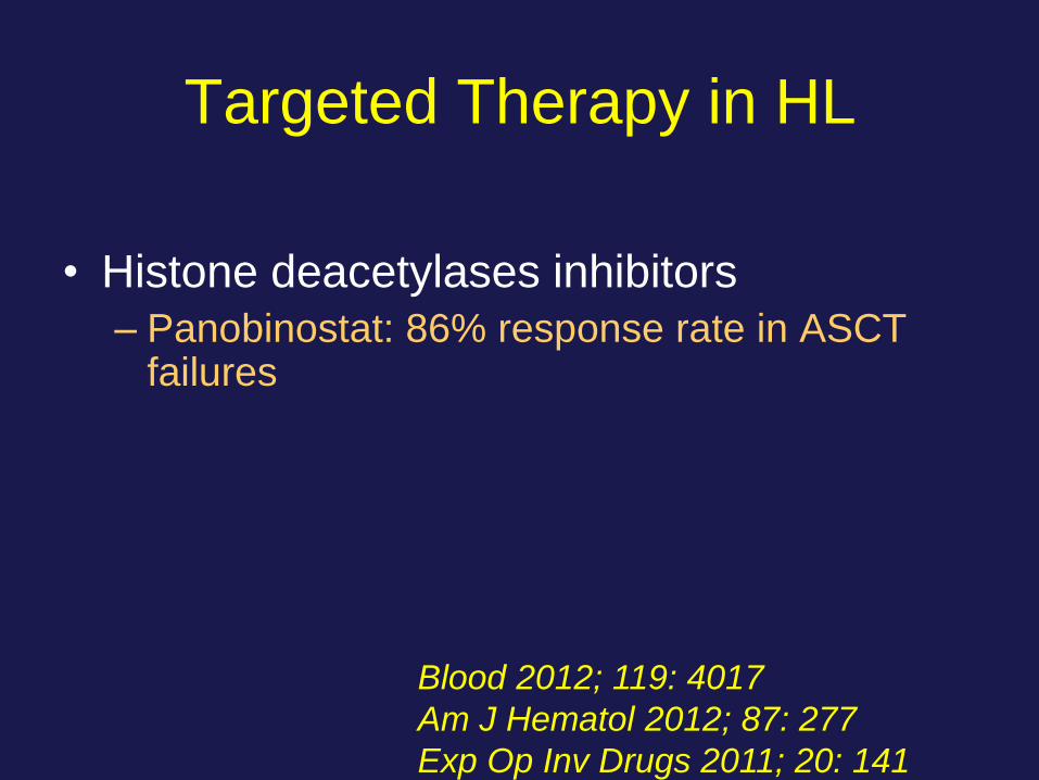
Targeted Therapy in HL
• Histone deacetylases inhibitors
– Panobinostat: 86% response rate in ASCT failures
Blood 2012; 119: 4017
Am J Hematol 2012; 87: 277
Exp Op Inv Drugs 2011; 20: 141

Consequences of Apoptosis and
B-Cell Downregulation
• Cell death
• However HRS cells survive
– Antiapoptosis • Extrinsic Pathway
• Intrinsic Pathway
– Proliferation signals • NFkB
Canonical pathway
Alternative pathway
Hum Pathol 2007; 38:103
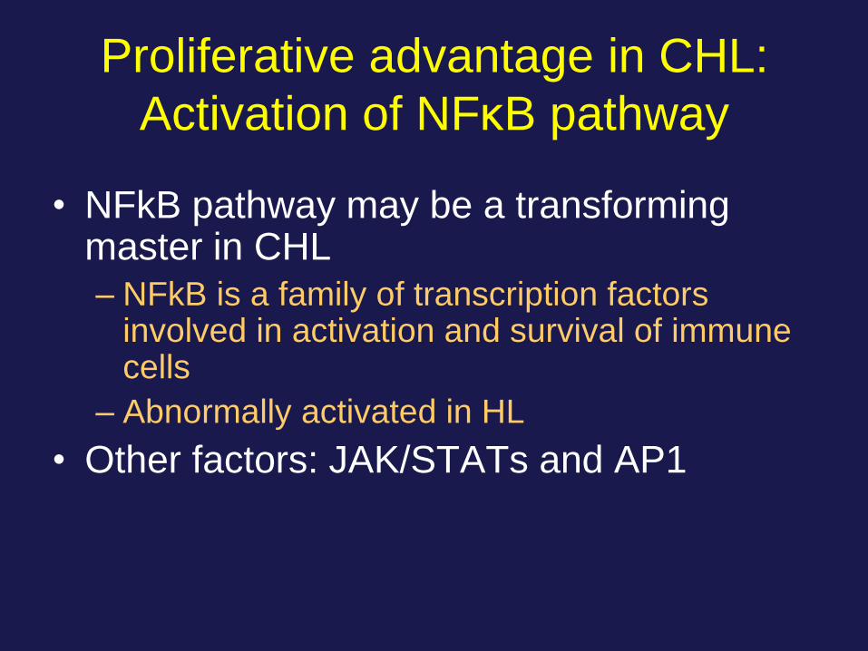
Proliferative advantage in CHL:
Activation of NFκB pathway
• NFkB pathway may be a transforming master in CHL
– NFkB is a family of transcription factors involved in activation and survival of immune cells
– Abnormally activated in HL
• Other factors: JAK/STATs and AP1
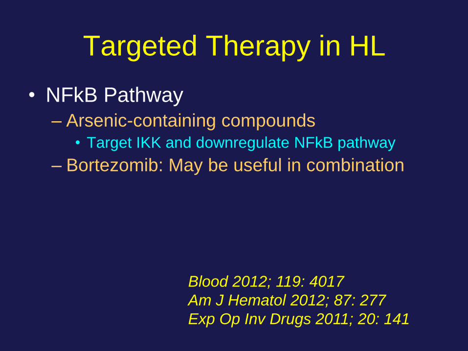
Targeted Therapy in HL
• NFkB Pathway
– Arsenic-containing compounds • Target IKK and downregulate NFkB pathway
– Bortezomib: May be useful in combination
Blood 2012; 119: 4017
Am J Hematol 2012; 87: 277
Exp Op Inv Drugs 2011; 20: 141
The Microenvironment in HL
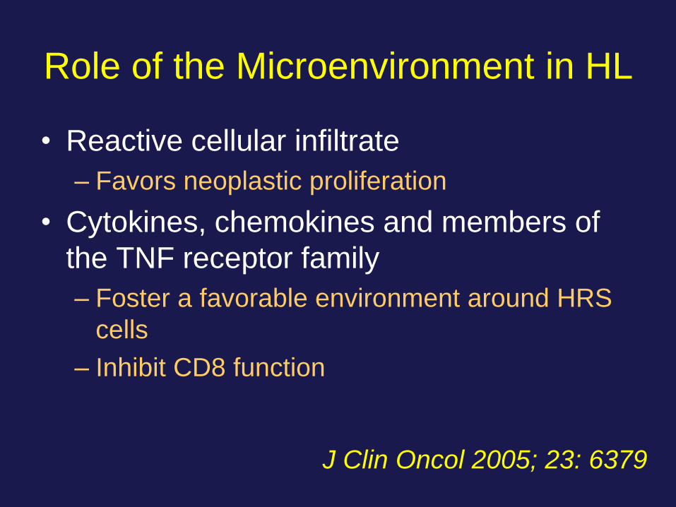
Role of the Microenvironment in HL
• Reactive cellular infiltrate
– Favors neoplastic proliferation
• Cytokines, chemokines and members of
the TNF receptor family
– Foster a favorable environment around HRS
cells
– Inhibit CD8 function
J Clin Oncol 2005; 23: 6379
PD1, PDL-1 and the Immune
Checkpoint Inhibitors in CHL • PD1 is normally expressed in effector T cells,
but inhibited through PDL-1/2 by APC cells
• HRS cells express
– PDL-1/CD274, PDL-2/CD273
– JAK2 (JAK/STAT)
• Tumor cells overexpress PDL-1 to evade
immune response
Viviani et al. Tumori 2017; 103: 101
Ok & Young; J Hem Oncol 2017; 10: 103
Immune Checkpoint Inhibitors in HL
• PDL-1 or PDL-2 increase due to gains and
amplification of 9p24.1
• EBV can induce PDL-1 expression
• Immune checkpoint inhibitors nivolumab and
pembrolizumab restore immune response
– Block interaction of PD1 with PDL-1
– FDA approved in 2016 for refractory HL
Green et al. Clin Cancer Res 2012; 18: 1611
Ok & Young; J Hem Oncol 2017; 10: 103
Jelinek T et al. Immunology 2017; 1
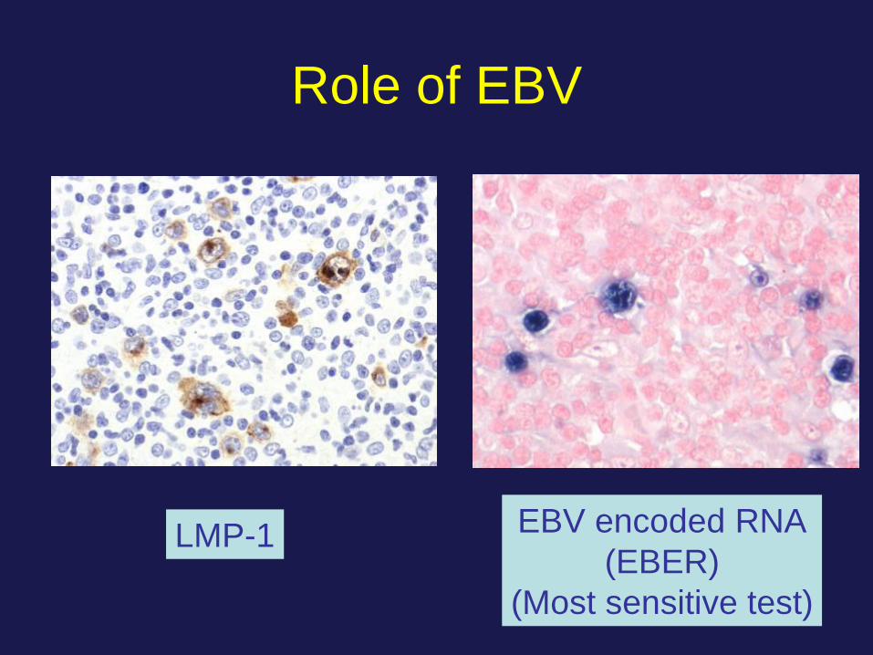
Role of EBV
LMP-1 EBV encoded RNA
(EBER)
(Most sensitive test)
CHL: EBV
• 70% in MC and LD CHL
• 20% in NS CHL
• EBV infected HRS
– Are monoclonal: Infection occurred before
clonal expansion
– LMP1: Activation (~CD40)
– LMP2A: Rescue from apoptosis (~BCR)
J Clin Pathol 2007; 60: 1342

Targeted Therapy for EBV
• EBV
– LMP2A-specific cytotoxic lymphocytes • Useful in relapsed EBV+ cases
• Microenvironment
– Immunomodulators: Thalidomide, lenalidomide
Exp Op Inv Drugs 2011; 20: 141

Cellular Changes and Possible
Mechanisms
Feature Mechanism
B-cell Neoplasm Monoclonal IGH GR
HRS Cell GC cell rescued from apoptosis
CD45 (-) Inactivation of b2M
Absent Ig Absent OCT2, BOB.1, PU.1
B-cell Downregulation NOTCH1, ID2
Antiapoptosis c-FLIP, XIAP, LMP2A
Increased proliferation NFkB
Activation: CD40 LMP1

Evolving Concept
HL and Immunodeficiency

HL and Immunodeficiency
• CHL is variable
– Immunocompetent: NS > MC
– Immunosuppressed: MC > NS
– HIV: If CD4+: 0.2 x 10⁹/L: HL
<0.05 x 10⁹/L: BL or DLBCL
• HL may regress in:
– Patients with autoimmune diseases receiving
MTX, anti-TNF
– Post-transplant LPD
• Decrease with reduction of immunosuppression
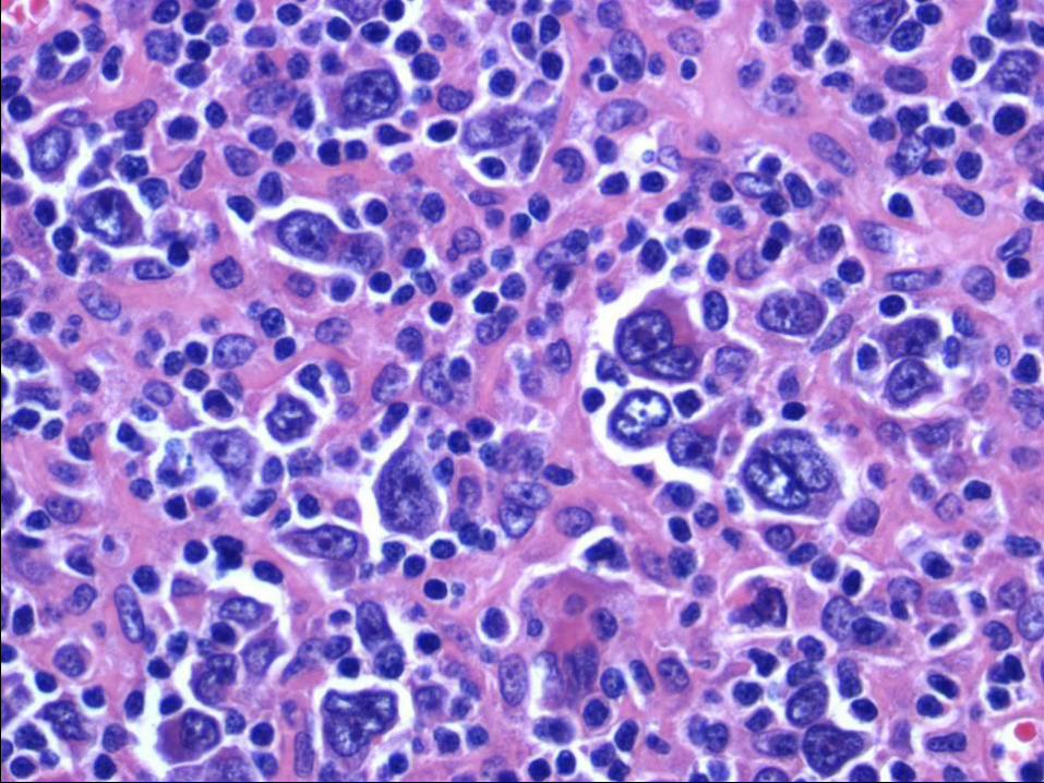
Case Discussion
• Adult patient with a history of
rheumatoid arthritis
• Therapy with methotrexate and
etanercept (anti-TNFα)
• Lymphadenopathy with B-symptoms
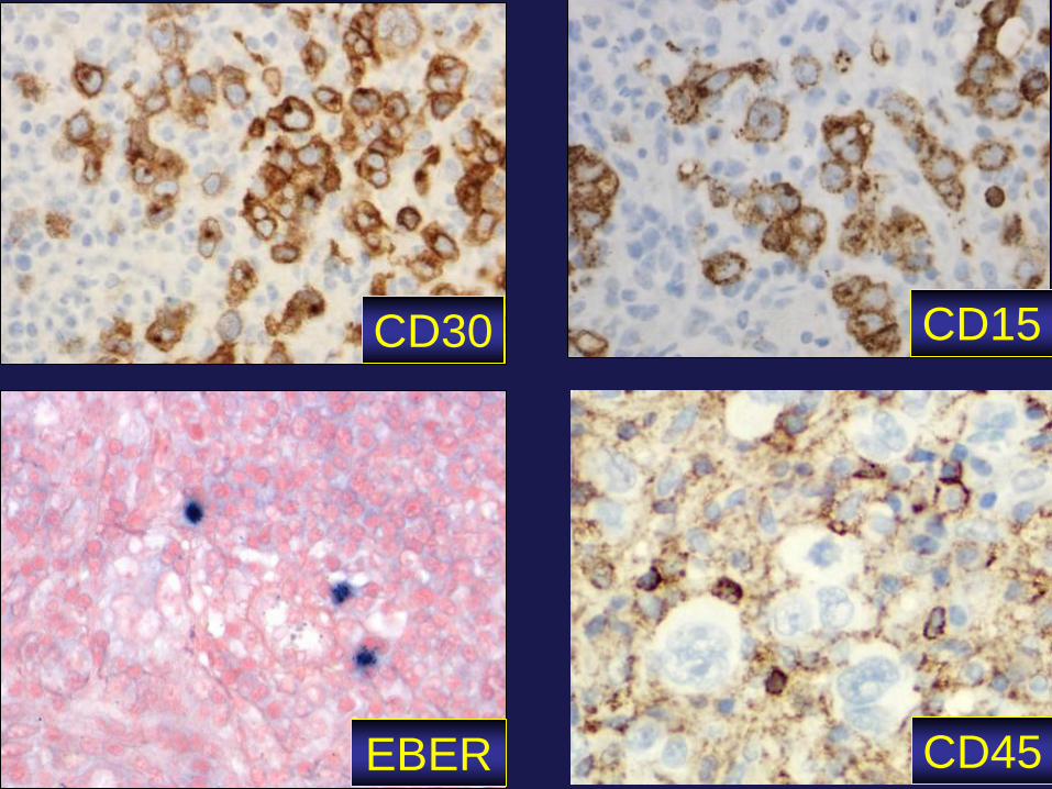
CD45 EBER
CD30 CD15
• Other iatrogenic immunodeficiency-
associated LPD
– Immunosuppression other than in transplant
– MTX
– Immunomodulators: Anti-TNFα
• Polymorphic to full-blown NHL or HL
• 40% extranodal: GI, skin, liver, spleen
WHO Entity:
Nodular Lymphocyte Predominant
Hodgkin Lymphoma
(NLPHL)

NLPHL
• Sites: Cervical, axillary, inguinal nodes
• Mostly males in 4th and 5th decades
• Rare in mediastinum, spleen and BM
• Most patients present in stage I or II
• 5 – 20 % present with stage III or IV
• Natural history:
– Slow development
– Frequent relapses, but rarely fatal
WHO, 2008

NLPHL • Nodular or nodular and diffuse pattern
• Large cells: LP or “popcorn” cells
• Contained within large nodular meshworks of
dendritic cells
• LP cell is a B cell, CD20 in 100% of cases
• Reactive background: mainly B lymphocytes
• CD15 (-) / CD30 (-)

Histopathology
Nodules are larger than follicles of
follicular lymphoma or follicular hyperplasia
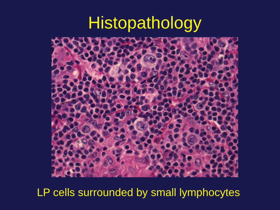
Histopathology
LP cells surrounded by small lymphocytes
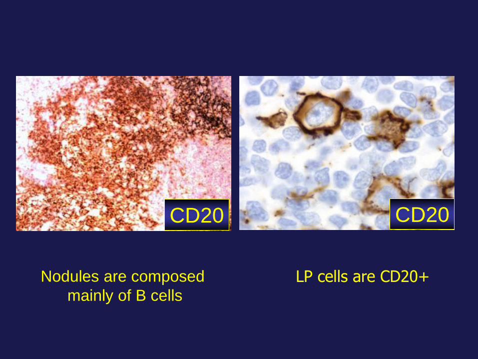
Nodules are composed
mainly of B cells
LP cells are CD20+
CD20 CD20
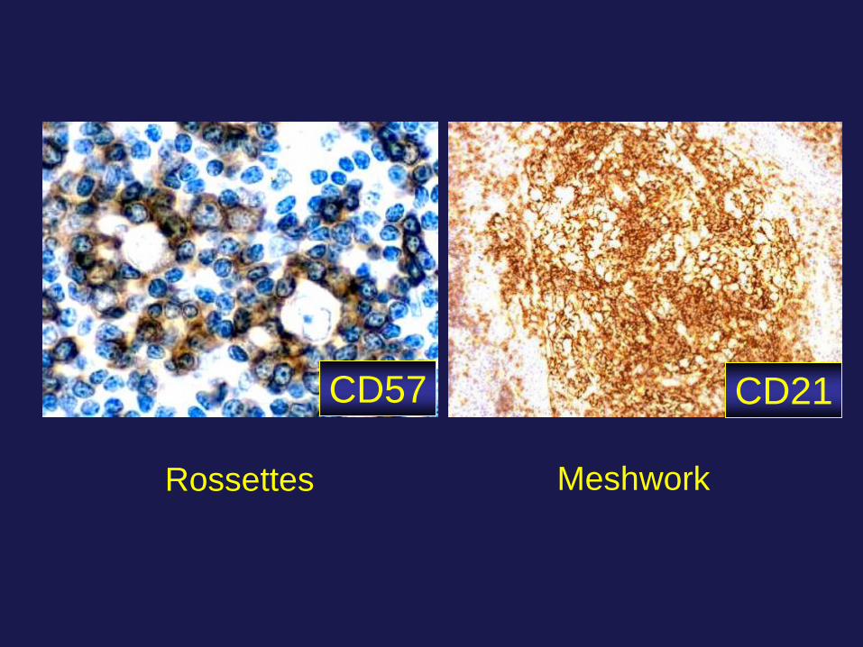
Rossettes Meshwork
CD57 CD21

OCT-2 and BOB.1
• OCT-2 is a transcription factor that
induces Ig synthesis by activating the
promoter of the Ig genes in conjunction
with BOB.1
• (+) 100 % in NLPHL
– Stronger in LP cells > surrounding small B-
cells
• (+) 20 % in CHL Blood 2001; 97: 496
Eur J Haematol 2000; 30: 458 - 469

NLPHL: Variant Patterns
• 137 biopsies
• Used H&E, CD3, CD20 and CD21
• 6 immunoarchitectural patterns
A. Nodular B-cell rich
B. Serpiginous
C. Nodular with prominent extranodular LP cells
D. Nodular T-cell rich
E. Diffuse with increased T-cells: THRBCL-like
F. Diffuse with B-cell rich pattern
Fan et al, Am J Surg Pathol 2003: 27: 1346
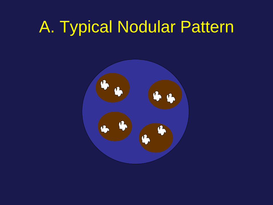
A. Typical Nodular Pattern
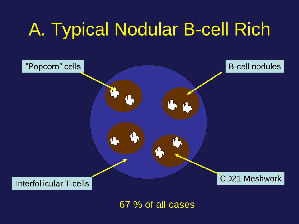
A. Typical Nodular B-cell Rich
B-cell nodules
CD21 Meshwork
“Popcorn” cells
Interfollicular T-cells
67 % of all cases
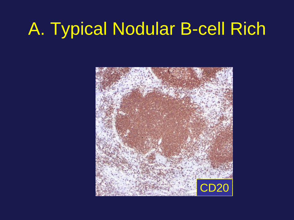
A. Typical Nodular B-cell Rich
CD20
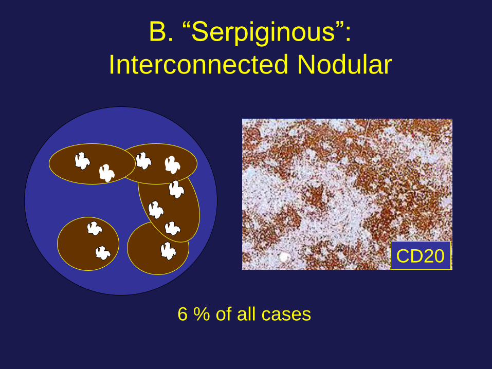
B. “Serpiginous”:
Interconnected Nodular
6 % of all cases
CD20

C. Nodular with prominent
extranodular L&H cells
7 % of all cases
CD20

D. Nodular T-Cell Rich
12 % of all cases
CD3

E. Diffuse T-Cell Rich
(THRBCL-like)
12 % of all cases
More common in patients with recurrent disease (p<.003)
CD20

E. Diffuse T-Cell Rich-like vs
THRBCL
• The detection of one nodule typical of NLPHL in an otherwise diffuse THRBCL excludes the diagnosis of THRBCL
• Reactive lymphocytes are – CD8 (+), TIA-1 (+) in TCRBCL vs
– CD4 (+), CD57 (+) in NLPHL
WHO 2008, 2016
Blood 2000; 96: 1889 – 1899
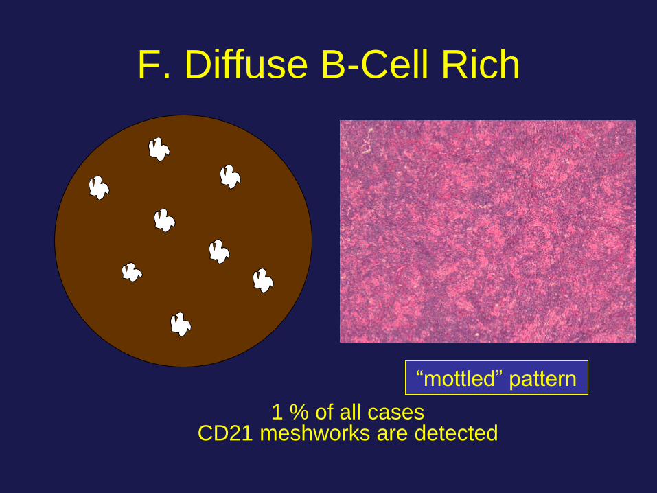
F. Diffuse B-Cell Rich
1 % of all cases CD21 meshworks are detected
“mottled” pattern

NLPHL: Prognosis
• Stage I and II: > 90 % survival at 10 years
• Not established if immediate therapy is
required for stage I disease in children
• Stage III or IV: Unfavorable prognosis
• Progression to DLBCL: 3 – 5 %
– Good prognosis if localized
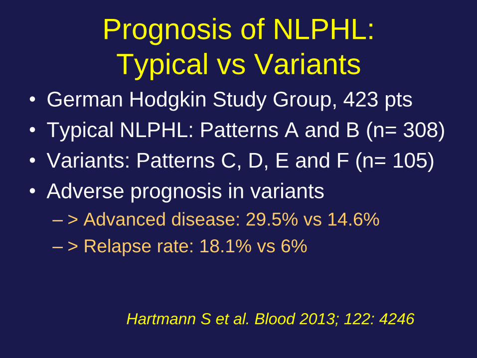
Prognosis of NLPHL:
Typical vs Variants • German Hodgkin Study Group, 423 pts
• Typical NLPHL: Patterns A and B (n= 308)
• Variants: Patterns C, D, E and F (n= 105)
• Adverse prognosis in variants
– > Advanced disease: 29.5% vs 14.6%
– > Relapse rate: 18.1% vs 6%
Hartmann S et al. Blood 2013; 122: 4246
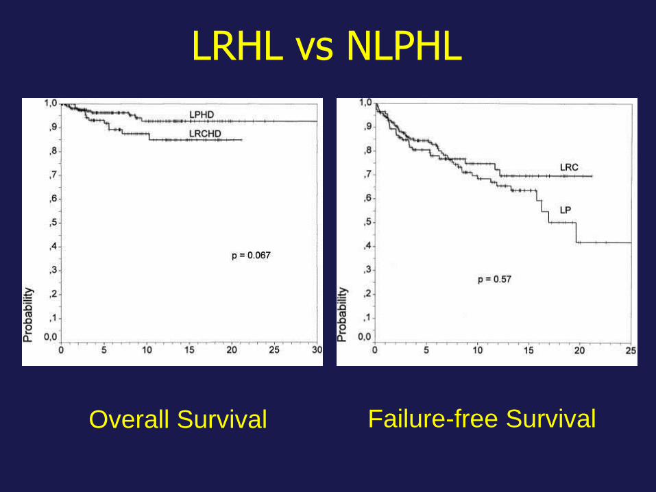
LRHL vs NLPHL
Overall Survival Failure-free Survival
Summary
• HLs are heterogeneous B-cell neoplasms
• HRS cell: Pre-apoptotic cell
– Does not produce Ig and lacks many B cell genes
– Rescued with anti-apoptotic mechanisms
– Important role of targeted therapy
• NLPHL cell: Ag selected B-cell
– Produces Ig and has a full set of B cell functioning
genes
– Predictive value of variants