Embed Size (px)
Citation preview
REVIEW Open Access
History of pelvic fracture management:a reviewPhilip F. Stahel1,2* and E. Mark Hammerberg1
Abstract
High-energy pelvic fractures represent potentially life-threatening injuries due to the risk of acute exsanguinatingretroperitoneal hemorrhage. The first report of a severe pelvic ring disruption dates back to Charles HewittMoore’s seminal publication from 1851. Significant advantages in the understanding of injury mechanisms andtreatment concepts of pelvic ring injuries evolved in the 20th century, and provided the basis to currentclassification-guided treatment and life-saving “damage control” concepts. However, there is a paucity of reportsin the current literature focused on the historic background on the treatment of pelvic ring injuries. The presentreview was designed to summarize the history and evolution of our current understanding of the mechanismsand management strategies for severe pelvic ring injuries (excluding acetabular fractures which represent adifferent entity outside of the scope of this article).
Keywords: Pelvic fracture, History, Management strategies, Retroperitoneal bleeding, Damage control
BackgroundThe concept of fracture stabilization for pain con-trol, hemostasis, reduction of deformity and fracturehealing dates back about 5,000 years to the ancientEgyptians who splinted fractures with wooden sticksand roller bandages [1]. The oldest documented sur-gical text in history is represented by the “EdwinSmith Papyrus” which dates back to the Old Kingdomin ancient Egypt, around 3,000–2,500 BC (Fig. 1).The papyrus is named after an American Egyptolo-gist who purchased it in Luxor in 1862, and repre-sents a scroll of 4.68 m in length. The text providesan outline on the diagnosis, management principles,and expected outcome of 48 different surgical condi-tions, including soft tissue injuries, fractures, jointdislocations, and tumors. The management of pelvicfractures is not specifically mentioned in the EdwinSmith Papyrus.
The ‘Malgaigne era’ (19th century)The modern history of pelvic fracture managementbegins with the seminal work of Joseph-Franҫois
Malgaigne (1806–1865), a French surgeon and world-renowned medical historian (Fig. 2). Malgaigne publishedmultiple influential textbooks on the management strat-egies of fractures and dislocations, including “Manuel demédicine opératoire fondée sur l’anatomie normale atl’anatomie pathologique” (1834), and “Traité des fractureset des luxations” (1847), which was published inEnglish translation (‘A treatise on fractures’) in 1859(Fig. 3). Multiple medical eponyms are associated withthe French pioneer, including ‘Malgaigne’s amputation’(subastragalar amputation with conservation of thetalus/astragalus), ‘Malgaigne’s hernia’ (infantile inguinalhernia), ‘Malgaigne’s luxation’ (radial head dislocation/‘nursemaid’s elbow’), and ‘Malgaigne’s fracture’. Thelatter is the first historic description of a “verticalshear” pelvic ring injury with bilateral sacro-iliac jointdislocations and associated anterior fractures of thepubic rami [2]. Several of these patients sustained in-juries after falling or jumping from heights, whileothers were crushed or run over by horse-drawn car-riages. Malgaigne described the resulting injury as a“double fracture” of the anterior and posterior pelvicring, with displacement of the hemipelvis and short-ening of the affected extremity, a case report whichwas recently made available in English translation [3].
* Correspondence: [email protected] of Orthopaedics, Univesity of Colorado, School of Medicine,Denver Health Medical Center, 777 Bannock Street, Denver, CO 80204, USA2Department of Neurosurgery, Univesity of Colorado, School of Medicine,Denver Health Medical Center, 777 Bannock Street, Denver, CO 80204, USA
© 2016 Stahel and Hammerberg. Open Access This article is distributed under the terms of the Creative CommonsAttribution 4.0 International License (http://creativecommons.org/licenses/by/4.0/), which permits unrestricted use, distribution,and reproduction in any medium, provided you give appropriate credit to the original author(s) and the source, provide a linkto the Creative Commons license, and indicate if changes were made. The Creative Commons Public Domain Dedicationwaiver (http://creativecommons.org/publicdomain/zero/1.0/) applies to the data made available in this article, unless otherwisestated.
Stahel and Hammerberg World Journal of Emergency Surgery (2016) 11:18 DOI 10.1186/s13017-016-0075-4

In the absence of radiographic imaging, accuratediagnosis in the 19th century was established by phys-ical exam. Malgaigne described palpation and manipu-lation to reveal crepitation at the fracture site, andcareful estimation of the height of the iliac crest tohelp rule out a more common fracture pattern associ-ated with lower extremity shortening, i.e. a fracture ofthe femoral neck. Malgaigne noted that vertically dis-placed pelvic fractures were often accompanied byimpairment or complete loss of lower extremity func-tion. The ‘key’ to successful management of these in-juries was the restoration and maintenance of lowerextremity length. To this end, Malgaigne advocatedan accurate closed reduction maneuver, aided by vagi-nal and/or rectal palpation. The reduction maneuverwas followed by maintenance in a modified tractionbed, with application of a pelvic sling, for a minimumof 45 to 50 days. As many patients could not toleratethe prolonged immobilization in traction, most fractureshealed with significant limb shortening. Malgaignenoted that many patients would not survive thissevere injury, and understood that there was a signifi-cant association between this fracture pattern, bleed-ing, and visceral injuries. The more fortunate patientswho survived the initial injury remained at significantrisk of delayed suppuration and sepsis, which waslikely the result of contaminated open fracture
Fig. 1 The ‘Edwin Smith Papyrus’ – the oldest documented surgical text in history
Fig. 2 Joseph-Franҫois Malgaigne (1806–1865)
Stahel and Hammerberg World Journal of Emergency Surgery (2016) 11:18 Page 2 of 6

wounds and associated visceral injuries. Therefore,those patients who survived had a grim prognosis ingeneral. As Malgaigne observed: “If life is preserved,lameness is very apt to ensue.”
Charles Hewitt Moore’s case report (1851)In parallel to Malgaigne’s seminal work in Paris,France, a British surgeon named Charles Hewitt Moore(1821–1870) conducted similar research on pelvic ringdisruptions in Plymouth, England [4]. Characterizedas a modest person who would “never speak unless hehad something of value to say” [5], much of CharlesHewitt Moore’s work never met the public eye. A rarecase report published in 1851 describes the deformingforces of a severe pelvic ring injury associated with afemoral head protrusion through an acetabular frac-ture (central hip dislocation) [6]. The injury patterndescribed in the case report by Dr. Moore repre-sented a rare entity in the 19th century since mostpatients would either sustain minor pelvic fractures,
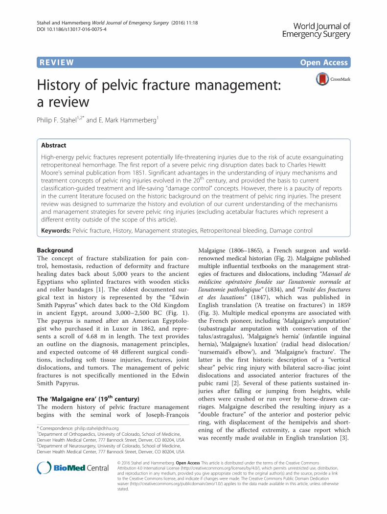
or succumbed rapidly after major trauma – typicallyfalls from heights – due to associated visceral injuriesand acute exsanguination. Moore described the pelvicfracture pattern in excruciating scientific detail, andhe emphasized the rare nature of multiple vectors ofimpacting and deforming forces (Fig. 4): “Examplesare exceedingly rare, however, in which more thanone cause of deformity exists in the same pelvis, andthere is, I believe, no instance in which so many ofthe principles of deformity are illustrated as in theaccompanying specimen (…)” [6].
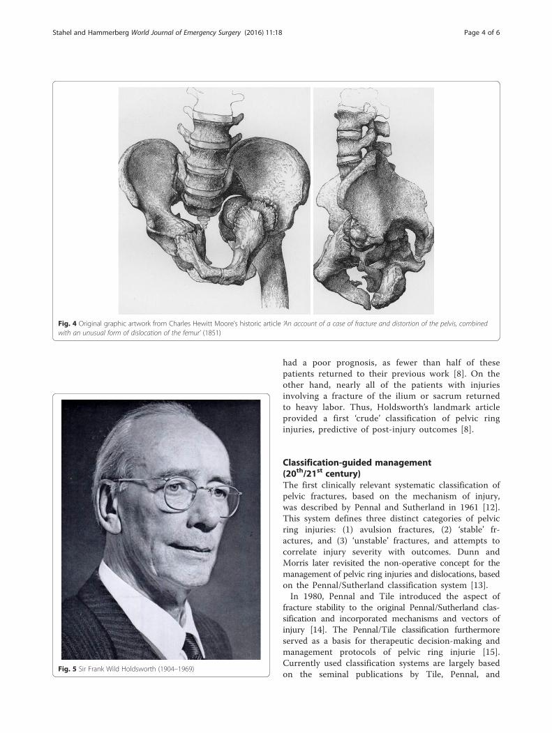
The ‘Holdsworth era’ (early 20th century)During the first half of the 20th century, the treat-ment protocols for pelvic ring injuries remained inline with Malgaigne’s general concept. The introduc-tion of X-ray technology in 1895 by the Germanphysicist Wilhelm Conrad Röntgen (1845–1923) dra-matically improved the diagnostic accuracy and classi-fication of these injuries, and allowed to monitor thehealing process. Yet, the hallmark of treatment of pel-vic ring injuries continued to consist of non-operativemanagement with closed fracture reduction and pro-longed bed rest. The application of a pelvic sling withskeletal traction was further refined by Sir Frank WildHoldsworth (1904–1969), a Professor of Orthopaedicsin Yorkshire, England (Fig. 5). Holdsworth’s legacy ismainly funded on the first spine fracture classifica-tion, however, he also dedicated significant work torefining the diagnostic and therapeutic strategies forpelvic fractures [7]. In a landmark article from 1948,Holdsworth reported his study of 50 patients withtraumatic pelvic ring disruptions during the years1937–1946 [8]. He described two distinct entities ofpelvic ring disruptions, as such: “1) dislocation of thesacro-iliac joint; 2) fracture of the ilium or sacrumadjacent to the sacro-iliac joint. In both types, there isseparation of the symphysis pubis, or fracture of bothpubic rami. In both varieties there is displacement of one-half of the pelvis outwards, or outwards and upwards.”[8]. Holdsworth’s detailed observation reflected the“open book” pattern, the “crescent” lateral comp-ression pattern, and the “vertical shear” injury. Hefurthermore provided technical recommendations forfracture reduction and immobilization, citing previousseminal work by Sir Astley Cooper [9], Sir ReginaldWatson-Jones [10], and Lorenz Böhler [11]. Theconcept of applying a sling and skeletal traction fortreatment of pelvic ring injuries is illustrated in a his-toric photograph from Holdsworth’s original publica-tion [8] (Fig. 6). Patients were maintained recumbentin a pelvic sling for 12 weeks. Using return to previ-ous employment as a marker for functional recovery,Holdsworth noted that pure sacro-iliac dislocations
Fig. 3 The English translation of Malgaigne’s landmark textbook‘A treatise on fractures’ (1859)
Stahel and Hammerberg World Journal of Emergency Surgery (2016) 11:18 Page 3 of 6
had a poor prognosis, as fewer than half of thesepatients returned to their previous work [8]. On theother hand, nearly all of the patients with injuriesinvolving a fracture of the ilium or sacrum returnedto heavy labor. Thus, Holdsworth’s landmark articleprovided a first ‘crude’ classification of pelvic ringinjuries, predictive of post-injury outcomes [8].
Classification-guided management(20th/21st century)The first clinically relevant systematic classification ofpelvic fractures, based on the mechanism of injury,was described by Pennal and Sutherland in 1961 [12].This system defines three distinct categories of pelvicring injuries: (1) avulsion fractures, (2) ‘stable’ fr-actures, and (3) ‘unstable’ fractures, and attempts tocorrelate injury severity with outcomes. Dunn andMorris later revisited the non-operative concept for themanagement of pelvic ring injuries and dislocations, basedon the Pennal/Sutherland classification system [13].In 1980, Pennal and Tile introduced the aspect of
fracture stability to the original Pennal/Sutherland clas-sification and incorporated mechanisms and vectors ofinjury [14]. The Pennal/Tile classification furthermoreserved as a basis for therapeutic decision-making andmanagement protocols of pelvic ring injurie [15].Currently used classification systems are largely basedon the seminal publications by Tile, Pennal, and
Fig. 4 Original graphic artwork from Charles Hewitt Moore’s historic article ‘An account of a case of fracture and distortion of the pelvis, combinedwith an unusual form of dislocation of the femur’ (1851)
Fig. 5 Sir Frank Wild Holdsworth (1904–1969)
Stahel and Hammerberg World Journal of Emergency Surgery (2016) 11:18 Page 4 of 6

Sutherland. For example, the AO/OTA classification forpelvic ring injuries [16] is mainly based on MarvinTile’s original classification system from 1980, and theclassification by Young & Burgess [17] is based on theoriginal Pennal/Sutherland description from 1961.Both the Tile and Young & Burgess classificationsystems are still widely used in the 21st century fordecision-making and guidance of therapeutic proto-cols in the acute management of patients with pelvicring disruptions [18, 19].By the middle part of the 20th century, with a
growing number of high-speed motor vehicle acci-dents, it had become clear that pelvic fractures wereinvolved in a significant number of fatal injuries, mostlyrelated to exsanguinating retroperitoneal hemorrhage[20]. At this time, resuscitation strategies were in theirinfancy, and there was ongoing debate regarding theappropriate sequence and surgical priorities in theacute management of pelvic hemorrhage [21]. Therole of the orthopaedic surgeon in the acute manage-ment and resuscitation of patients with pelvic ringdisruptions continued to grow in the second half ofthe 20th century. External fixation of pelvic ring injur-ies was introduced and applied increasingly in theearly management of hemodynamically unstable pa-tients [22–24]. The underlying theory was that exter-nal fixation might decrease ongoing blood loss byeliminating motion at the fracture site. In addition, byreducing ‘open book’ injuries, external fixation wasthought to reduce the intrapelvic volume and to helpreducing retroperitoneal blood loss [25].
While there are some early reports from the 1950son internal fixation for acute pelvic fractures [26], themajority of pelvic ring injuries were managed non-operatively at the time. During the second half of the20th century, treatment protocols moved beyondconservative treatment strategies, as a number of sur-geons began to recommend surgical fixation for se-lected pelvic ring injuries. Marvin Tile was a pioneerin this field and he used his own classification systemto guide treatment recommendations [25]. Initially,definitive internal fixation was reserved for verticallyunstable fractures [25]. However, into the 1980s, indi-cations for definitive internal fixation were broadenedto include rotationally unstable fractures as well[27–29]. The notion that surgical fixation of unstablepelvic ring injuries allows early mobilization of pa-tients and provides superior clinical outcomes be-came prevalent towards the end of the 20th century inNorth American and European countries [30, 31]. Thisexperience solidified the concept of early internal fix-ation of unstable pelvic ring injuries as a new inter-national standard of care in the 21st century.
ConclusionsSignificant progress has been made in recent years inthe acute management of severe pelvic ring disrup-tions by mitigating the risk of acute exsanguinatinghemorrhage and associated post-injury mortality. Thehistoric evolution related to our understanding ofunderlying injury mechanisms has provided the basisfor classification-guided management strategies for
Fig. 6 Original photograph depicting the concept of skeletal traction for treatment of a pelvic ring injury in Holdsworth’s original publication (1948)
Stahel and Hammerberg World Journal of Emergency Surgery (2016) 11:18 Page 5 of 6

high-energy pelvic injuries. Future innovations on thehorizon include less-invasive management strategies,e.g. by early definitive care with percutaneous fixationof unstable pelvic ring disruptions, and bedside point-of-care resuscitation of hemorrhagic shock and post-injurycoagulopathy which represents the current “frontier” ofcutting-edge research in the 21st century [32–38].
Competing interestsThe authors declare that they have no competing interests.
Authors’ contributionsBoth authors contributed equally to the design and writing of this reviewarticle. Both authors read and approved the final manuscript.
Received: 17 February 2016 Accepted: 29 April 2016
References1. Croce MA, Magnotti LJ, Savage SA, Wood GWN, Fabian TC. Emergent pelvic
fixation in patients with exsanguinating pelvic fractures. J Am Coll Surg.2007;204:935–9.
2. Peltier LF. Joseph Franҫois Malgaigne and Malgaigne’s fracture. Surgery.1958;44(4):777–84.
3. Malgaigne JF. Double vertical fractures of the pelvis (1859). Clin OrthopRelat Res. 2007;458:17–9.
4. Brand RA. Biographical sketch - Charles Hewitt Moore, FRCS (1821–1870).Clin Orthop Relat Res. 2012;470:2075–6.
5. Anonymous. Obituary: Charles Hewitt Moore, FRCS. Brit Med J. 1870;1:641–2.6. Moore CH. An account of a case of fracture and distortion of the pelvis,
combined with an unusual form of dislocation of the femur. Med ChirTrans. 1851;34:107–19.
7. Brand RA. Biographical sketch - Frank Wild Holdsworth, FRCS (1904–1969).Clin Orthop Relat Res. 2012;470:2083–4.
8. Holdsworth FW. Dislocation and fracture-dislocation of the pelvis. J BoneJoint Surg (Br). 1948;30:461–6.
9. Cooper A. A treatise on dislocations and fractures of the joints, Americanedition edn. Philadelphia: Blanchard and Lea; 1851.
10. Watson-Jones R. Fractures and joint injuries, 3rd edition edn. Edinburgh:Livingstone; 1943.
11. Böhler L. Die Technik der Knochenbruchbehandlung. Vienna: W. Maudrich;1953.
12. Fakler JKM, Stahel PF, Lundy DW. Classification of pelvic ring injuries. In:Fractures of the Pelvis and Acetabulum. Edited by Smith WR, Ziran BH,Morgan SJ. New York/London: CRC Press, Taylor & Francis Group; 2007. p.11–25.
13. Dunn AW, Morris HD. Fractures and dislocations of the pelvis. J Bone JointSurg Am. 1968;50(8):1639–48.
14. Pennal GF, Tile M, Waddell JP, Garside H. Pelvic disruption: assessment andclassification. Clin Orthop Relat Res. 1980;151:12–21.
15. Tile M, Pennal GF. Pelvic disruption: principles of management. Clin OrthopRelat Res. 1980;151:56–64.
16. Marsh JL, Slongo TF, Agel J, et al. Fracture and dislocation classificationcompendium - 2007: Orthopaedic Trauma Association classification, databaseand outcomes committee. J Orthop Trauma. 2007;21 Suppl 10:S1–S133.
17. Young JW, Burgess AR, Brumback RJ, Poka A. Pelvic fractures: value of plainradiography in early assessment and management. Radiology. 1986;160(2):445–51.
18. Stahel PF, Mauffrey C, Smith WR, McKean J, Hao J, Burlew CC, Moore EE.External fixation for acute pelvic ring injuries: decision making and technicaloptions. J Trauma Acute Care Surg. 2013;75(5):882–7.
19. Osterhoff G, Scheyerer MJ, Fritz Y, Bouaicha S, Wanner GA, Simmen HP,Werner CM. Comparing the predicitive value of the pelvic ring injuryclassification systems by Tile and Young and Burgess. Injury. 2014;45:742–7.
20. Peltier LF. Fractures of the pelvis: a report of eighty cases treated atuniversity hospitals. Minn Med. 1955;38(8):563–4.
21. Peltier LF. Complications associated with fractures of the pelvis. J Bone JointSurg Am. 1965;47:1060–9.
22. Slätis P, Karaharju EO. External fixation of the pelvic girdle with a trapezoidcompression frame. Injury. 1975;7(1):53–6.
23. Peltier LF. Treatment of trauma. The fractured pelvis. Med Times. 1976;104(1):76–8.
24. Bonnel F. External fixation in fractures of the pelvis [French]. Ann Chir. 1976;30(2):131–4.
25. Tile M. Pelvic fractures: operative versus nonoperative treatment. OrthopClin North Am. 1980;11(3):423–64.
26. Whiston G. Internal fixation for fractures and dislocations of the pelvis. JBone Joint Surg Am. 1953;35:701–6.
27. Tile M. Pelvic ring fractures: should they be fixed? J Bone Joint Surg (Br).1988;70:1–12.
28. Goldstein A, Phillips T, Sclafani SJ, Scalea T, Duncan A, Goldstein J, Panetta T,Shaftan G. Early open reduction and internal fixation of the disrupted pelvicring. J Trauma. 1986;26:325–33.
29. Matta JM, Saucedo T. Internal fixation of pelvic fractures. Clin Orthop RelatRes. 1989;242:83–97.
30. Bosch U, Pohlemann T, Haas N, Tscherne H. Classification and managementof complex pelvic trauma. Unfallchirurg. 1992;95:189–96.
31. Käch K, Trentz O. Distraction spondylodesis of the sacrum in “vertical shearlesions” of the pelvis. Unfallchirurg. 1994;97:28–38.
32. Mauffrey C, Cuellar DO, Pieracci F, Hak DJ, Hammerberg EM, Stahel PF,Burlew CC, Moore EE. Strategies for the management of haemorrhagefollowing pelvic fractures and associated trauma-induced coagulopathy.Bone Joint J. 2014;96-B(9):1143–54.
33. Stahel PF, Smith WR, Moore EE. Current trends in resuscitation strategy forthe multiply injured patient. Injury. 2009;40 Suppl 4:S27–35.
34. Rossaint R, Bouillon B, Cerny V, Coats TJ, Duranteau J, Fernández-Mondéjar E,Hunt BJ, Komadina R, Nardi G, Neugebauer E et al. Management of bleedingfollowing major trauma: an updated European guideline. Crit Care. 2010;14(2):R52.
35. Kashuk JL, Moore EE, Sawyer M, Le T, Johnson J, Biffl WL, Cothren CC,Barnett C, Stahel P, Silliman CC, et al. Postinjury coagulopathy management:goal directed resuscitation via POC thrombelastography. Ann Surg. 2010;251(4):604–14.
36. Zhu L, Wang L, Shen D, Ye TW, Zhao LY, Chen AM. Treatment of pelvicfractures through a less invasive ilioinguinal approach combined with aminimally invasive posterior approach. BMC Musculoskelet Disord.2015;16:167.
37. McDonald E, Theologis AA, Horst P, Kandemir U, Pekmezci M. When doanterior external or internal fixators provide additional stability in anunstable (Tile C) pelvic fracture? A biomechanical study. Eur J TraumaEmerg Surg. 2015;41(6):665–71.
38. Halawi MJ. Pelvic ring injuries: Emergency assessment and management.J Clin Orthop Trauma. 2015;6(4):252–8.
• We accept pre-submission inquiries
• Our selector tool helps you to find the most relevant journal
• We provide round the clock customer support
• Convenient online submission
• Thorough peer review
• Inclusion in PubMed and all major indexing services
• Maximum visibility for your research
Submit your manuscript atwww.biomedcentral.com/submit
Submit your next manuscript to BioMed Central and we will help you at every step:
Stahel and Hammerberg World Journal of Emergency Surgery (2016) 11:18 Page 6 of 6