Embed Size (px)
Citation preview
7327 November 14, 2013|Volume 19|Issue 42|WJG|www.wjgnet.com
Hepatobiliary manifestations in inflammatory bowel disease: The gut, the drugs and the liver
María Rojas-Feria, Manuel Castro, Emilio Suárez, Javier Ampuero, Manuel Romero-Gómez
María Rojas-Feria, Manuel Castro, Emilio Suárez, Javier Ampuero, Manuel Romero-Gómez, Unit for Medical and Sur-gical Management of Digestive Diseases and CIBERehd, Valme University Hospital, University of Seville, E-41014 Sevilla, SpainAuthor contributions: Romero-Gómez M designed the concept for this review and contributed to the writing and reviewing of the final revision of the manuscript; Rojas-Feria M, Castro M, Ampuero J, and Suárez E contributed to the study design, litera-ture search, manuscript writing, and final revision of the article; all authors approved the final version of the manuscript.Correspondence to: Manuel Romero-Gómez, MD, PhD, Professor of Medicine, Unit for Medical and Surgical Manage-ment of Digestive Diseases and CIBERehd, Valme University Hospital, University of Seville, Avda. Bellavista s/n, E-41014 Sevilla, Spain. [email protected] Telephone: +34-95-5015761 Fax: +34-95-5015899Received: June 27, 2013 Revised: September 23, 2013Accepted: September 29, 2013Published online: November 14, 2013
AbstractAbnormal liver biochemical tests are present in up to 30% of patients with inflammatory bowel disease (IBD), and therefore become a diagnostic challenge. Liver and biliary tract diseases are common extraintestinal manifestations for both Crohn’s disease and ulcerative colitis (UC), and typically do not correlate with intesti-nal activity. Primary sclerosing cholangitis (PSC) is the most common hepatobiliary manifestation of IBD, and is more prevalent in UC. Approximately 5% of patients with UC develop PSC, with the prevalence reaching up to 90%. Cholangiocarcinoma and colon cancer risks are increased in these patients. Less common disorders include autoimmune hepatitis/PSC overlap syndrome, IgG4-associated cholangiopathy, primary biliary cir-rhosis, hepatic amyloidosis, granulomatous hepatitis, cholelithiasis, portal vein thrombosis, liver abscess, and non-alcoholic fatty liver disease. Hepatitis B reactivation during immunosuppressive therapy is a major concern, with screening and vaccination being recommended in
serologically negative cases for patients with IBD. Re-activation prophylaxis with entecavir or tenofovir for 6 to 12 mo after the end of immunosuppressive therapy is mandatory in patients showing as hepatitis B surface antigen (HBsAg) positive, independently from viral load. HBsAg negative and anti-HBc positive patients, with or without anti-HBs, should be closely monitored, measur-ing alanine aminotransferase and hepatitis B virus DNA within 12 mo after the end of therapy, and should be treated if the viral load increases. On the other hand, immunosuppressive therapy does not seem to promote reactivation of hepatitis C, and hepatitis C antiviral treatment does not influence IBD natural history either. Most of the drugs used for IBD treatment may induce hepatotoxicity, although the incidence of serious ad-verse events is low. Abnormalities in liver biochemical tests associated with aminosalicylates are uncommon and are usually not clinically relevant. Methotrexate-related hepatotoxicity has been described in 14% of patients with IBD, in a dose-dependent manner. Liver biopsy is not routinely recommended. Biologics-related hepatotoxicity is rare, but has been shown most fre-quently in patients treated with infliximab. Thiopurines have been associated with veno-occlusive disease, regenerative nodular hyperplasia, and liver peliosis. Routine liver biochemical tests are recommended, es-pecially during the first month of treatment. All these conditions should be considered in IBD patients with clinical or biochemical features suggestive of hepatobili-ary involvement. Diagnosis and management of these disorders usually involve hepatologists and gastroenter-ologists due to its complexity.
© 2013 Baishideng Publishing Group Co., Limited. All rights reserved.
Key words: Inflammatory bowel disease; Hepatobiliary disorders; Extraintestinal manifestations; Primary scle-rosing cholangitis; Drug-induced liver injury; Hepato-toxicity; Hepatitis B; Hepatitis C
Core tip: Hepatobiliary disorders are common extrain-
REVIEW
Online Submissions: http://www.wjgnet.com/esps/[email protected]:10.3748/wjg.v19.i42.7327
World J Gastroenterol 2013 November 14; 19(42): 7327-7340 ISSN 1007-9327 (print) ISSN 2219-2840 (online)
© 2013 Baishideng Publishing Group Co., Limited. All rights reserved.

testinal manifestations of inflammatory bowel disease (IBD) that become a diagnostic challenge for the gas-troenterologist. In this review, we have summarized the main diseases involving the hepatobiliary system in IBD and secondary liver toxicity to IBD treatment. This review also highlights the impact of immunosuppressive and anti-tumor necrosis factor treatment in hepatitis B and C, as well as its prophylaxis and treatment, accord-ing to current clinical practice guidelines.
Rojas-Feria M, Castro M, Suárez E, Ampuero J, Romero-Gómez M. Hepatobiliary manifestations in inflammatory bowel disease: The gut, the drugs and the liver. World J Gastroenterol 2013; 19(42): 7327-7340 Available from: URL: http://www.wjgnet.com/1007-9327/full/v19/i42/7327.htm DOI: http://dx.doi.org/10.3748/wjg.v19.i42.7327
INTRODUCTIONHepatobiliary diseases are relatively common in inflam-matory bowel disease (IBD) and therefore become a diag-nostic challenge. Liver and biliary tract disorders are typi-cal extraintestinal manifestations in both Crohn’s disease (CD) and ulcerative colitis (UC). In patients receiving immunosuppressive therapy, including biologics, the risk of hepatitis B reactivation is high, so patients undergoing this therapy should be screened for hepatitis B surface antigen (HBsAg) and anti-HBc prior to starting the treat-ment. Most of the drugs used for IBD treatment have also been associated with hepatotoxicity. All these condi-tions should be ruled out in IBD patients with clinical or biochemical features suggestive of liver involvement, as summarized in this review.
Bibliographic searches were performed in the MED-LINE electronic database up to February 2013 using the Medical Subject Headings terms: (“inflammatory bowel disease” OR “Crohn’s disease” OR “ulcerative colitis”) AND (“liver” OR “biliary tract” OR “primary sclerosing cholangitis” OR “hepatobiliary disorders” OR “small-duct PSC” OR “PSC/AIH overlap syndrome” OR “IgG4-associated cholangitis” OR “primary biliary cir-rhosis” OR “hepatic amyloidosis” OR “granulomatous hepatitis” OR “cholelithiasis” OR “portal vein thrombo-sis” OR “liver abscess” OR “non-alcoholic fatty liver dis-ease” OR “viral hepatitis” OR “hepatitis B” OR “hepatitis C” OR “drug-induced liver injury” OR “drug-induced hepatitis” OR “hepatotoxicity”).
HEPATOBILIARY DISORDERS ASSOCIATED WITH INFLAMMATORY BOWEL DISEASEHepatobiliary manifestations constitute some of the most common extraintestinal manifestations of IBD. They typically adopt an independent course irrespective of intestinal activity and are present in both UC and CD.
Primary sclerosing cholangitisPrimary sclerosing cholangitis (PSC) is a chronic fibroes-clerotic disorder of the intrahepatic and extrahepatic biliary tree. and is the most common hepatobiliary mani-festation of IBD. The association of PSC and IBD was described for first time in 1965[1].
Epidemiology: Approximately 70%-80% of patients with PSC have concomitant IBD and about 1.4%-7.5% of patients with IBD will develop PSC[2]; however the course of IBD is not related to PSC. It is more prevalent in males, in UC, and in young and middle-aged patients[3]. UC has been reported in 25 to 90 percent of patients with PSC[4]. In a Spanish multicenter study, based on a survey, UC was present in 44%[5]. Nevertheless, the real prevalence of UC in PSC is up to 90% when rectal and sigmoid biopsies are routinely obtained[6].
PSC is typically characterized by progressive inflam-mation, obliterative fibrosis, and destruction of intra- and extrahepatic bile ducts, leading to end-stage liver disease and portal hypertension[7]. Patients with PSC may also de-velop complications such as cholestasis-associated mani-festations, biliary stricture, cholangitis, cholelithiasis, chol-angiocarcinoma, and colon cancer. The diagnosis of PSC is usually previous to IBD, but PSC may be diagnosed over time, after a proctocolectomy in UC patients[8].
Etiology: The etiology of PSC remains unclear. Ge-netic, immunological, and environmental factors seem to contribute to its pathogenesis. First-degree relatives of patients with PSC show an increased risk of PSC and UC, supporting a genetic predisposition to these conditions[9]. Multiple genetic factors associated with susceptibility have been described, like HLA-B8, HLA-DRB1*0301 (DR3), HLADRB3*0101 (DRw52a), and HLA-DRB1*0401 (DR4)[10,11]. In addition, three UC sus-ceptibility loci have been associated with PSC, harboring the presumed candidate genes REL, IL2, and CARD9[12]. An autoimmune mechanism has been suggested, since both are immune-mediated disorders, and are also as-sociated with other autoimmune diseases. Several auto-antibodies may be present, such as antinuclear antibodies (ANA) in 24%-53%, smooth muscle antibodies (SMA) in 13%-20%, and anti-perinuclear cytoplasmic antibodies (pANCA) in 65%-88% of patients[13-15]. Other autoan-tibodies, including anticardiolipin, thyroperoxidase, and rheumatoid factor may be present, but show uncertain clinical significance. In one study, 97% of cases with PSC were positive for, at least, one autoantibody, while 81% were positive for three or more[16]. An inflammatory re-sponse to chronic or recurrent bacterial infection into the portal circulation or ischemic damage to the bile ducts has also been postulated[17]. Therefore, the most plausible theory involves the exposure of genetically predisposed individuals to an environmental agent that provokes an anomalous immune response, leading to disease develop-ment.
IBD in PSC patients has a distinct behavior, as it shows a higher incidence of rectal sparing, backwash
7328 November 14, 2013|Volume 19|Issue 42|WJG|www.wjgnet.com
Rojas-Feria M et al . Hepatobiliary manifestations in IBD

ileitis, extensive colitis, pouchitis after ileal pouch anal anastomosis, colon dysplasia, colon cancer, and poorer prognosis[18-21]. Patients with UC and PSC usually have a lower grade of colon inflammation and a milder course, compared to patients without PSC[22]. In addition, severe progressive PSC requiring liver transplantation appears to reduce histological activity and the need for colectomy in UC[23,24].
Diagnosis: Most patients with PSC are asymptomatic at diagnosis. This disease should be considered in patients with IBD and abnormal liver biochemical tests, where a marked elevation of serum alkaline phosphatase is com-monly found[25]. In symptomatic patients, fatigue and pruritus are common. Other features include abdominal pain, jaundice, and weight loss. Cholangitis occurs in 10%-15% of patients during the course of the disease. Biochemical tests usually show a cholestatic pattern. Aminotransferases levels are typically lower than 300 IU/L. Additional biochemical parameters are hypergamma-globulinemia (30% of cases), increased serum IgM levels (40%-50%), and p-ANCA (30%-80%). Serum albumin levels later decrease during the course of the disease, and the presence of hypoalbuminemia earlier may indicate ac-tive IBD.
Diagnosis is established by the demonstration of dif-fuse, multifocal strictures and dilations in the intra- and extrahepatic bile ducts. In 41% of cases, the gallbladder and cystic duct may also be involved[26]. In the early stages of the disease, superficial ulcerations of the bile ducts may be the only manifestation found. Endoscopic retro-grade cholangiopancreatography (ERCP) is considered the gold standard technique for PSC diagnosis. It can be both diagnostic and therapeutic, and also may be useful in the early diagnosis of cholangiocarcinoma. Magnetic resonance cholangiography (MRCP) is a non-invasive al-ternative with high sensitivity and specificity, and without the risks related to the technique[27]. Liver biopsy is only recommended in cases of clinical suspicion of small-duct PSC, as it is rarely diagnostic of PSC[28]. The most spe-cific histologic finding in PSC is fibrous obliteration of small bile ducts, with periductal concentric fibrosis in an “onion skin” pattern. Other abnormalities are non-specif-ic and similar to those in primary biliary cirrhosis. Liver biopsy is helpful for staging the disease and determining prognosis. Ludwig described 4 stages of PSC based on morphologic features[29].
Prognosis: PSC is a progressive disease that, ultimately, results in portal hypertension, cirrhosis, and hepatic fail-ure. The median survival time without liver transplanta-tion is approximately 12 years. Survival is significantly worse in symptomatic patients at the time of diagnosis[30]. Coexisting IBD may also be related to a poorer prog-nosis, as it has been associated with a younger age at diagnosis, the development of malignant complications, dysplasia and/or colon cancer[31,32]. Patients with PSC usually develop complications of end-stage liver disease
with portal hypertension, such as varices, ascites, and hepatic encephalopathy. The Mayo Risk Score based on age, serum bilirubin, albumin, aspartate aminotransfer-ase, and the presence of variceal bleeding, has been used to assess disease progression and prognosis[33]. Other complications include steatorrhea and fat-soluble vitamin deficiency, secondary to chronic cholestasis, amyloidosis secondary to amyloid A protein deposition in tissues due to a progressive inflammatory process[34], dominant bili-ary strictures, cholangiocarcinoma, and colon cancer. The risk for cholangiocarcinoma is significantly increased in PSC and its development remains unpredictable. The an-nual incidence has been estimated as 1.5%[35]. Risk factors include the presence of IBD, cirrhosis, variceal bleeding, a dominant stricture in the bile duct, and alcohol intake[36]. Worsening jaundice, weight loss, and abdominal discom-fort are suspicion symptoms. Diagnosis may be difficult, as imaging techniques and brush cytology show a lack of sensitivity for early detection. However, ERCP and cytol-ogy of bile duct strictures is highly specific[37]. Prognosis is devastating, with a survival rate of 10% two years after diagnosis[38] and a recurrence rate in the transplanted liver of about 20%-25%[39]. Patients with PSC have an increased risk for gallbladder cancer, pancreatic cancer and, in cirrhotic patients, hepatocellular carcinoma. A higher risk of colorectal dysplasia/cancer has also been described among UC patients with PSC[21], even after liver transplantation[40]. The severity and the duration of PSC have not been significantly associated with the risk of colon cancer[41]. In patients with ileal pouch-anal anas-tomosis, the risk for dysplasia persists after colectomy[42]. Therefore, surveillance for colorectal cancer should be strongly recommended in PSC patients with UC[43].
Treatment: Treatment of PSC associated with UC does not differ from PSC without IBD. As no pharmacologic therapy has proven effective for PSC, treatment goals are the control of symptoms and the management of complications. Ursodeoxycholic acid (UDCA) has been shown as effective in liver function improvement based on biochemical tests but it had no effect on liver histol-ogy, liver transplant-free survival, requirements for liver transplantation, development of cholangiocarcinoma, or incidence of death[44,45]. In a meta-analysis, UDCA does not appear to decrease either the risk of adenomas or colon cancer[46]. Immunosuppressants, chelators, and ste-roids have been used without any benefit.
Liver transplantation is the only therapy that can change the inevitable outcome. The appropriate moment for liver transplantation can be difficult to determine, as patients with advanced disease may not show signs of liver failure. Survival rates after hepatic transplant at 5 and 10 years are 85% and 70%, respectively[47]. However, in 20%-25% of cases, PSC recurs in the transplanted liver[39]
.
Endoscopic management of PSC is indicated in cases of cholangitis, exacerbate jaundice, or suspicion of cholangiocarcinoma. Endoscopic dilation of dominant
7329 November 14, 2013|Volume 19|Issue 42|WJG|www.wjgnet.com
Rojas-Feria M et al . Hepatobiliary manifestations in IBD
7330 November 14, 2013|Volume 19|Issue 42|WJG|www.wjgnet.com
cell infiltrating the bile duct and other organs is decisive in reaching the diagnosis[58,59]. Clinically, patients with IAC are older at diagnosis compared to patients with PSC. Obstructive jaundice can be the first symptom, whereas it is rarely present in PSC[62]. Steroids are the first-choice therapy of IAC, as they result in the resolution of jaun-dice, improve liver laboratory parameters, and reduce serum IgG4 levels and the reversal of strictures on chol-angiogram[63]. Azathioprine (AZA) should be considered alongside those with proximal and intrahepatic stenosis, and those that relapse during and ⁄ or after corticosteroid therapy[64].
Primary biliary cirrhosisPrimary biliary cirrhosis (PBC) frequently accompanies various autoimmune diseases including Sjögren syn-drome, chronic thyroiditis, and rheumatoid arthritis, but rarely IBD[65]. There are a few reported cases of both dis-eases in the literature[66,67]. The clinical presentation varies from typical PBC; affecting males more frequently, being diagnosed at younger age and at earlier stages of PBC, and usually associated with previously diagnosed mild left-side UC. Although the pathogenesis of this disease has not yet been clarified, environmental and genetic fac-tors are considered important in the susceptibility to both diseases.
Hepatic amyloidosisSecondary amyloidosis is an unusual complication of IBD, more frequent in CD than in UC (0.9% vs 0.07%)[68]. Chronic activity in the bowel contributes to amyloid de-position in the vasculatures and sinusoids of almost any organ, including the liver. It may present as asymptomatic hepatomegaly and is more common in men with colonic diseases. Treatment is based on controlling gut inflam-mation, thereby decreasing the release of the acute phase reactant serum amyloid A[69]. In some cases, colchicine can be effective.
Granulomatous hepatitisGranulomatous hepatitis is another rare complication of CD, which is characterized by granulomas on the liver bi-opsy. The main manifestation is an increase in cholestatic enzymes such as alkaline phosphatase. Granulomatous hepatitis is often secondary to different medications, including sulfasalazine[70]. Other causes are malignancies, infections, or CD metastasis[71]. Corticosteroids and im-munosuppressive drugs have been used in its treatment.
CholelithiasisIt has been estimated that patients with CD have a doubled risk for gallstones comparing to IBD-free controls, while UC is not associated with an increased risk[72]. The inci-dence of gallstones is raised in patients with Crohn’s ileitis or ileal resection, ranging from 13% to 34%[73]. Risk factors associated with its development are CD loca-tion at diagnosis, surgery, and extent of ileal resection. Other factors include the age of the patient, frequency
strictures, with or without stenting, has been shown to al-leviate cholestasis and to improve laboratory test results, although it does not prevent disease progression[48]
.
Small-duct PSCSmall-duct PSC is characterized by laboratory and histo-logical findings similar to PSC but with normal cholan-giogram. The presence of coexisting IBD is required for the diagnosis of this entity[49]. In a large multicenter study, 80% of patients with small-duct PSC had concurrent IBD (78% UC and 21% CD)[50]. Progression of small-duct PSC to PSC was observed in 12%-23% of cases. Small-duct PSC has been associated with a better long-term prognosis as compared with large-duct PSC. Chol-angiocarcinoma has not been previously described. Some patients may require liver transplantation for end-stage liver disease, and the disease may recur after liver trans-plantation. In IBD patients with cholestatic liver function tests altered and a normal cholangiogram by ERCP/MRCP, a biopsy is recommended to rule out small-duct PSC, after excluding other hepatobiliary disorders.
AIH/PSC overlap syndrome: AIH/PSC overlap syn-drome has been described in patients with IBD, espe-cially UC[51]. The diagnosis is suspected when features of AIH and PSC are present in the same patient, requiring a definitive diagnosis of AIH based on the International Autoimmune Hepatitis Group Criteria, which includes demographic, histologic, and laboratory markers[52]. Diagnosis, treatment, and prognosis of the overlap syn-drome are controversial and so standardized diagnostic criteria are needed[53,54]. Previous studies have reported cases that initially presented with laboratory markers and histologic features of either AIH or both diseases with a normal cholangiography, only to develop pathologic characteristics of PSC during the follow-up[52,55,56]. In-trahepatic and extrahepatic bile ducts may be affected. Conventional corticosteroid therapy, alone or in conjunc-tion with UDCA (13-15 mg/kg daily), has been variably effective, and cyclosporine, mycophenolate mofetil, and budesonide have been beneficial in selected patients. The cholestatic features that influence the prognosis of auto-immune hepatitis must be defined and incorporated into the definition of the syndrome[57].
IgG4-associated cholangiopathyIgG4-associated cholangiopathy (IAC) is a biliary disease of unknown immunopathogenesis. Indistinguishable from PSC according to cholangiographic characteris-tics, it shows distinct histological findings. It is one of a variety of IgG4-related systemic disease and has been described in patients with concurrent UC[58]. Clinical di-agnostic criteria for IgG4-related disease require systemic organ involvement, elevated serum IgG4 levels (≥ 135 mg/dL), and histopathological findings[59]. IgG4 levels have also been reported in 9%-36% of patients with PSC, although these levels are usually lower than in pa-tients with IAC[60,61]. The identification of IgG4 plasma
Rojas-Feria M et al . Hepatobiliary manifestations in IBD
7331 November 14, 2013|Volume 19|Issue 42|WJG|www.wjgnet.com
of clinical recurrences, length of hospital stay, and the use of total parenteral nutrition. The pathophysiology of cholelithiasis in CD is not well defined. Abnormal mal-absorption of bile acids that interfere with enterohepatic circulation has been proposed. Moreover, reduced gall-bladder motility has been described in CD and increased gallstone cholesterol concentrations have been identified in patients with ileoanal anastomosis[74].
Portal vein thrombosisIBD is associated with an increased risk of vascular complications, such as arterial and venous thromboem-bolisms, which are considered extraintestinal manifesta-tions. Portal vein thrombosis is a rare but potentially life-threatening complication, with an incidence in IBD patients higher than that of the general population. In a Mayo Clinic study, portal/mesenteric vein thrombosis was reported in 1.3% of IBD cases, with a mortality of 50%[75]. Recent abdominal surgery, younger age, and female gender are associated with a higher incidence of portal vein thrombosis[76]. The factors involved in this pathogenesis are diverse. Acquired prothrombotic fac-tors can be identified, such as inflammation, immobiliza-tion, extent of colon disease, surgery, central catheters, corticosteroids, and smoking[77,78]. Furthermore, patients with IBD have increased platelet counts, factor Ⅴ and Ⅷ levels, and fibrinogen, along with decreased antithrombin Ⅲ levels. Anticoagulants, such as low-molecular-weight heparin and warfarin, are mainstays of therapy, even in the setting of gastrointestinal bleeding[73]. In the presence of a congenital hypercoagulable state, lifelong systemic anticoagulation should be considered, although in other prothrombotic conditions, a six-month course provides adequate coverage[79].
Liver abscessThe association between liver abscesses and IBD is uncommon[80,81], but hepatic abscesses can be an initial manifestation of CD[82]. The mechanism of abscess de-velopment may be related to direct extension of intra-abdominal abscesses or due to portal pyemia, secondary to an increase in intestinal mucosa permeability. Among associated risk factors, intra-abdominal abscesses, fistuliz-ing disease, malnutrition, and treatment with steroids and metronidazole have been reported.
Non-alcoholic fatty liver disease Non-alcoholic fatty liver disease (NAFLD) is a clinic-pathological syndrome with a histologic spectrum rang-ing from benign steatosis to non-alcoholic steatohepatitis (NASH)[83]. Steatosis is described in up to 50% of abnor-mal liver biopsies in IBD patients and has been related to colitis severity. It was presumed secondary to severe illness, with malnutrition, hypoproteinemia, and corti-costeroids primarily responsible[84]. On the other hand, NAFLD occurs in 8.2% of the IBD population, which is much lower than the frequency reported in the United States general population (33.6%). Those patients who
developed NAFLD tended to be older, and developing IBD at an older age normally requires small bowel sur-gery[85]. It has been reported that IBD patients develop NAFLD with fewer metabolic risk factors than non-IBD NAFLD patients. In multivariate analysis, hypertension (OR = 3.5), obesity (OR = 2.1), small bowel surgeries (OR = 3.7), and use of steroids at the time of imaging (OR = 3.7) were independent factors associated with NAFLD. NAFLD is also less common among patients who re-ceived antibodies against tumor necrosis factor alpha (anti-TNF-α) therapy.
VIRAL HEPATITIS AND IBDChronic hepatitis B and C are two common diseases. The WHO estimates that 350 million people in the world suf-fer from chronic hepatitis B, and more than 200 million from hepatitis C[86,87]. Hepatitis B infection is transmitted during delivery or early childhood. Hepatitis C is a blood-borne disease spread mainly via blood product transfu-sion and misuse of illegal drugs. Around 20% of patients with chronic hepatitis B show cirrhosis progression and 5% are at risk of developing hepatocellular carcinoma.
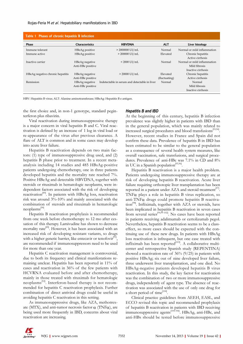
Chronic hepatitis B infection is a dynamic process, owing to the interaction between the hepatitis B virus and the host immune system. Its natural history is covered by five different phases[88] (Table 1): (1) Immunotoler-ant phase: characterized by positive hepatitis B e antigen (HBeAg), higher HBVDNA titre, and normal or near normal ALT levels. Liver biopsy at this phase shows mild or no inflammatory lesions and scarce non-progressive fibrosis; (2) Immunoclearance phase: positive HBeAg, lower HBVDNA, and raised aminotransferase levels. Liv-er histology shows necroinflammatory activity together with fibrosis progression. Spontaneous HBeAg clearance and anti-HBe seroconversion could occur at this phase; (3) HBsAg inactive carrier: characterized by negative HBeAg, positive anti-HBe, residual viremia (HBVDNA lower than 2000 IU/mL), and ALT levels under the nor-mal limit. Liver histology shows minimal or no lesions; (4) Chronic hepatitis B: HBeAg negative could appear after unsuccessful seroconversion. HBVDNA remains quanti-fiable and ALT levels are fluctuant. Fibrosis progression is common; and (5) Resolved infection: characterized by loss of HBsAg and could be a stable phase, with negative HBsAg and non-detectable HBVDNA with normal ALT levels and excellent prognosis.
Chronic hepatitis C is a progressive liver disease that could evolve to cirrhosis. Risk factors associated with fibrosis progression are alcohol consumption, HIV co-infection, and adult age at infection.
Chronic hepatitis B patients showing HBVDNA > 2000 IU/mL and at least moderate necroinflammatory activity or fibrosis in a liver biopsy should be treated with entecavir or tenofovir and, in selected cases, with pegin-terferon α-2a. In patients with chronic hepatitis C and positive HCVRNA, antiviral treatment should be started. In genotype 1, protease inhibitor-based triple therapy is
Rojas-Feria M et al . Hepatobiliary manifestations in IBD
7332 November 14, 2013|Volume 19|Issue 42|WJG|www.wjgnet.com
the first choice and, in non-1 genotype, standard pegin-terferon plus ribavirin.
Viral reactivation during immunosuppressive therapy is a major concern in viral hepatitis B and C. Viral reac-tivation is defined by an increase of 1 log in viral load or re-appearance of the virus after previous clearance. A flare of ALT is common and in some cases may develop into acute liver failure.
Hepatitis B reactivation depends on two main fac-tors: (1) type of immunosuppressive drug used, and (2) hepatitis B phase prior to treatment. In a recent meta-analysis including 14 studies and 485 HBsAg-positive patients undergoing chemotherapy, one in three patients developed hepatitis and the mortality rate reached 7%. Positive HBeAg and detectable HBVDNA, together with steroids or rituximab in hematologic neoplasms, were in-dependent factors associated with the risk of developing reactivation[89]. In patients with HBsAg loss, reactivation risk was around 3%-10% and mainly associated with the combination of steroids and rituximab in hematologic neoplasms[90]
.
Hepatitis B reactivation prophylaxis is recommended from one week before chemotherapy to 12 mo after ces-sation of this therapy. Lamivudine has shown to decrease mortality rate[89]. However, it has been associated with an increased risk of developing resistant variants, so drugs with a higher genetic barrier, like entecavir or tenofovir[91], are recommended if immunosuppressors need to be used for more than one year.
Hepatitis C reactivation management is controversial, due to both its frequency and clinical manifestations re-maining unclear. Hepatitis has been reported in 11% of cases and reactivation in 36% of the few patients with HCVRNA evaluated before and after chemotherapy, mainly in those treated with rituximab for hematologic neoplasms[92]. Interferon-based therapy is not recom-mended for hepatitis C reactivation prophylaxis. Further combination of direct antiviral drugs could be useful in avoiding hepatitis C reactivation in this setting.
As immunosuppressive drugs, like AZA, methotrex-ate (MTX), and anti-tumor necrosis factor α (TNFα), are being used more frequently in IBD, concerns about viral reactivation are increasing.
Hepatitis B and IBD At the beginning of this century, hepatitis B infection prevalence was slightly higher in patients with IBD than in the general population, which was mainly related to increased surgical procedures and blood transfusions[93,94]. However, recent studies in France and Spain did not confirm these data. Prevalence of hepatitis B in IBD has been estimated to be similar to the general population as a consequence of several health system measures, like overall vaccination, safe transfusions, and surgical proce-dures. Prevalence of anti-HBc was 7.1% in CD and 8% in UC in a Spanish population[95,96].
Hepatitis B reactivation is a major health problem. Patients undergoing immunosuppressive therapy are at risk of developing hepatitis B reactivation. Acute liver failure requiring orthotopic liver transplantation has been reported in a patient under AZA and steroid treatment[97]. TNFα plays a role in hepatitis B virus replication, so anti-TNFα drugs could promote hepatitis B reactiva-tion[98]. Infliximab, together with AZA or steroids, have been implicated in hepatitis B reactivation in seven cases from several series[94,99-102]. No cases have been reported in patients receiving adalimumab or certolizumab pegol. Nevertheless, hepatitis B reactivation seems to be a class-effect, so more cases should be expected with the con-tinuing use of these new drugs. In patients with HBsAg loss reactivation is infrequent, but one case treated with infliximab has been reported[103]. A collaborative multi-center and retrospective Spanish study (REPENTINA) showed a reactivation rate of 36% (9/25) in patients with positive HBsAg; six out of nine developed liver failure, three underwent liver transplantation, and one died. No HBsAg-negative patients developed hepatitis B virus reactivation. In this study, the key factor for reactivation was the combination of two or more immunosuppressive drugs, independently of agent type. The absence of reac-tivation was associated with the use of only one drug for a short period of time[104].
Clinical practice guidelines from AEEH, EASL, and ECCO revised this topic and recommended prophylaxis of hepatitis B reactivation in patients with IBD receiving immunosuppressive agents[104-106]. HBsAg, anti-HBc, and anti-HBs should be tested before immunosuppressive
Table 1 Phases of chronic hepatitis B infection
Phase Characteristics HBVDNA ALT Liver histology
Immune tolerant HBeAg positive > 2000000 UI/mL Normal Normal or mild inflammationImmune active HBeAg positive > 200000 UI/mL Elevated Chronic hepatitis
Active cirrhosisInactive carrier HBeAg negative < 2000 UI/mL Normal Normal or mild inflammation
Anti-HBe positive Mild fibrosisInactive cirrhosis
HBeAg negative chronic hepatitis HBeAg negative > 20000 UI/mL Elevated Chronic hepatitisAnti-HBe positive (fluctuating) Active cirrhosis
Remission HBsAg negative Indetectable in serum and detectable in liver Normal NormalAnti-HBc positive Mild fibrosis
Inactive cirrhosis
HBV: Hepatitis B virus; ALT: Alanine aminotransferase; HBeAg: Hepatitis B e antigen.
Rojas-Feria M et al . Hepatobiliary manifestations in IBD

7333 November 14, 2013|Volume 19|Issue 42|WJG|www.wjgnet.com
therapy with two goals: avoid possible fatal complications and use antiviral drugs to control hepatitis B virus rep-lication: (1) HBV serologically-negative patients should receive vaccination. Vaccination rates in IBD are variable. In Spain, only 56% of young people showed anti-HBs antibodies[95]. Vaccine response was around 46% using double doses in patients with IBD receiving anti-TNF drugs. However, AZA seems not to influence vaccine response. In non-responders a new complete vaccination course should be recommended. Hepatitis B screening is highly recommended immediately after the diagnosis of IBD, and vaccination is indicated in serologically-negative patients[107]; (2) Patients showing HBsAg positive and HBVDNA > 2000 IU/mL should be treated with tenofovir or entecavir as chronic hepatitis B patients; (3) Patients showing HBsAg positive and HBVDNA < 2000 IU/mL or undetectable levels, and patients with HBsAg negative and HBVDNA positive should be treated with tenofovir or entecavir for 6 to 12 mo after the end of immunosuppressive therapy. ALT levels and HBVDNA titre should be monitored every three months during treatment; and (4) Patients showing HBsAg negative and anti-HBc positive with or without anti-HBs should be closely monitored every 1 to 3 mo, measuring ALT and HBVDNA until 6 to 12 mo after the end of therapy. For patients with an increase in viral load, entecavir or teno-fovir therapy should be immediately started.
Hepatitis C and IBDThe prevalence of hepatitis C infection was increased in IBD patients younger than 50-year-old in comparison with the general population in studies of the last decade of the twentieth century[94]. However, recent studies have demonstrated a similar prevalence to the general popula-tion both in Spain (2.3% in CD and 1.3% in UC) and France (0.79% in CD and 1.59% in UC)[96,97].
The impact of immunosuppressive therapy for IBD on hepatitis C remains controversial. Steroids could pro-mote viral replication, as demonstrated after liver trans-plantation. However, steroids have been used to treat hepatitis C without success or adverse events. ALT flares have been reported after stopping steroid therapy in pa-tients with IBD[108]. Steroid therapy should be avoided in patients with hepatitis C, and patients should be closely monitored after any withdrawal or tapering process. Im-munosuppressive drugs like AZA, MTX, cyclosporine, and mycophenolate mofetil have been largely used in the liver transplantation setting, showing a slight antivi-ral activity against hepatitis C[109-111]. MTX did not affect hepatitis C disease in patients with arthritis[112]. Therefore, immunosuppressive therapy with these drugs seems to be safe. Indeed, in a Spanish study, 16% (8/51) of patients with chronic hepatitis C and IBD receiving immunosup-pressive therapy developed non-severe liver dysfunction, seven related to steroids, and one case with AZA[104].
TNFα plays a major role in the pathogenesis of chronic hepatitis C and associated metabolic abnor-malities. TNFα is crucial in hepatitis C-induced insulin resistance, but could also modulate interferon response.
Increased TNFα levels have been associated with im-paired sustained virological response (SVR)[113]. Thus, TNFα inhibition could be more beneficial than harmful in the management of chronic hepatitis C. Etanercept improved SVR in patients with chronic hepatitis C re-ceiving peginterferon plus ribavirin[114]. Patients suffering from rheumatoid arthritis and hepatitis C treated with etanercept or infliximab did not show any changes in transaminases level or viral load[115]. Although etanercept is not effective on IBD, a class-effect should be expected, and the use of anti-TNFα could be safe in hepatitis C as-sociated with IBD.
CD is characterized by a Th1 response. Interferon seems to play an immunomodulatory activity-enhancing Th1 response, and can cause outbreak of CD[116]. On the other hand, some authors have suggested that inter-feron could be safely used in patients with IBD, due to a lack of negative impact on the gut[117,118]. In a prospec-tive study including 11 patients with IBD and hepatitis C, peginterferon plus ribavirin achieved SVR in a similar way to non-IBD patients. During treatment six patients developed gastrointestinal symptoms that required opti-mization of immunosuppressive agents without impact-ing antiviral treatment[119]. Several case reports showed a beneficial effect of interferon-alpha and beta on patients with UC[120,121]. A systematic review, including three pro-spective studies, demonstrated no effect of interferon-alpha on UC[122], although new cases or exacerbation of UC have been seen in patients treated with interferon-alpha[123,124]. Higher doses of interferon used in hepatitis C, in comparison with lower doses in UC trials, could partially explain this discrepancy. Finally, 10 patients with CD and 10 with UC in remission or showing mild bowel activity underwent antiviral therapy for hepatitis C. No patient developed reactivation of IBD during treatment or the 12 mo of follow-up[125], confirming data from a recent review concluding that interferon does not impair IBD course[126].
In summary, hepatitis C antiviral treatment has no in-fluence on IBD natural history, and immunosuppressive therapy for IBD does not promote reactivation of hepa-titis C. Currently, no therapeutic option for reactivation prophylaxis nor vaccination are available for hepatitis C, but in the near future interferon-free regimen combining protease, polymerase, and NS5A inhibitors could be use-ful in the management of hepatitis C in IBD.
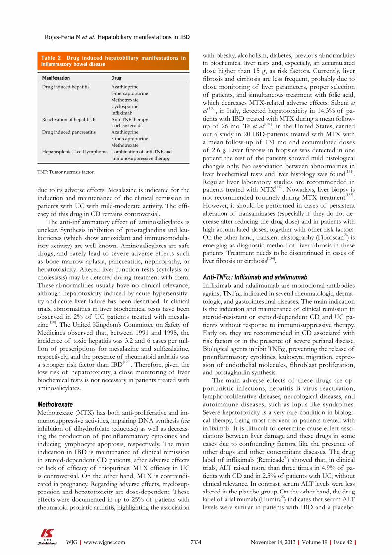
DRUG-INDUCED LIVER INJURY IN IBDApproximately 30% of patients with IBD show abnor-malities in liver biochemical tests during the course of the disease. Most of the drugs used in IBD have potential hepatotoxicity[127] (Table 2).
Aminosalicylates: Sulfasalazine and mesalazine Sulfasalazine, an association between sulfapyridine and 5-aminosalicylate (5-ASA), was the first aminosalicylate used in the treatment of IBD. This drug has been re-placed in the last two decades by mesalazine or 5-ASA,
Rojas-Feria M et al . Hepatobiliary manifestations in IBD
7334 November 14, 2013|Volume 19|Issue 42|WJG|www.wjgnet.com
due to its adverse effects. Mesalazine is indicated for the induction and maintenance of the clinical remission in patients with UC with mild-moderate activity. The effi-cacy of this drug in CD remains controversial.
The anti-inflammatory effect of aminosalicylates is unclear. Synthesis inhibition of prostaglandins and leu-kotrienes (which show antioxidant and immunomodula-tory activity) are well known. Aminosalicylates are safe drugs, and rarely lead to severe adverse effects such as bone marrow aplasia, pancreatitis, nephropathy, or hepatotoxicity. Altered liver function tests (cytolysis or cholestasis) may be detected during treatment with them. These abnormalities usually have no clinical relevance, although hepatotoxicity induced by acute hypersensitiv-ity and acute liver failure has been described. In clinical trials, abnormalities in liver biochemical tests have been observed in 2% of UC patients treated with mesala-zine[128]. The United Kingdom’s Committee on Safety of Medicines observed that, between 1991 and 1998, the incidence of toxic hepatitis was 3.2 and 6 cases per mil-lion of prescriptions for mesalazine and sulfasalazine, respectively, and the presence of rheumatoid arthritis was a stronger risk factor than IBD[129]. Therefore, given the low risk of hepatotoxicity, a close monitoring of liver biochemical tests is not necessary in patients treated with aminosalicylates.
MethotrexateMethotrexate (MTX) has both anti-proliferative and im-munosuppressive activities, impairing DNA synthesis (via inhibition of dihydrofolate reductase) as well as decreas-ing the production of proinflammatory cytokines and inducing lymphocyte apoptosis, respectively. The main indication in IBD is maintenance of clinical remission in steroid-dependent CD patients, after adverse effects or lack of efficacy of thiopurines. MTX efficacy in UC is controversial. On the other hand, MTX is contraindi-cated in pregnancy. Regarding adverse effects, myelosup-pression and hepatotoxicity are dose-dependent. These effects were documented in up to 25% of patients with rheumatoid psoriatic arthritis, highlighting the association
with obesity, alcoholism, diabetes, previous abnormalities in biochemical liver tests and, especially, an accumulated dose higher than 15 g, as risk factors. Currently, liver fibrosis and cirrhosis are less frequent, probably due to close monitoring of liver parameters, proper selection of patients, and simultaneous treatment with folic acid, which decreases MTX-related adverse effects. Sabeni et al[130], in Italy, detected hepatotoxicity in 14.3% of pa-tients with IBD treated with MTX during a mean follow-up of 26 mo. Te et al[131], in the United States, carried out a study in 20 IBD-patients treated with MTX with a mean follow-up of 131 mo and accumulated doses of 2.6 g. Liver fibrosis in biopsies was detected in one patient; the rest of the patients showed mild histological changes only. No association between abnormalities in liver biochemical tests and liver histology was found[131]. Regular liver laboratory studies are recommended in patients treated with MTX[132]. Nowadays, liver biopsy is not recommended routinely during MTX treatment[133]. However, it should be performed in cases of persistent alteration of transaminases (especially if they do not de-crease after reducing the drug dose) and in patients with high accumulated doses, together with other risk factors. On the other hand, transient elastography (Fibroscan®) is emerging as diagnostic method of liver fibrosis in these patients. Treatment needs to be discontinued in cases of liver fibrosis or cirrhosis[134].
Anti-TNFα: Infliximab and adalimumabInfliximab and adalimumab are monoclonal antibodies against TNFα, indicated in several rheumatologic, derma-tologic, and gastrointestinal diseases. The main indication is the induction and maintenance of clinical remission in steroid-resistant or steroid-dependent CD and UC pa-tients without response to immunosuppressive therapy. Early on, they are recommended in CD associated with risk factors or in the presence of severe perianal disease. Biological agents inhibit TNFα, preventing the release of proinflammatory cytokines, leukocyte migration, expres-sion of endothelial molecules, fibroblast proliferation, and prostaglandin synthesis.
The main adverse effects of these drugs are op-portunistic infections, hepatitis B virus reactivation, lymphoproliferative diseases, neurological diseases, and autoimmune diseases, such as lupus-like syndromes. Severe hepatotoxicity is a very rare condition in biologi-cal therapy, being most frequent in patients treated with infliximab. It is difficult to determine cause-effect asso-ciations between liver damage and these drugs in some cases due to confounding factors, like the presence of other drugs and other concomitant diseases. The drug label of infliximab (Remicade®) showed that, in clinical trials, ALT raised more than three times in 4.9% of pa-tients with CD and in 2.5% of patients with UC, without clinical relevance. In contrast, serum ALT levels were less altered in the placebo group. On the other hand, the drug label of adalimumab (Humira®) indicates that serum ALT levels were similar in patients with IBD and a placebo.
Table 2 Drug induced hepatobiliary manifestations in inflammatory bowel disease
Manifestation Drug
Drug induced hepatitis Azathioprine6-mercaptopurineMethotrexateCyclosporineInfliximab
Reactivation of hepatitis B Anti-TNF therapyCorticosteroids
Drug induced pancreatitis Azathioprine6-mercaptopurineMethotrexate
Hepatosplenic T-cell lymphoma Combination of anti-TNF and immunosuppressive therapy
TNF: Tumor necrosis factor.
Rojas-Feria M et al . Hepatobiliary manifestations in IBD

7335 November 14, 2013|Volume 19|Issue 42|WJG|www.wjgnet.com
According to infliximab indications, jaundice has been an uncommon finding, as well as infectious hepatitis, with liver failure being a very rare condition[135]. The Food and Drug Administration considers infliximab a hepatotoxic drug[136]. Recently, hepatotoxicity by these drugs has been evaluated in the United States (2003-2011), where only 34 cases were found, confirming the peculiarity of this adverse effect. Most cases (76%) were related to inflix-imab, showing a hepatocellular or cholestatic pattern with autoimmune characteristics, and improving after discon-tinuation of the drug[137]. Cross hepatotoxicity has not been documented in anti-TNF agents. In fact, in cases of infliximab-induced hepatotoxicity, adalimumab has been shown to be safe[138].
Thiopurines (azathioprine and 6-mercaptopurine) AZA and its metabolite, 6-mercaptopurine (MP), are the immunosuppressive agents most commonly used in IBD. They are purine analogues, which interfere in nucleic acid synthesis and inhibit the proliferation of B and T lym-phocytes, although the most relevant action is the apop-totic activation of T lymphocytes. The main indication of these drugs in CD and UC is the maintenance of clinical remission, preventing the use of steroids.
The active metabolites of AZA and MP are the 6-thioguanine nucleotides. In the liver, AZA is modi-fied to MP, which is metabolized by xanthine oxidase and thiopurine methyltransferase (TPMT) in 6-thiouric acid and 6-methylmercaptopurine, resulting ultimately in 6-thioguanine nucleotides by hypoxanthine phosphoribo-syltransferase. The decreased activity of TPMT facilitates the increasing in 6-thioguanine nucleotide levels, which are related to adverse effects. In fact, the efficacy of AZA and MP is limited owing to their adverse effects, which are responsible for treatment discontinuations in up to 15% of patients. Adverse effects are classified as dose-independent, dose-dependent, or idiosyncratic (which appears during the first two weeks of treatment). Regard-ing dose-independent adverse effects, the most common are allergic reactions (fever, exanthema, myalgias, and ar-thralgias) and acute pancreatitis. Among dose-dependent adverse effects, gastrointestinal intolerance and myelotox-icity are present in 2%-5% of patients. In retrospective studies, hepatotoxicity affected 3% of patients with an annual incidence of 1.4%, while these results are higher in prospective studies (10%)[139]. AZA and MP are able to damage the vascular endothelium, especially sinusoids and terminal veins, promoting veno-occlusive disease, regenerative nodular hyperplasia, and liver peliosis. These complications could be detected between 3 mo and 3 years after the beginning of treatment, and generate portal hypertension[140,141]. In general, mechanisms for AZA and MP hepatotoxicity remain unclear. It is thought that the main reason is the intracellular accumulation of 6-thioguanine nucleotides due to the decreased activity of TPMT.
It is recommended to determine levels of TPMT before the beginning of treatment with AZA or MP and
routinely perform liver biochemical tests, especially dur-ing the first months of treatment, to detect myelotoxicity and/or hepatotoxicity. Mild abnormalities in liver pa-rameters, without clinical relevance, allow the continua-tion of treatment at a lower dose. However, jaundice or persistent alterations in spite of reduced dose require an immediate stop to treatment[142].
CONCLUSIONHepatobiliary disorders are common extraintestinal mani-festations of IBD, and PSC represents the most prevalent disease among them. Abnormal liver biochemical tests are present in up to 30 percent of patients with IBD and emerge as a diagnostic challenge. Drug-induced hepa-totoxicity should always be rule out, as most IBD treat-ments have been associated with liver toxicity, although the incidence of serious complications is low. Hepatitis B screening and vaccination is recommended in patients with IBD. Reactivation prophylaxis with entecavir or tenofovir is mandatory in patients under immunosup-pressive therapy showing HBsAg positive, independently from viral load. HBsAg negative and anti-HBc positive patients, with or without anti-HBs, should be closely monitored, and treated if the viral load increase. Diag-nosis complexity often requires a joint gastroenterologist and hepatologist approach.
REFERENCES1 Smith MP, Loe RH. Sclerosing cholangitis; review of recent
case reports and associated diseases and four new cases. Am J Surg 1965; 110: 239-246 [PMID: 14313190 DOI: 10.1016/0002-9610(65)90018-8]
2 Broomé U, Bergquist A. Primary sclerosing cholangitis, in-flammatory bowel disease, and colon cancer. Semin Liver Dis 2006; 26: 31-41 [PMID: 16496231 DOI: 10.1055/s-2006-933561]
3 Wiesner RH, Grambsch PM, Dickson ER, Ludwig J, Mac-Carty RL, Hunter EB, Fleming TR, Fisher LD, Beaver SJ, LaRusso NF. Primary sclerosing cholangitis: natural history, prognostic factors and survival analysis. Hepatology 1989; 10: 430-436 [PMID: 2777204 DOI: 10.1002/hep.1840100406]
4 Bambha K, Kim WR, Talwalkar J, Torgerson H, Benson JT, Therneau TM, Loftus EV, Yawn BP, Dickson ER, Melton LJ. Incidence, clinical spectrum, and outcomes of primary scle-rosing cholangitis in a United States community. Gastroen-terology 2003; 125: 1364-1369 [PMID: 14598252 DOI: 10.1016/j.gastro.2003.07.011]
5 Escorsell A, Parés A, Rodés J, Solís-Herruzo JA, Miras M, de la Morena E. Epidemiology of primary sclerosing cholangitis in Spain. Spanish Association for the Study of the Liver. J Hepatol 1994; 21: 787-791 [PMID: 7890895 DOI: 10.1016/S0168-8278(94)80240-8]
6 Tung BY, Brentnall TA, Kowdley KV, Emond M, Kimmey MB, Stevens AC, Rubin CE, Haggitt RC. Diagnosis and prevalence of ulcerative colitis in patients with sclerosing cholangitis (abstract). Hepatology 1996; 24: 169A
7 Lee YM, Kaplan MM. Primary sclerosing cholangitis. N Engl J Med 1995; 332: 924-933 [PMID: 7877651 DOI: 10.1056/NEJM199504063321406]
8 Joo M, Abreu-e-Lima P, Farraye F, Smith T, Swaroop P, Gardner L, Lauwers GY, Odze RD. Pathologic features of ulcerative colitis in patients with primary sclerosing cholan-gitis: a case-control study. Am J Surg Pathol 2009; 33: 854-862
Rojas-Feria M et al . Hepatobiliary manifestations in IBD
7336 November 14, 2013|Volume 19|Issue 42|WJG|www.wjgnet.com
[PMID: 19295408 DOI: 10.1097/PAS.0b013e318196d018]9 Bergquist A, Montgomery SM, Bahmanyar S, Olsson R,
Danielsson A, Lindgren S, Prytz H, Hultcrantz R, Lööf LA, Sandberg-Gertzén H, Almer S, Askling J, Ehlin A, Ekbom A. Increased risk of primary sclerosing cholangitis and ulcer-ative colitis in first-degree relatives of patients with primary sclerosing cholangitis. Clin Gastroenterol Hepatol 2008; 6: 939-943 [PMID: 18674735 DOI: 10.1016/j.cgh.2008.03.016]
10 Farrant JM, Doherty DG, Donaldson PT, Vaughan RW, Hayllar KM, Welsh KI, Eddleston AL, Williams R. Amino acid substitutions at position 38 of the DR beta polypeptide confer susceptibility to and protection from primary scleros-ing cholangitis. Hepatology 1992; 16: 390-395 [PMID: 1639348 DOI: 10.1002/hep.1840160217]
11 Olerup O, Olsson R, Hultcrantz R, Broome U. HLA-DR and HLA-DQ are not markers for rapid disease progression in primary sclerosing cholangitis. Gastroenterology 1995; 108: 870-878 [PMID: 7875491 DOI: 10.1016/0016-5085(95)90463-8]
12 Janse M, Lamberts LE, Franke L, Raychaudhuri S, Elling-haus E, Muri Boberg K, Melum E, Folseraas T, Schrumpf E, Bergquist A, Björnsson E, Fu J, Jan Westra H, Groen HJ, Feh-rmann RS, Smolonska J, van den Berg LH, Ophoff RA, Porte RJ, Weismüller TJ, Wedemeyer J, Schramm C, Sterneck M, Günther R, Braun F, Vermeire S, Henckaerts L, Wijmenga C, Ponsioen CY, Schreiber S, Karlsen TH, Franke A, Weersma RK. Three ulcerative colitis susceptibility loci are associated with primary sclerosing cholangitis and indicate a role for IL2, REL, and CARD9. Hepatology 2011; 53: 1977-1985 [PMID: 21425313 DOI: 10.1002/hep.24307]
13 Mulder AH, Horst G, Haagsma EB, Limburg PC, Kleibeuker JH, Kallenberg CG. Prevalence and characterization of neu-trophil cytoplasmic antibodies in autoimmune liver diseases. Hepatology 1993; 17: 411-417 [PMID: 8444414 DOI: 10.1002/hep.1840170310]
14 Bansi DS, Fleming KA, Chapman RW. Importance of antineutrophil cytoplasmic antibodies in primary scleros-ing cholangitis and ulcerative colitis: prevalence, titre, and IgG subclass. Gut 1996; 38: 384-389 [PMID: 8675091 DOI: 10.1136/gut.38.3.384]
15 Terjung B, Spengler U. Role of auto-antibodies for the di-agnosis of chronic cholestatic liver diseases. Clin Rev Allergy Immunol 2005; 28: 115-133 [PMID: 15879618 DOI: 10.1385/CRIAI:28:2:115]
16 Angulo P, Peter JB, Gershwin ME, DeSotel CK, Shoe-nfeld Y, Ahmed AE, Lindor KD. Serum autoantibod-ies in patients with primary sclerosing cholangitis. J Hepatol 2000; 32: 182-187 [PMID: 10707856 DOI: 10.1016/S0168-8278(00)80061-6]
17 Aoki CA, Bowlus CL, Gershwin ME. The immunobiology of primary sclerosing cholangitis. Autoimmun Rev 2005; 4: 137-143 [PMID: 15823499 DOI: 10.1016/j.autrev.2004.09.003]
18 Loftus EV, Harewood GC, Loftus CG, Tremaine WJ, Harm-sen WS, Zinsmeister AR, Jewell DA, Sandborn WJ. PSC-IBD: a unique form of inflammatory bowel disease associated with primary sclerosing cholangitis. Gut 2005; 54: 91-96 [PMID: 15591511 DOI: 10.1136/gut.2004.046615]
19 Heuschen UA, Hinz U, Allemeyer EH, Stern J, Lucas M, Autschbach F, Herfarth C, Heuschen G. Backwash ileitis is strongly associated with colorectal carcinoma in ulcerative colitis. Gastroenterology 2001; 120: 841-847 [PMID: 11231938 DOI: 10.1053/gast.2001.22434]
20 Penna C, Dozois R, Tremaine W, Sandborn W, LaRusso N, Schleck C, Ilstrup D. Pouchitis after ileal pouch-anal anasto-mosis for ulcerative colitis occurs with increased frequency in patients with associated primary sclerosing cholangi-tis. Gut 1996; 38: 234-239 [PMID: 8801203 DOI: 10.1136/gut.38.2.234]
21 Soetikno RM, Lin OS, Heidenreich PA, Young HS, Black-stone MO. Increased risk of colorectal neoplasia in patients with primary sclerosing cholangitis and ulcerative colitis:
a meta-analysis. Gastrointest Endosc 2002; 56: 48-54 [PMID: 12085034 DOI: 10.1067/mge.2002.125367]
22 Lundqvist K, Broomé U. Differences in colonic disease ac-tivity in patients with ulcerative colitis with and without primary sclerosing cholangitis: a case control study. Dis Co-lon Rectum 1997; 40: 451-456 [PMID: 9106695 DOI: 10.1007/BF02258391]
23 Navaneethan U, Venkatesh PG, Mukewar S, Lashner BA, Remzi FH, McCullough AJ, Kiran RP, Shen B, Fung JJ. Progressive primary sclerosing cholangitis requiring liver transplantation is associated with reduced need for colec-tomy in patients with ulcerative colitis. Clin Gastroenterol Hepatol 2012; 10: 540-546 [PMID: 22245961 DOI: 10.1016/j.cgh.2012.01.006]
24 Marelli L, Xirouchakis E, Kalambokis G, Cholongitas E, Hamilton MI, Burroughs AK. Does the severity of primary sclerosing cholangitis influence the clinical course of as-sociated ulcerative colitis? Gut 2011; 60: 1224-1228 [PMID: 21402617 DOI: 10.1136/gut.2010.235408]
25 Uko V, Thangada S, Radhakrishnan K. Liver disorders in in-flammatory bowel disease. Gastroenterol Res Pract 2012; 2012: 642923 [PMID: 22474447 DOI: 10.1155/2012/642923]
26 Said K, Glaumann H, Bergquist A. Gallbladder disease in patients with primary sclerosing cholangitis. J Hepatol 2008; 48: 598-605 [PMID: 18222013 DOI: 10.1016/j.jhep.2007.11.019]
27 Moff SL, Kamel IR, Eustace J, Lawler LP, Kantsevoy S, Kalloo AN, Thuluvath PJ. Diagnosis of primary sclerosing cholangitis: a blinded comparative study using magnetic resonance cholangiography and endoscopic retrograde chol-angiography. Gastrointest Endosc 2006; 64: 219-223 [PMID: 16860072 DOI: 10.1016/j.gie.2005.12.034]
28 Chapman R, Fevery J, Kalloo A, Nagorney DM, Boberg KM, Shneider B, Gores GJ. Diagnosis and management of prima-ry sclerosing cholangitis. Hepatology 2010; 51: 660-678 [PMID: 20101749 DOI: 10.1002/hep.23294]
29 Ludwig J, Barham SS, LaRusso NF, Elveback LR, Wiesner RH, McCall JT. Morphologic features of chronic hepatitis associated with primary sclerosing cholangitis and chronic ulcerative colitis. Hepatology 1981; 1: 632-640 [PMID: 7308996 DOI: 10.1002/hep.1840010612]
30 Broomé U, Olsson R, Lööf L, Bodemar G, Hultcrantz R, Danielsson A, Prytz H, Sandberg-Gertzén H, Wallerstedt S, Lindberg G. Natural history and prognostic factors in 305 Swedish patients with primary sclerosing cholangitis. Gut 1996; 38: 610-615 [PMID: 8707097 DOI: 10.1136/gut.38.4.610]
31 Ngu JH, Gearry RB, Wright AJ, Stedman CA. Inflamma-tory bowel disease is associated with poor outcomes of pa-tients with primary sclerosing cholangitis. Clin Gastroenterol Hepatol 2011; 9: 1092-107; quiz e135 [PMID: 21893134 DOI: 10.1016/j.cgh.2011.08.027]
32 Navaneethan U, Venkatesh PG, Lashner BA, Shen B, Kiran RP. The Impact of ulcerative colitis on the long-term out-come of patients with primary sclerosing cholangitis. Aliment Pharmacol Ther 2012; Epub ahead of print [PMID: 22428605 DOI: 10.1111/j.1365-2036.2012.05063]
33 Kim WR, Therneau TM, Wiesner RH, Poterucha JJ, Benson JT, Malinchoc M, LaRusso NF, Lindor KD, Dickson ER. A revised natural history model for primary sclerosing cholan-gitis. Mayo Clin Proc 2000; 75: 688-694 [PMID: 10907383]
34 Kato T, Komori A, Bae SK, Migita K, Ito M, Motoyoshi Y, Abiru S, Ishibashi H. Concurrent systemic AA amyloidosis can discriminate primary sclerosing cholangitis from IgG4-associated cholangitis. World J Gastroenterol 2012; 18: 192-196 [PMID: 22253527 DOI: 10.3748/wjg.v18.i2.192]
35 Bergquist A, Ekbom A, Olsson R, Kornfeldt D, Lööf L, Danielsson A, Hultcrantz R, Lindgren S, Prytz H, Sandberg-Gertzén H, Almer S, Granath F, Broomé U. Hepatic and extrahepatic malignancies in primary sclerosing cholangitis. J Hepatol 2002; 36: 321-327 [PMID: 11867174 DOI: 10.1016/S0168-8278(01)00288-4]
Rojas-Feria M et al . Hepatobiliary manifestations in IBD

7337 November 14, 2013|Volume 19|Issue 42|WJG|www.wjgnet.com
36 Burak K, Angulo P, Pasha TM, Egan K, Petz J, Lindor KD. Incidence and risk factors for cholangiocarcinoma in primary sclerosing cholangitis. Am J Gastroenterol 2004; 99: 523-526 [PMID: 15056096 DOI: 10.1111/j.1572-0241.2004.04067.x]
37 Boberg KM, Jebsen P, Clausen OP, Foss A, Aabakken L, Schrumpf E. Diagnostic benefit of biliary brush cytology in cholangiocarcinoma in primary sclerosing cholangitis. J Hepatol 2006; 45: 568-574 [PMID: 16879890 DOI: 10.1016/j.jhep.2006.05.010]
38 Marsh JW, Iwatsuki S, Makowka L, Esquivel CO, Gordon RD, Todo S, Tzakis A, Miller C, Van Thiel D, Starzl TE. Orthotopic liver transplantation for primary sclerosing cholangitis. Ann Surg 1988; 207: 21-25 [PMID: 2827593 DOI: 10.1097/00000658-198801000-00005]
39 Graziadei IW, Wiesner RH, Batts KP, Marotta PJ, LaRusso NF, Porayko MK, Hay JE, Gores GJ, Charlton MR, Ludwig J, Poterucha JJ, Steers JL, Krom RA. Recurrence of primary sclerosing cholangitis following liver transplantation. Hepa-tology 1999; 29: 1050-1056 [PMID: 10094945 DOI: 10.1002/hep.510290427]
40 Bleday R, Lee E, Jessurun J, Heine J, Wong WD. Increased risk of early colorectal neoplasms after hepatic transplant in patients with inflammatory bowel disease. Dis Colon Rectum 1993; 36: 908-912 [PMID: 8404380 DOI: 10.1007/BF02050624]
41 Navaneethan U, Kochhar G, Venkatesh PG, Lewis B, Lash-ner BA, Remzi FH, Shen B, Kiran RP. Duration and severity of primary sclerosing cholangitis is not associated with risk of neoplastic changes in the colon in patients with ulcerative colitis. Gastrointest Endosc 2012; 75: 1045-1054.e1 [PMID: 22405258 DOI: 10.1016/j.gie.2012.01.015]
42 Ståhlberg D, Veress B, Tribukait B, Broomé U. Atrophy and neoplastic transformation of the ileal pouch mucosa in pa-tients with ulcerative colitis and primary sclerosing cholan-gitis: a case control study. Dis Colon Rectum 2003; 46: 770-778 [PMID: 12794579]
43 Chaparro M, Trapero-Marugán M, Guijarro M, López C, Moreno-Otero R, Gisbert JP. Dysplasia and colorectal cancer in a patient with ulcerative colitis and primary sclerosing cholangitis: a case report and a short review of the litera-ture. J Crohns Colitis 2013; 7: e61-e65 [PMID: 22552273 DOI: 10.1016/j.crohns.2012.04.005]
44 Lindor KD, Kowdley KV, Luketic VA, Harrison ME, Mc-Cashland T, Befeler AS, Harnois D, Jorgensen R, Petz J, Keach J, Mooney J, Sargeant C, Braaten J, Bernard T, King D, Miceli E, Schmoll J, Hoskin T, Thapa P, Enders F. High-dose ursodeoxycholic acid for the treatment of primary sclerosing cholangitis. Hepatology 2009; 50: 808-814 [PMID: 19585548 DOI: 10.1002/hep.23082]
45 Shi J, Li Z, Zeng X, Lin Y, Xie WF. Ursodeoxycholic acid in primary sclerosing cholangitis: meta-analysis of random-ized controlled trials. Hepatol Res 2009; 39: 865-873 [PMID: 19467021 DOI: 10.1111/j.1872-034X.2009.00527.x]
46 Ashraf I, Choudhary A, Arif M, Matteson ML, Hammad HT, Puli SR, Bechtold ML. Ursodeoxycholic acid in patients with ulcerative colitis and primary sclerosing cholangitis for prevention of colon cancer: a meta-analysis. Indian J Gastroenterol 2012; 31: 69-74 [PMID: 22528343 DOI: 10.1007/s12664-012-0175-3]
47 Graziadei IW, Wiesner RH, Marotta PJ, Porayko MK, Hay JE, Charlton MR, Poterucha JJ, Rosen CB, Gores GJ, LaRusso NF, Krom RA. Long-term results of patients undergoing liver transplantation for primary sclerosing cholangitis. Hepatology 1999; 30: 1121-1127 [PMID: 10534330]
48 Baluyut AR, Sherman S, Lehman GA, Hoen H, Chalasani N. Impact of endoscopic therapy on the survival of patients with primary sclerosing cholangitis. Gastrointest Endosc 2001; 53: 308-312 [PMID: 11231388]
49 Angulo P, Maor-Kendler Y, Lindor KD. Small-duct primary sclerosing cholangitis: a long-term follow-up study. Hepa-tology 2002; 35: 1494-1500 [PMID: 12029635 DOI: 10.1053/
jhep.2002.33202]50 Björnsson E, Olsson R, Bergquist A, Lindgren S, Braden B,
Chapman RW, Boberg KM, Angulo P. The natural history of small-duct primary sclerosing cholangitis. Gastroenter-ology 2008; 134: 975-980 [PMID: 18395078 DOI: 10.1053/j.gastro.2008.01.042]
51 Woodward J, Neuberger J. Autoimmune overlap syn-dromes. Hepatology 2001; 33: 994-1002 [PMID: 11283866 DOI: 10.1053/jhep.2001.23316]
52 Gregorio GV, Portmann B, Karani J, Harrison P, Donald-son PT, Vergani D, Mieli-Vergani G. Autoimmune hepati-tis/sclerosing cholangitis overlap syndrome in childhood: a 16-year prospective study. Hepatology 2001; 33: 544-553 [PMID: 11230733 DOI: 10.1053/jhep.2001.22131]
53 Czaja AJ. Frequency and nature of the variant syndromes of autoimmune liver disease. Hepatology 1998; 28: 360-365 [PMID: 9695997 DOI: 10.1002/hep.510280210]
54 Lüth S, Kanzler S, Frenzel C, Kasper HU, Dienes HP, Sch-ramm C, Galle PR, Herkel J, Lohse AW. Characteristics and long-term prognosis of the autoimmune hepatitis/primary sclerosing cholangitis overlap syndrome. J Clin Gastroenterol 2009; 43: 75-80 [PMID: 18769363 DOI: 10.1097/MCG.0b013e318157c614]
55 Santos OM, Muñoz Ortiz E, Pérez C, Restrepo JC. [Auto-immune hepatitis/primary sclerosing cholangitis overlap syndrome in adults: report of three cases]. Gastroenterol Hepatol 2012; 35: 254-258 [PMID: 22284044 DOI: 10.1016/j.gastrohep.2011.12.003]
56 Malik TA, Gutierrez AM, McGuire B, Zarzour JG, Mukhtar F, Bloomer J. Autoimmune hepatitis-primary scleros-ing cholangitis overlap syndrome complicated by Crohn’s disease. Digestion 2010; 82: 24-26 [PMID: 20160443 DOI: 10.1159/000273735]
57 Czaja AJ. The overlap syndromes of autoimmune hepatitis. Dig Dis Sci 2013; 58: 326-343 [PMID: 22918690 DOI: 10.1007/s10620-012-2367-1]
58 Dastis SN, Latinne D, Sempoux C, Geubel AP. Ulcerative colitis associated with IgG4 cholangitis: similar features in two HLA identical siblings. J Hepatol 2009; 51: 601-605 [PMID: 19615774 DOI: 10.1016/j.jhep.2009.05.032]
59 Okazaki K, Uchida K, Koyabu M, Miyoshi H, Takaoka M. Recent advances in the concept and diagnosis of autoim-mune pancreatitis and IgG4-related disease. J Gastroenterol 2011; 46: 277-288 [PMID: 21452084 DOI: 10.1007/s00535-011-0386-x]
60 Hirano K, Kawabe T, Yamamoto N, Nakai Y, Sasahira N, Tsujino T, Toda N, Isayama H, Tada M, Omata M. Serum IgG4 concentrations in pancreatic and biliary diseases. Clin Chim Acta 2006; 367: 181-184 [PMID: 16426597 DOI: 10.1016/j.cca.2005.11.031]
61 Mendes FD, Jorgensen R, Keach J, Katzmann JA, Smyrk T, Donlinger J, Chari S, Lindor KD. Elevated serum IgG4 con-centration in patients with primary sclerosing cholangitis. Am J Gastroenterol 2006; 101: 2070-2075 [PMID: 16879434 DOI: 10.1111/j.1572-0241.2006.00772.x]
62 Novotný I, Dítě P, Trna J, Lata J, Husová L, Geryk E. Immu-noglobulin G4-related cholangitis: a variant of IgG4-related systemic disease. Dig Dis 2012; 30: 216-219 [PMID: 22722442 DOI: 10.1159/000336706]
63 Culver EL, Chapman RW. Systematic review: management options for primary sclerosing cholangitis and its variant forms - IgG4-associated cholangitis and overlap with auto-immune hepatitis. Aliment Pharmacol Ther 2011; 33: 1273-1291 [PMID: 21501198 DOI: 10.1111/j.1365-2036.2011.04658.x]
64 Silveira MG. IgG4-associated cholangitis. Clin Liver Dis 2013; 17: 255-268 [PMID: 23540501 DOI: 10.1016/j.cld.2012.11.007]
65 Tada F, Abe M, Nunoi H, Azemoto N, Mashiba T, Furukawa S, Kumagi T, Murakami H, Ikeda Y, Matsuura B, Hiasa Y, Onji M. Ulcerative colitis complicated with primary biliary cirrhosis. Intern Med 2011; 50: 2323-2327 [PMID: 22001458
Rojas-Feria M et al . Hepatobiliary manifestations in IBD
7338 November 14, 2013|Volume 19|Issue 42|WJG|www.wjgnet.com
DOI: 10.2169/internalmedicine.50.5919]66 Xiao WB, Liu YL. Primary biliary cirrhosis and ulcerative
colitis: a case report and review of literature. World J Gastro-enterol 2003; 9: 878-880 [PMID: 12679954]
67 Ohge H, Takesue Y, Yokoyama T, Hiyama E, Murakami Y, Imamura Y, Shimamoto F, Matsuura Y. Progression of pri-mary biliary cirrhosis after proctocolectomy for ulcerative colitis. J Gastroenterol 2000; 35: 870-872 [PMID: 11085498 DOI: 10.1007/s005350070026]
68 Greenstein AJ, Sachar DB, Panday AK, Dikman SH, Meyers S, Heimann T, Gumaste V, Werther JL, Janowitz HD. Amy-loidosis and inflammatory bowel disease. A 50-year experi-ence with 25 patients. Medicine (Baltimore) 1992; 71: 261-270 [PMID: 1522802 DOI: 10.1097/00005792-199209000-00001]
69 Wester AL, Vatn MH, Fausa O. Secondary amyloidosis in in-flammatory bowel disease: a study of 18 patients admitted to Rikshospitalet University Hospital, Oslo, from 1962 to 1998. Inflamm Bowel Dis 2001; 7: 295-300 [PMID: 11720318 DOI: 10.1097/00054725-200111000-00003]
70 Braun M, Fraser GM, Kunin M, Salamon F, Tur-Kaspa R. Mesalamine-induced granulomatous hepatitis. Am J Gastro-enterol 1999; 94: 1973-1974 [PMID: 10406274 DOI: 10.1111/j.1572-0241.1999.01245.x]
71 Venkatesh PG, Navaneethan U, Shen B. Hepatobiliary dis-orders and complications of inflammatory bowel disease. J Dig Dis 2011; 12: 245-256 [PMID: 21791019 DOI: 10.1111/j.1751-2980.2011.00511.x]
72 Parente F, Pastore L, Bargiggia S, Cucino C, Greco S, Molteni M, Ardizzone S, Porro GB, Sampietro GM, Giorgi R, Moretti R, Gallus S. Incidence and risk factors for gallstones in pa-tients with inflammatory bowel disease: a large case-control study. Hepatology 2007; 45: 1267-1274 [PMID: 17464998 DOI: 10.1002/hep.21537]
73 Navaneethan U, Shen B. Hepatopancreatobiliary manifes-tations and complications associated with inflammatory bowel disease. Inflamm Bowel Dis 2010; 16: 1598-1619 [PMID: 20198712 DOI: 10.1002/ibd.21219]
74 Mibu R, Makino I, Chijiiwa K. Gallstones and their compo-sition in patients with ileoanal anastomosis. J Gastroenterol 1995; 30: 413-415 [PMID: 7647911 DOI: 10.1007/BF02347521]
75 Talbot RW, Heppell J, Dozois RR, Beart RW. Vascular complications of inflammatory bowel disease. Mayo Clin Proc 1986; 61: 140-145 [PMID: 3080643 DOI: 10.1016/S0025-6196(12)65200-8]
76 Sridhar AR, Parasa S, Navaneethan U, Crowell MD, Olden K. Comprehensive study of cardiovascular morbidity in hospitalized inflammatory bowel disease patients. J Crohns Colitis 2011; 5: 287-294 [PMID: 21683298 DOI: 10.1016/j.crohns.2011.01.011]
77 Danese S, Papa A, Saibeni S, Repici A, Malesci A, Vecchi M. Inflammation and coagulation in inflammatory bowel dis-ease: The clot thickens. Am J Gastroenterol 2007; 102: 174-186 [PMID: 17100967 DOI: 10.1111/j.1572-0241.2006.00943.x]
78 Solem CA, Loftus EV, Tremaine WJ, Sandborn WJ. Venous thromboembolism in inflammatory bowel disease. Am J Gastroenterol 2004; 99: 97-101 [PMID: 14687149 DOI: 10.1046/j.1572-0241.2003.04026.x]
79 Sinagra E, Aragona E, Romano C, Maisano S, Orlando A, Virdone R, Tesè L, Modesto I, Criscuoli V, Cottone M. The role of portal vein thrombosis in the clinical course of in-flammatory bowel diseases: report on three cases and review of the literature. Gastroenterol Res Pract 2012; 2012: 916428 [PMID: 23093957 DOI: 10.1155/2012/916428]
80 Molina Infante J, Bañares Cañizares R, Gómez Camarero J, Pérez Calle JL. [Liver abscess and Crohn’ disease. Report of 3 cases]. Gastroenterol Hepatol 2004; 27: 317-319 [PMID: 15117612]
81 Bernabeu JL, Leo E, Trigo C, Herrera JM, Sousa JM, Marquez JL. Crohn’s disease and liver abscess due to Pediococcus sp. Inflamm Bowel Dis 2011; 17: 2207-2208 [PMID: 21287670 DOI:
10.1002/ibd.21622]82 Aguas M, Bastida G, Nos P, Beltrán B, Grueso JL, Grueso J.
Septic thrombophlebitis of the superior mesenteric vein and multiple liver abscesses in a patient with Crohn’s disease at onset. BMC Gastroenterol 2007; 7: 22 [PMID: 17565671 DOI: 10.1186/1471-230X-7-22]
83 Ludwig J, Viggiano TR, McGill DB, Oh BJ. Nonalcoholic steatohepatitis: Mayo Clinic experiences with a hitherto unnamed disease. Mayo Clin Proc 1980; 55: 434-438 [PMID: 7382552]
84 McGowan CE, Jones P, Long MD, Barritt AS. Changing shape of disease: nonalcoholic fatty liver disease in Crohn’s disease-a case series and review of the literature. Inflamm Bowel Dis 2012; 18: 49-54 [PMID: 21351214 DOI: 10.1002/ibd.21669]
85 Sourianarayanane A, Garg G, Smith TH, Butt MI, Mc-Cullough AJ, Shen B. Risk factors of non-alcoholic fatty liver disease in patients with inflammatory bowel disease. J Crohns Colitis 2013; 7: e279-e285 [PMID: 23158500 DOI: 10.1016/j.crohns.2012.10.015]
86 World Health Organization. Immunization, vaccines and biologicals, hepatitis B. Available from: URL: http://www.who.int/immunization/topics/hepatitis_b/en/index.html Accessed November 11, 2008
87 NIH Consensus Statement on Management of Hepatitis C: 2002. NIH Consens State Sci Statements 2002; 19: 1-46 [PMID: 14768714]
88 Buti M, García-Samaniego J, Prieto M, Rodríguez M, Sánchez-Tapias JM, Suárez E, Esteban R. [Consensus docu-ment of the Spanish Association for the Study of the Liver on the treatment of hepatitis B infection (2012)]. Gastroenterol Hepatol 2012; 35: 512-528 [PMID: 22749508 DOI: 10.1016/j.gastrohep.2012.04.006]
89 Katz LH, Fraser A, Gafter-Gvili A, Leibovici L, Tur-Kaspa R. Lamivudine prevents reactivation of hepatitis B and reduces mortality in immunosuppressed patients: systematic review and meta-analysis. J Viral Hepat 2008; 15: 89-102 [PMID: 18184191 DOI: 10.1111/j.1365-2893.2007.00902.x]
90 Hui CK, Cheung WW, Zhang HY, Au WY, Yueng YH, Leung AY, Leung N, Luk JM, Lie AK, Kwong YL, Liang R, Lau GK. Kinetics and risk of de novo hepatitis B infection in HBsAg-negative patients undergoing cytotoxic chemother-apy. Gastroenterology 2006; 131: 59-68 [PMID: 16831590 DOI: 10.1053/j.gastro.2006.04.015]
91 Li HR, Huang JJ, Guo HQ, Zhang X, Xie Y, Zhu HL, Zhai LZ, Pu XX, Huang Y, Guo CC, Lin TY. Comparison of en-tecavir and lamivudine in preventing hepatitis B reactiva-tion in lymphoma patients during chemotherapy. J Viral Hepat 2011; 18: 877-883 [PMID: 21054683 DOI: 10.1111/j.1365-2893.2010.01386.x]
92 Mahale P, Kontoyiannis DP, Chemaly RF, Jiang Y, Hwang JP, Davila M, Torres HA. Acute exacerbation and reactiva-tion of chronic hepatitis C virus infection in cancer patients. J Hepatol 2012; 57: 1177-1185 [PMID: 22871500 DOI: 10.1016/j.jhep.2012.07.031]
93 Biancone L, Pavia M, Del Vecchio Blanco G, D’Incà R, Casti-glione F, De Nigris F, Doldo P, Cosco F, Vavassori P, Bresci GP, Arrigoni A, Cadau G, Monteleone I, Rispo A, Fries W, Mallardi B, Sturniolo GC, Pallone F. Hepatitis B and C vi-rus infection in Crohn’s disease. Inflamm Bowel Dis 2001; 7: 287-294 [PMID: 11720317]
94 Esteve M, Saro C, González-Huix F, Suarez F, Forné M, Viver JM. Chronic hepatitis B reactivation following inflix-imab therapy in Crohn’s disease patients: need for primary prophylaxis. Gut 2004; 53: 1363-1365 [PMID: 15306601 DOI: 10.1136/gut.2004.040675]
95 Loras C, Saro C, Gonzalez-Huix F, Mínguez M, Merino O, Gisbert JP, Barrio J, Bernal A, Gutiérrez A, Piqueras M, Cal-vet X, Andreu M, Abad A, Ginard D, Bujanda L, Panés J, Torres M, Fernández-Bañares F, Viver JM, Esteve M. Preva-
Rojas-Feria M et al . Hepatobiliary manifestations in IBD

7339 November 14, 2013|Volume 19|Issue 42|WJG|www.wjgnet.com
lence and factors related to hepatitis B and C in inflammato-ry bowel disease patients in Spain: a nationwide, multicenter study. Am J Gastroenterol 2009; 104: 57-63 [PMID: 19098850 DOI: 10.1038/ajg.2008.4]
96 Chevaux JB, Bigard MA, Bensenane M, Oussalah A, Jarlot S, Belle A, Nani A, Bronowicki JP, Peyrin-Biroulet L. Inflam-matory bowel disease and hepatitis B and C. Gastroenterol Clin Biol 2009; 33: 1082-1093 [PMID: 19896313 DOI: 10.1016/j.gcb.2009.03.021]
97 Zeitz J, Mullhaupt B, Fruehauf H, Rogler G, Vavricka SR. Hepatic failure due to hepatitis B reactivation in a patient with ulcerative colitis treated with prednisone. Hepatology 2009; 50: 653-654 [PMID: 19575458 DOI: 10.1002/hep.23035]
98 Kim YJ, Lee HS, Yoon JH, Kim CY, Park MH, Kim LH, Park BL, Shin HD. Association of TNF-alpha promoter polymor-phisms with the clearance of hepatitis B virus infection. Hum Mol Genet 2003; 12: 2541-2546 [PMID: 12915457]
99 Millonig G, Kern M, Ludwiczek O, Nachbaur K, Vogel W. Subfulminant hepatitis B after infliximab in Crohn’s disease: need for HBV-screening? World J Gastroenterol 2006; 12: 974-976 [PMID: 16521231 DOI: 10.1093/hmg/ddg262]
100 Colbert C, Chavarria A, Berkelhammer C. Fulminant he-patic failure in chronic hepatitis B on withdrawal of corti-costeroids, azathioprine and infliximab for Crohn’s disease. Inflamm Bowel Dis 2007; 13: 1453-1454 [PMID: 17600380 DOI: 10.1002/ibd.20216]
101 Ojiro K, Naganuma M, Ebinuma H, Kunimoto H, Tada S, Ogata H, Iwao Y, Saito H, Hibi T. Reactivation of hepatitis B in a patient with Crohn’s disease treated using infliximab. J Gastroenterol 2008; 43: 397-401 [PMID: 18592158 DOI: 10.1007/s00535-008-2165-x]
102 Ueno Y, Tanaka S, Shimamoto M, Miyanaka Y, Hiyama T, Ito M, Kitadai Y, Yoshihara M, Sumii M, Chayama K. Inflix-imab therapy for Crohn’s disease in a patient with chronic hepatitis B. Dig Dis Sci 2005; 50: 163-166 [PMID: 15712655 DOI: 10.1007/s10620-005-1295-8]
103 Madonia S, Orlando A, Scimeca D, Olivo M, Rossi F, Cot-tone M. Occult hepatitis B and infliximab-induced HBV reactivation. Inflamm Bowel Dis 2007; 13: 508-509 [PMID: 17206687 DOI: 10.1002/ibd.20035]
104 Loras C, Gisbert JP, Mínguez M, Merino O, Bujanda L, Saro C, Domenech E, Barrio J, Andreu M, Ordás I, Vida L, Bastida G, González-Huix F, Piqueras M, Ginard D, Calvet X, Gutiérrez A, Abad A, Torres M, Panés J, Chaparro M, Pascual I, Rodri-guez-Carballeira M, Fernández-Bañares F, Viver JM, Esteve M. Liver dysfunction related to hepatitis B and C in patients with inflammatory bowel disease treated with immunosup-pressive therapy. Gut 2010; 59: 1340-1346 [PMID: 20577000 DOI: 10.1136/gut.2010.208413]
105 European Association For The Study Of The Liver. EASL clinical practice guidelines: Management of chronic hepatitis B virus infection. J Hepatol 2012; 57: 167-185 [PMID: 22436845 DOI: 10.1016/j.jhep.2012.02.010]
106 Rahier JF, Ben-Horin S, Chowers Y, Conlon C, De Munter P, D’Haens G, Domènech E, Eliakim R, Eser A, Frater J, Gas-sull M, Giladi M, Kaser A, Lémann M, Moreels T, Moschen A, Pollok R, Reinisch W, Schunter M, Stange EF, Tilg H, Van Assche G, Viget N, Vucelic B, Walsh A, Weiss G, Yazdan-panah Y, Zabana Y, Travis SP, Colombel JF. European evidence-based Consensus on the prevention, diagnosis and management of opportunistic infections in inflammatory bowel disease. J Crohns Colitis 2009; 3: 47-91 [PMID: 21172250 DOI: 10.1016/j.crohns.2009.02.010]
107 Gisbert JP, Villagrasa JR, Rodríguez-Nogueiras A, Chaparro M. Efficacy of hepatitis B vaccination and revaccination and factors impacting on response in patients with inflammatory bowel disease. Am J Gastroenterol 2012; 107: 1460-1466 [PMID: 23034605 DOI: 10.1038/ajg.2012.79]
108 Biancone L, Del Vecchio Blanco G, Pallone F, Castiglione F, Bresci G, Sturniolo G. Immunomodulatory drugs in Crohn’s
disease patients with hepatitis B or C virus infection. Gastro-enterology 2002; 122: 593-594 [PMID: 11845808 DOI: 10.1053/gast.2002.31602]
109 Schiavon LL, Carvalho-Filho RJ, Narciso-Schiavon JL, Barbo-sa DV, Lanzoni VP, Ferraz ML, Silva AE. Impact of cyclospo-rine-based immunosuppressive therapy on liver histology of hepatitis C virus-infected renal transplant patients. Hepatology 2008; 48: 348-349 [PMID: 18521870 DOI: 10.1002/hep.22331]
110 Samonakis DN, Triantos CK, Thalheimer U, Quaglia A, Le-andro G, Teixeira R, Papatheodoridis GV, Sabin CA, Rolan-do N, Davies S, Dhillon AP, Griffiths P, Emery V, Patch DW, Davidson BR, Rolles K, Burroughs AK. Immunosuppression and donor age with respect to severity of HCV recurrence after liver transplantation. Liver Transpl 2005; 11: 386-395 [PMID: 15776454 DOI: 10.1002/lt.20344]
111 Zekry A, Gleeson M, Guney S, McCaughan GW. A prospec-tive cross-over study comparing the effect of mycophenolate versus azathioprine on allograft function and viral load in liver transplant recipients with recurrent chronic HCV in-fection. Liver Transpl 2004; 10: 52-57 [PMID: 14755778 DOI: 10.1002/lt.20000]
112 Nissen MJ, Fontanges E, Allam Y, Zoulim F, Trépo C, Miossec P. Rheumatological manifestations of hepatitis C: incidence in a rheumatology and non-rheumatology setting and the effect of methotrexate and interferon. Rheumatology (Oxford) 2005; 44: 1016-1020 [PMID: 15855185 DOI: 10.1093/rheumatology/keh668]
113 Fukuda R, Ishimura N, Ishihara S, Chowdhury A, Mor-lyama N, Nogami C, Miyake T, Niigaki M, Tokuda A, Satoh S, Sakai S, Akagi S, Watanabe M, Fukumoto S. Intrahepatic expression of pro-inflammatory cytokine mRNAs and inter-feron efficacy in chronic hepatitis C. Liver 1996; 16: 390-399 [PMID: 9021719 DOI: 10.1111/j.1600-0676.1996.tb00768.x]
114 Zein NN. Etanercept as an adjuvant to interferon and ribavi-rin in treatment-naive patients with chronic hepatitis C virus infection: a phase 2 randomized, double-blind, placebo-controlled study. J Hepatol 2005; 42: 315-322 [PMID: 15791697 DOI: 10.1016/j.jhep.2004.11.025]
115 Peterson JR, Hsu FC, Simkin PA, Wener MH. Effect of tumour necrosis factor alpha antagonists on serum trans-aminases and viraemia in patients with rheumatoid arthritis and chronic hepatitis C infection. Ann Rheum Dis 2003; 62: 1078-1082 [PMID: 14583571 DOI: 10.1136/ard.62.11.1078]
116 Villa F, Rumi MG, Signorelli C, Saibeni S, Del Ninno E, Ferrero Bogetto S, de Franchis R, Vecchi M. Onset of inflam-matory bowel diseases during combined alpha-interferon and ribavirin therapy for chronic hepatitis C: report of two cases. Eur J Gastroenterol Hepatol 2005; 17: 1243-1245 [PMID: 16215439 DOI: 10.1097/00042737-200511000-00015]
117 Abdelmalek MF, Liu C, Valentine JF. Successful treatment of chronic hepatitis C with pegylated interferon, ribavi-rin, and infliximab in a patient with Crohn’s disease. Am J Gastroenterol 2007; 102: 1333-1334 [PMID: 17531027 DOI: 10.1111/j.1572-0241.2007.01131.x]
118 Salcedo-Mora X, Maté J, Medina J, Nam Cha SJ, Gisbert JP, Moreno-Otero R. Chronic hepatitis C and Crohn’s disease: nosocomial infection treatment with PEG-interferon plus ribavirin. Digestion 2006; 73: 210-214 [PMID: 16883071 DOI: 10.1159/000094787]
119 Scherzer TM, Staufer K, Novacek G, Steindl-Munda P, Schumacher S, Hofer H, Ferenci P, Vogelsang H. Efficacy and safety of antiviral therapy in patients with Crohn’s disease and chronic hepatitis C. Aliment Pharmacol Ther 2008; 28: 742-748 [PMID: 19145730 DOI: 10.1111/j.1365-2036.2008.03779.x]
120 Sümer N, Palabiyikoğlu M. Induction of remission by interferon-alpha in patients with chronic active ulcerative colitis. Eur J Gastroenterol Hepatol 1995; 7: 597-602 [PMID: 8590152]
121 Musch E, Andus T, Malek M. Induction and maintenance of
Rojas-Feria M et al . Hepatobiliary manifestations in IBD
7340 November 14, 2013|Volume 19|Issue 42|WJG|www.wjgnet.com
clinical remission by interferon-beta in patients with steroid-refractory active ulcerative colitis-an open long-term pilot trial. Aliment Pharmacol Ther 2002; 16: 1233-1239 [PMID: 12144572 DOI: 10.1046/j.1365-2036.2002.01264.x]
122 Seow CH, Benchimol EI, Griffiths AM, Steinhart AH. Type I interferons for induction of remission in ulcerative coli-tis. Cochrane Database Syst Rev 2008; (3): CD006790 [PMID: 18646167 DOI: 10.1002/14651858.CD006790.pub2]
123 De Diego Lorenzo A, Kashoob M, Romero M, Parera A, Durán F, Piqueras B, Santos L, Cos E, Clemente G. [Treatment with recombinant interferon alfa-2b of a patient with chronic hepatitis C and concomitant ulcerative colitis]. Rev Esp En-ferm Dig 1997; 89: 399-401 [PMID: 9280430]
124 Mitoro A, Yoshikawa M, Yamamoto K, Mimura M, Yoshi-kawa Y, Shiroi A, Mochi T, Sakamoto T, Yamao J, Kikuchi E. Exacerbation of ulcerative colitis during alpha-interferon therapy for chronic hepatitis C. Intern Med 1993; 32: 327-331 [PMID: 8102914 DOI: 10.2169/internalmedicine.32.327]
125 Bargiggia S, Thorburn D, Anderloni A, Ardizzone S, Giorgi A, Bianchi Porro G, Parente F. Is interferon-alpha therapy safe and effective for patients with chronic hepatitis C and inflammatory bowel disease? A case-control study. Ali-ment Pharmacol Ther 2005; 22: 209-215 [PMID: 16091058 DOI: 10.1111/j.1365-2036.2005.02556.x]
126 Horn TL, Reynolds J, de Villiers W, Peña LR. Hepatitis C virus and inflammatory bowel disease. Dig Dis Sci 2009; 54: 1171-1177 [PMID: 18975088 DOI: 10.1007/s10620-008-0489-2]
127 Gisbert JP, Luna M, González-Lama Y, Pousa ID, Velasco M, Moreno-Otero R, Maté J. Liver injury in inflammatory bowel disease: long-term follow-up study of 786 patients. Inflamm Bowel Dis 2007; 13: 1106-1114 [PMID: 17455203 DOI: 10.1002/ibd.20160]
128 Loftus EV, Kane SV, Bjorkman D. Systematic review: short-term adverse effects of 5-aminosalicylic acid agents in the treatment of ulcerative colitis. Aliment Pharmacol Ther 2004; 19: 179-189 [PMID: 14723609 DOI: 10.1111/j.0269-2813.2004.01827.x]
129 Ransford RA, Langman MJ. Sulphasalazine and mesalazine: serious adverse reactions re-evaluated on the basis of sus-pected adverse reaction reports to the Committee on Safety of Medicines. Gut 2002; 51: 536-539 [PMID: 12235076 DOI: 10.1136/gut.51.4.536]
130 Saibeni S, Bollani S, Losco A, Michielan A, Sostegni R, Devani M, Lupinacci G, Pirola L, Cucino C, Meucci G, Bas-ilisco G, D’Incà R, Bruno S. The use of methotrexate for treat-ment of inflammatory bowel disease in clinical practice. Dig Liver Dis 2012; 44: 123-127 [PMID: 22051323 DOI: 10.1016/j.dld.2011.09.015]
131 Te HS, Schiano TD, Kuan SF, Hanauer SB, Conjeevaram HS, Baker AL. Hepatic effects of long-term methotrexate use in the treatment of inflammatory bowel disease. Am J Gastro-
enterol 2000; 95: 3150-3156 [PMID: 11095334 DOI: 10.1111/j.1572-0241.2000.03287.x]
132 Chande N, Ponich T, Gregor J. A survey of Canadian gastro-enterologists about the use of methotrexate in patients with Crohn’s disease. Can J Gastroenterol 2005; 19: 553-558 [PMID: 16151547]
133 Chande N, Abdelgadir I, Gregor J. The safety and tolerabili-ty of methotrexate for treating patients with Crohn’s disease. J Clin Gastroenterol 2011; 45: 599-601 [PMID: 21224737 DOI: 10.1097/MCG.0b013e3181f593f9]
134 Barbero-Villares A, Mendoza J, Trapero-Marugan M, Gonzalez-Alvaro I, Daudén E, Gisbert JP, Moreno-Otero R. Evaluation of liver fibrosis by transient elastography in methotrexate treated patients. Med Clin (Barc) 2011; 137: 637-639 [PMID: 21719043 DOI: 10.1016/j.medcli.2010.12.024]
135 Dominique L. Liver toxicity of TNFalpha antagonists. Joint Bone Spine 2008; 75: 636-638 [PMID: 18952478 DOI: 10.1016/j.jbspin.2008.07.005]
136 FDA Briefing Document. Arthritis Advisory Committee (15 Jun 2009). Available from: URL: http://ww.fda.gov/ohrms/dockets/act/03/transcripts/3930T1.htm
137 Ghabril M, Bonkovsky HL, Kum C, Davern T, Hayashi PH, Kleiner DE, Serrano J, Rochon J, Fontana RJ, Bonacini M. Liver injury from tumor necrosis factor-α antagonists: analy-sis of thirty-four cases. Clin Gastroenterol Hepatol 2013; 11: 558-564.e3 [PMID: 23333219 DOI: 10.1016/j.cgh.2012.12.025]
138 Titos Arcos JC, Hallal H, Robles M, Andrade RJ. Recurrent hepatotoxicity associated with etanercept and adalimumab but not with infliximab in a patient with rheumatoid arthri-tis. Rev Esp Enferm Dig 2012; 104: 282-284 [PMID: 22662786 DOI: 10.4321/S1130-01082012000500014]
139 Bastida G, Nos P, Aguas M, Beltrán B, Rubín A, Dasí F, Ponce J. Incidence, risk factors and clinical course of thiopu-rine-induced liver injury in patients with inflammatory bow-el disease. Aliment Pharmacol Ther 2005; 22: 775-782 [PMID: 16225485 DOI: 10.1111/j.1365-2036.2005.02636.x]
140 Russmann S, Zimmermann A, Krähenbühl S, Kern B, Reichen J. Veno-occlusive disease, nodular regenerative hy-perplasia and hepatocellular carcinoma after azathioprine treatment in a patient with ulcerative colitis. Eur J Gastroen-terol Hepatol 2001; 13: 287-290 [PMID: 11293451 DOI: 10.1097/00042737-200103000-00013]
141 Haboubi NY, Ali HH, Whitwell HL, Ackrill P. Role of en-dothelial cell injury in the spectrum of azathioprine-induced liver disease after renal transplant: light microscopy and ultrastructural observations. Am J Gastroenterol 1988; 83: 256-261 [PMID: 3278593]
142 Gisbert JP, González-Lama Y, Maté J. Thiopurine-induced liver injury in patients with inflammatory bowel disease: a systematic review. Am J Gastroenterol 2007; 102: 1518-1527 [PMID: 17391318 DOI: 10.1111/j.1572-0241.2007.01187.x]
P- Reviewers: Ingle SB, Thorne K S- Editor: Song XX L- Editor: Rutherford A E- Editor: Zhang DN
Rojas-Feria M et al . Hepatobiliary manifestations in IBD
© 2013 Baishideng Publishing Group Co., Limited. All rights reserved.
Published by Baishideng Publishing Group Co., LimitedFlat C, 23/F., Lucky Plaza,
315-321 Lockhart Road, Wan Chai, Hong Kong, ChinaFax: +852-65557188
Telephone: +852-31779906E-mail: [email protected]
http://www.wjgnet.com
I S S N 1 0 0 7 - 9 3 2 7
9 7 7 1 0 07 9 3 2 0 45
4 2