Embed Size (px)
Citation preview
Hemolytic Disease of the Newborn Caused by a New
Deletion of the Entire f-Globin Cluster
MARIOPIRASTU and YUETWAI KAN, Howard Hughes Medical Institute Laboratoryand the Division of Medical Genetics and Molecular Hematology,Department of Medicine, University of California,San Francisco, California 94143
C. C. LIN, Department of Pediatrics and Medical Biochemistry, University ofCalgary, Calgary, Alberta, Canada T2N 4NI
ROSALIE M. BAINE, Department of Health and Human Services, Centers forDisease Control, Atlanta, Georgia 30333
C. TATE HOLBROOK,Department of Pediatrics, East Carolina University Schoolof Medicine, Greenville, North Carolina 27834
A B S T R A C T We describe a new type of 76,-thal-assemia in four generations of a family of Scotch-Irishdescent. The proposita presented with hemolytic dis-ease of the newborn, which was characterized by amicrocytic anemia. Initial restriction endonucleaseanalysis of the DNAshowed no grossly abnormal pat-terns, but studies of polymorphic restriction sites andgene dosage revealed an extensive deletion that re-moved all the A- and ,B-like globin genes from theaffected chromosome. In situ hybridization of chro-mosome preparations with radioactive f3-globin geneprobes showed that only one lip homolog containedthe f3-globin gene cluster in the affected family mem-bers.
INTRODUCTION
The human ,B-globin gene cluster lies on the short armof chromosome 11 (1, 2) in the order _(3). Many different DNAdeletions, varying in lengthfrom a single nucleotide to thousands of base pairs,affect the globin genes (4). In the ,3-globin cluster,deletion produces either the genotypes of #l-thalasse-mia (5), b5f-thalassemia (6, 7), hereditary persistence
Dr. Kan is an investigator of the Howard Hughes MedicalInstitute.
Received for publication 27 December 1982 and in re-vised form 6 April 1983.
of fetal hemoglobin (HPFH)' (6-9), or y6fl-thalassemia(10, 11).
-ybl3-Thalassemia is an uncommon clinical syndromethat produces hemolytic disease in the newborn (10,12). Unlike the common form of #-thalassemia thathas no clinical manifestations at birth, newborns het-erozygous for y65f-thalassemia present with microcytichemolytic anemia and normoblastemia. The acute he-molytic episodes usually subside after the neonatalperiod, but the erythrocyte morphology of thalassemiatrait persists. Adults with this disorder have normal HbA2 and F levels and an a/,B globin chain synthetic ratioof -2.0. Two extensive deletions involving more than60,000 base pairs (60 kb) of DNAon chromosome 11have been associated with this syndrome. Both involvethe e, G0_y Aty, Of-, and 5-globin loci; one stops justupstream from the ,8-locus and leaves the entire ,8-globin gene intact (10) and the other includes the 5'portion of the ,B-globin gene but leaves the 3' portion(11). Southern analysis of genomic DNA from thesesyndromes using a ,B-globin gene probe revealed ab-normal patterns with many restriction enzymes.
This report describes an extensive new DNAdele-tion that also produces the yb#-thalassemia syndrome.The deletion removes the entire f,-globin gene clusterfrom the affected chromosome.
1 Abbreviations used in this paper: HPFH, hereditarypersistence of fetal hemoglobin; IVS, intervening sequence.
602 J. Clin. Inivest. © The Amterican Society for Clinical Investigation, Inc. * 0021-9738/83/08/0602/08 $1.00Volume 72 August 1983 602-609
METHODS
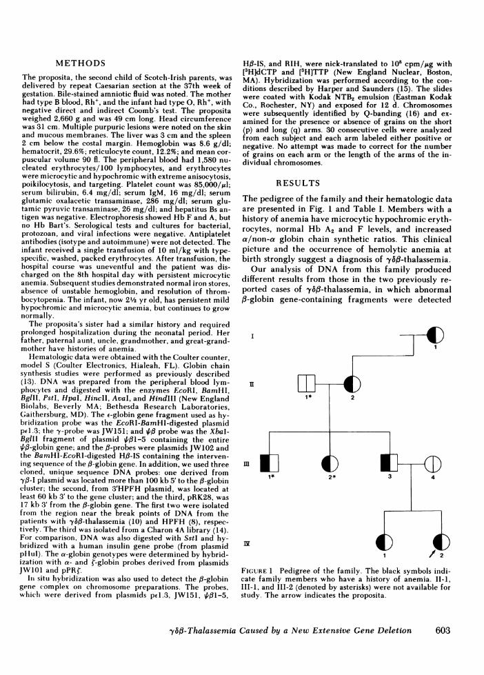
The proposita, the second child of Scotch-Irish parents, wasdelivered by repeat Caesarian section at the 37th week ofgestation. Bile-stained amniotic fluid was noted. The motherhad type B blood, Rh', and the infant had type 0, Rh+, withnegative direct and indirect Coomb's test. The propositaweighed 2,660 g and was 49 cm long. Head circumferencewas 31 cm. Multiple purpuric lesions were noted on the skinand mucous membranes. The liver was 3 cm and the spleen2 cm below the costal margin. Hemoglobin was 8.6 g/dl;hematocrit, 29.6%; reticulocyte count, 12.2%; and mean cor-puscular volume 90 fl. The peripheral blood had 1,580 nu-cleated erythrocytes/100 lymphocytes, and erythrocyteswere microcytic and hypochromic with extreme anisocytosis,poikilocytosis, and targeting. Platelet count was 85,000/al;serum bilirubin, 6.4 mg/dl; serum IgM, 16 mg/dl; serumglutamic oxalacetic transaminase, 286 mg/dl; serum glu-tamic pyruvic transaminase, 26 mg/dl; and hepatitus Bs an-tigen was negative. Electrophoresis showed Hb F and A, butno Hb Bart's. Serological tests and cultures for bacterial,protozoan, and viral infections were negative. Antiplateletantibodies (isotype and autoimmune) were not detected. Theinfant received a single transfusion of 10 ml/kg with type-specific, washed, packed erythrocytes. After transfusion, thehospital course was uneventful and the patient was dis-charged on the 8th hospital day with persistent microcyticanemia. Subsequent studies demonstrated normal iron stores,absence of unstable hemoglobin, and resolution of throm-bocytopenia. The infant, now 2½/2 yr old, has persistent mildhypochromic and microcytic anemia, but continues to grownormally.
The proposita's sister had a similar history and requiredprolonged hospitalization during the neonatal period. Herfather, paternal aunt, uncle, grandmother, and great-grand-mother have histories of anemia.
Hematologic data were obtained with the Coulter counter,model S (Coulter Electronics, Hialeah, FL). Globin chainsynthesis studies were performed as previously described(13). DNA was prepared from the peripheral blood lym-phocytes and digested with the enzymes EcoRI, BamHI,BglII, PstI, HpaI, HincII, AvaI, and HindIII (New EnglandBiolabs, Beverly MA; Bethesda Research Laboratories,Gaithersburg, MD). The e-globin gene fragment used as hy-bridization probe was the EcoRI-BamHI-digested plasmidpd.3; the y-probe was JW151; and 4,'1 probe was the XbaI-BgllI fragment of plasmid 4,/1-5 containing the entire{B-globin gene; and the ,3-probes were plasmids JW102 andthe BamHI-EcoRI-digested H#-IS containing the interven-ing sequence of the ,B-globin gene. In addition, we used threecloned, unique sequence DNA probes: one derived from-to-I plasmid was located more than 100 kb 5' to the f3-globincluster; the second, from 3'HPFH plasmid, was located atleast 60 kb 3' to the gene cluster; and the third, pRK28, was17 kb 3' from the f3-globin gene. The first two were isolatedfrom the region near the break points of DNA from thepatients with -y5b-thalassemia (10) and HPFH (8), respec-tively. The third was isolated from a Charon 4A library (14).For comparison, DNAwas also digested with SstI and hy-bridized with a human insulin gene probe (from plasmidpHul). The a-globin genotypes were determined by hybrid-ization with a- and ¢-globin probes derived from plasmidsJW1I1 and pPRl.
In situ hybridization was also used to detect the j3-globingene complex on chromosome preparations. The probes,whicli were derived from plasmids p4.3, JW151, 4,#1-5,
Hfl-IS, and RIH, were nick-translated to 108 cpm/,ug with[3H]dCTP and [3H]TTP (New England Nuclear, Boston,MA). Hybridization was performed according to the con-ditions described by Harper and Saunders (15). The slideswere coated with Kodak NTB2 emulsion (Eastman KodakCo., Rochester, NY) and exposed for 12 d. Chromosomeswere subsequently identified by Q-banding (16) and ex-amined for the presence or absence of grains on the short(p) and long (q) arms. 30 consecutive cells were analyzedfrom each subject and each arm labeled either positive ornegative. No attempt was made to correct for the numberof grains on each arm or the length of the arms of the in-dividual chromosomes.
RESULTS
The pedigree of the family and their hematologic dataare presented in Fig. 1 and Table I. Members with ahistory of anemia have microcytic hypochromic eryth-rocytes, normal Hb A2 and F levels, and increaseda/non-a globin chain synthetic ratios. This clinicalpicture and the occurrence of hemolytic anemia atbirth strongly suggest a diagnosis of "y6fB-thalassemia.
Our analysis of DNA from this family produceddifferent results from those in the two previously re-ported cases of yf6f-thalassemia, in which abnormal,B-globin gene-containing fragments were detected
I )
nI
1* 2~~~~~~~~~~
FIGURE 1 Pedigree of the family. The black symbols indi-cate family members who have a history of anemia. II-1,III-1, and III-2 (denoted by asterisks) were not available forstudy. The arrow indicates the proposita.
,yb3-Thalassemia Caused by a New Extensive Gene Deletion 603

TABLE IHematologic Data of the Family
Subject Age Jib Hct RBC MCV M(CII NICHiC: Retic 11i) A2 Jib F a/non-a ratio
g .'dl % X1O8/MI fl pg g/dl % % %
I-1 76 12.8 39.6 6.08 65 21.1 32.4 2.0 2.9 0.5 1.811-2 50 11.6 35.8 6.00 60 19.3 32.4 1.0 2.9 0.9 2.3111-3 30 13.2 41.1 6.92 59 19.1 32.1 2.2 3.0 1.3 2.1111-4 29 13.0 38.6 4.08 90 30.5 33.8 1.0 2.9 0.6 1.0IV-1 4 10.6 32.5 5.53 58 19.2 32.7 2.5 3.2 0.6 1.8IV-2 2 9.0 29.0 4.65 62 19.4 31.3 5.0 2.9 2.2 2.1
° Globin chain synthesis study performed at 2 mo of age.
(10, 11). In this family, the DNA from the affectedsubjects produced normal-sized restriction fragmentsthat contained the f-globin gene when digested withEcoRI, HpaI, PstI, and BamHI, and hybridized withthe f3-globin probe. The a-globin genotypes were nor-mal and additional hybridizations with y- and E-globingene probes were also normal. However, the intensityof hybridization with the d- and p3-like globin geneprobes was lighter than normal and this raised thepossibility of an extensive deletion involving the entiref3-globin gene cluster. Such a deletion would producenormal globin gene restriction patterns because theDNA fragments detected would be derived from theunaffected chromosome.
Several restriction sites in the 0-globin gene clusterare polymorphic; a given individual is usually hetero-zygous for some of these sites because the paternal andmaternal chromosomes are different in them (17). Westudied seven of these polymorphic sites: the HincIIsite 5' to the e-globin gene (17), the HindIII sites inthe second intervening sequence (IVS) of the Gy_ andAy-globin genes (18), the two HincII sites in and 3' tothe 4/13-globin gene (17), the AvaIl site in the secondIVS of the ,B-globin gene (17), and the BamHI site 3'to the ,B-globin gene (Table II; 19). Whereas the nor-
mocytic family member was heterozygous for severalof these sites, all members with microcytosis exhibiteda single restriction pattern at each of the sites. Thissuggested that the ,B-globin gene cluster was presenton one homologous chromosome only and that the af-fected members were hemizygous for these sites.
Detailed analysis of several sites supported this con-tention. Results from the BamHI analysis are shownin Fig. 2. The DNAdigest from the proposita's father(II1-3) showed only a 22-kb fragment which containedthe 3' portion of the f,-globin gene due to the absence
A Bam Hi
1 2 3 4B SstI
1 2 3 4
22
9.3 -
a . 4.8
TABLE IIHaplotypes of the Family
IIindlIl HinclIIfincll Avall BamHI
, y Aty T 3
I-1 -/ -/ -/ -/ +/ -/II-2 +/ +/ / +/ +/ +/ +/111-3 -/ +/ -/ +/ +/ +/ /I11-4 -/+ +/- +/- -/- +/- -/+ +/+IV-1 -/ +/ +/ -/ +/ -/ +/IV-2 +/ -/ -/ -/ -/ +/ +/
1 g
pglobin Insulin
FIGURE 2 Autoradiographs of DNAdigested with BamHIand SstI and hybridized with the ,B-globin and insulin geneprobes, respectively. The 1.8-kb fragment contains the 5'portion of the f,-globin gene, while the polymorphic BamHIsite 3' to the (3-globin gene results in a 9.3- or 22-kb fragmentcontaining the 3' portion of the gene. (1) III-4, (2) III-3, (3)IV-1, (4) IV-2.
604 M. Pirastu, Y. W. Kan, C. C. Lin, R. M. Baine, and C. T. Holbrook

of the BamHI site 3' to the gene. The mother's (III-4)DNAproduced a 9.3-kb fragment with this enzymebecause the BamHI site is present. If both parents arehomozygous for these patterns, DNA from the pro-posita (IV-2) and her sister (IV-1) should show boththe 9.3- and 22-kb fragments. In fact, only the 9.3-kbfragment from the mother was observed, indicatingthat neither child inherited a ,-globin gene from thefather. Analysis of the HincII site at the 4,/q-globingene points to a similar conclusion. In fact, haplotypeanalysis of all seven restriction sites showed that thepatterns carried by the two children were inheritedfrom the two different maternal chromosomes(Table II).
As a control, we studied the inheritance of the hu-man insulin gene. The insulin gene is also located onthe short arm of chromosome 11 (20). Restriction anal-ysis has shown a high degree of polymorphism due tovariations in the length of DNAsequences 5' to it (21).This polymorphism is detectable by digestion withSstI. When we digested the DNAwith this enzyme,the proposita's father had a 4.8-kb SstI fragment, themother had a 6.0-kb fragment, and the proposita andher sister inherited the 4.8- and 6.0-kb fragments fromthe father and mother, respectively. These results dem-
-b
._
0:0m
0I
A a1
a2
B
onstrate that both offspring inherited one insulin genefrom each parent, but only one ,-globin gene clusterfrom the mother.
Weconfirmed the decrease in ,8-globin gene dosageby quantitating the intensity of hybridization of theglobin gene bands. A filter containing BglII-digestedDNAwas blotted with mixed a- and ,-globin probes.In all family members, the 03-globin gene resided ina 5.2-kb fragment and the two a-globin loci, al anda2, were on 7.5-kb and 12-kb fragments, respectively.Densitometric scans of the autoradiographs supportedour conclusion that only one fl-globin locus is presentper diploid genome in the microcytic family members.The intensity of the f,-globin band relative to the twoa-globin bands in these individuals was similar to thatfound in a heterozygote with the deletion type of b60#-thalassemia (6, 7) and was half that of a normal subject(Fig. 3).
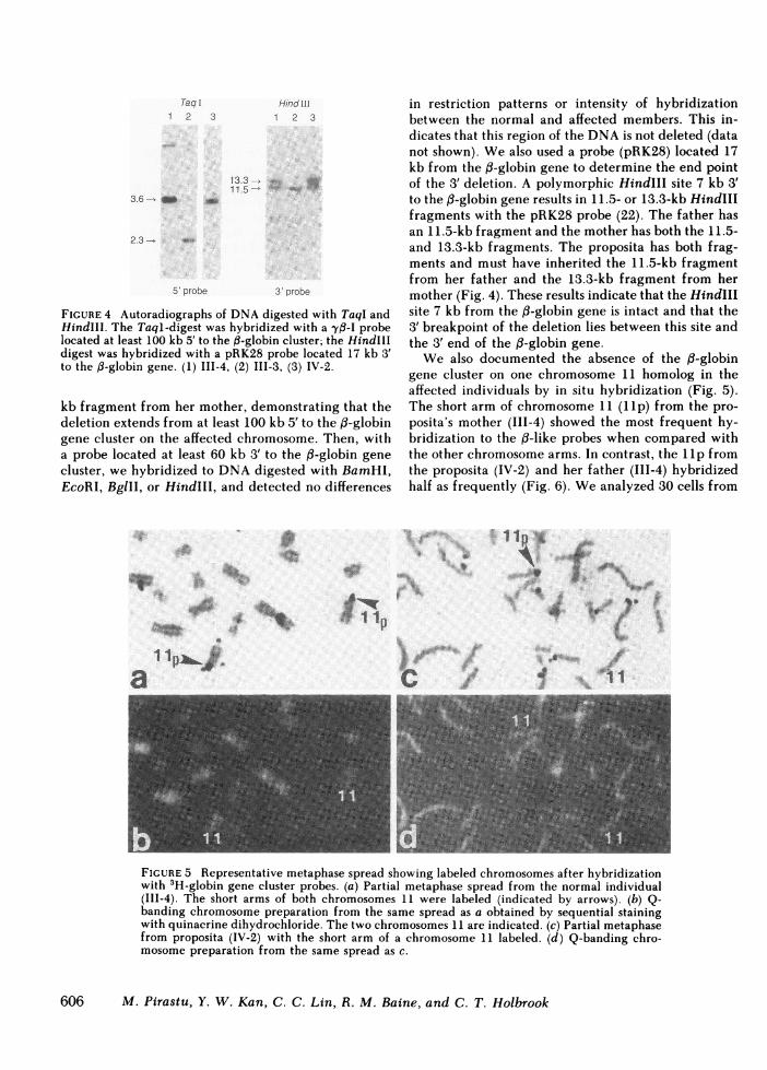
Weestimated the extent of the deletion by hybrid-ization with probes situated 5' and 3' to the ,3-globingene cluster. Hybridization of TaqI-digested DNAwith the yB-I probe located over 100 kb 5' to the ,#-globin gene cluster yielded a 3.6-kb fragment from theproposita's mother and a 2.3-kb fragment from herfather (Fig. 4). The proposita inherited only the 3.6-
C aI
-y6b-Thalassemia Caused by a New Extensive Gene Deletion
FIGURE 3 Densitometric scanning of autoradiographs from DNAdigested with BglII and hy-bridized with mixed a- and ,-globin probes. The arrow shows the direction of electrophoresis.The sizes of the DNAfragments are: P, 5.2 kb; al, 7.5 kb; and a2, 12 kb. (A) normal (111-4),(B) proposita (IV-2), 2, and (C) a heterozygote for the deletion type of P#'B thalassemia.
I
605
Taq1 2 3
3.6 - _M o4
Hind ill1 2 3
13.3 , .. t11.5 - ~
2.3 -
5' probe 3' probe
FIGURE 4 Autoradiographs of DNAdigested with TaqI andHindIII. The Taql-digest was hybridized with a -yf-I probelocated at least 100 kb 5' to the ,B-globin cluster; the HindIIIdigest was hybridized with a pRK28 probe located 17 kb 3'to the ,B-globin gene. (1) III-4, (2) III-3, (3) IV-2.
kb fragment from her mother, demonstrating that thedeletion extends from at least 100 kb 5' to the f3-globingene cluster on the affected chromosome. Then, witha probe located at least 60 kb 3' to the f3-globin genecluster, we hybridized to DNAdigested with BamHI,EcoRI, BglII, or HindIII, and detected no differences
in restriction patterns or intensity of hybridizationbetween the normal and affected members. This in-dicates that this region of the DNAis not deleted (datanot shown). Wealso used a probe (pRK28) located 17kb from the 13-globin gene to determine the end pointof the 3' deletion. A polymorphic HindIll site 7 kb 3'to the f3-globin gene results in 11.5- or 13.3-kb HindIIIfragments with the pRK28 probe (22). The father hasan 11.5-kb fragment and the mother has both the 11.5-and 13.3-kb fragments. The proposita has both frag-ments and must have inherited the 11.5-kb fragmentfrom her father and the 13.3-kb fragment from hermother (Fig. 4). These results indicate that the HindIIIsite 7 kb from the 0B-globin gene is intact and that the3' breakpoint of the deletion lies between this site andthe 3' end of the ,B-globin gene.
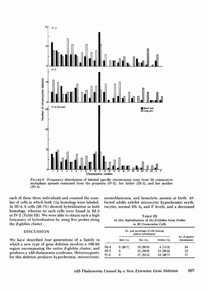
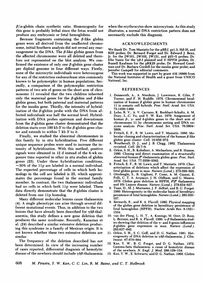
We also documented the absence of the fp-globingene cluster on one chromosome 11 homolog in theaffected individuals by in situ hybridization (Fig. 5).The short arm of chromosome 11 (llp) from the pro-posita's mother (111-4) showed the most frequent hy-bridization to the d-like probes when compared withthe other chromosome arms. In contrast, the lIp fromthe proposita (IV-2) and her father (III-4) hybridizedhalf as frequently (Fig. 6). Weanalyzed 30 cells from
Y
la CI
4pn,
FIGURE 5 Representative metaphase spread showing labeled chromosomes after hybridizationwith 3H-globin gene cluster probes. (a) Partial metaphase spread from the normal individual(III-4). The short arms of both chromosomes 11 were labeled (indicated by arrows). (b) Q-banding chromosome preparation from the same spread as a obtained by sequential stainingwith quinacrine dihydrochloride. The two chromosomes 11 are indicated. (c) Partial metaphasefrom proposita (IV-2) with the short arm of a chromosome 11 labeled. (d) Q-banding chro-mosome preparation from the same spread as c.
606 M. Pirastu, Y. W. Kan, C. C. Lin, R. M. Baine, and C. T. Holbrook
4,
20 V20-IV-2
10
0 E
111-3
C 10 3
0
° 0 111-4 (Norffal) *Short armD C~~~~~~~~~~~~~~~~~~~~~Long arm
E
00
20
10
tI. mILuniJl12 3 4 5 6 7 8 9 0 1 12 13 14 15 16 17 18 19 20 21 22 X Y
Chromosome number
FIGURE 6 Frequency distribution of labeled specific chromosome arms from 30 consecutivemetaphase spreads examined from the proposita (IV-2), her father (III-3), and her mother(III-4).
each of these three individuals and counted the num-ber of cells in which both llp homologs were labeled.In III-4, 8 cells (26.7%) showed hybridization in bothhomologs, whereas no such cells were found in III-3or IV-2 (Table III). Wewere able to obtain such a highfrequency of hybridization by using five probes alongthe ,B-globin cluster.
DISCUSSION
We have described four generations of a family inwhich a new type of gene deletion involves a 100-kbregion encompassing the entire ,B-globin cluster, andproduces a 'ybf-thalassemia syndrome. Heterozygosityfor this deletion produces hypochromia, microcytosis,
normoblastemia, and hemolytic anemia at birth. Af-fected adults exhibit microcytic hypochromic eryth-rocytes, normal Hb A2 and F levels, and a decreased
TABLE IIIIn Situ Hybridization of the (3-Globin Gene Probes
in 30 Consecutive Cells
No. (and percentage) of cells showingpositive hybridization
No. of positiveBoth lIp One lip Neither lip chromosomes
III-4 8 (26.7) 18 (60.0) 4 (13.3) 34III-3 0 15 (50.0) 15 (50.0) 15IV-2 0 17 (53.3) 13 (46.7) 17
y6#-Thalassemia Caused by a New Extensive Gene Deletion
r _r.L-
607
f3/a-globin chain synthetic ratio. Homozygosity forthis gene is probably lethal since the fetus would notproduce any embryonic or fetal hemoglobins.
Because fragments containing the ,B-like globingenes were all derived from the unaffected chromo-some, initial Southern analysis did not reveal any rear-rangement in the DNA. The ,8-like globin genes fromthe affected chromosome were all deleted and there-fore not represented on the blot analysis. We con-firmed the existence of only one ,3-globin gene clusterper diploid genome in four different ways. Firstly,none of the microcytic individuals were heterozygousfor any of the restriction endonuclease sites commonlyknown to be polymorphic in human populations. Sec-ondly, a comparison of the polymorphic restrictionpatterns of two sets of genes on the short arm of chro-mosome 11 revealed that the two children inheritedonly the maternal genes for the 13-globin and ,8-likeglobin genes, but both paternal and maternal patternsfor the insulin gene. Thirdly, the intensity of hybrid-ization of the ,B-globin probe with the DNAfrom af-fected individuals was half the normal level. Hybrid-ization with DNAprobes upstream and downstreamfrom the ,B-globin gene cluster demonstrated that thedeletion starts over 100 kb 5' to the 13-globin gene clus-ter and extends to within 7 kb 3' to it.
Finally, we studied the abnormal chromosomes inthis family by in situ hybridization. Five differentunique sequence probes were used to increase the in-tensity of hybridization. With this method, positivesignals were obtained in 12 d instead of the 6-wk ex-posure time reported in other in situ studies of globingenes (23). Under these hybridization conditions,-50% of the lip are labeled in a normal individual.
The expected percentage of cells in which both ho-mologs in the cell are labeled is 25, which approxi-mates the percentage found in the normal familymember. In contrast, the two thalassemic individualshad no cells in which both Ilp were labeled. Thesedata directly demonstrate that the ,B-globin cluster isdeleted from one Ilp homolog.
Many different molecular lesions cause thalassemia(4). A single phenotype can arise through several dif-ferent mutational events. Thus, in addition to the twolesions that have already been described for y6fl-thal-assemia, this study defines a new gene deletion thatproduces the same syndrome. Recently, Kazazian etal. (24) described a similar extensive deletion produc-ing this syndrome in a family of Mexican origin. It isnot known whether these two extensive deletions aresimilar.
The frequency of the deletion described has notbeen determined. In view of the increasing numberof cases reported, differential diagnosis of hemolyticdisease of the newborn should include '61B-thalassemia
when the erythrocytes show microcytosis. As this studyillustrates, a normal DNArestriction pattern does notnecessarily exclude this diagnosis.
ACKNOWLEDGMENTSWethank Dr. Tom Maniatis for the pBRr, pel.3, HP8-IS, andRIH probes; Dr. Bernard Forget and Dr. Edward J. Benz,Jr. for the JW101, JW102, JW151, and 4,#1-5 probes; Dr.Elio Vanin for the 'y-I plasmid and 3' HPFHprobes; Dr.Russell Kaufman for the pRK28 probe; Dr. Howard Good-man and Dr. Barbara Cordell for the insulin gene probe; andJennifer Gampell for editorial comments.
This work was supported in part by grant AM16666 fromthe National Institutes of Health and a grant from UNICONational, Inc.
REFERENCES1. Deisseroth, A., A. Nienhuis, J. Lawrence, R. Giles, P.
Turner, and F. H. Ruddle. 1978. Chrornosomal local-ization of human , globin gene in human chromosome11 in somatic cell hybrids. Proc. Nati. Acad. Sci. USA.75:1456-1460.
2. Lebo, R. V., A. V. Carrano, K. Burkhart-Schultz, A. M.Dozy, L.-C. Yu, and Y. W. Kan. 1979. Assignment ofhuman ,B-, -y- and 6-globin genes to the short arm ofchromosome 11 by chromosome sorting and DNAen-zyme analysis. Proc. Natl. Acad. Sci. USA. 76:5804-5808.
3. Fritsch, E. F., R. M. Lawn, and T. Maniatis. 1980. Mo-lecular cloning and characterization of the human #-likeglobin gene cluster. Cell. 19:959-972.
4. Weatherall, D. J., and J. B. Clegg. 1982. Thalassemiarevisited. Cell. 29:7-9.
5. Orkin, S. H., R. Kolodner, A. Michaelson, and R. Husson.1980. Cloning and direct examination of a structurallyabnormal human ,3°-thalassemia globin gene. Proc. Natl.Acad. Sci. USA. 77:3558-3562.
6. Fritsch, E. F., R. M. Lawn, and T. Maniatis. 1979. Char-acterization of deletions which affect the expression offetal globin genes in man. Nature (Lond.). 279:598-603.
7. Ottolenghi, S., B. Giglioni, P. Comi, A. M. Gianni, E.Polli, C. T. A. Acquaye, J. H. Oldham, and G. Masera.1979. Globin gene deletion in HPFH, 8ot0 thalassemiaand Hb Lepore disease. Nature (Lond.). 278:654-657.
8. Tuan, D., M. J. Murname, J. F. deRiel, and B. G. Forget.1980. Heterogeneity in the molecular basis of hereditarypersistence of fetal hemoglobin. Nature (Lond.). 285:335-337.
9. Bernards, R., and R. A. Flavell. 1980. Physical mappingof the globin gene deletion in hereditary persistence offetal hemoglobin (HPFH). Nucleic Acids Res. 8:1521-1534.
10. van der Ploeg, L. H. T., A. Konings, M. Oort, D. Roos,L. Bernini, and R. A. Flavell. 1980. y-ft thalassemia stud-ies showing that deletion of the y- and 6-genes influences#-globin gene expression in man. Nature (Lond.).283:637-642.
11. Orkin, S. H., S. C. Goff, and D. G. Nathan. 1981. Het-erogeneity of DNAdeletion in y6,f-thalassemia. J. Clin.Invest. 67:878-884.
12. Kan, Y. W., B. G. Forget, and D. G. Nathan. 1972.Gamma-beta thalassemia: a cause of hemolytic diseaseof the newborn. N. Engl. J. Med. 286:129-134.
13. Kan, Y. W., E. Schwartz, and D. G. Nathan. 1968. Globin
608 M, Pirastu, Y. W. Kan, C. C. Lin, R. M. Baine, and C. T. Holbrook

chain synthesis in the alpha thalassemia syndromes. J.Clin. Invest. 47:2515-2522.
14. Kaufman, R. E., P. J. Kretschmer, J. W. Adam, H. C.Coon, W. F. Anderson, and A. W. Nienhuis. 1980. Clon-ing and characterization of DNAsequences surroundingthe human y-, b-, and ,B-globin genes. Proc. Natl. Acad.Sci. USA. 77:4229-4237.
15. Harper, M. E., and C. F. Saunders. 1981. Localizationof single copy DNAsequences on Q-banded human chro-mosomes by in situ hybridization. Chromosoma (Berl.).83:431-439.
16. Lin, C. C., J. H. van de Sande, W. K. Smink, and D. R.Newton. 1975. Quinacrine fluorescence and Q-bandingpatterns of human chromosomes. I. Effects of varyingfactors. Can. J. Genet. Cytol. 17:81-92.
17. Antonarakis, S. E., C. D. Boehm, P. J. V. Giardina, andH. H. Kazazian, Jr. 1982. Non-random association ofpolymorphic restriction sites in the f3-globin gene cluster.Proc. Natl. Acad. Sci. USA. 79:137-141.
18. Jeffreys, A. J. 1979. DNA sequence variants in theG(y_, Ay_, b-, and f-globin genes of man. Cell. 18:1-10.
19. Kan, Y. W., K. Y. Lee, M. Furbetta, A. Angius, and A.Cao. 1980. Polymorphismn of DNA sequence in the ,B-globin gene region. N. Engl. J. Med. 302:185-189.
20. Owerbach, D., G. I. Bell, W. J. Rutter, and T. B. Shows.1980. The insulin gene is located on the short arm ofchromosome 11 in humans. Diabetes. 30:267-270.
21. Bell, G. I., J. H. Karam, and W. J. Rutter. 1981. Poly-morphic DNAregion adjacent to the 5' end of the humaninsulin gene. Proc. Natl. Acad. Sci. USA. 78:5759-5763.
22. Forget, B. G., D. Tuan, M. V. Newman, E. A. Finegold,F. Collins, Y. Fukumaki, P. Jagadeeswaran, and S. M.Weissman. 1983. Molecular studies of mutations that in-crease Hb F productivity in man. In HemoglobinSwitching. G. Stamatoyannopoulos, A. Nienhuis, editors.Alan R. Liss, New York. In press.
23. Malcolm, S., P. Barton, C. Murphy, and M. A. Ferguson-Smith. 1981. Chromosomal localization of a single copygene by in situ hybridization-human /3-globin genes onthe short arm of chromosome 11. Ann. Hum. Genet.45: 135-141.
24. Kazazian, H. H., Jr., E. R. Fearon, P. G. Waber, J. 1.Lee, S. E. Antonarakis, S. H. Orkin, E. F. Vanin, P. S.Heathorn, F. C. Grosveld, and G. R. Buchanan. 1982.-y,b thalasseniia. Deletion of the entire ,B-globin genecluster. Blood. 60(Suppl.):54a.
y6,-Thalassemia Caused by a New Extensive Gene Deletion 609