Embed Size (px)
Citation preview
Universidad Autónoma de Madrid
Departamento de Bioquímica
Heart Specific ScoX Knockdown Induces p53
Dependent Apoptosis and Cardiomyopathy
Leticia Martínez Morentin
Madrid, 2014

Departamento de Bioquímica Facultad de Medicina
Universidad Autónoma de Madrid
Heart Specific ScoX Knockdown Induces p53 Dependent Apoptosis and
Cardiomyopathy
Memoria de Tesis Doctoral presentada por:
Leticia Martínez Licenciada en Bioquímica, para optar al grado de Doctor
por la Universidad de Madrid
Dr. Juan Jose Arredondo Profesor Contratado Doctor
Directores de la Tesis:
Dra. Margarita Cervera Catedrática de Universidad
Departamento de Bioquímica. Facultad de Medicina Universidad Autónoma de Madrid
Instituto de investigaciones Biomédicas “Alberto Sols”. CSIC-UAM.


Facultad de Medicina
Departamento de Bioquímica
Margarita Cervera Jover, Catedrática de Bioquímica y Biología Molecular de la UAM, como Directora de Tesis, y Juan Jose Arredondo Lamas Profesor Contratado Doctor como Director de Tesis, CERTIFICAN: Que Doña Leticia Martínez con D.N.I.: 16064390V, licenciada en Bioquímica ha realizado, bajo la dirección de los Directores de Tesis, en el Departamento de Bioquímica de la Facultad de Medicina de la Universidad Autónoma de Madrid / Instituto de Investigaciones Biomédicas “Alberto Sols”, el trabajo titulado:
Heart Specific ScoX Knockdown Induces p53 Dependent Apoptosis and Cardiomyopathy
Una vez supervisado el trabajo, consideramos que reúne todos los requisitos necesarios en cuanto a originalidad y calidad para ser presentado como Tesis Doctoral con el objeto de optar al título de Doctor en Ciencias por la Universidad Autónoma de Madrid.
Madrid, 7 de Mayo de 2014 Madrid, 7 de Mayo de 2014 Fdo. Juan Jose Arredondo Lamas Margarita Cervera Jover Director de la Tesis Directora de Tesis Profesor Contratado Doctor Catedrática de Bioquímica

6

7
Esta tesis doctoral ha sido realizada realizado en el Departamento de Bioquímica del Instituto de Investigaciones Biomédicas “Alberto Sols” (UAM-CSIC), gracias a la concesión de un Beca de Formación de Personal Investigador por parte del Ministerio de Economía y Competitividad a Leticia Martínez Morentin.

8

9
A mis padres y mi hermana

10

11
“ LOS CIENTÍFICOS DICEN QUE ESTAMOS HECHOS DE ÁTOMOS, PERO A MÍ UN PAJARITO
ME CONTÓ QUE ESTAMOS HECHOS DE HISTORIAS”
EDUARDO GALEANO

12
13

14
15
AGRADECIMIENTOS

AGRADECIMIENTOS
16
Benjamin Franklin dijo, “Either write something worth reading or do something worth
writing”. Espero haber conseguido las dos cosas.
Creo que no hay apartado en toda tesis que genere tanta expectación como la de
los agradecimientos. Sé que es la única parte que muchos de vosotros os vais a leer, “ay
pillines, qué poco interés por la ciencia…jajajaja”, así que por favor, si no consigo
expresar bien con mis palabras todo lo que os tengo que agradecer no os lo toméis
como algo personal, achacadlo a mi incapacidad como buena redactora.
Durante todos estos años que he pasado en Madrid haciendo la tesis, reconozco que no
ha habido mejor periodo de formación que éste, no sólo desde el punto de vista
científico, sino también desde el personal. Porque todo el que ha pasado por esto, sabe
que no es un camino fácil y sin ninguno de vosotros (creedme, es verdad), nada de esto
habría sido posible. Gracias de verdad a todos los que habéis “perdido” el tiempo
enseñándome, por las risas (y hasta por los lloros), por esas bromas en la lupa de “bueno,
me voy mosquear un rato” pero sobre todo, por eso apoyo incondicional que he sentido
en el transcurso de todos estos años.
Rompiendo un poco con la tradición, no puedo comenzar estos agradecimientos
con otras personas que no sean mis padres, Mercedes y Fernando. Si de alguien es esta
tesis, es de ellos. Gracias mamá y papá simplemente, por todo. Tengo tanto que
agradeceros que no sé ni por dónde empezar. Sé que a veces soy una hija un poco
ingrata, pero espero algún día poder devolveros de alguna forma todo lo que hacéis por
mí. Gracias de todo corazón, por todo vuestro apoyo incondicional, por creer y confiar en
mí y por transmitirme vuestro apoyo y confianza. Gracias por hacer de mi la persona que
soy (creo que no he salido tan mal, ¿no? Jejeje), fuerte y luchadora. Pero sobre todo,
gracias a ti, mamá. Gracias por tu paciencia infinita y por hacer y conseguir, que esa niña
tan revoltosa e inquieta se sentara a estudiar. Sin ti, no habría conseguido nada. Os
quiero.
Los siguientes en la lista son por supuesto mis directores de tesis, Marga y Juan.
Gracias Marga, por darme la oportunidad de ser parte del laboratorio ya hace tantos
años. Por tu ayuda, apoyo y tus siempre valiosos consejos a lo largo de todo este tiempo
y por cuidar de todos nosotros. A ti Juan, gracias por ayudarme y enseñarme ciencia; por
tu confianza y por haber creído en mí (lo tuyo sí que ha sido un acto de fe). Por tu tesón

AGRADECIMIENTOS
17
y perseverancia en intentar convertirme en científica y no morir en el intento, y por tu
paciencia… ya sabes lo cabezota que soy y que no siempre tengo un carácter fácil, sino a
veces más bien todo lo contrario.
Muchas han sido las personas que han pasado por el labo y con las que he
compartido tantas cosas. Jorge, gracias por ayudarme tanto. Por enseñarme a trabajar
con embriones de mosca, por tantas horas en la lupa compartidas. Por las risas, las
infinitas charlas y todo tu apoyo, y por ese corazón tan grande que te caracteriza. Pedrito,
a ti por estar siempre ahí cuando he necesitado hablar. No te preocupes, todo llega y si
yo he llegado hasta aquí tu también lo harás ;) . A Bego y Lidia, gracias chicas por todo
vuestro apoyo y ayuda. Bego, siempre he pensado que las personas buenas se merecen
cosas buenas y sé que te llegará. Por las risas y esos momentos “gangnam style” tan
divertidos. Lidia, pero niña qué habría hecho sin ti??? Gracias por tu constante ayuda, por
tu paciencia cada vez que te pedía algo y por haber sido un pilar importante en este
trabajo aunque no lo creas. A ti Berti, por ser ese aire fresco que tanta falta nos hacía, por
tu alegría y fuerza contagiosas.
Por supuesto, gracias a nuestros compañeros del B-19. En especial a los
profesores Rafael Garesse y Miguel Fernández. Gracias Rafa por tu ayuda y por transmitir
ese entusiasmo y esa pasión por la mitocondria, por conseguir que nos parezca a todos
el orgánulo más maravilloso del mundo. A ti Miguel, gracias no sólo por tus consejos
científicos y tu sabiduría, sino por estar ahí. Por tu cercanía y tu cariño, y por preocuparte
por mí cuando me has visto mal. No todo el mundo tiene esa capacidad que tu tienes
para saber cuando algo nos preocupa y transmitirnos tranquilidad. Gracias.
A todas las personas que formáis parte del B19: las nuevas generaciones Sara, Teresa;
Cristina, Rosana, por esa amabilidad que te caracteriza. Esther, por tus consejos y tu
ayuda. Por todas esas risas, creo que nadie más que tu puede tener una foto de Antonio
Resines en su mesa, y que no parezca algo raro jajajajajajajaja.
A vosotros, Rami y Alberto. Chicos gracias por ser mucho más que simples compañeros
de trabajo, sino mis amigos. Por estar ahí para todo, siempre que os he necesitado y por
vuestras palabras de ánimo. Por todos esos viernes de risas en la Yoli y tonterías al salir
del labo. Rami, pocas personas he conocido con un corazón tan grande. Vales mucho, no
sólo desde el punto de vista personal, sino también del científico. Sólo espero que algún
día te lo termines creyendo. Alberto, cari, mucho ánimo en ésta etapa que ya sabemos
que es dura pero lo conseguirás y sé que lograrás todo lo que te propongas.

AGRADECIMIENTOS
18
A vosotros, Álvaro, Oihane, Paula, Verito, Lucy. Chicos, ¡no sabéis lo que se os echa de
menos! Esto va para todos, gracias por vuestra amistad y cariño, y por haber sido unos
compañeros geniales. Por vuestro apoyo, tanto en Madrid como en la distancia. Por ser
unos amigos increíbles y por estar ahí cuando os he necesitado. Sé que a todos os va a ir
genial, porque todos lo valéis.
Y por supuesto, gracias a todos los compañeros de pasillo pero en especial a ti Carlos.
Carlos, cari, mucha suerte en tu nueva aventura por Denver y no dudes de que
tardaremos poco en vernos.
No puedo continuar, sin antes agradecerles a Lucia y a Diego, del servicio de Microscopía.
Gracias chicos, por enseñarme todo lo que sé sobre el Zeiss que no es poco!!! Por vuestra
ayuda y por dejarme utilizar todas las horas del mundo en el confocal, había veces que
parecía que vivía allí jajajaja.
Por supuesto a Ana y Lola, del servicio de microscopía de la UAM. Chicas, no hay mejor
confesionario que el vuestro.
Quiero agradecer al Dr. Ernesto Sánchez Herrero por su amabilidad y por hacerme un
hueco en el laboratorio. A ti Guarner, gracias por tu paciencia (que fue mucha). Por todos
esos tubos de moscas y por ayudar a que mis experimentos de TUNEL quedaran tan
rebonikos!! A ti Manolo, gracias por tu apoyo, comprensión y consejo. Por esas palabras
de ánimo.
Gracias al Dr. Rolf Bodmer y a la Dra. Karen Ocoor, del Sanford Burnham Medical
Research Institute por acogerme en el laboratorio. Thank you, Rolf and Karen for giving
me the opportunity of being part of the lab. For teaching me everything I know about
Drosophila´s heart physiology. And to the members of the lab: Mayuko, such a
sweetheart, please do not never change. Soda, Leah, Santiago, Srheehari, Takeshi, Min,
Ying, Jerome, it´s been a pleasure. But, specially to Sarah and Jumana. Sarah, thanks a lot
for your support and for helping me out with the dissections and the filming, you had so
much patient hahaha. I can´t wait to see you guys again and meet the new baby girl, she
is such a lucky girl. Jumana, hun, thank you so much for your friendship, all the laughter
and for being my “beach sister”. Your gonna became a great Doctor, I´m so proud of you.

AGRADECIMIENTOS
19
También me gustaría dar las gracias a todos los amigos que tuve la oportunidad de
conocer alli. Evie, what can I say apart from “double rainbow all the way across the sky!!”.
Can´t believe I´m not gonna be able to make it for your wedding!! Juli, Sheena, Jeff,
thanks a lot. Keau, for all our conversations and our coffee breaks!!
No solo los amigos científicos son importantes, la familia y los amigos fuera del gremio
también lo son. Gracias a mis tíos Rosa y Angel, por hacer de mi la sobrina más mimada
del mundo. A David, mi “primi”, por ese apoyo que siempre me has dado, a parte de por
acogerme en tu casa por tantooo tiempo jajajaja. Por presentarme a tus amigos, Pable,
tantas risas y por estar siempre ahí.
A toda mi gente de Madrid, bueno y de no Madrid. Chicos gracias a todos por
preocuparos y por los ratos, excursiones, cenas, comidas (más cenas y comidas) y
palabras de ánimo. A mis compi de piso, pero en especial a ti Vir. Por esos desayunos,
charlas y tus siempre sabiossss consejosssss (creo que ya sabes de que te hablo no?
Jajaja).
A mis amigos: Vir, se te echa de menos; Rebe, Merce, chicas no cambies; T-ñinnnnn, no
hay nadie al que le sienten los sombreros de paja tan bien como a ti RAWRRRRR!!. A Patri
y Robert, gracias por estar siempre ahí y por hacerme sentir siempre como en casa. A
Lore, Aitor y Cepe. Qué deciros chicos, tantos años juntos ya y todos los que nos
quedan!! Gracias por ese cariño y estar ahí en todo momento, y que sepáis que os gano a
Padel fijo! Paula, compi de pelu!!! Gracias por ser mi amiga y compartir conmigo “locura”
y aficiones. Como diría David, “nos va la marcha pero bien” ;) . A ti mokito, gracias por
todas esas risas en clase y en prácticas de bioquímica Madre mía, todo lo que ha llovido!
Gracias por ser tan buena amiga y estar ahí en los momentos duros. Mucha suerte a
Rodri y a ti en vuestra aventura Danesa y no dudes, de que en cuanto podamos, iremos a
veros. A mis “vecis”, Bruno y Ianire. Chicos, ya sabéis que sois mis diseñadores favoritos.
Pocas personas hay tan auténtica como vosotros. Por favor, no cambiéis nunca.
A ti turrita, qué decirte que no sepas ya. Tengo tanto que agradecerte que no sé ni por
donde empezar. Gracias por haber sido casi como una extensión de mi misma durante
todos estos años y por ese cariño y apoyo tan incondicional que siempre me das. Sabes
lo orgullosísima que estoy de ti y sé que vas a llegar muy lejos, no lo dudes nunca. Y
tranquila, en menos de lo que piensas nos tienes ahí molestándote otra vez (es que eres
de las pocas que se ríe con mis chistes XDXD).
AGRADECIMIENTOS
20
A mi cuñado, Javier, por darme un abrazo cuando lo he necesitado. A mi hermana,
Mercedes. Sis, gracias por ser la mejor hermana mayor del mundo. Sé que eres la única
que tengo pero da igual jajajajaja. Gracias por acompañarme y estar ahí siempre que te
necesito y por ese amor tan incondicional que siempre me das. No sé qué habría hecho
sin ti. Maite zaitut.
Y por último, a ti David. Peke, sé que unas meras palabras en unos agradecimientos son
incapaces de plasmar todo lo que tengo que decirte y agradecerte, porque sin tu apoyo
y confianza, creo que no habría sido capaz de llegar hasta aquí. Gracias por hacerme feliz
cada minuto del día, por todas las risas, todos los besos y ese amor que sólo tu sabes
darme. Por darme tranquilidad y querer acompañarme en este camino, que ya sabemos
que no va a ser fácil. Pero sobre todo, gracias peketroki por ser como eres, no podía
haber tenido más suerte. Garrotxa, Pinotxa, Skronky… I love you Till the end.

22

21
RESUMEN/SUMMARY

Resumen/Summary
23
La citocromo c oxidasa o complejo IV es el ultimo elemento de la cadena de
transporte de electrones mitocondrial. Cataliza la oxidación del citocromo c transfiriendo
sus electrones al oxígeno. El déficit de complejo IV debido a mutaciones en factores de
ensamblaje es uno de los defectos más comunes de la cadena respiratoria. Estas
patologías se caracterizan por su aparición a una edad temprana y cursar con un amplio
espectro de manifestaciones clínicas como encefalopatía, cardiomiopatía, hepatopatía o
leucodistrofia.
SCO1 y SCO2 son dos metalochaperonas responsables de la formación del centro
de cobre CuA en el ensamblaje del complejo IV. Mutaciones en SCO1 causan hepato-
encefalomiopatía aunque se ha descrito un caso que también presenta cardiomiopatía
hipertrófica. Las mutaciones en SCO2 causan cardiomiopatía hipertrófica y
encefalopatía infantil. A excepción de un caso, todos los pacientes portan la mutación
E140K.
En esta tesis hemos utilizado Drosophila melanogaster como sistema modelo para
el estudio de las bases genéticas y moleculares de la cardiopatía causada por mutaciones
en las proteínas SCO. Los mecanismos genéticos que controlan la especificación de los
cardiomiocitos y numerosos aspectos de la fisiología del corazón están conservados en
Drosophila lo que hace de este organismo un excelente modelo para el estudio de la
función cardiaca y las cardiomiopatías humanas. Además, recientemente se han
desarrollado técnicas que permiten caracterizar la fisiología del corazón de Drosophila,
facilitando el estudio de cómo defectos en la cadena de transporte de electrones,
afectan a la función cardiaca.
Drosophila presenta un solo ortólogo para los genes de mamífero Sco1 y Sco2,
ScoX. Los resultados obtenidos demuestran que la interferencia de ScoX en el corazón
causa una cardiomiopatía dilatada, viéndose gravemente afectada tanto la función como
la estructura del corazón. Los cardiomiocitos sufren un cambio metabólico favoreciendo
la glicólisis frente a la fosforilación oxidativa, causando, probablemente, acidosis láctica y
emulando los síntomas clínicos observados en pacientes con mutaciones en Sco1 y Sco2.
El fenotipo observado es debido a una activación, dependiente de dp53, de la muerte
celular. Estos resultados sugieren que dp53 contribuye directamente en el desarrollo de
la cardiomiopatía.

Summary/Resumen
24
Cytochrome c oxidase or complex IV is the terminal component of the mitochondrial
electron transport chain. It catalyses the transfer of electrons from reduced cytochrome c
to molecular oxygen. Complex IV deficiency due to mutations in assembly factors is one
of the most frequent defects of respiratory chain in humans. These pathologies are
characterized by a very early age of onset and the display of different clinical
presentations, as encephalopathy, cardiomyopathy, hepatic failure and leukodystrophy.
SCO1 and SCO2 are two metallochaperones playing a key role in the formation of
cooper centre CuA during complex IV assembly. Mutations in SCO1 cause hepato-
encephalomyopathy although one case has been reported which also presents
hypertrophic cardiomyopathy. Mutations in SCO2 cause hypertrophic cardiomyopathy
and infantile encephalomyophathy and with but one exception, all patients harbour the
E140K mutation.
In this thesis, we have used Drosophila melanogaster as model system to
investigate the genetic and molecular mechanisms that underlie the cardiomyopathy
associated with SCO deficiency. The genetic network controlling cardiac specification
and differentiation as well as many aspects of heart function are conserved from flies to
mammals. Thus, Drosophila has become a powerful model system for the study and
understanding of cardiac function and human cardiomyopathies. Furthermore, the
recently established heart function assays in Drosophila make it possible to characterize
how mitochondrial electron transport chain defects affect heart function.
In Drosophila there is a single ortholog of mammalian Sco1 and 2, ScoX. Our
findings demonstrate that cardiac-specific knockdown of ScoX causes dileted
cardiomyopathy severely compromising heart function and structure. Cardiomyocytes
undergo a metabolic switch from oxidative phosphorylation to glycolysis probably
accompanied by lactic acidosis and therefore mimicking the clinical features found in
patients with mutations in Sco1 and Sco2. The observed phenotype is result of dp53-
dependent cell death activation. These results strongly suggest that dp53 is directly
involved in cardiomyopathy development.
25
INDEX

26
27
ABBREVIATIONS ................................................................................................. 29
INTRODUCTION ................................................................................................... 35 1.THE MITOCHONDRIA ....................................................................................................................................... 37
1.1. Mitochondrial structure ................................................................................................................................ 39 2. MITOCHONDRIAL ENERGETICS: The mitochondrial respiratory chain ......................................... 41 3. CYTOCROME C OXIDASE ............................................................................................................................... 42
3.1. Structure of Cytochrome c Oxidase ........................................................................................................... 42 3.3. Cytochrome c Oxidase Deficiency ............................................................................................................. 45 5.1. The heart of Drosophila melanogaster ................................................................................................... 47 5.2. Drosophila melanogaster as a model to study human cardiomyopathies ............................... 49
6. p53 ........................................................................................................................................................................ 50 6.1. Regulation of energy metabolism by p53 ............................................................................................... 51 6.2. p53, mitochondria and apoptosis ............................................................................................................. 53
AIMS ....................................................................................................................... 57
MATERIALS AND METHODS ............................................................................. 61 1. Materials ............................................................................................................................................................. 63
1.1 Reagents, solutions and buffers .................................................................................................................. 63 1.2. Drosophila melanogaster ............................................................................................................................. 63 1.3. Oligonucleotides .............................................................................................................................................. 65 1.6. Dyes ....................................................................................................................................................................... 66
2. Methods .............................................................................................................................................................. 66 2.1. GAL4/UAS system ............................................................................................................................................. 66 2.2. DNA ISOLATION ................................................................................................................................................ 67 2.3. Drosophila cardiac analysis ......................................................................................................................... 67 2.4. Inmunostaining of the Adult Drosophila heart .................................................................................... 68 2.5. TUNEL Staining of adult hearts ................................................................................................................... 68 2.6. TUNEL Staining of Mouse esqueletal muscle and liver ...................................................................... 69 2.7. DHE Stainning ................................................................................................................................................... 69 2.8. Respiratory Complexes enzyme activity measurement .................................................................... 69 2.9. Inmunohistochemical Staining of Adul hearts ..................................................................................... 69 2.10. mRNA extraction and Q-RT-PCR of Drosophila cardiac associated transcripts .................... 70 2.11. Statistical Analyses ....................................................................................................................................... 70
RESULTS ................................................................................................................ 71 1. ScoX knockdown in Drosophila melanogaster .................................................................................... 73 2. Cardiac specific interference of SCOX cause mitochondrial impairment. .................................. 74
2.1. Mitochondrial complexes activity staining in ScoX knockdown hearts ...................................... 75 2.2. Characterization of metabolic state in Scox knockdown cardiomyocytes ................................ 76
3. ScoX RNAi knockdown causes dilated cardiomyopathy in Drosophila melanogaster ............ 77 3.1. Analysis of heart structure ............................................................................................................................ 77 3.2. Scox RNAi knockdown compromises heart function ......................................................................... 78 3.3. ScoX knockdown affects heart structure. ............................................................................................... 80
4. COX deficiency leads to an increased production of reactive oxygen species ......................... 81 5. ScoX cardiomyopathy is p53-dependent ............................................................................................... 82
5.1. Overexpression of dp53 in ScoX knockdown hearts ........................................................................... 83 5.2. Overexpression of dp53 in Surf1 knockdown hearts .......................................................................... 84
6. Cardiac specific ScoX knockdown induces apoptosis ....................................................................... 85 6.2. TUNEL staining in Scox RNAi knockdown hearts ................................................................................. 86 6.3. Reaper overexpression ................................................................................................................................... 87
28
7. Blockage of dp53 activity rescues SCOX cardiomyopathy ............................................................... 87 7.1. Overexpression of a dominant negative form of dp53 rescues heart dysfunction and heart structure phenotype ................................................................................................................................................ 87
8. Inhibition of apoptosis rescues structural degeneration .................................................................. 91 9. Sco2KIKO mice undergoes apoptosis ........................................................................................................... 93
DISCUSSION .......................................................................................................... 95 1. Drosophila melanogaster Scox .................................................................................................................. 98 3. COX deficiency in fly heart results in ROS production ....................................................................... 99 4. dp53 is involved in cardiomyopathy development ......................................................................... 100 5. dp53 contributes in the development of ScoX mediated cardiomyopathy ............................ 100
5.1 dp53 plays a role in the maintenance of metabolic homeostasis. .............................................. 103 5.2. Connecting mitochondria and dp53 to cell death. .......................................................................... 103
CONCLUSIONES ................................................................................................. 107
REFERENCES ..................................................................................................... 111

29
ABBREVIATIONS

30
ABBREVIATIONS
31
H Electrochemical potencial
Electrical gradient
AIF Apoptosis-inducing factor
CI Complex I
CII Complex II
CIII Complex III
CIV Complex IV
CoQ Coenzyme Q
COX Cytochrome c oxidase
CsA Cyclosprin A
CV ATP synthase
CypD Cyclophilin D
cyt c Cytochrome c
DHE Dihydroethidium
DI Diastolic interval
dpp Decapentaplegic
ABBREVIATIONS
32
ETC Electron Transport Chain
FS Fractional Shortening
GPI Glucose-6-phosphate isomerase
HFD High fat diet
HK2 Hexokinase II
HP Heart period
IMM Inner mitochondrial membrane
IMS intermembrane space
LDH Lactate dehydrogenase
LS Leigh Syndrome
MOMP Mitochondrial outer membrane permeabilization
mPTP Mitochondrial permeability transition pore
MRC Mitochondrial respiratory chain
MRCD Mitochondrial Respiratory Chain Disorders
mtDNA Mitochondrial DNA
OMM Outer mitochondrial membrane

ABBREVIATIONS
33
OXPHOS Oxidative phosphorylation
p53R2 p53-inducible ribonucleotide reductase
PDC Pyruvate dehydrogenase complex
PDH Pyruvate dehydrogenase
PDK2 Pyruvate dehydrogenase kinase 2
PDK Pyruvate dehydrogenase kinase
PFK Phosphofructokinase
PGM Phophoglycerate mutase
PGM-M Muscle isoform of PGM
POLG DNA polymerase
PPP Pentose phosphate pathway
ROS Reactive oxygen species
RPL0 Ribosomal protein L0
SDH Succinate Dehydrogenase
TIGAR TP53-induced glycolysis and apoptosis regulator
Tin Tinman

34

35
INTRODUCTION

36
INTRODUCTION
37
1.THE MITOCHONDRIA
In 1970, Lynn Margulis published Origin of Eukariotic Cells, where she postulated
the endosymbiotic hypothesis about a prokaryotic origin for eukaryotic mitochondria
(Lynn Margulis 1970). According to this theory, present eukaryotic cells originated from a
beneficial symbiosis between two free-living cells. Indeed, a -proteobacterium was
supposedly taken inside the pre-eukaryotic host cell and then formed an obligate
endosymbiont (Lynn Margulis 1970; Martin et al. 2001). The discovery of DNA within
mitochondrion (mtDNA), together with the finding that they contain a translation
system distinct from that of the cytosol, were two of the observations that Margulis
marshalled in support of the endosymbiont hypothesis. Indeed, throughout her career,
INTRODUCTION
38
Margulis forcefully argued that simbiosis is a potent but largely unrecognized and
unappreciated force in evolution (L. Margulis and Bermudes 1985).
Mitochondria have always being termed the powerhouses of eukaryotic cells due
to their central role in ATP production through a process called oxidative
phosphorylation (OXPHOS) and carried out by the electron transport chain (ETC, see
below). However, mitochondria play a very important role in cell metabolism. Not only it
is responsible of energy production but it also has crucial roles in amino acid and lipid
metabolism and the biosynthesis of heme group and iron-sulphur clusters (Schmidt et al.
2010). Mitochondria is a major site of reactive oxygen species (ROS) production (Starkov
2008) and a key player in apoptosis (Tait and Green 2010) and calcium homeostasis
(Mammucari et al. 2011).
The mitochondrial OXPHOS machinery is essential for cell function, maintenance,
and survival. In mammals, OXPHOS provides more than 90% of cellular energy from the
oxidation of different substrates, mainly pyruvate, the product of glycolysis, and fatty
acids. During the Krebs cycle and the -oxidation of fatty acids electrons are transferred
from the intermediates being oxidized to the hydrogen carriers NAD+ and FAD+ which, in
turn, feed the reducing equivalents into the respiratory chain in the inner mitochondrial
membrane (Drose and Brandt 2012).
One of the consequences of oxidative phosphorylation is the generation of
mitochondrial ROS. Mitochondria constitute a major source of ROS within cells (Murphy
2009). At high concentrations, ROS can damage lipids, proteins and DNA, and can induce
cell death (Spierings et al. 2005). But even if ROS have been mostly associated with
oxidative damage and pathology, they can also function as regulatory molecules or
“second messengers”, thus playing an important role in normal cell function and
physiology (Murphy et al. 2011).
Mitochondria are highly dynamic organelles, which ultrastructure, morphology
and distribution varies enormously among cell types. Mitochondria are often organized
in the cytoplasm as a network, a reticulum of interconnected organelles shaped by
fusion and fission events (Bereiter-Hahn and Voth 1994; Scorrano 2013). Its cytosolic
localization is not random; these organelles accumulate where high amounts of ATP are
required or where Ca2+ signalling needs to be tightly regulated. Thanks to the
continuous studies in the field, we have learned that changes in mitochondrial shape
influence crucial cellular functions, from Ca2+ signalling to ROS generation, neuronal
plasticity, muscle atrophy, lymphocyte migration and even lifespan (Campello and

INTRODUCTION
39
Scorrano 2010).
Mitochondrial malfunction causes a wide range of syndromes associated with
metabolic and degenerative diseases as well as cancer and aging (Wallace 2007). They
can have its origin in mutations in mtDNA or in nuclear-encoded mitochondrial genes
(Koopman et al. 2012; Schapira 2006). The currently accepted unifying point of
mitochondrial disease is that mutations in either mtDNA or nDNA mitochondrial genes
lead to a decreased respiratory chain/OXPHOS performance thus, causing diseases
associated to energy deficiency (Smeitink et al. 2006).
Mitochondrial disorders due to OXPHOS dysfunction are one of the most
frequent inborn errors of metabolism, with an incidence of 1:5000 live births (Schaefer et
al. 2004; Skladal et al. 2003). They can be caused by mutations in the nuclear or the
mitochondrial genome and are characterized for being associated with a broad
spectrum of clinical presentations. They might affect single or multiple organs,
particularly those with a high energy demand such as brain, heart, skeletal muscle, liver,
and endocrine systems (Schapira 2006). Cardiac involvement is a common feature
associated with Mitochondrial Respiratory Chain Disorders (MRCD), being neonatal
manifestation the most prevailing feature. When MRCD affects the heart may cause
Dilated (with abnormal chamber size) or Hypertrophic (with abnormal wall thickness)
cardiomyopathies. Neonatal cardiac abnormalities can be either isolated or
accompanied with multi-organ involvement being frequently associated to metabolic
crises and lactic acidosis, having fatal outcome (Schiff et al. 2011).
Although many mutations have been found to be responsible for OXPHOS
defects, their pathogenic mechanisms are still poorly understood. In the last decade,
numerous animal models ranging from invertebrates to mammals have been developed
in order to understand their pathophysiology. In most cases, models mimic the
pathological features observed in humans and are paving the way for the development
of new alternative treatments for mitochondrial diseases.
1.1. Mitochondrial structure
Mitochondria have a structure distinct from that of other organelles since they
contain two membranes: the outer mitochondrial membrane (OMM) and the inner
mitochondrial membrane (IMM), which separates the intermembrane space (IMS) from
the matrix. The IMM is the active site for the electron transport chain and ATP production,
being its integrity crucial for mitochondrial function (Schenkel and Bakovic 2014). The
INTRODUCTION
40
IMM is highly folded and protrudes into the matrix by invaginations called cristae. In
consequence, the IMM has a large surface area that increases the efficiency of the
chemical reactions occurring at its inner surface (Mannella 2008).
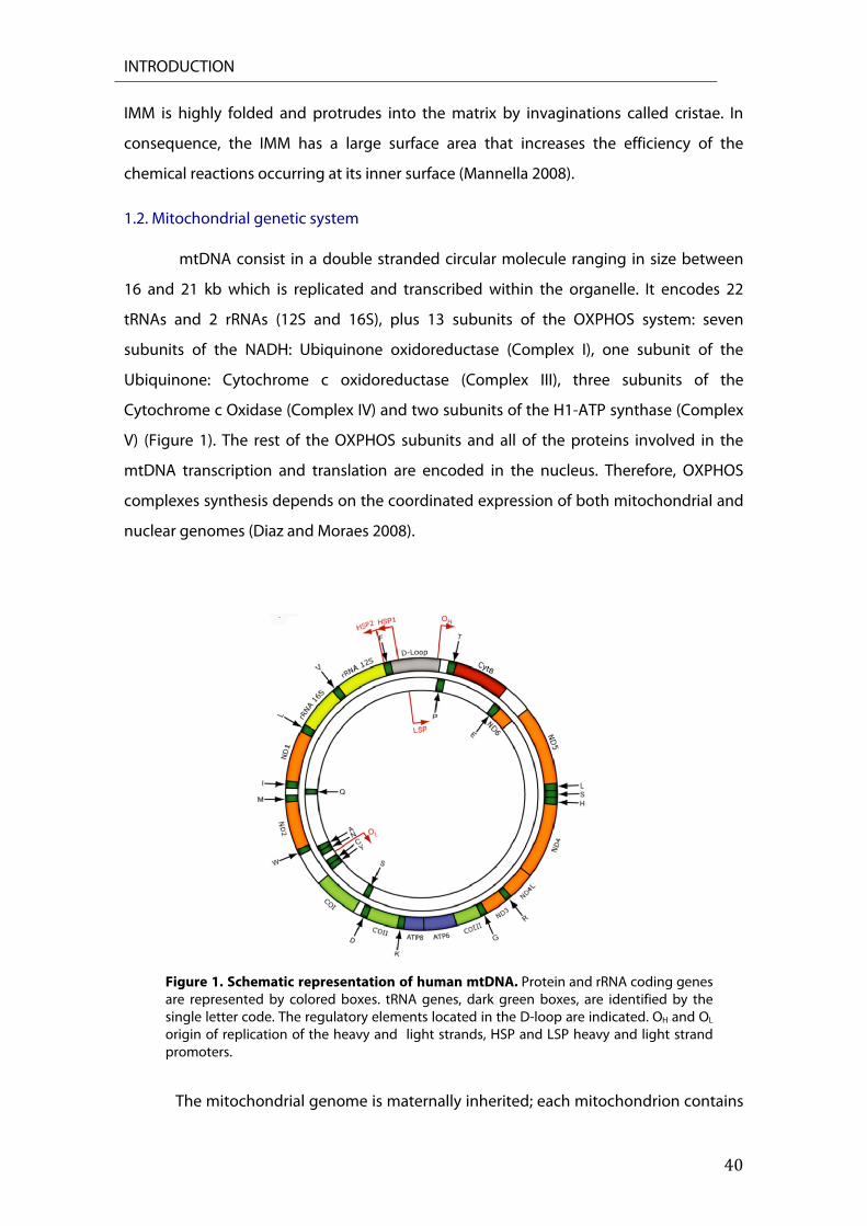
1.2. Mitochondrial genetic system mtDNA consist in a double stranded circular molecule ranging in size between
16 and 21 kb which is replicated and transcribed within the organelle. It encodes 22
tRNAs and 2 rRNAs (12S and 16S), plus 13 subunits of the OXPHOS system: seven
subunits of the NADH: Ubiquinone oxidoreductase (Complex I), one subunit of the
Ubiquinone: Cytochrome c oxidoreductase (Complex III), three subunits of the
Cytochrome c Oxidase (Complex IV) and two subunits of the H1-ATP synthase (Complex
V) (Figure 1). The rest of the OXPHOS subunits and all of the proteins involved in the
mtDNA transcription and translation are encoded in the nucleus. Therefore, OXPHOS
complexes synthesis depends on the coordinated expression of both mitochondrial and
nuclear genomes (Diaz and Moraes 2008).
Figure 1. Schematic representation of human mtDNA. Protein and rRNA coding genes are represented by colored boxes. tRNA genes, dark green boxes, are identified by the single letter code. The regulatory elements located in the D-loop are indicated. OH and OL origin of replication of the heavy and light strands, HSP and LSP heavy and light strand promoters.
The mitochondrial genome is maternally inherited; each mitochondrion contains
INTRODUCTION
41
several copies of mtDNA (Shuster et al. 1988). In a given individual, all mtDNA copies are
thought to be identical, a condition known as homoplasmy. Mutations can arise, be
maintained or amplified to different levels and coexist with wild-type mtDNA, giving rise
to heteroplasmy (Lightowlers et al. 1997). In consequence, it is common to find a
threshold effect in mtDNA-linked human diseases; the number of mutated molecules
has to reach a certain percentage, usually higher than 60-80%, in order to manifest
pathological effects (Fernandez-Silva et al. 2003).
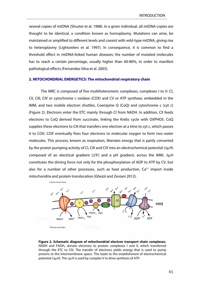
2. MITOCHONDRIAL ENERGETICS: The mitochondrial respiratory chain
The MRC is composed of five multiheteromeric complexes, complexes I to V: CI,
CII, CIII, CIV or cytochrome c oxidase (COX) and CV or ATP synthase, embedded in the
IMM, and two mobile electron shuttles, Coenzyme Q (CoQ) and cytochrome c (cyt c)
(Figure 2). Electrons enter the ETC mainly through CI from NADH. In addition, CII feeds
electrons to CoQ derived from succinate, linking the Krebs cycle with OXPHOS. CoQ
supplies these electrons to CIII that transfers one electron at a time to cyt c, which passes
it to COX. COX eventually fixes four electrons to molecular oxygen to form two water
molecules. This process, known as respiration, liberates energy that is partly converted
by the proton pumping activity of CI, CIII and CIV into an electrochemical potential (H)
composed of an electrical gradient () and a pH gradient, across the IMM. H
constitutes the driving force not only for the phosphorylation of ADP to ATP by CV, but
also for a number of other processes, such as heat production, Ca2+ import inside
mitochondria and protein translocation (Ghezzi and Zeviani 2012).
Figure 2. Schematic diagram of mitochondrial electron transport chain complexes. NADH and FADH2 donate electrons to protein complexes I and II, which transferred through the ETC to CIV. The transfer of electrons yields energy that is used to pump protons to the intermembrane space. This leads to the establishment of electrochemical potential (H). The H is used by complex V to drive synthesis of ATP.

INTRODUCTION
42
3. CYTOCROME C OXIDASE
3.1. Structure of Cytochrome c Oxidase
Cytochrome c oxidase is the terminal component of MRC. It catalyzes the transfer
of electrons from reduced cyt c to molecular oxygen. Mammalian COX is a 200 kDa
multimeric protein complex comprised of 13 structural subunits, embedded in the IMM,
and being active as a dimer. The enzyme core is composed by the three mitochondrially
encoded subunits (CO I-III) (Tsukihara et al. 1996). It has two copper binding sites (CuA
and CuB), two hemes groups found exclusively in COX (a and a3), and magnesium and
zinc ions (Tsukihara et al. 1996; Yoshikawa et al. 1996). Both heme a and the binuclear
center a3-CuB are located in COX1 whereas the binuclear CuA centre is located in COX2. A
Zn2+ ion is in COX5b on the matrix side of the complex. The Mg2+ ion is close to the a3-
CuA site, between COX1 and COX2. Complex IV can also bind calcium and sodium ions
(Tsukihara et al. 1995, 1996).
3.2. Assembly of Cytochrome c Oxidase
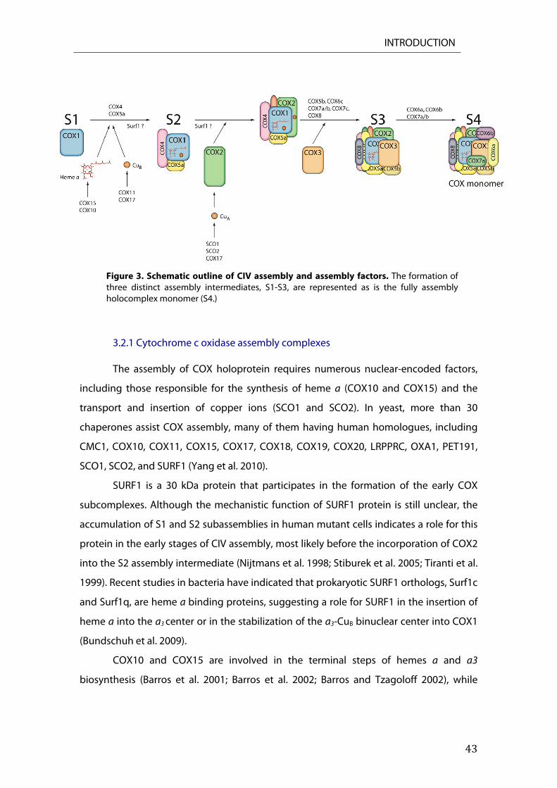
The assembly of individual structural subunits into a fully functional holoenzyme
is a rather complicated process, with the formation of three distinct assembly
intermediates, S1-S3, preceding that of the mature holoenzyme and which requires
nuclear-encoded accessory factors (Leary 2010; Nijtmans et al. 1998). The assembly starts
with the insertion of newly synthesized COX1 into the inner mitochondrial membrane.
This first, crucial, step (S1) is followed by the incorporation of subunits COX4 and COX5a,
to form a second assembly intermediate, S2. Insertion of heme a is likely to occur just
after the formation of S1 or during the formation of S2, together with the insertion of CuB
and heme a3 into COX1 (Antonicka et al. 2003b; Antonicka et al. 2003a; S. L. Williams et al.
2004). The formation of the COX2-associated CuA center is followed by the incorporation
of COX2 into the S2 intermediate (Figure 3).
Next, rapid sequential incorporation of COX3 and the smaller nuclear encoded
subunits, COX5b and COX8, to form S3 leads to the formation of a quasi-complete
assembly intermediate (subcomplex b) (Massa et al. 2008; Stiburek et al. 2005; S. L.
Williams et al. 2004). The addition of the remaining subunits, including COX6a, COX6b,
COX7a and COX7b, results in the formation of holocomplex monomer (S4) (Figure 3)
(Massa et al. 2008; Nijtmans et al. 1998). Finally, monomeric COX dimerizes in an active
structure containing the cyt c binding site (Tsukihara et al. 1996).
INTRODUCTION
43
Figure 3. Schematic outline of CIV assembly and assembly factors. The formation of three distinct assembly intermediates, S1-S3, are represented as is the fully assembly holocomplex monomer (S4.)
3.2.1 Cytochrome c oxidase assembly complexes The assembly of COX holoprotein requires numerous nuclear-encoded factors,
including those responsible for the synthesis of heme a (COX10 and COX15) and the
transport and insertion of copper ions (SCO1 and SCO2). In yeast, more than 30
chaperones assist COX assembly, many of them having human homologues, including
CMC1, COX10, COX11, COX15, COX17, COX18, COX19, COX20, LRPPRC, OXA1, PET191,
SCO1, SCO2, and SURF1 (Yang et al. 2010).
SURF1 is a 30 kDa protein that participates in the formation of the early COX
subcomplexes. Although the mechanistic function of SURF1 protein is still unclear, the
accumulation of S1 and S2 subassemblies in human mutant cells indicates a role for this
protein in the early stages of CIV assembly, most likely before the incorporation of COX2
into the S2 assembly intermediate (Nijtmans et al. 1998; Stiburek et al. 2005; Tiranti et al.
1999). Recent studies in bacteria have indicated that prokaryotic SURF1 orthologs, Surf1c
and Surf1q, are heme a binding proteins, suggesting a role for SURF1 in the insertion of
heme a into the a3 center or in the stabilization of the a3-CuB binuclear center into COX1
(Bundschuh et al. 2009).
COX10 and COX15 are involved in the terminal steps of hemes a and a3
biosynthesis (Barros et al. 2001; Barros et al. 2002; Barros and Tzagoloff 2002), while
INTRODUCTION
44
COX19 is a cytosolic protein with a similar structure to that of COX17, suggesting a role
in copper translocation to mitochondria (Ghezzi and Zeviani 2012).
SCO1 and SCO2 are paralog genes encoding two metallochaperones playing a
role in the formation of the CuA site on COX2. They harbour a highly conserved Cx3C
domain that is thought to bind cooper (Horng et al. 2005). SCO2 synthesis is
transcriptionally activated by p53, which modulates the balance between OXPHOS and
glycolysis (Matoba et al. 2006). Both SCO proteins are essential for the assembly of
complex IV catalitic core and play a role in the maintenance of cellular copper
homeostasis (Leary et al. 2007).
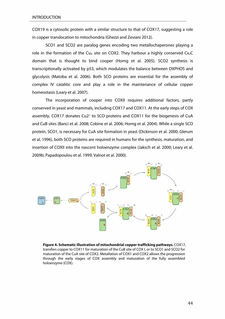
The incorporation of cooper into COXII requires additional factors, partly
conserved in yeast and mammals, including COX17 and COX11. At the early steps of COX
assembly, COX17 donates Cu2+ to SCO proteins and COX11 for the biogenesis of CuA
and CuB sites (Banci et al. 2008; Cobine et al. 2006; Horng et al. 2004). While a single SCO
protein, SCO1, is necessary for CuA site formation in yeast (Dickinson et al. 2000; Glerum
et al. 1996), both SCO proteins are required in humans for the synthesis, maturation, and
insertion of COXII into the nascent holoenzyme complex (Jaksch et al. 2000; Leary et al.
2009b; Papadopoulou et al. 1999; Valnot et al. 2000).
Figure 4. Schematic illustration of mitochondrial copper trafficking pathways. COX17, transfers copper to COX11 for maturation of the CuB site of COX I, or to SCO1 and SCO2 for maturation of the CuA site of COX2. Metallation of COX1 and COX2 allows the progression through the early stages of COX assembly and maturation of the fully assembled holoenzyme (COX).
INTRODUCTION
45
Despite the requirement of both SCO proteins in humans, studies in SCO1 and
SCO2 patient derived cell lines have demonstrated that SCO proteins have non-
overlapping but cooperative roles in CuA biogenesis (Leary et al. 2004). Human SCO2
interacts with newly synthesized COX2, as it is inserted into the inner membrane or
immediately thereafter, an association that depends on its prior metallation by COX17
(Figure 4). The physical interaction between SCO2 and COX2 triggers the metallation of
SCO1 by COX17 and its recruitment to the SCO2-COX17 complex. Each SCO protein
sequentially delivers a copper ion to COX2 to form the CuA site. The mature polypeptide
is then incorporated into the nascent holoenzyme. Initially, SCO2 donates its copper with
its cysteine thiols becoming oxidazed in the process. After the subsequent transfer of
copper from SCO1 to COX2, SCO2 acts as a thiol-disulfide oxidoreductase to reoxidize
the cysteines in SCO1. Alternatively, SCO2 may induce disulfide bond formation in
copper-loaded SCO1 to facilitate its transfer to COXII (Figure 4) (Leary et al. 2004; Leary et
al. 2009b).
3.3. Cytochrome c Oxidase Deficiency
Cytochrome c oxidase deficiencies are one of the most common defects of the
respiratory chain found in mitochondrial diseases. Just one pathogenic mutation has
been reported in a nuclear-encoded structural CIV subunit, COX6B1, in two cases of
mitochondrial encephalomyopathy with isolated COX deficiency (Massa et al. 2008).
In humans, most syndromes with isolated complex IV deficiency are caused by
mutations in genes encoding COX assembly factors that affect the stability and
incorporation of COX subunits into the assembled complex such as SURF1 (Pequignot et
al. 2001; Tiranti et al. 1998; Zhu et al. 1998), SCO1 and SCO2 (Leary et al. 2004;
Papadopoulou et al. 1999; Valnot et al. 2000), COX15 (Antonicka et al. 2003a; Bugiani et
al. 2005; Oquendo 2004), COX10 (Antonicka et al. 2003b; Coenen et al. 2004) and LRPPRC
(Mootha et al. 2003; Xu et al. 2004). They are associated with different clinical
presentations, including encephalopathies, Leigh Syndrome (LS), hypertrophic
cardiomyopathies, fatal lactic acidosis, hepatic failure and leukodystrophy.
More that 40 different pathogenic mutations have been reported in the Surf1
gene, all causing LS associated with COX deficiency (Pecina et al. 2004; Pequignot et al.
2001). Almost all Surf1 mutations reported to date cause the complete absence of the
protein, a marked reduction in the amount of fully assembled COX and the accumulation
of assembly intermediates S1 and S2 (Stiburek et al. 2005; Tiranti et al. 1999).
INTRODUCTION
46
Mutations in COX10 are associated with a wide spectrum of conditions including
LS; encephalopathy with proximal renal tubulopathy; sensorineural deafness, metabolic
acidosis, hypotonia and hypertrophic cardiomyopathy. Likewise, mutations in COX15
gene can cause fatal infantile hypertrophic cardiomyopathy, as well as LS (Antonicka et
al. 2003b; Antonicka et al. 2003a; Bugiani et al. 2005).
Pathogenic mutations in either Sco1 or Sco2 result in severe COX deficiency with
different early onset and fatal clinical phenotypes. Mutations in Sco1 cause fatal infantile
hepato-encephalomyopathy (Leary et al. 2013) and one case has been reported with
hypertrophic cardiomyopathy, encephalopathy and hepatomegaly with fatal outcome
(Stiburek et al. 2009). Mutations in Sco2 cause a fatal infantil cardio-
encephalomyophathy and, but with one exception, all patients harboured the missense
mutation E140K (Mobley et al. 2009; Papadopoulou et al. 1999).
Heart hypertrophy, which has a direct life-threatening impact, is usually severe in
all Sco2 mutant patients. Despite the similar functional involvement of SCO1 and SCO2
as copper-binding proteins, the precise molecular function of the two human SCO
proteins in COX assembly remains unknown. Moreover, their ubiquitous expression with
no strict tissue specificity for either protein arise the question of why mutations in the
two genes lead to different tissue-specific deficiencies in COX with distinct clinical
phenotype (Brosel et al. 2010).
5. DROSOPHILA MELANOGASTER
The common fruit fly, Drosophila melanogaster, is a well studied, highly tractable
genetic model organism for the understanding of the molecular mechanisms underlying
human diseases. Drosophila melanogaster have a short life cycle, approximately 10 days
from egg lay to adult hatching from the puparium at 25ºC. The embryo completes its
development in just 24 hours which ends with the hatching of a wormlike first instar
larva which grows and increases its body size to produce second and third instar larva.
About three days later, third instar larvae complete their growth and pupate,
undergoing metamorphosis. This process last four days in which most larval tissues
disintegrate and are replaced, through the proliferation and differentiation of adult
precursor tissues as imaginal discs, to produce adult structures. After metamorphosis
completion, imagoes emerge from the pupa case.

INTRODUCTION
47
5.1. The heart of Drosophila melanogaster
Drosophila’s heart is a simple linear tube that pumps and delivers hemolymph
through the organism in an open circulatory system (Curtis et al. 1999). It is constituted
of two major cell types: the inner contractile muscle cells (cardiomyocytes) are flanked
on each side by pericardial cells, which do not express muscle-specific structural proteins
and are implicated in hemolymph detoxification. The dorsal vessel is surrounded by an
extracellular matrix composed of pericardin, a type IV collagen-like protein (Chartier et al.
2002). The adult heart forms during metamorphosis by a remodelling of the larval
cardiomyocytes without cell proliferation or migration (Bodmer 2006). The dorsal vessel
is divided into heart proper and aorta.
Cardiogenesis in flies begins with cell fate specification in the developing
mesoderm where secreted ligands, including decapentaplegic (dpp), regulate
embryonic heart development and trigger cell fate specification (Bodmer and Venkatesh
1998; Cripps and Olson 2002; Zaffran and Frasch 2002). Cardiac precursor cells
expressing specific transcription factors including tinman (tin), the fly orthologue of
Nkx2.5, and GATA family members migrate along the mesoderm and form the
recognizable single layered dorsal vessel at stage 16 of embryonic development (Figure
5) (Bodmer 1993). It is worth mentioning that many of the temporal and tissue specific
signals required for heart development are evolutionarily conserved from flies to
mammals.
Figure 5. The embryonic Drosophila circulatory system. (A) The developing embryonic circulatory system arises from cardiac precursor cells that migrate to form the dorsal vessel at Stage 16. Stages 12, 13, and 17 are shown. Figure adapted from Fly Embryo RNAi Project . (http://flyembryo.nhlbi.nih.gov). (B) The embryo cardiac organ is constituted by a simple tube, divided into an anterior “aorta”, and a posterior “heart”. Ostia cells are represented in purple; Tin positive cells are represented in green. Lower Panel: Dorsal view of a stage 16 labeled with anti-Mef2 (Blue).
INTRODUCTION
48
The newly hatched larvae possess a functional heart that pumps hemolymph
from the posterior to the anterior region of the animal. During larval growth
cardiomyocyte number remains constant but cells elongate dramatically, suffering
changes at the ultrastructural level while the luminal space build by cardiomyocytes
increases significantly (Curtis et al. 1999; Monier et al. 2005).
During metamorphosis, the heart undergoes a remodelling process to give rise to
the adult heart wich remains unchanged until death (Monier et al. 2005; Zeitouni et al.
2007). It consists in a single layer of cardiomyocytes with circumferentially oriented
myofibers and it is closely juxtaposed to the ventral longitudinal muscle and sets of
suspensory muscles arising from the dorsal cuticle called alary muscles (Figure 6 A). The
ventral longitudinal muscle is a non-cardiac muscle type. It arises from lymph cells via
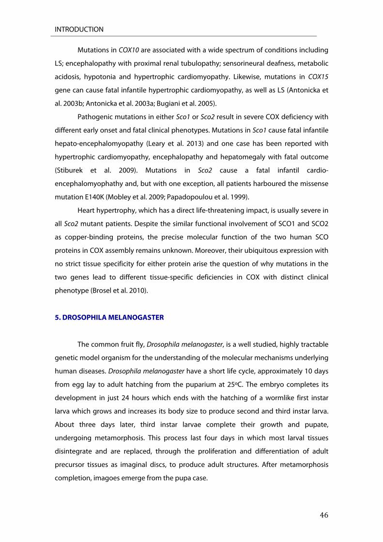
trans-differentiation during pupal stages of development (Shah et al. 2011).
Figure 6. Adult Drosophila circulatory system. (A) The adult fly circulatory system consists of an open system with the main conical chamber, heart, located along the dorsal aspect of the A1 abdominal segment. Confocal imaging of the dorsal vessel labeled with anti-Myosine (Green), Alexa Fluor 594-phalloidin and Dapi (blue). (B) Ostia cells of A3 abdominal segment labeled with anti-Myosine (Green), Alexa Fluor 594-phalloidin and Dapi (blue).
The 104 embryonic cardiomyoblasts differentiate into three different cell types,
contractile cardiomyocytes forming the heart lumen, segmentally arranged pairs of ostia
cells, hemolymph inflow devices, and the intracardiac valves that divide the heart in
chambers. Hemolymph enters throught four pairs of ostia cells (Figure 6B), which open
during diastole and close during systole. As the heart contracts, hemolymph passes
through the aorta and is dispersed into the body cavity close to the brain to re-enter the
INTRODUCTION
49
dorsal vessel through the ostia (Shah et al. 2011).
5.2. Drosophila melanogaster as a model to study human cardiomyopathies
Drosophila melanogaster has been used to study genetic, development, and
signalling for nearly a century but only over the past few decades has this tremendous
resource been focus on cardiovascular research. The fruit fly is a powerful genetic model
system with multitude of tools for manipulating genes and gene expression. Thus
several groups have begun to examine heart function in Drosophila with the goal of
using this system as a physiological model (Birse et al. 2010; Melkani et al. 2013; Tang et
al. 2013).
Drosophila is an advantageous model for studying cardiac development and
cardiac dysfunction since the genetic network controlling cardiac specification and
differentiation as well as many aspects of heart function, are conserved from flies to
mammals (Bodmer 1995; Bodmer and Venkatesh 1998; Cripps and Olson 2002; Harvey
1996; E. N. Olson 2006; Wolf and Rockman 2011). In addition, it is now clear that the
Drosophila heart undergoes cardiac aging. The fly heartbeat becomes irregular with
increased episodes of arrhythmias (Ocorr et al. 2007a), reminiscent of increased atrial
fibrillation and heart failure in aged humans (Lakatta 2003). Most importantly, unlike in
vertebrates, the fly heart is not needed for oxygen delivery and its function can therefore
be significantly compromised without causing immediate death, making it an excellent
model to study cardiomyopathies (Ocorr et al. 2007a).
Genetic manipulations of ion channel genes in Drosophila, including L-type Ca2+
channels and several types of K+ channels, perturb proper heart function, further
underlining the remarkable functional parallels in basic cardiac physiology between flies
and humans (Ocorr et al. 2007b; Ray and Dowse 2005; Sanguinetti and Tristani-Firouzi
2006). Furthermore, Drosophila constitute a useful and emerging model for the study of
high fat diet induced obesity and cardiac dysfunction, one of the main causes of
cardiovascular diseases in North America and Europe (Birse et al. 2010) and for the
screening and identification of candidate genes involved in heart function and disease
(Neely et al. 2010). Thus, Drosophila has become a powerful genetic model system for the
study and understanding of cardiomyopathies (10.1038/ng.2610 2013) and, the recently
established heart function assays make it possible to analyse and characterize adult
cardiac function and functional abnormalities (Fink et al. 2009; Ocorr et al. 2007b; Ocorr
et al. 2009).
INTRODUCTION
50
Human cardiomyopathies can be categorized as dilated, hypertrophic and
restrictive (Wolf 2012). Despite the simplicity of Drosophila´s heart, the concept of
cardiomyopathies in the fly requires further definition. Dilated cardiomyopathies, the
best characterized cardiomyopathies in Drosophila, can be defined as an “eccentric”
hypertrophy in which contractile fibers are added in series and cause an enlarged
chamber at end diastole and impaired systolic function. Hypertrophic cardiomyopathies
can be thought of as a “concentric” hypertrophy in which contractile fibers are added in
parallel, causing increased cardiomyocyte size and decreased chamber lumens.
Restrictive cardiomyopathy can be thought as a process that impair heart relaxation
(Wolf 2012).
6. p53 The tumor suppressor protein p53 is undoubtedly one of the most studied
proteins to date being frequently mutated in cancer (Greenblatt et al. 1994). p53 is a
transcription factor that functions in a complex signalling network to mediate cellular
adaptation to stress (reviewed in (Jones and Thompson 2009). In the absence of cellular
stress, p53 is present in the cell at low levels, being in an inactive state. Stimulus as
genetic or metabolic stress and DNA damage lead to p53 protein activation (Harris and
Levine 2005). In response to stress, p53 selectively regulates the expression of its target
genes, inducing cell cycle arrest, apoptosis or senescence (Vogelstein et al. 2000).
p53 protein levels are regulated primarily by the ubiquitin ligase Mdm2, which
binds to its transactivation domain and ubiquitylates the protein targeting it for
degradation (Hu et al. 2007). Because p53 transcriptionally activates Mdm2, expression
levels of p53 and Mdm2 are balanced through a negative feedback loop which is altered
by an increase in p53 levels as result of a stress response (Hu et al. 2007). Additional
Mdm2-like proteins as Mdm4, also known as MdmX, have been described to regulate
p53 functions (Marine and Jochemsen 2004).
The interplay between p53, Mdm2 and Mdm4 at molecular level is complex.
Mdm4 represses p53 transactivation but neither can stimulate p53 nuclear export nor its
degradation, appearing to protect p53 from Mdm2-mediated suppression (Jackson and
Berberich 2000). In response to cellular stress, protein-protein interactions between p53
and Mdm2 diminish, resulting in reduced p53 ubiquitination and accumulation of p53
protein (Momand et al. 2000; Shvarts et al. 1996). Once stabilized, p53 undergoes various

INTRODUCTION
51
posttranslational modifications, including acetylation, methylation, ubiquitination and
phosphorylation of specific p53 residues, that affect its activity and subcellular
localization (Kruse and Gu 2008).
Although the primordial function of p53 is that of a stress-response transcription
factor, recent studies indicate that it also contributes to the maintenance of intracellular
homeostasis, regulating metabolism even in the absence of acute stress (Olovnikov et al.
2009; Vousden and Ryan 2009) thereby exerting prosurvival functions and regulating a
number of physiological and pathological processes (Lassus et al. 1996).
6.1. Regulation of energy metabolism by p53
p53 is an emerging regulator for metabolic homeostasis, coordinating stress
responses with changes in cellular metabolism. In 1956, Otto Warburg found that unlike
the majority of normal cells which depend on mitochondrial oxidative phosphorylation
to provide energy, most tumor cells primarily utilize glycolysis for their energy needs
even under normal oxygen concentrations. This shift from mitochondrial respiration to
aerobic glycolysis is known as the Warburg effect (Warburg 1956). The metabolic
alterations in cancer cells determine how cells respond to variable nutrient and oxygen
availability and promote cell proliferation, growth and survival (Vousden and Ryan 2009).
Recent findings indicate that p53 plays an important role in metabolic shifting in
cancerous cells, suggesting a new function of p53 as a cell metabolism regulator
(Bensaad et al. 2006; Feng and Levine 2010; Momota et al. 2013; Vousden and Ryan 2009).
In fact, p53 activation by metabolic stress is regulated by AMPK-dependent
phosphorylation and influenced by mTOR (Hardie 2004), two master regulators of
cellular metabolism. Thus, p53 is able to contribute to the regulation of glycolysis,
oxidative phosphorylation, glutaminolysis, insulin sensitivity, nucleotide biosynthesis,
mitochondrial integrity, fatty acid oxidation, antioxidant response, autophagy and mTOR
signalling (Inoki et al. 2003; Maddocks and Vousden 2011)
6.1.1. The role of p53 in regulating glycolysis and mitochondrial oxidative phosphorylation Recent studies indicate that p53 plays a major role in suppressing glucose
consumption and antagonizing the Warburg effect. It modulates glucose consumption

INTRODUCTION
52
and glycolysis at multiple levels. p53 regulates glucose uptake by reducing, through the
inhibition of NF-B, the expression of GLUT3 glucose transporter which, together with
GLUT1, is expressed in most mammalian cells (Kawauchi et al. 2008a, 2008b). It directly
suppresses GLUT1 and GLUT4 expression, the later an insulin-regulated glucose
transporter (Schwartzenberg-Bar-Yoseph et al. 2004). Furthermore, p53 represses the
insulin receptor promoter providing another mechanism by which p53 can limit the
transport of glucose into the cells (Webster et al. 1996).
Figure 7. Regulation of energy production by p53. Several functions of p53 reduce the flux through the glycolytic pathway and increase oxidative phosphorylation, thereby opposing the Warburg effect, in which cancer cells predominantly use glycolysis for energy production. However, there are also activities of p53, such as the activation of hexokinase and phosphoglycerate mutase (PGM), which could increase glycolysis under some circumstances. GLUT, glucose transporter; IKK, IκB kinase; NF-κB, nuclear factor-κB; SCO2, synthesis of cytochrome c oxidase 2; TCA, tricarboxylic acid.
Through post-translational modifications, p53 decreases the levels of
phosphoglycerate mutase (PGM), which catalyzes the conversion of 3-phosphoglycerate
to 2-phosphoglycerate, promoting it´s degradation (Kondoh et al. 2005). p53
transactivates TP53-induced glycolysis and apoptosis regulator (TIGAR) (Bensaad et al.
2006), which functions as a fructose-2,6-bisphosphatase. TIGAR limits the activity of
phosphofructokinase 1 (PFK1), the most important control site for the glycolytic flux,

INTRODUCTION
53
blocking glycolysis at the fructose-6-phosphate stage and promoting the diversion of
glycolytic intermediates into the pentose phosphate pathway (PPP) (Bensaad et al. 2006)
(Figure 7). p53 deficient cells shows an increased PPP flux, NADPH production and
glucose consumption, which can be largely reversed via the inhibition of Glucose 6-
Phosphate Dehydrogenase (Jiang et al. 2011). p53 also negatively regulates the
expression of pyruvate dehydrogenase kinase 2 (PDK2), which inactivates pyruvate
dehydrogenase complex (PDC), a protein that converts pyruvate to acetyl-CoA,
favouring the production of acetyl-CoA at the expense of lactate production (Contractor
and Harris 2012).
Despite the convincing evidence that p53 can be a negative regulator of
glycolyisis, there is also evidence that suggest that p53 can enhance some steps on this
pathway. Hence, while p53 reduces the protein levels of PGM in embryonic fibroblast
cells, in cardiac myocytes it activates the transcription of the muscle isoform of PGM
(PGM-M) and hexokinase II (HK2), which catalyses the first step in glycolysis (Ruiz-Lozano
et al. 1999) .
On the other hand, p53 plays a critical role in the maintenance of oxidative
phosphorylation and mitochondrial integrity. It has been shown to promote OXPHOS
through mechanisms that include the transcriptional activation SCO2 (Matoba et al.
2006). In addition to SCO2, p53 appears to promote mitochondrial function through
different target genes, including AIF (apoptosis-inducing factor), required to maintain
the integrity of mitochondrial CI (Stambolsky et al. 2006; Vahsen et al. 2004), GLS2
(glutaminase 2) (Hu et al. 2010), parkin (Zhang et al. 2011), p53R2 (p53-inducibel
ribonucleotide reductase), TFAM, DNA polymerase (POLG) and PGC1 (Achanta et al.
2005; Bourdon et al. 2007; Kulawiec et al. 2009; Lebedeva et al. 2009; Park et al. 2009;
Sahin et al. 2011) required for mitochondrial dynamics regulation and the maintenance
of the mitochondrial genome.
In conclusion, given that p53 helps to shift ATP production from glycolysis to
oxidative phosphorylation and since they have a homeostatic relationship, in order to
regulate global changes in cellular activity the existence of a feedback mechanism
between mitochondria and p53 is expected.
6.2. p53, mitochondria and apoptosis
Apoptosis, a type of programmed cell death, is a highly regulated process used to
INTRODUCTION
54
remove unwanted cells. Apoptosis is required for normal development, maintenance of
tissue homeostasis and regulation of certain disease states (Thompson 1995; Vaux and
Korsmeyer 1999).
The proapoptotic function of p53 constitutes the best characterized facet of its
oncosuppressive activities. Mitochondria is central for carrying out the intrinsic apoptosis
pathway, and p53 participates in executing the intrinsic pathway through transcription
dependent and independent mechanisms (Moll and Zaika 2001). p53 has been shown to
induce apoptosis through the expression of a variety of pro-apoptotic genes, as Apaf-1,
PUMA, Noxa, Bax, Bid, Bad and p53IAP1 (Brady and Attardi 2010; Miyashita and Reed
1995; Nakano and Vousden 2001; Robles et al. 2001) and the repression of antiapoptotic
genes such as bcl-2 and survivin (Hoffman et al. 2002; Shen and Shenk 1994).
The last decade of research, has revealed the transcription independent
proapoptotic activities of p53, which involves its direct action at the mitochondria
(Green and Kroemer 2009; Vaseva and Moll 2009). In response to stress stimuli, a pool of
cytoplasmic p53 rapidly translocates to the mitocondrial surface where it physically
interacts with both anti-apoptotic (Bcl-xL, Bcl-2, Mcl-1) and proapoptotic (PUMA, Bax,
Bak) members of the Bcl-2 family to inhibit or activate their respective functions, leading
to mitochondrial outer membrane pore (MOMP) opening and apoptosis (Vaseva and
Moll 2009).
p53 gene is a major proapoptotic factor in both Drosophila and vertebrates
(Kanda and Miura 2004; Steller 2000). The Drosophila homolog of p53, dp53, has mostly a
proapoptotic role, having been reported to activate the expression of Reaper and Hid
proapoptotic proteins upon different insults (Brodsky et al. 2000; Fan et al. 2010; Grether
et al. 1995a).
Similar to the mammalian cell death machinery, Drosophila possesses also a
complicated cell-death regulatory system: the fly has an Apaf-1-like protein (dARK)
(Kanuka et al. 1999; Rodriguez et al. 1999), seven caspases (Chen et al. 1998; Dorstyn et al.
1999; Hay and Guo 2006; Song et al. 1997) and two Bcl-2 family members (reviewed in
(Igaki and Miura 2004). Drosophila also has unique killer proteins, Reaper, Hid and Grim
(Chen et al. 1996; Grether et al. 1995b; White et al. 1994) thought to be functional
orthologes of mammalian mitochondrial IAP inhibitors such as Smac/DIABLO and
HtrA2/Omi.

INTRODUCTION
55
Figure 8. Schematic representation of apoptosis pathway in Drosophila. The adaptor protein dARK promotes activation of the apical caspase DRONC. DIAP1-binding proteins such as RPR, HID, GRIM partly promote death by disrupting 'the anti-caspase' function of DIAP1.
INTRODUCTION
56
Caspases are the core of the cell-death machinery (Degterev et al. 2003),
becoming activated in response to different death signals. As it has been mentioned
above, seven Drosophila caspases have been identified. Like the mammalian ones, these
can be divided into initiator and executioner caspases. DRONC, the fly homolog of
mammalian caspases-2 and -9, has emerged as the essential apoptotic initiator caspase
in Drosophila. DRONC interacts with dARK, and once is activated it cleaves and activates
the executioner caspases DCP-1 and DRICE (Igaki et al. 2002; Kanuka et al.
1999)(reviewed in (Salvesen and Abrams 2004). This activation of DRONC might be
regulated by the pro- and anti-apoptotic multidomain Bcl2 family members DEBCL and
Buffy (reviewed in (Igaki and Miura 2004). Genetic studies have demonstrated that dARK
and caspases act downstream of Reaper, Hid, and Grim. Thus, in cells committed to die,
apoptosis is initiated by Reaper, Hid, and Grim, which bind to apoptosis inhibitor DIAP1
(Wang et al. 1999), a fly homolog of the mammalian IAP proteins, inducing its
autoubiquitination and proteasome-mediated degradation, thus realising caspases from
DIAP1 inhibition (Figure 8) (Steller 2008).
Although the role of mitochondria in Drosophila apoptosis its still unclear and
despite the striking differences, there are some clear evidence suggesting that
mitochondria play an important role in cell death in flies as it does in mammals. In fact, it
is known that Rpr, Hid and Grim localization in the mitochondria is essential to promote
cell death (Claveria et al. 2002; Haining et al. 1999; M. R. Olson et al. 2003), and it has
been shown that under apoptotic stimulus, fly mitochondria undergoes Rpr, Hid and
Drp1-dependent morphological changes and disruption during cell death (Abdelwahid
et al. 2007). The participation of Drp1, a mitochondrial fission protein, in cell death is
conserved in worms and mammals (Frank et al. 2001; Jagasia et al. 2005).
57
AIMS

58

AIMS
59
COX deficiency due to mutations in COX assembly factors is one of the most
frequent causes of MRC defects in humans. Usually, it has a very early age of onset and
fatal outcome, displaying different clinical presentations, including encephalopathies
such as Leigh Syndrome, fatal cardiomyopathy, hepatic failure and leukodystrophy.
Pathogenic mutations in human Sco1 and Sco2 have been reported to cause
hypertrophic cardiomyopathy among other clinical symptoms. Although the role of Sco
proteins as copper metallochaperones is largely documented, the molecular mechanism
underlying the cardiac dysfunction caused by mutations in human Sco1 and Sco2, have
yet to be elucidated.
Thus, the generation of animal models is of vital importance. Although
unfortunately there are no Sco1 KO mice available, a Sco2KIKO human disease mice model
has been recently developed. Despite Sco2KIKO mice model present most features of the
human disease they showed no evidence of cardiomyopathy.
Therefore, the specific aims of the present study were:
1. Generation of a Drosophila cardiomyopathy model to study cardiomyopathies with a
mitochondrial origin.
2. Investigate the genetic and molecular mechanisms that underlie ScoX induced
cardiomyopathy.

60

61
MATERIALS AND METHODS

62

MATERIALS AND METHODS
63
1. Materials
1.1 Reagents, solutions and buffers
All the reagents used in the different experiments performed in this thesis are
described in methods or as described in the references.
1.2. Drosophila melanogaster
1.2.1. Standard condition for Drosophila growth All fly stocks were raised on standard Drosophila medium; yeast 10%, sucrose
7.5%, wheat flour 3.5%, propionic acid and agar 1.25%.
Fly culture and crosses throughout this thesis were performed at 25ºC and 60%
relative humidity.
1.2.2. Fly Stocks All fly stocks used in this thesis are listed in the below, tables 1 to 3. For RNAi
silencing, Scox-RNAi or Surf1-RNAi males or virgin females were crossed to TinC4-Gal4
flies and incubated at 25ºC throughout development. Female F-1 progeny were

MATERIALS AND METHODS
64
collected for posterior analysis and aged at 25ºC. w1118 flies crossed to TinC4-Gal4 or
each UAS-RNAi line were used as control flies.
1.2.2.1 Balancers and control stocks
Table 1.
1.2.2.2. Gal4 stock
Table 2.
1.2.2.3 UAS stocks
Name Origin Observations
yellow white R. Garesse Control stock
w1118 R. Garesse Parental transgenic stock
Double Balancers R. Garesse Used for the mapping of P elements in the transgenic
lines generated
Name Origen Observations
daugtherless-GAL4 Bloomington Stock Centre nº 8641 Ubiquitous
TinC4-Gal4 Manfred Frasch Cardiac-tissue specific
Nombre Origen Observaciones
UAS - ScoxRNAi Vienna Drosophila RNAi Center (VDRC)
Scox RNAi of Drosophila melanogaster
UAS – Surf1RNAi Vienna Drosophila RNAi
Center (VDRC) Surf1 RNAi of Drosophila
melanogaster
UAS – p53 Rolf Bodmer Overexpression of p53

MATERIALS AND METHODS
65
Table 3.
1.3. Oligonucleotides
The oligonucleotides used in the development of this thesis were synthesized by
Sigma-Aldrich. Oligonucleotides names and 5´3´ sequences are listed in Table 4.
UAS – p53DN Rolf Bodmer Overexpression of a DN form of p53
UAS- p53 mut Bloomington Drosophila Stock Center
Deletion of dp53 gen
UAS- GFP Stringer Rolf Bodmer Green fluorescent protein
UAS-p35 Manuel Calleja Inhibitor of apoptosis
UAS-Diap1 Manuel Calleja Inhibitor of apoptosis
UAS-Rpr Manuel Calleja Pro-apoptotic gene
Nombre Secuencia
Rpl10 Fw AAGAAGGTGCTCTGCCTGTC
Rpl10Rv CGCACATTCTGCCAGTTCT
Scox Fw CTCCCGCAGATTCCACTAAA
Scox Rw GCTCTTCACGTACAGCATGA
IMPL3 Fw GTGTGCCTCATCGATGTCTG
IMPL3 Rw GTGCTGGCCGTGATCTG
GPI Fw GGCCTTCACTCCCACTTTTGT
GPI Rw CCACGGACTCAGGCTCCT
Pdk Fw ATCTCATTAGGAATCGGCACAA
Pdk Rw CTGAATGGAACTCTCTGTAGGC

MATERIALS AND METHODS
66
Table 4.
1.6. Dyes
- Dapi (Molecular Probes)
-Phalloidin-TRITC (Molecular Probes)
-Phalloiding-488 (Molecular Probes)
2. Methods
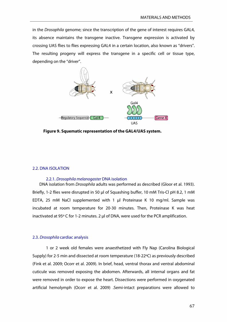
2.1. GAL4/UAS system
In this thesis, we have used the GAL4/UAS system for targeted gene expression in
Drosophila melanogaster (A. H. Brand and Perrimon 1993a). This system allows the
ectopic expression of a gene of interest (gene X) in a specific tissue or cell type (Figure 9).
The expression of the gene of interest is controlled by the UAS element, consisting of
GAL4-binding sites. GAL4 is a yeast transcriptional activator with no known binding sies
Pfk Fw CGCCTAGCTGTGATGCATATT
Pfk Rw GACACCGTCGTTGATTCCATAA
Rpr Fw GAGTCACAGTGGAGATTCCT
Rpr Rw AATCCTCATTGCGATGC
Hid Fw ACGGCCATCCGAATCCGAAC
Hid Rw TGCTGCTGCCGGAAGAAGAAGTT
Grim Fw CATCAGCAACAGCGCCCA
Grim Rw
UAS Fw
UAS Rw
Gal4 Fw
Gal4 Rw
GCTGGCTCGAACTGTAGCTG
CACACCACAGAAGTAAGGTTCCTTCAC
GAACACGTCGCTAAGCGAAAGCTAAG
TCCTCGAGAAGACCTTG
GGTCCGTTTTCAGGAAG
MATERIALS AND METHODS
67
in the Drosophila genome; since the transcription of the gene of interest requires GAL4,
its absence maintains the transgene inactive. Transgene expression is activated by
crossing UAS flies to flies expressing GAL4 in a certain location, also known as “drivers”.
The resulting progeny will express the transgene in a specific cell or tissue type,
depending on the “driver”.
Figure 9. Squematic representation of the GAL4/UAS system.
2.2. DNA ISOLATION
2.2.1. Drosophila melanogaster DNA isolation DNA isolation from Drosophila adults was performed as described (Gloor et al. 1993).
Briefly, 1-2 flies were disrupted in 50 μl of Squashing buffer, 10 mM Tris-Cl pH 8.2, 1 mM
EDTA, 25 mM NaCl supplemented with 1 μl Proteinase K 10 mg/ml. Sample was
incubated at room temperature for 20-30 minutes. Then, Proteinase K was heat
inactivated at 95º C for 1-2 minutes. 2 μl of DNA, were used for the PCR amplification.
2.3. Drosophila cardiac analysis
1 or 2 week old females were anaesthetized with Fly Nap (Carolina Biological
Supply) for 2-5 min and dissected at room temperature (18-22ºC) as previously described
(Fink et al. 2009; Ocorr et al. 2009). In brief, head, ventral thorax and ventral abdominal
cuticule was removed exposing the abdomen. Afterwards, all internal organs and fat
were removed in order to expose the heart. Dissections were performed in oxygenated
artificial hemolymph (Ocorr et al. 2009) .Semi-intact preparations were allowed to

MATERIALS AND METHODS
68
equilibrate with oxygenation for 20 min prior to filming. Movies of beating hearts were
recorded for 30 seconds with a high-speed Hamamatsu EM-CCD digital camara at rates
of 100-200 frames/sec in conjunction with 10x immersion lens. Movies were analyzed
with Semi-automatic Optical Heartbeat Analysis software to quantify heart periods,
systolic and diastolic intervals, systolic and diastolic diameters, cardiac rhythmicity,
frantional shortening and to produce M-mode records (Fink et al. 2009).
2.4. Inmunostaining of the Adult Drosophila heart
Fluorescence imaging of Drosophila heart tubes were performed as described
(Alayari et al. 2009). Briefly, semi-intact Drosophila hearts were placed in artificial
hemolymph containing 10mM EGTA for 5-10 min to inhibit cardiac tube contractions.
Hearts were then fixed in situ for 20 min with 4% paraformaldehyde in PBS at room
temperature with gentle shaking. Then, hearts were washed three times in PBST (PBS -
0.1% Triton X-100). After washing, hearts were incubated with Alexa Fluor-594 Phalloidin
in PBST (1:200) for 2h. Hearts were washed three times for 10 min in PBST, rinsed in 100
μl of PBS for 10 min and mounted on Vectashield (Vector Laboratories). Images were
acquired using a Zeiss LSM 710 fluorescent microscope.
2.5. TUNEL Staining of adult hearts
TUNEL staining was essentially performed according to the instructions from the
manufacturer (In Situ Cell Death Detection Kit, TMR Red, Roche, Germany). Briefly, semi-
intact hearts were prepared as described above. Then, hearts were incubated with PBST
plus Na-Citrate 100mM at 65ºC for 30 min. Then the hearts were washed trice 10 minutes
in PBST, incubated in TUNEL-Dilution buffer two times for 5 min at room temperature
and afterwards in Labeling solution for 30 mins at 37ºC. Then, hearts were incubated in
Enzyme solution at 37ºC for 2h and washed three times, 10 min in PBST, and mounted on
Vectashield plus DAPI (Vector Laboratories). For positive control, hearts were pretreated
with DNAse I to introduce nonspecific strand breaks.

MATERIALS AND METHODS
69
2.6. TUNEL Staining of Mouse esqueletal muscle and liver
Muscle and liver tissues were cryo-sectioned (8μm thick) and tissue sections were
fixed for 20 min at room temperature in 4% paraformaldehyde-PBS and stained for
TUNEL as described above. For positive control, tissues were pretreated with DNAse I to
introduce nonspecific strand breaks.
2.7. DHE Stainning
Dihydroethidium (DHE) was employed for the detection of oxidative stress. Briefly semi-
intact hearts were prepared as described above and stained with 3mM DHE (Molecular
Probes, Carlsbad, CA) in PBS for 1h, followed by three washes in PBS. Hearts were relaxed
with 10mM EGTA and fixed in 7% paraformaldehyde-PBS for 4 min at room temperature.
Hearts were mounted in Vectashield . Fluorescence imaging was carried out using a Zeiss
LSM 710 fluorescent microscope.
2.8. Respiratory Complexes enzyme activity measurement
Mitochondrion-enriched homogenates were prepared from 30 third instard
larvae ground in SETH buffer (250 mM sucrose, 2mM EDTA, 100 units/liter heparin, 10
mM Tris-HCL, pH 7.4), in a glass-glass homogenizer (5-8 strokes on ice). The homgenate
was centrifugated at 600g for 5 min at 4ºC. The supernatant was collected and
centrifugated a second time at 8000g for 15 min at 4ºC. The pellet containing the crude
mitochondria was resuspended in SETH buffer. Protein concentration was determined by
the Bradford method using bovine serum albumin as a standard using DC Protein Assay
Kit (Bio-Rad). Respiratory chain complexes Inad IV and mitochondrial mass marker Citrate
Syntase activities were measured spectrophotometricaly as described previously (Perez-
Carreras et al. 2003). Activities are expressed in nanomoles of substrate catalyzed/minute,
milligram of protein.
2.9. Inmunohistochemical Staining of Adul hearts
Briefly, semi-intact hearts were prepared as described above and stained for SDH
and COX activities (Sciacco and Bonilla 1996). For SDH staining, hearts were incubated in
MATERIALS AND METHODS
70
SDH staining solution (0.01M PBS, 5mM Na-EDTA, 1mM Potassium cyanide, 0.2mM
Phenazine Methasulfate, 50mM Succinic acid, 1.5mM Nitro Blue Tetrazolium, pH 7.0) at
37ºC for 20 min. For control hearts, SDH staining solution was supplemented with 10mM
sodium malonate. For COX activity, hearts were incubated at 37ºC in the dark for 3h with
COX staining solution (0.1M PBS, 5mg 3,3´-diaminobenzidine (DAB), 2mg Cytochrome c,
0.4g Sucrose, pH 7.4). For control hearts, COX staining solution was supplemented with
10 mM potassium cyanide. Afterwards, hearts were washed three times for 10 min in PBS
and fixed with 4%paraformaldehyde-PBS at room temperature for 20 min. Optical
imaging was carried out using a Nikon 90i.
2.10. mRNA extraction and Q-RT-PCR of Drosophila cardiac associated transcripts
Total RNA was extracted from 8-10 hearts per genotype dissected from female
flies. RNA extractions, Reverse Transcription (RT) and Quantitative RT-PCR were
performed with the Fast SYBR Green Cells-to-CTTM KIT (Ambion, Applied Biosystems) in a
7900 Fast Real Time PCR System (Applied Biosystems) according to manufacturer´s
protocol.
2.11. Statistical Analyses
Data are presented as mean values ± SEM. Q-PCRs were analyzed by a 2-tailed,
unpaired Student´s t-test between two groups and ANOVA for multiple comparisons. For
heart function, data sets were tested for normal (Gaussian) distributions using the
D´Agostino and Pearson ómnibus normality test. For data sets that passed this test, we
used a regular 1-way ANOVA followed by a Tukey multiple comparisons post hoc test of
significance. In all cases, P<0.05 were taken as significant. All statistical analyses were
performed using Prism Statistical Software (Graph Pad, Inc, versión 6).

71
RESULTS

72
RESULTS
73
1. ScoX knockdown in Drosophila melanogaster
Mitochondrial diseases are characterized for having a whole body impact,
therefore it is very difficult to distinguish tissue specific phenotypes from systemic ones.
In particular, pathogenic mutations in the human SCO genes, and Sco2 in particular,
cause fatal infantile mitochondrial diseases associated with COX deficiency. Mutations in
the human Sco2 have been found to cause fatal cardio-encephalomyopathy (Jaksch et al.
2000; Knuf et al. 2007; Papadopoulou et al. 1999). Patients with mutations in Sco1 display
fatal infantile encephalopathy and lactic acidosis (Leary et al. 2013) and in some cases,
early onset hypertrophic cardiomyopathy, hypotoina and hepatopathy (Stiburek et al.
2009).
In Drosophila there is a single Sco1 and 2 ortholog, ScoX, which has been recently
identified and characterized (Porcelli et al. 2010). ScoX null mutant flies are homozygous
lethal at larval stages, while weaker mutants are associated with motor dysfunction and
female sterility. They also present a strong disruption of Complex IV assembly and a
concomitant reduction of enzyme activity (Porcelli et al. 2010). To analyze the tissue
specific role played by Sco proteins in the human cardiomyopathy, we have generated a
Drosophila heart specific Gal4 driven RNAi model to knock down ScoX protein levels
specifically in cardiomyocytes (A. H. Brand and Perrimon 1993b).
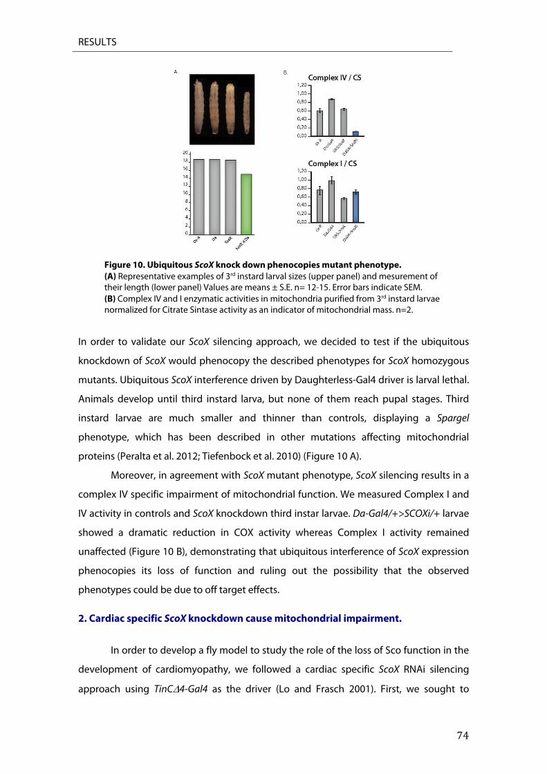
RESULTS
74
Figure 10. Ubiquitous ScoX knock down phenocopies mutant phenotype. (A) Representative examples of 3rd instard larval sizes (upper panel) and mesurement of their length (lower panel) Values are means ± S.E. n= 12-15. Error bars indicate SEM. (B) Complex IV and I enzymatic activities in mitochondria purified from 3rd instard larvae normalized for Citrate Sintase activity as an indicator of mitochondrial mass. n=2.
In order to validate our ScoX silencing approach, we decided to test if the ubiquitous
knockdown of ScoX would phenocopy the described phenotypes for ScoX homozygous
mutants. Ubiquitous ScoX interference driven by Daughterless-Gal4 driver is larval lethal.
Animals develop until third instard larva, but none of them reach pupal stages. Third
instard larvae are much smaller and thinner than controls, displaying a Spargel
phenotype, which has been described in other mutations affecting mitochondrial
proteins (Peralta et al. 2012; Tiefenbock et al. 2010) (Figure 10 A).
Moreover, in agreement with ScoX mutant phenotype, ScoX silencing results in a
complex IV specific impairment of mitochondrial function. We measured Complex I and
IV activity in controls and ScoX knockdown third instar larvae. Da-Gal4/+>SCOXi/+ larvae
showed a dramatic reduction in COX activity whereas Complex I activity remained
unaffected (Figure 10 B), demonstrating that ubiquitous interference of ScoX expression
phenocopies its loss of function and ruling out the possibility that the observed
phenotypes could be due to off target effects.
2. Cardiac specific ScoX knockdown cause mitochondrial impairment.
In order to develop a fly model to study the role of the loss of Sco function in the
development of cardiomyopathy, we followed a cardiac specific ScoX RNAi silencing
approach using TinC4-Gal4 as the driver (Lo and Frasch 2001). First, we sought to

RESULTS
75
determine if heart specific ScoX knockdown compromises Drosophila lifespan. TinC4-
Gal4>ScoXi flies show shortened lifespan as compared to controls (Figure 11). Since ScoX
interfered flies start to die after 2 weeks, all experiments were carried out in 1 or 2 week
old flies.
Figure 11. Survival curve. Cardiac specific ScoX knockdown affects the life span of the flies. Graph plots % survival (n=200 for each genotype).
2.1. Mitochondrial complexes activity staining in ScoX knockdown hearts
Next, we measured ScoX interference strength by assessing mRNA levels in fly
hearts by qRT-PCR and, given the small size of this organ in Drosophila, performing
histochemical staining of heart complex IV activity. Hearts from one week old TinC4-
Gal4>ScoXi flies showed a 50% reduction in ScoX mRNA levels as compared to controls,
while complex IV activity histochemical staining of hearts from 1-week old control and
knockdown flies demonstrated a clear deficiency in COX activity in the later (Figure 12 A
and B).
Figure 12. Cardiac specific ScoX knockdown cause a mitochondrial impairment. (A) ScoX mRNA expression levels in TinCΔ4::G4, UAS-ScoXi and TinCΔ4-G4>ScoXi hearts normalized to RPL0 and relative to TinCΔ4::G4. Values are means ± S.E. n= 6-10. *p < 0.05. (B) Enzyme histochemical staining for COX and SDH activities from TinCΔ4::G4, UAS-ScoXi and TinCΔ4>ScoXi hearts in absence or presence of their respective inhibitors KCN (COX) and malonate (CII).

RESULTS
76
The observed decrease in COX activity was not due to a general respiratory chain
mal function since we observed no differences in succinate dehydrogenase (SDH)
(Figure 12 B), or complex I (data not shown) activity staining between controls and ScoX
interfered hearts. These results indicate that a decrease of just 50% of ScoX mRNA levels
is enough to compromise COX activity in the Drosophila heart, and that this defect is
specific for Complex IV.
2.2. Characterization of metabolic state in Scox knockdown cardiomyocytes
It is well described that mitochondrial dysfunction leads to an alteration in
energy metabolism in many forms of heart disease (Masoud et al. 2013; Nascimben et al.
2004). We therefore hypothesized that COX deficiency displayed by TinC4-Gal4>ScoXi
hearts would alter oxidative phosphorylation resulting in a compensatory up-regulation
of glycolysis and partial blockage of the Krebs cycle. To address this supposition we
measured by qRT-PCR the mRNA relative levels of key enzymes involved in different
steps of these process. As show in figure 13, mRNA expression levels of gpi, pfk and pgk1
were upregulated in TinC4-Gal4>ScoXi hearts (Figure 13).
Figure 13. Cardiac specific ScoX knockdown cause a metabolic switch. mRNA expression levels of PDK, GPI, PFK, PGK1 and IMPL3 mRNA in hearts from 2-week old TinCΔ4::G4, UAS-ScoXi and TinCΔ4G4>ScoXi flies normalized to RPL0 and relative to TinCΔ4::G4. Values are means ± S.E. n= 6-10. ***p < 0.001.
RESULTS
77
Furthermore, in these hearts we observed an increased expression of impl3, the
Drosophila ortholog of human lactate dehydrogenase (LDH) (10.1038/ng.2610), and the
mitochondrial matrix enzyme pdk (Figure 13). LDH enzyme converts pyruvate to lactate,
the final product of the non-respiratory consumption of glucose while PDK is a key
regulator of glucose oxidation. It phosphorylates and inhibits pyruvate dehydrogenase
(PDH), therefore blocking the kreb´s cycle (Piao et al. 2010; Sugden et al. 2000).
Our data strongly suggest that TinC4-Gal4>ScoXi cardiomyocytes undergo a
metabolic switch from glucose oxidation to glycolysis. Since the pyruvate cannot be
used by mitochondrial PDH (inhibited by PDK high levels), the concomitant increase in
impl3 mRNA relative levels led us to hypothesize that, in order to maintain the glycolytic
flux, pyruvate is mostly converted to lactate resulting in lactic acidosis. Our data suggest
that heart specific interference of ScoX results in a mitochondrial dysfunction due to the
alteration of oxidative phosphorylation and promotes upregulation of glycolysis.
3. ScoX RNAi knockdown causes dilated cardiomyopathy in Drosophila melanogaster
3.1. Analysis of heart structure
To explore the impact of the mitochondrial dysfunction caused by ScoX silencing
in the fly heart, we analyzed the structure of controls and TinC4-Gal4>ScoXi hearts from
1 and 2 week old flies by confocal microscopy. Although overall heart structure seemed
to be fairly normal, ScoX RNAi animals exhibited myofibrillar disarray and narrowing of
the heart tube from abdominal segment 4. This degeneration of the heart tube became
more noticeable with age, with 2 week old flies having a much stronger phenotype
(Figure 14 A). Intriguingly, TinC4-Gal4>ScoXi heart tubes had an apparent increase in
conical chamber diameter as compared to controls. We measured the diameter of the
conical chamber from 1-week old TinC4-Gal4>ScoXi and controls hearts. As shown in
figure 14 B, knocked down hearts had increased conical chamber diameter, suggesting
that in Drosophila heart specific ScoX interference causes dilated cardiomyopathy.
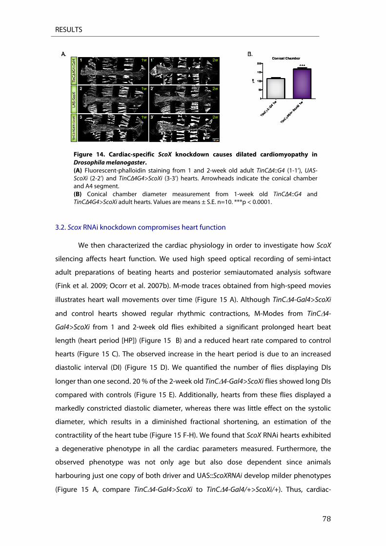
RESULTS
78
Figure 14. Cardiac-specific ScoX knockdown causes dilated cardiomyopathy in Drosophila melanogaster. (A) Fluorescent-phalloidin staining from 1 and 2-week old adult TinCΔ4::G4 (1-1’), UAS-ScoXi (2-2’) and TinCΔ4G4>ScoXi (3-3’) hearts. Arrowheads indicate the conical chamber and A4 segment. (B) Conical chamber diameter measurement from 1-week old TinCΔ4::G4 and TinCΔ4G4>ScoXi adult hearts. Values are means ± S.E. n=10. ***p < 0.0001.
3.2. Scox RNAi knockdown compromises heart function
We then characterized the cardiac physiology in order to investigate how ScoX
silencing affects heart function. We used high speed optical recording of semi-intact
adult preparations of beating hearts and posterior semiautomated analysis software
(Fink et al. 2009; Ocorr et al. 2007b). M-mode traces obtained from high-speed movies
illustrates heart wall movements over time (Figure 15 A). Although TinC4-Gal4>ScoXi
and control hearts showed regular rhythmic contractions, M-Modes from TinC4-
Gal4>ScoXi from 1 and 2-week old flies exhibited a significant prolonged heart beat
length (heart period [HP]) (Figure 15 B) and a reduced heart rate compared to control
hearts (Figure 15 C). The observed increase in the heart period is due to an increased
diastolic interval (DI) (Figure 15 D). We quantified the number of flies displaying DIs
longer than one second. 20 % of the 2-week old TinC4-Gal4>ScoXi flies showed long DIs
compared with controls (Figure 15 E). Additionally, hearts from these flies displayed a
markedly constricted diastolic diameter, whereas there was little effect on the systolic
diameter, which results in a diminished fractional shortening, an estimation of the
contractility of the heart tube (Figure 15 F-H). We found that ScoX RNAi hearts exhibited
a degenerative phenotype in all the cardiac parameters measured. Furthermore, the
observed phenotype was not only age but also dose dependent since animals
harbouring just one copy of both driver and UAS::ScoXRNAi develop milder phenotypes
(Figure 15 A, compare TinC4-Gal4>ScoXi to TinC4-Gal4/+>ScoXi/+). Thus, cardiac-

RESULTS
79
specific knockdown of ScoX results in severe cardiac dysfunction and dilated
cardiomyopathy.
Figure 15. Cardiac-specific ScoX knockdown causes heart dysfunction in Drosophila melanogaster. (A) Representative M-Mode traces (10s) from high-speed movies of 1 and 2 weeks old semi-intact flies. (B-H) Physiological parameter measurement in hearts from 1 and 2 weeks old semi-intact flies. (B) Heart Period, (C) Heart Rate, (D) Diastolic Interval, (E) % of Long diastoles, (F) Diastolic Diameter, (G) Systolic Diameter, (H) Fractional Shortening. Differences are relative to TinCΔ4::Gal4. Values are means ± S.E. n= 20-40 flies. *=p<0.05, **=p<0.01 and ***=p<0.001).
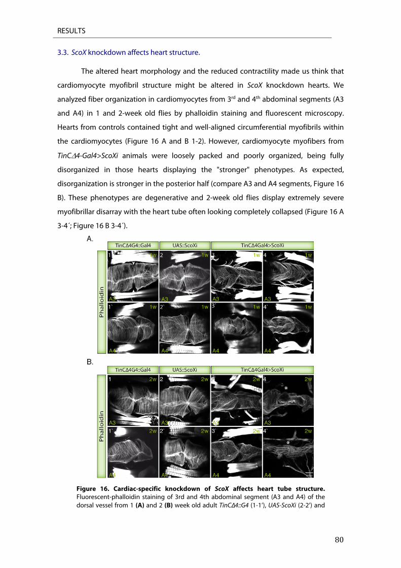
RESULTS
80
3.3. ScoX knockdown affects heart structure.
The altered heart morphology and the reduced contractility made us think that
cardiomyocyte myofibril structure might be altered in ScoX knockdown hearts. We
analyzed fiber organization in cardiomyocytes from 3rd and 4th abdominal segments (A3
and A4) in 1 and 2-week old flies by phalloidin staining and fluorescent microscopy.
Hearts from controls contained tight and well-aligned circumferential myofibrils within
the cardiomyocytes (Figure 16 A and B 1-2). However, cardiomyocyte myofibers from
TinC4-Gal4>ScoXi animals were loosely packed and poorly organized, being fully
disorganized in those hearts displaying the "stronger" phenotypes. As expected,
disorganization is stronger in the posterior half (compare A3 and A4 segments, Figure 16
B). These phenotypes are degenerative and 2-week old flies display extremely severe
myofibrillar disarray with the heart tube often looking completely collapsed (Figure 16 A
3-4´; Figure 16 B 3-4´).
Figure 16. Cardiac-specific knockdown of ScoX affects heart tube structure. Fluorescent-phalloidin staining of 3rd and 4th abdominal segment (A3 and A4) of the dorsal vessel from 1 (A) and 2 (B) week old adult TinCΔ4::G4 (1-1’), UAS-ScoXi (2-2’) and
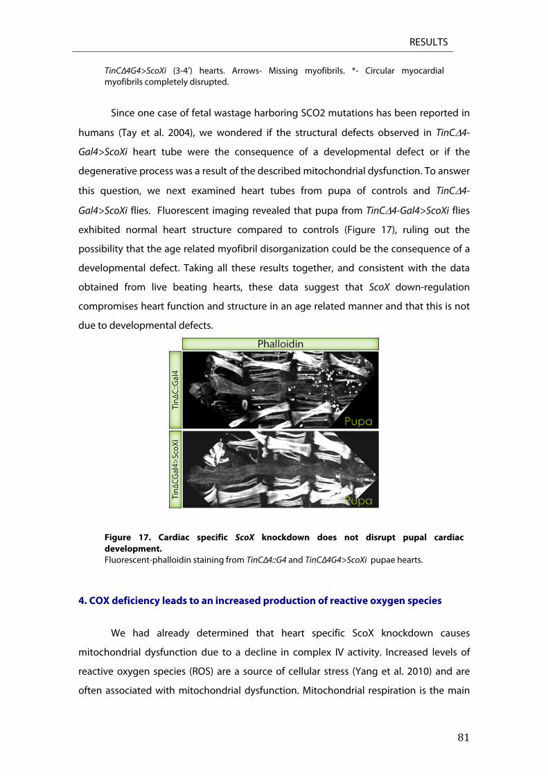
RESULTS
81
TinCΔ4G4>ScoXi (3-4’) hearts. Arrows- Missing myofibrils. *- Circular myocardial myofibrils completely disrupted.
Since one case of fetal wastage harboring SCO2 mutations has been reported in
humans (Tay et al. 2004), we wondered if the structural defects observed in TinC4-
Gal4>ScoXi heart tube were the consequence of a developmental defect or if the
degenerative process was a result of the described mitochondrial dysfunction. To answer
this question, we next examined heart tubes from pupa of controls and TinC4-
Gal4>ScoXi flies. Fluorescent imaging revealed that pupa from TinC4-Gal4>ScoXi flies
exhibited normal heart structure compared to controls (Figure 17), ruling out the
possibility that the age related myofibril disorganization could be the consequence of a
developmental defect. Taking all these results together, and consistent with the data
obtained from live beating hearts, these data suggest that ScoX down-regulation
compromises heart function and structure in an age related manner and that this is not
due to developmental defects.
Figure 17. Cardiac specific ScoX knockdown does not disrupt pupal cardiac development. Fluorescent-phalloidin staining from TinCΔ4::G4 and TinCΔ4G4>ScoXi pupae hearts.
4. COX deficiency leads to an increased production of reactive oxygen species
We had already determined that heart specific ScoX knockdown causes
mitochondrial dysfunction due to a decline in complex IV activity. Increased levels of
reactive oxygen species (ROS) are a source of cellular stress (Yang et al. 2010) and are
often associated with mitochondrial dysfunction. Mitochondrial respiration is the main

RESULTS
82
source of ROS in all eukaryotic cells, with two major superoxide producing sites: complex
I and III of the ETC (St-Pierre et al. 2002). In order to determine whether reduced COX
activity leads to an increased ROS production, we analyzed ROS levels in hearts from
control and from 1- and 2-week-old TinC4-Gal4>ScoXi flies using dihydroethidium
(DHE) as an indicator (Figure 18). Hearts from TinC4-Gal4>ScoXi displayed significantly
increased ROS levels compared with control at both tested ages. Note that 2-week-old
ScoX down-regulated cardiomyocytes displayed higher levels of DHE staining than did 1-
week-old flies.
Figure 18. Cardiac-specific ScoX knockdown induces oxidative stress. DHE staining in hearts from TinCΔ4::G4 and TinCΔ4G4>ScoXi 1and 2-week-old flies. Cardiomyocyte nuclei are encircled in white.
5. ScoX cardiomyopathy is p53-dependent
p53 is a tumor suppressor with a central role in cancer development, apoptosis,
senescence and differentiation (Vogelstein et al. 2000). Furthermore, a number of studies
have implicated p53 in different types of cardiomyopathy (Birks et al. 2008; Nakamura et
al. 2012). p53 also has been shown to have an important role in cellular stress responses
regulating metabolic pathways like glycolysis (Mathupala et al. 2001; Ruiz-Lozano et al.
1999) and OXPHOS (Matoba et al. 2006). Interestingly, they found that in stress response,
p53 directly regulates aerobic respiration through the modulation of Sco2. dp53, the
Drosophila homolog of p53, shares structural and functional properties with mammalian
p53 (Jin et al. 2000; Ollmann et al. 2000). Given that p53 regulates stress response
through the modulation of Sco2 expression and since it is associated to the development
of various cardiomyopathies, we speculated that dp53 might also have a role in D.
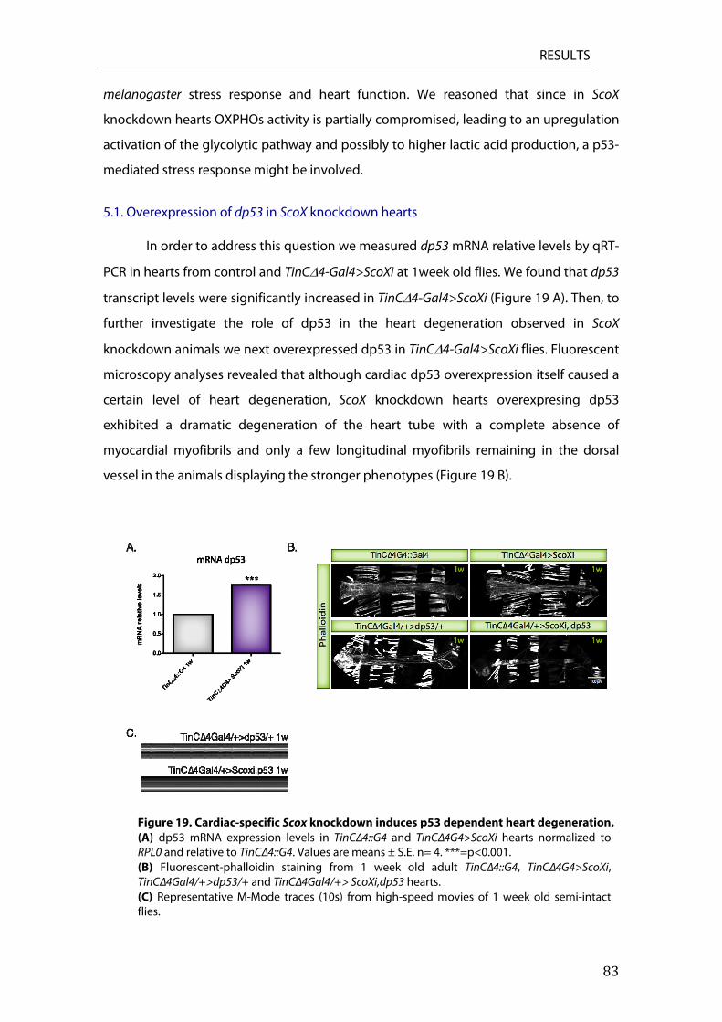
RESULTS
83
melanogaster stress response and heart function. We reasoned that since in ScoX
knockdown hearts OXPHOs activity is partially compromised, leading to an upregulation
activation of the glycolytic pathway and possibly to higher lactic acid production, a p53-
mediated stress response might be involved.
5.1. Overexpression of dp53 in ScoX knockdown hearts
In order to address this question we measured dp53 mRNA relative levels by qRT-
PCR in hearts from control and TinC4-Gal4>ScoXi at 1week old flies. We found that dp53
transcript levels were significantly increased in TinC4-Gal4>ScoXi (Figure 19 A). Then, to
further investigate the role of dp53 in the heart degeneration observed in ScoX
knockdown animals we next overexpressed dp53 in TinC4-Gal4>ScoXi flies. Fluorescent
microscopy analyses revealed that although cardiac dp53 overexpression itself caused a
certain level of heart degeneration, ScoX knockdown hearts overexpresing dp53
exhibited a dramatic degeneration of the heart tube with a complete absence of
myocardial myofibrils and only a few longitudinal myofibrils remaining in the dorsal
vessel in the animals displaying the stronger phenotypes (Figure 19 B).
Figure 19. Cardiac-specific Scox knockdown induces p53 dependent heart degeneration. (A) dp53 mRNA expression levels in TinCΔ4::G4 and TinCΔ4G4>ScoXi hearts normalized to RPL0 and relative to TinCΔ4::G4. Values are means ± S.E. n= 4. ***=p<0.001. (B) Fluorescent-phalloidin staining from 1 week old adult TinCΔ4::G4, TinCΔ4G4>ScoXi, TinCΔ4Gal4/+>dp53/+ and TinCΔ4Gal4/+> ScoXi,dp53 hearts. (C) Representative M-Mode traces (10s) from high-speed movies of 1 week old semi-intact flies.
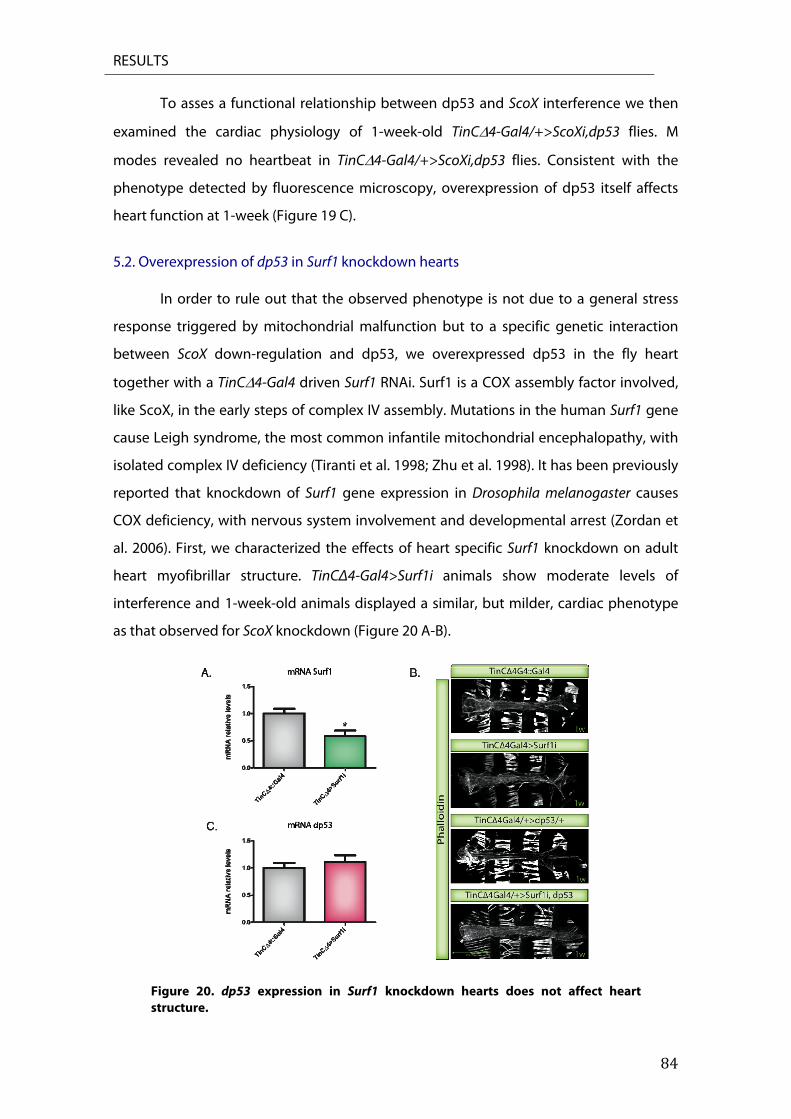
RESULTS
84
To asses a functional relationship between dp53 and ScoX interference we then
examined the cardiac physiology of 1-week-old TinC4-Gal4/+>ScoXi,dp53 flies. M
modes revealed no heartbeat in TinC4-Gal4/+>ScoXi,dp53 flies. Consistent with the
phenotype detected by fluorescence microscopy, overexpression of dp53 itself affects
heart function at 1-week (Figure 19 C).
5.2. Overexpression of dp53 in Surf1 knockdown hearts
In order to rule out that the observed phenotype is not due to a general stress
response triggered by mitochondrial malfunction but to a specific genetic interaction
between ScoX down-regulation and dp53, we overexpressed dp53 in the fly heart
together with a TinC4-Gal4 driven Surf1 RNAi. Surf1 is a COX assembly factor involved,
like ScoX, in the early steps of complex IV assembly. Mutations in the human Surf1 gene
cause Leigh syndrome, the most common infantile mitochondrial encephalopathy, with
isolated complex IV deficiency (Tiranti et al. 1998; Zhu et al. 1998). It has been previously
reported that knockdown of Surf1 gene expression in Drosophila melanogaster causes
COX deficiency, with nervous system involvement and developmental arrest (Zordan et
al. 2006). First, we characterized the effects of heart specific Surf1 knockdown on adult
heart myofibrillar structure. TinCΔ4-Gal4>Surf1i animals show moderate levels of
interference and 1-week-old animals displayed a similar, but milder, cardiac phenotype
as that observed for ScoX knockdown (Figure 20 A-B).
Figure 20. dp53 expression in Surf1 knockdown hearts does not affect heart structure.
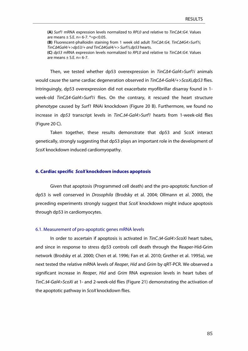
RESULTS
85
(A) Surf1 mRNA expression levels normalized to RPL0 and relative to TinCΔ4::G4. Values are means ± S.E. n= 6-7. *=p<0.05. (B) Fluorescent-phalloidin staining from 1 week old adult TinCΔ4::G4, TinCΔ4G4>Surf1i, TinCΔ4Gal4/+>dp53/+ and TinCΔ4Gal4/+> Surf1i,dp53 hearts. (C) dp53 mRNA expression levels normalized to RPL0 and relative to TinCΔ4::G4. Values are means ± S.E. n= 6-7.
Then, we tested whether dp53 overexpression in TinCΔ4-Gal4>Surf1i animals
would cause the same cardiac degeneration observed in TinCΔ4-Gal4/+>ScoXi,dp53 flies.
Intringuingly, dp53 overexpression did not exacerbate myofibrillar disarray found in 1-
week-old TinCΔ4-Gal4>Surf1i flies. On the contrary, it rescued the heart structure
phenotype caused by Surf1 RNAi knockdown (Figure 20 B). Furthermore, we found no
increase in dp53 transcript levels in TinC4-Gal4>Surf1 hearts from 1-week-old flies
(Figure 20 C).
Taken together, these results demonstrate that dp53 and ScoX interact
genetically, strongly suggesting that dp53 plays an important role in the development of
ScoX knockdown induced cardiomyopathy.
6. Cardiac specific ScoX knockdown induces apoptosis
Given that apoptosis (Programmed cell death) and the pro-apoptotic function of
dp53 is well conserved in Drosophila (Brodsky et al. 2004; Ollmann et al. 2000), the
preceding experiments strongly suggest that ScoX knockdown might induce apoptosis
through dp53 in cardiomyocytes.
6.1. Measurement of pro-apoptotic genes mRNA levels
In order to ascertain if apoptosis is activated in TinC4-Gal4>ScoXi heart tubes,
and since in response to stress dp53 controls cell death through the Reaper-Hid-Grim
network (Brodsky et al. 2000; Chen et al. 1996; Fan et al. 2010; Grether et al. 1995a), we
next tested the relative mRNA levels of Reaper, Hid and Grim by qRT-PCR. We observed a
significant increase in Reaper, Hid and Grim RNA expression levels in heart tubes of
TinC4-Gal4>ScoXi at 1- and 2-week-old flies (Figure 21) demonstrating the activation of
the apoptotic pathway in ScoX knockdown flies.
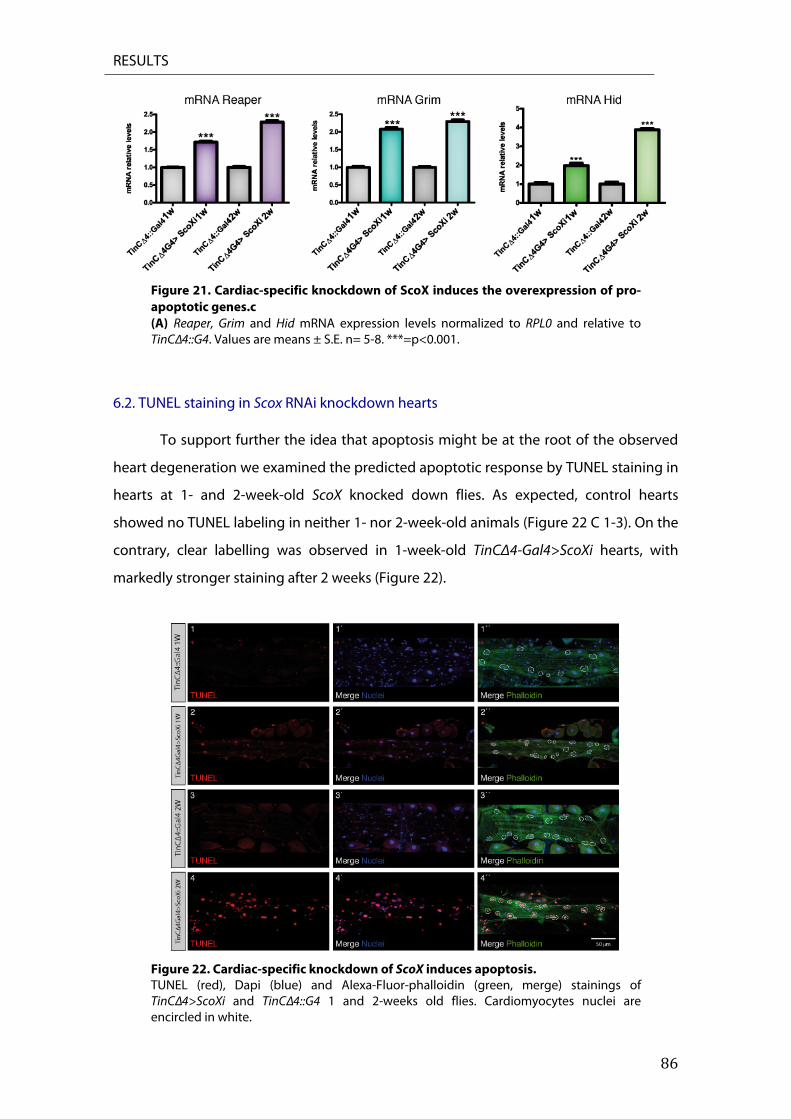
RESULTS
86
Figure 21. Cardiac-specific knockdown of ScoX induces the overexpression of pro-apoptotic genes.c (A) Reaper, Grim and Hid mRNA expression levels normalized to RPL0 and relative to TinCΔ4::G4. Values are means ± S.E. n= 5-8. ***=p<0.001.
6.2. TUNEL staining in Scox RNAi knockdown hearts
To support further the idea that apoptosis might be at the root of the observed
heart degeneration we examined the predicted apoptotic response by TUNEL staining in
hearts at 1- and 2-week-old ScoX knocked down flies. As expected, control hearts
showed no TUNEL labeling in neither 1- nor 2-week-old animals (Figure 22 C 1-3). On the
contrary, clear labelling was observed in 1-week-old TinCΔ4-Gal4>ScoXi hearts, with
markedly stronger staining after 2 weeks (Figure 22).
Figure 22. Cardiac-specific knockdown of ScoX induces apoptosis. TUNEL (red), Dapi (blue) and Alexa-Fluor-phalloidin (green, merge) stainings of TinCΔ4>ScoXi and TinCΔ4::G4 1 and 2-weeks old flies. Cardiomyocytes nuclei are encircled in white.
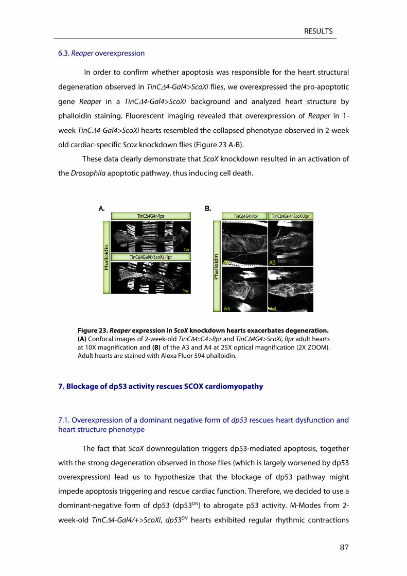
RESULTS
87
6.3. Reaper overexpression
In order to confirm whether apoptosis was responsible for the heart structural
degeneration observed in TinC4-Gal4>ScoXi flies, we overexpressed the pro-apoptotic
gene Reaper in a TinC4-Gal4>ScoXi background and analyzed heart structure by
phalloidin staining. Fluorescent imaging revealed that overexpression of Reaper in 1-
week TinC4-Gal4>ScoXi hearts resembled the collapsed phenotype observed in 2-week
old cardiac-specific Scox knockdown flies (Figure 23 A-B).
These data clearly demonstrate that ScoX knockdown resulted in an activation of
the Drosophila apoptotic pathway, thus inducing cell death.
Figure 23. Reaper expression in ScoX knockdown hearts exacerbates degeneration. (A) Confocal images of 2-week-old TinCΔ4::G4>Rpr and TinCΔ4G4>ScoXi, Rpr adult hearts at 10X magnification and (B) of the A3 and A4 at 25X optical magnification (2X ZOOM). Adult hearts are stained with Alexa Fluor 594 phalloidin.
7. Blockage of dp53 activity rescues SCOX cardiomyopathy
7.1. Overexpression of a dominant negative form of dp53 rescues heart dysfunction and heart structure phenotype
The fact that ScoX downregulation triggers dp53-mediated apoptosis, together
with the strong degeneration observed in those flies (which is largely worsened by dp53
overexpression) lead us to hypothesize that the blockage of dp53 pathway might
impede apoptosis triggering and rescue cardiac function. Therefore, we decided to use a
dominant-negative form of dp53 (dp53DN) to abrogate p53 activity. M-Modes from 2-
week-old TinC4-Gal4/+>ScoXi, dp53DN hearts exhibited regular rhythmic contractions
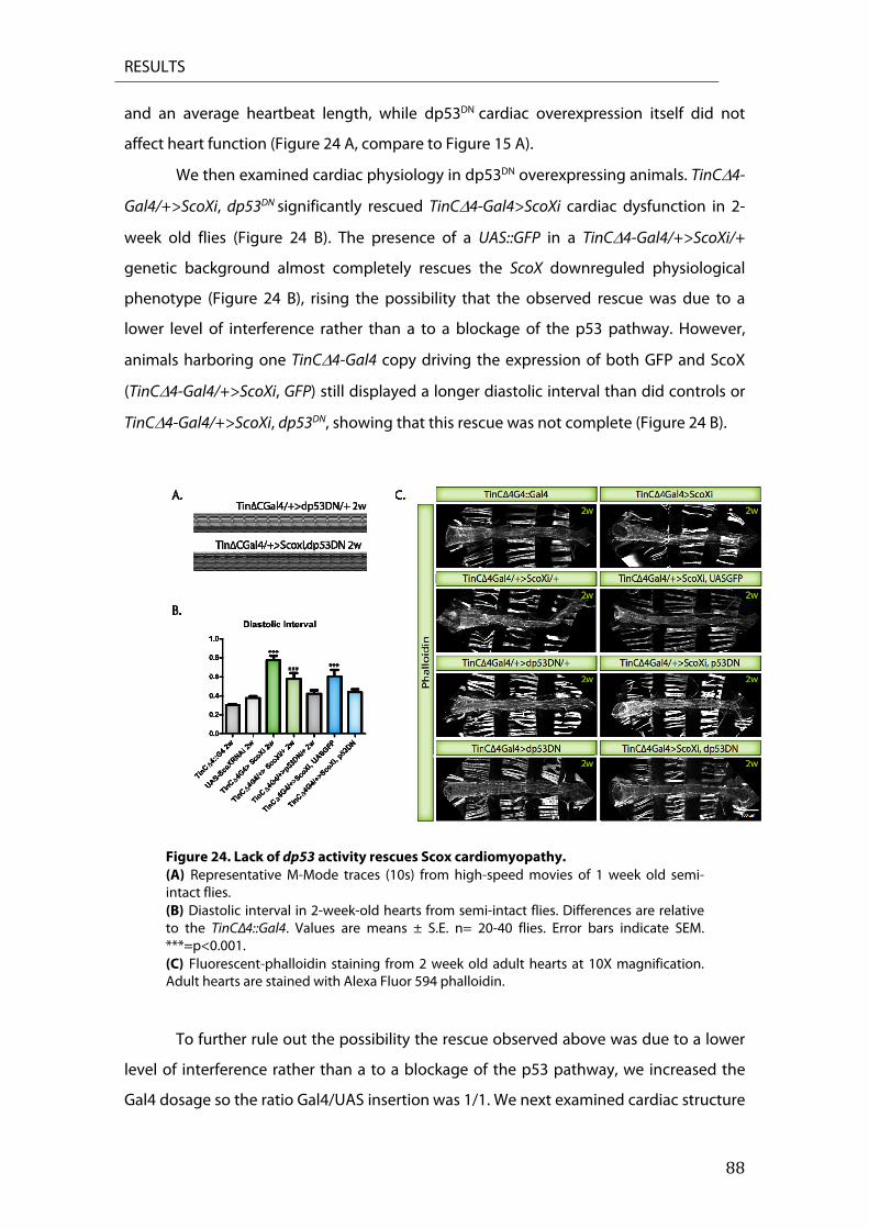
RESULTS
88
and an average heartbeat length, while dp53DN cardiac overexpression itself did not
affect heart function (Figure 24 A, compare to Figure 15 A).
We then examined cardiac physiology in dp53DN overexpressing animals. TinC4-
Gal4/+>ScoXi, dp53DN significantly rescued TinC4-Gal4>ScoXi cardiac dysfunction in 2-
week old flies (Figure 24 B). The presence of a UAS::GFP in a TinC4-Gal4/+>ScoXi/+
genetic background almost completely rescues the ScoX downreguled physiological
phenotype (Figure 24 B), rising the possibility that the observed rescue was due to a
lower level of interference rather than a to a blockage of the p53 pathway. However,
animals harboring one TinC4-Gal4 copy driving the expression of both GFP and ScoX
(TinC4-Gal4/+>ScoXi, GFP) still displayed a longer diastolic interval than did controls or
TinC4-Gal4/+>ScoXi, dp53DN, showing that this rescue was not complete (Figure 24 B).
Figure 24. Lack of dp53 activity rescues Scox cardiomyopathy. (A) Representative M-Mode traces (10s) from high-speed movies of 1 week old semi-intact flies. (B) Diastolic interval in 2-week-old hearts from semi-intact flies. Differences are relative to the TinCΔ4::Gal4. Values are means ± S.E. n= 20-40 flies. Error bars indicate SEM. ***=p<0.001. (C) Fluorescent-phalloidin staining from 2 week old adult hearts at 10X magnification. Adult hearts are stained with Alexa Fluor 594 phalloidin.
To further rule out the possibility the rescue observed above was due to a lower
level of interference rather than a to a blockage of the p53 pathway, we increased the
Gal4 dosage so the ratio Gal4/UAS insertion was 1/1. We next examined cardiac structure
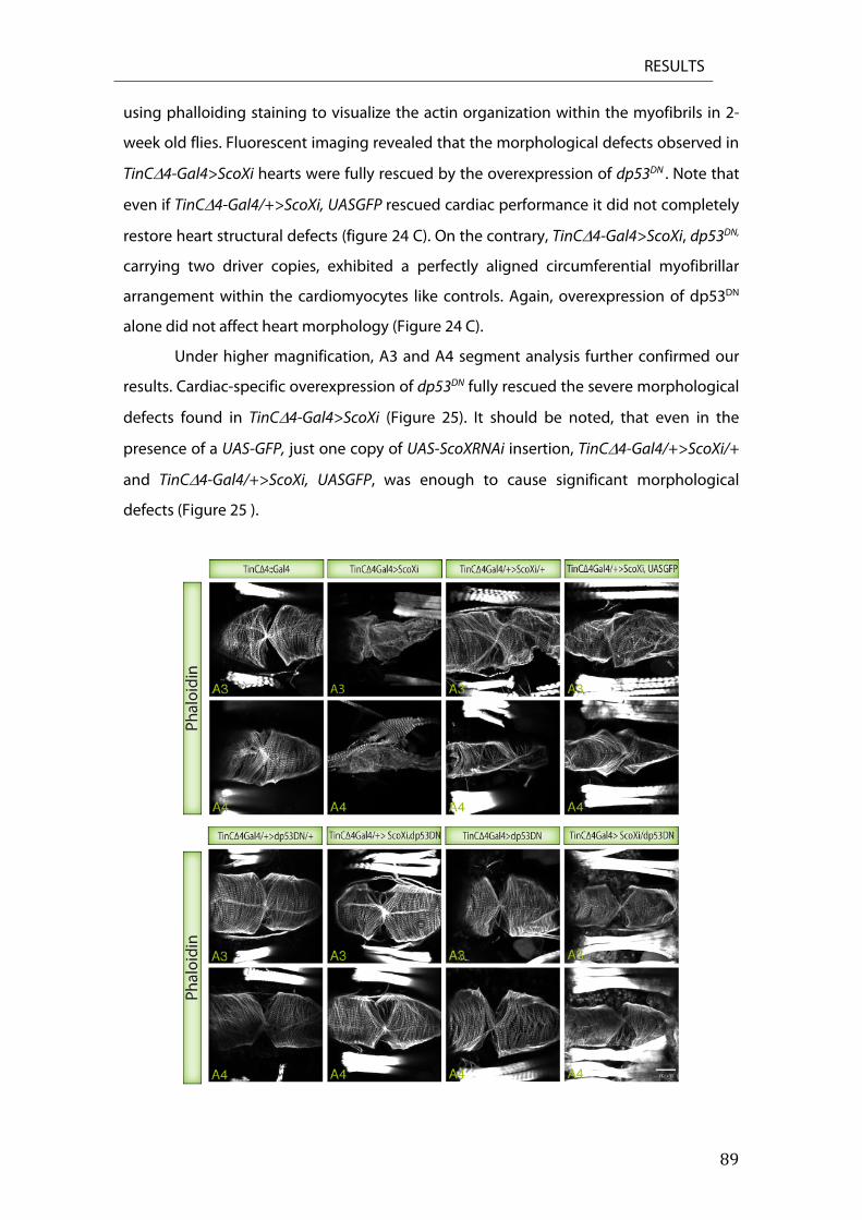
RESULTS
89
using phalloiding staining to visualize the actin organization within the myofibrils in 2-
week old flies. Fluorescent imaging revealed that the morphological defects observed in
TinC4-Gal4>ScoXi hearts were fully rescued by the overexpression of dp53DN . Note that
even if TinC4-Gal4/+>ScoXi, UASGFP rescued cardiac performance it did not completely
restore heart structural defects (figure 24 C). On the contrary, TinC4-Gal4>ScoXi, dp53DN,
carrying two driver copies, exhibited a perfectly aligned circumferential myofibrillar
arrangement within the cardiomyocytes like controls. Again, overexpression of dp53DN
alone did not affect heart morphology (Figure 24 C).
Under higher magnification, A3 and A4 segment analysis further confirmed our
results. Cardiac-specific overexpression of dp53DN fully rescued the severe morphological
defects found in TinC4-Gal4>ScoXi (Figure 25). It should be noted, that even in the
presence of a UAS-GFP, just one copy of UAS-ScoXRNAi insertion, TinC4-Gal4/+>ScoXi/+
and TinC4-Gal4/+>ScoXi, UASGFP, was enough to cause significant morphological
defects (Figure 25 ).
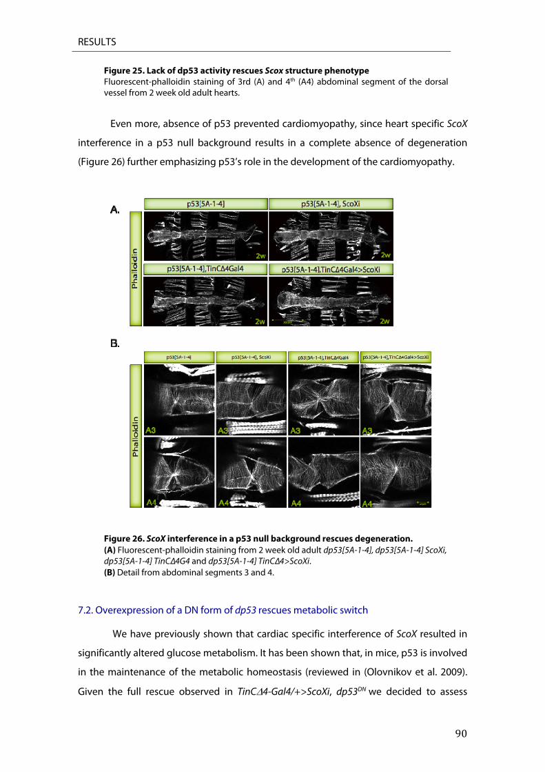
RESULTS
90
Figure 25. Lack of dp53 activity rescues Scox structure phenotype Fluorescent-phalloidin staining of 3rd (A) and 4th (A4) abdominal segment of the dorsal vessel from 2 week old adult hearts.
Even more, absence of p53 prevented cardiomyopathy, since heart specific ScoX
interference in a p53 null background results in a complete absence of degeneration
(Figure 26) further emphasizing p53’s role in the development of the cardiomyopathy.
Figure 26. ScoX interference in a p53 null background rescues degeneration. (A) Fluorescent-phalloidin staining from 2 week old adult dp53[5A-1-4], dp53[5A-1-4] ScoXi, dp53[5A-1-4] TinCΔ4G4 and dp53[5A-1-4] TinCΔ4>ScoXi. (B) Detail from abdominal segments 3 and 4.
7.2. Overexpression of a DN form of dp53 rescues metabolic switch We have previously shown that cardiac specific interference of ScoX resulted in
significantly altered glucose metabolism. It has been shown that, in mice, p53 is involved
in the maintenance of the metabolic homeostasis (reviewed in (Olovnikov et al. 2009).
Given the full rescue observed in TinC4-Gal4/+>ScoXi, dp53DN we decided to assess
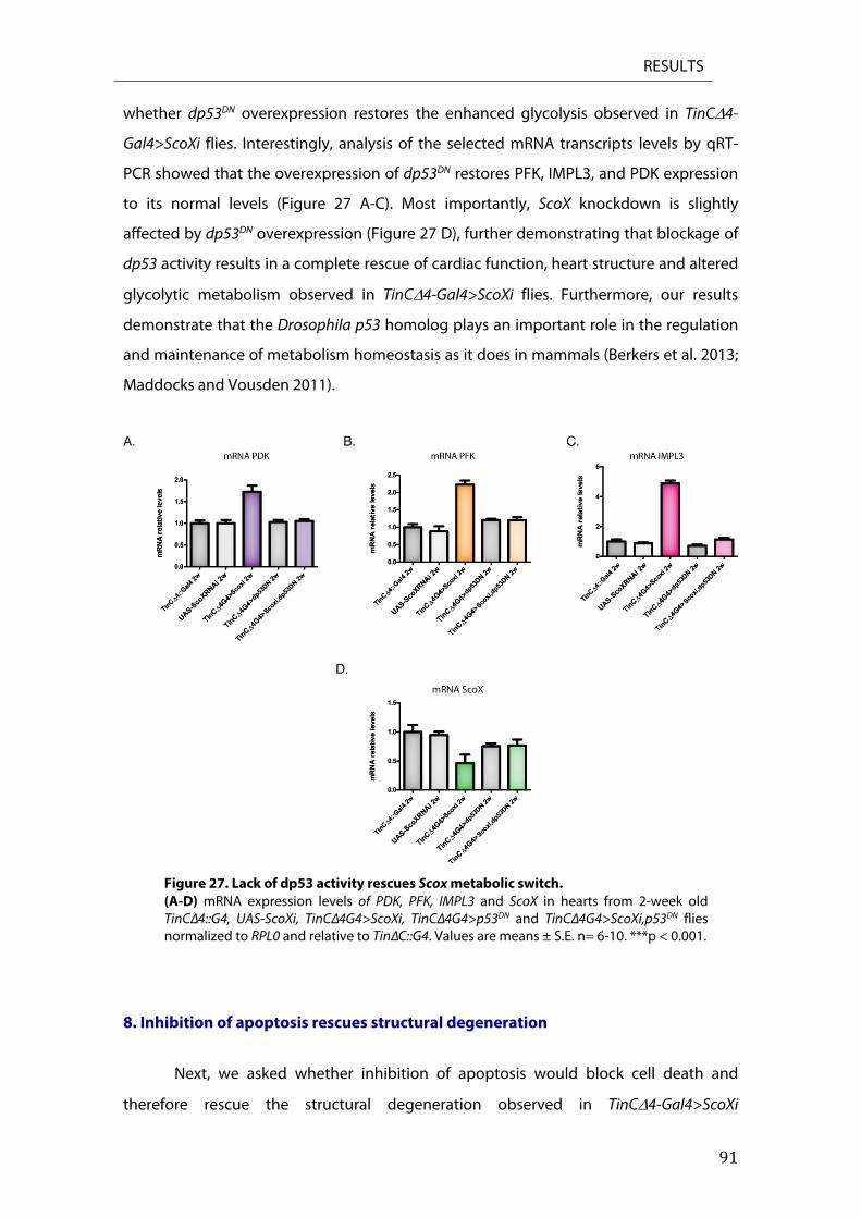
RESULTS
91
whether dp53DN overexpression restores the enhanced glycolysis observed in TinC4-
Gal4>ScoXi flies. Interestingly, analysis of the selected mRNA transcripts levels by qRT-
PCR showed that the overexpression of dp53DN restores PFK, IMPL3, and PDK expression
to its normal levels (Figure 27 A-C). Most importantly, ScoX knockdown is slightly
affected by dp53DN overexpression (Figure 27 D), further demonstrating that blockage of
dp53 activity results in a complete rescue of cardiac function, heart structure and altered
glycolytic metabolism observed in TinC4-Gal4>ScoXi flies. Furthermore, our results
demonstrate that the Drosophila p53 homolog plays an important role in the regulation
and maintenance of metabolism homeostasis as it does in mammals (Berkers et al. 2013;
Maddocks and Vousden 2011).
Figure 27. Lack of dp53 activity rescues Scox metabolic switch. (A-D) mRNA expression levels of PDK, PFK, IMPL3 and ScoX in hearts from 2-week old TinCΔ4::G4, UAS-ScoXi, TinCΔ4G4>ScoXi, TinCΔ4G4>p53DN and TinCΔ4G4>ScoXi,p53DN flies normalized to RPL0 and relative to TinΔC::G4. Values are means ± S.E. n= 6-10. ***p < 0.001.
8. Inhibition of apoptosis rescues structural degeneration
Next, we asked whether inhibition of apoptosis would block cell death and
therefore rescue the structural degeneration observed in TinC4-Gal4>ScoXi
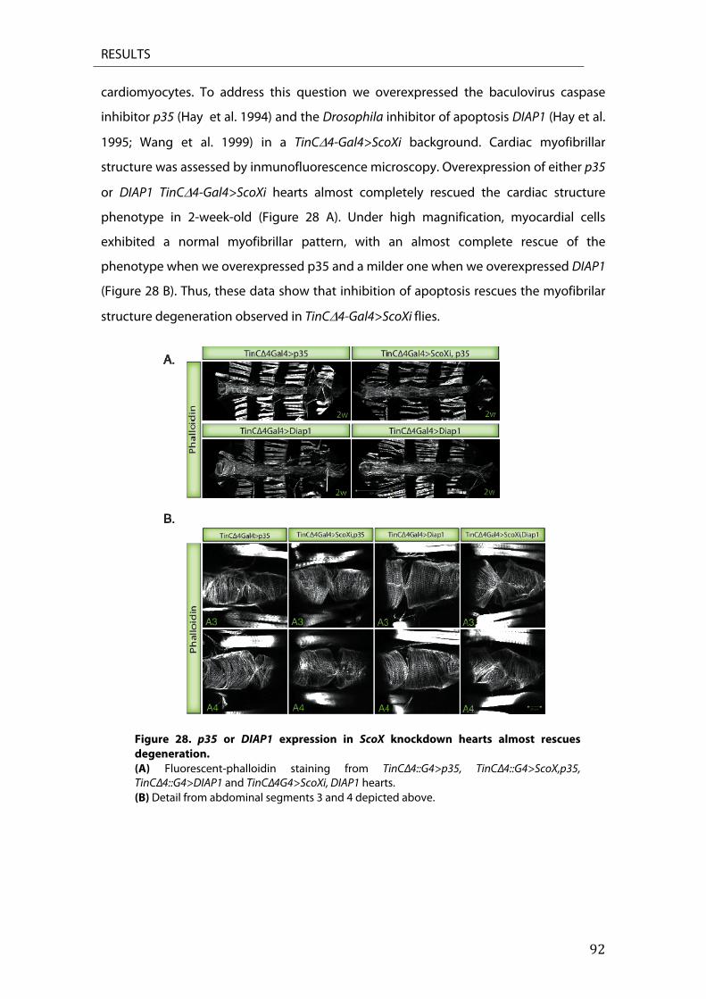
RESULTS
92
cardiomyocytes. To address this question we overexpressed the baculovirus caspase
inhibitor p35 (Hay et al. 1994) and the Drosophila inhibitor of apoptosis DIAP1 (Hay et al.
1995; Wang et al. 1999) in a TinC4-Gal4>ScoXi background. Cardiac myofibrillar
structure was assessed by inmunofluorescence microscopy. Overexpression of either p35
or DIAP1 TinC4-Gal4>ScoXi hearts almost completely rescued the cardiac structure
phenotype in 2-week-old (Figure 28 A). Under high magnification, myocardial cells
exhibited a normal myofibrillar pattern, with an almost complete rescue of the
phenotype when we overexpressed p35 and a milder one when we overexpressed DIAP1
(Figure 28 B). Thus, these data show that inhibition of apoptosis rescues the myofibrilar
structure degeneration observed in TinC4-Gal4>ScoXi flies.
Figure 28. p35 or DIAP1 expression in ScoX knockdown hearts almost rescues degeneration. (A) Fluorescent-phalloidin staining from TinCΔ4::G4>p35, TinCΔ4::G4>ScoX,p35, TinCΔ4::G4>DIAP1 and TinCΔ4G4>ScoXi, DIAP1 hearts. (B) Detail from abdominal segments 3 and 4 depicted above.

RESULTS
93
9. Sco2KIKO mice undergoes apoptosis
Thus, if partial loss of ScoX in Drosophila leads to apoptosis as a final consequence,
and since its function is conserved in mammals, we wondered whether our results in
Drosophila could be extended to mammalian system. Although unfortunately there are
no Sco1 KO mice available, a Sco2KIKO human disease mouse model has been recently
developed (Yang et al. 2010). Sco2KIKO mice harbor both a Sco2 knock-out allele and the
Sco2 knock-in E129K allele. The mice E129K allele corresponds to the E140K mutation
found in almost all human patients (Mobley et al. 2009; Yang et al. 2010).
Consistent with what it has been observed in patients with SCO2 deficiency,
Sco2KIKO mice model showed motor impairments and biochemical and functional defects.
In contrast to human, the reduction in COX activity in Sco2KIKO mice was less severe in
muscle than in other tissues, such as liver, and was accompanied by an unexpected
defect in complex III activity (Yang et al. 2010). Although Sco2KIKO mice do not present all
the features of the human disease, they are viable and fertile, which makes it an excellent
model for the search of pharmacological therapies. When Sco2KIKO mice were treated with
AICAR, an AMPK agonist, it leads to a partial rescue of COX activity with motor
improvement (Viscomi et al. 2011). Hence, we decided to use Sco2KIKO mice to validate
our Drosophila data in a mammalian model system. Sco2KIKO liver and skeletal muscle, but
not tissue from wt animals, had massive apoptosis as shown by TUNEL staining (Figure
29). Furthermore, Sco2KIKO liver seemed to have more apoptotic cells than did muscle, in
concordance with the lowest complex IV activity value observed in Sco2KIKO liver
compared with other examined tissues (Yang et al. 2010). We concluded that, as in
Drosophila, partial loss of Sco2 in mice function induces apoptosis, further validating our
findings.
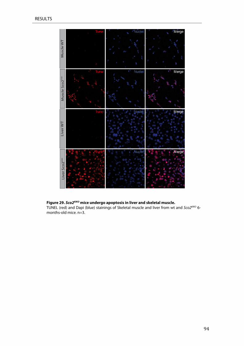
RESULTS
94
Figure 29. Sco2KIKO mice undergo apoptosis in liver and skeletal muscle. TUNEL (red) and Dapi (blue) stainings of Skeletal muscle and liver from wt and Sco2KIKO 6-months-old mice. n=3.

RESULTS
95
DISCUSSION

96
DISCUSSION
97
Mitochondrial diseases due to defects in cytochrome c oxidase (COX) are one of the most
frequent respiratory chain disorders in humans. Patients displaying these pathologies
present extreme heterogeneity of clinical manifestations with an onset varying from
neonatal to adult life, and multiple organ involvement such as brain, skeletal muscle and
heart. This heterogeneity makes considerably difficult the understanding of the
pathophysiology of these diseases. Pathogenic mutations in human SCO1 and SCO2 have
been reported to cause hypertrophic cardiomyopathy among other clinical symptoms
(Leary et al. 2013; Papadopoulou et al. 1999). However, the molecular mechanisms
underlying this cardiac dysfunction have yet to be elucidated.
Here, we present the first cardiac-specific animal model to study human Sco1/2
mediated cardiomyopathy. Cardiac-specific knockdown of ScoX results in severe dilated
cardiomyopathy due to mitochondrial dysfunction with a concomitant metabolic switch
from glucose oxidation to glycolysis and increased ROS levels, leading to p53 dependent
cell death as fatal outcome. Moreover, partial loss of Sco2 function induces apoptosis in
liver and skeletal muscle in a Sco2KIKO mouse model. We also report a new role for dp53 in
homeostasis regulation and maintenance of metabolism, a function conserved in
mammals.

DISCUSSION
98
1. Drosophila melanogaster ScoX
ScoX is a conserved complex IV assembly factor presenting homology with the
human Sco1 and Sco2 genes (Porcelli et al. 2010). Ubiquitious ScoX knockdown in
Drosophila is lethal in third instard larva, displaying a spargel phenotype and a severe
COX deficiency thus phenocoping its loss of function (Figure 10) (Porcelli et al. 2010).
Furthermore, this reduction in COX activity is in concordance to that observed in
patients displaying mutations in either Sco1 or Sco2 genes (Jaksch et al. 2001; Leary et al.
2013).
Although the role of Sco proteins as copper metallochaperones is largely
documented, they have been shown to be involved in the maintenance of cellular
copper homeostasis, presumably by controlling cellular copper export (Leary et al. 2007;
Leary et al. 2009a), the molecular mechanism underlying the cardiac dysfunction caused
by mutations in human Sco1 and Sco2, have yet to be elucidated (Leary et al. 2013;
Papadopoulou et al. 1999). In the present study, we got advantage of the genetic
versatility of the Drosophila model to investigate ScoX loss of function in the heart. A
cardiac-specific knockdown of ScoX by 50% at the RNA level was sufficient to cause a
deficiency in COX activity suggesting that the knockdown might be more efficient at the
protein activity level (Figure 12).
In order to maintain the glycolytic flux and compensate for the fall in ATP
production caused by COX deficiency, ScoX-knockdown hearts displayed a
compensatory upregulation of glycolysis, partial blockage of the Krebs cycle, and
increased Impl3 expression (the ortholog of the mammalian LDH), most probably
resulting in lactic acidosis (Figure 13). Interestingly, previous studies in patients and rat
models, have shown that mitochondrial dysfunction is associated with abnormalities in
cardiac function and changes in energy metabolism, resulting in glycolysis optimization
and lactic acidosis (Nascimben et al. 2004) reviewed in (Honzik et al. 2012; Schiff et al.
2011).
DISCUSSION
99
2. ScoX knockdown induces dilated cardiomyopathy in Drosophila.
Since all Sco2 patients die from cardiac insufficiency, we assessed cardiac function
in Scox knockdown flies using functional imaging. This experimental approach allows us
to analyze the physiological impact of ScoX interference in heart function and disease. In
contrast to what was observed in the mouse Sco2KO/KI model, where no evidence of
cardyomiopathy was described (Yang et al. 2010), cardiac-specific ScoX knockdown
resulted in a severe impairment of cardiac function. Flies developed dilated
cardiomyopathy resembling cardiomyopathies caused by mitochondrial fusion defects
in flies (Dorn et al. 2011). ScoX knockdown also caused a significant reduction of FS as
well as prolonged DIs (Figure 15) accompanied by an age-associated cardiac myofibril
disorganization (Figure 16). Furthermore, these cardiac defects are likely caused by a
degenerative process most likely due to an adult-onset mitochondrial dysfunction rather
than to a developmental defect, since pupae heart structure remained unaffected
(Figure 17).
3. COX deficiency in fly heart results in ROS production
The electron transport chain (ETC), within the mitochondrial inner membrane is
the main site of ROS production in the cell (reviewed in ( reviewed Koopman et al. 2010).
Many neurodegenerative diseases and the aging process has been link to mitochondrial
dysfunction that triggers excessive oxidative stress production, mostly due to an
impairment of the ETC (reviewed in M. T. Lin and Beal 2006). Previous studies in human
cells have shown that Sco2 -/- cells display an increased ROS formation with oxidative
DNA damage (Sung et al. 2010).
We found that cardiac-specific knockdown of ScoX induces oxidative stress
production (Figure 18). However, we cannot distinguish whether this increase in free
radical accumulation arises from the mitochondria, due to a loss of COX function, or if it
comes from non-mitochondrial sources as a consequence of the loss of cellular
homeostatic control, as it has been shown in yeast (Leadsham et al. 2013) or in a neuro-
specific COX-deficient Alzheimer disease mouse model (Fukui et al. 2007).

DISCUSSION
100
4. dp53 is involved in cardiomyopathy development
The p53 tumor suppressor, is a well described transcription factor known for its
participation in cancer development, DNA repair, cell cycle, autophagy, necrosis and
apoptosis and senescence (reviewed in Berkers et al. 2013; Vousden and Prives 2009).
Beyond these functions, increasing evidences of new roles for p53 have been reported,
including its ability to regulate both glycolyisis and OXPHOS, through the modulation of
Sco2 (Bensaad et al. 2006; Mathupala et al. 2001; Matoba et al. 2006; Ruiz-Lozano et al.
1999). Furthermore, the contribution of p53 in cardiovascular disorders has been
demonstrated (Birks et al. 2008; Nakamura et al. 2012; Sano et al. 2007; Shimizu et al.
2012).
In this scenario and since p53 activation in response to stress signals, like
increased oxidative stress or latic acidosis, is well documented (Johnson et al. 1996;
Olovnikov et al. 2009; Zhuang et al. 2012), we hypothesized that dp53 might play an
important role in the cellular response to ScoX loss of function. We found that p53
expression was upregulated in cardiac-specific ScoX knockdown hearts (Figure 18A).
Moreover, the overexpression of dp53 in TinC4-Gal4>ScoXi flies dramatically affects
heart structure and physiology, as demonstrated by the lack of heart beat in TinC4-
Gal4/+>ScoXi,dp53 flies (Figure 19 B-C). Interestingly, we observed that dp53
overexpression in a cardiac-specific Surf1 knockdown animals, displying a heart
phenotype similar to that observed in ScoX interfered flies, rescued the heart structure
phenotype (Figure 20), further stressing the specificity of dp53 involvement in the
development ScoX induced cardiomyopathy.
5. dp53 contributes in the development of ScoX mediated cardiomyopathy
The strong degeneration observed in TinC4-Gal4>ScoXi flies together with the
almost complete disappearance of the fly heart when p53 is overexpressed in these flies,
strongly suggests that the apoptotic pathway might be activated in TinC4-Gal4>ScoXi
hearts.
Interestingly, it has been recently reported that Sco2 overexpression induces
p53-mediated apoptosis in tumor xenografts and cancer cells (Madan et al. 2013).
DISCUSSION
101
Furthermore, in a previous report, it was shown that Sco2 knockdown in p53 +/+ glioma
cells, but not in those negative for it, sensitivized them to hypoxia-induced apoptosis in a
p53 dependent manner and induced necrosis in tumors expressing wild-type p53
(Wanka et al. 2012), further liking the Sco2/p53 axe to cell death.
In Drosophila, heart specific ScoX knockdown induces dp53 mediated cell death
in cardiomyocytes and it does so through the activation of the canonical apoptotic
pathway since dp53 target genes Reaper, Hid and Grim are upregulated (Figure 20-21).
Moreover, Reaper over-expression in adult heart enhances the structural defects
observed in TinCΔ4-Gal4>ScoXi flies (Figure 23). Consistent with the results in the fly
heart, we found that skeletal muscle and liver from Sco2KIKO mice undergoes massive cell
death as well (Figure 29). These data suggest a conserved response to Sco2 loss of
function mediated stress.
We provide evidence that TinC4-Gal4>ScoXi hearts exhibit a partial loss of COX
activity. Therefore, we hypothesize mitochondrial dysfunction not to be the only reason
of the profound cardiomyopathy and phenotype observed. Moreover, in contrast to our
results, prior evidence from vertebrate and invertebrate models has shown that a partial
inhibition of mitochondrial respiration, promotes longevity and metabolic health due to
a hormetic response (Lee et al. 2010; Liu et al. 2005; Rea et al. 2007). In fact, it has been
recently shown that a mild interference in OXPHOS system activity in Drosophila IFMs
preserves mitochondrial function, improves muscle performance, and increases life span
through the activation of the mitochondrial unfolded pathway response and IGF/like
signalling pathways (Owusu-Ansah et al. 2013).
Based upon these data, we speculate that cell death, rather than mitochondrial
dysfunction itself, is likely to be the main reason for the profound heart degeneration
detected in TinCΔ4-Gal4>ScoXi flies. Expression of a dp53DN in ScoX knocked-down hearts
rescues cardiac dysfunction and degeneration and TinC4-Gal4>ScoXi, dp53 null animals
display no apparent heart defects. Thus, we can attribute the observed rescue to a
blockage of the p53 pathway (Figure 24-25).
Moreover, to further rule out that the observed rescue was due to a competition
for the GAL4 among the two UAS insertions, we generated a TinC4-Gal4>ScoXi, dp53DN
DISCUSSION
102
line carrying two driver copies. Here, we find that the overexpression of dp53DN restore
the morphological defects observed in TinC4-Gal4>ScoXi hearts. Thus, we can attribute
the observed rescue to a blockage of the p53 pathway (Figure 23-24). Interestingly, in
vitro silencing of p53 in cardiomyocytes prevented the mitochondrial damage and
cardiomyocyte necrosis linked to monoamine oxidase- A (MAO-A) activation, further
validating our findings (Villeneuve et al. 2013)
In accordance, inhibition of apoptosis by p35 or Diap1 over-expression resulted in
an almost complete rescue of the ScoX knockdown morphological phenotype. Thus,
even if inhibition of apoptosis can significantly prevent cardiac dysfunction significantly,
it is not a full rescue (Figure 28). The inability of p35 and Diap1 to completely rescue the
morphological phenotype suggests that in addition to apoptosis induction, dp53 plays a
key role in cardyomyopathy development. Nevertheless, it remains to be determined if
the activation of dp53, and therefore cell death, is a side effect of COX dysfunction and
loss of cellular homeostasis or if the partial lack of ScoX itself triggers dp53 upregulation
and apoptosis.
Inhibition of complex IV by a morpholino antisense oligomer against Surf1 and
CoxVa in fish causes COX deficiency, organ development defect, activation of
programmed cell death and cardiovascular dysfunction (Baden et al. 2007). Interestingly,
apoptosis was just found in head region, neural tube and trunk but not in the heart of
COX-deficient fish, even though they display a strong cardiac dysfuntion. In contrast to
zebrafish, Surf1 KO mice despite showing reduced COX levels and increased blood
lactate levels, displayed increased longevity (Dell'agnello et al. 2007), enhanced
neuroprotection and cognitive function even though presenting increased ROS
generation and glucose metabolism (A. L. Lin et al. 2013). Since loss of Surf1 in humans
causes Leigh Syndrome (Tiranti et al. 1998; Zhu et al. 1998), these studies in the mice
support the idea that Surf1 might have additional roles, like its participation in Ca2+
homeostasis (Dell'agnello et al. 2007).
In this sense, it is tempting to hypothesize that ScoX might have another role
independent from its function as a COX assembly factor, perhaps as redox sensor (J. C.
Williams et al. 2005), and may act in conjunction with dp53 to accomplish this role. In this
context, is worth mentioning that it has been recently shown that Sco2 interference in
DISCUSSION
103
mammalian cells induces the relocalization of p53 from mitochondria to the nucleus
(Zhuang et al. 2013). On the other hand, it may be also possible that the threshold of
COX deficiency tolerable by the heart might be lower as compared with other tissues,
reflecting a tissue-dependent phenomenon or a tissue-dependent role of ScoX. More
work is necessary to test this hypothesis and try to understand how the partial lack of
ScoX induces cell death through dp53.
5.1 dp53 plays a role in the maintenance of metabolic homeostasis.
We found that ScoX knockdown in the heart of Drosophila leads to a
mitochondrial dysfunction, hence resulting in mitochondrial respiration deficiency and
the promotion glycolysis as major ATP source (Figure 13). Most probably, as suggested
by increased Impl3 expression, pyruvate formed during glycolysis will be transformed to
lactate resulting in lactic acidosis.
Since p53 has the ability to participate in different and apparently opposing
groups of responses regarding metabolic homeostasis, we examined whether blockage
of dp53 would have some impact in the altered glycolytic metabolism observed in heart-
specific ScoX knockdown flies. Loss of p53 compromises cytochrome c oxidase function
and supports a switch to glycolytic metabolism in both cultured cells and mouse models
(Matoba et al. 2006). Here, we found the opposing response, where the overexpression
of dp53DN restore glycolytic metabolism, rescuing the metabolic phenotype caused by
the loss of ScoX (Figure 26). Based on current evidence, we propose a new role for dp53
in the maintenance of metabolic homeostasis in Drosophila.
5.2. Connecting mitochondria and dp53 to cell death.
Mitochondria play multiple roles in apoptosis in many organisms (Kroemer and
Reed 2000). Although the apoptotic machinery in Drosophila appears to be superficially
similar to that of mammals, the role of mitochondria in Drosophila apoptosis is less well
understood. As it have been mentioned, in mammalian systems, cyt c is released on
MOMP. However, the role of cyt c in Drosophila apoptosis remains controversial since cyt
c is not released in drug- or UV-induced apoptosis in fly cells (Dorstyn et al. 2004; Means
et al. 2006; Zimmermann et al. 2002). On the other hand, in vivo data support a role for
DISCUSSION
104
cyt c in caspase activation during spermatogenesis and death of inter ommatidial cells
(Arama et al. 2003; Arama et al. 2006).
Although the role of mitochondria in Drosophila apoptosis is still unclear there
are some clear evidences suggesting that mitochondria play an important role in flies
cell death as it does in mammals. In fact, it is known that Rpr, Hid and Grim localization in
the mitochondria is essential to promote cell death (Claveria et al. 2002; Haining et al.
1999; M. R. Olson et al. 2003). Hid has a mitochondrial targeting sequence localized at its
C-terminus (Haining et al. 1999) and it seems to be responsible for Reaper recruitment to
mitochondria (M. R. Olson et al. 2003). The proapoptotic GH3 domain of Reaper, which is
also found in Grim (Claveria et al. 2002) is required for mitochondrial localization and
promotion of apoptosis. Moreover, it has been reported that expression of Reaper and
Hid induce cyt c release into the cytosol of S2 cells and that under apoptotic stimulus, fly
mitochondria undergoes Rpr, Hid and Drp1-dependent morphological changes and
disruption during cell death (Abdelwahid et al. 2007). The participation of Drp1, a
mitochondrial fission protein, in cell death shows a conserved role for mitochondria
between flies and mammals which is also conserved in worms (Frank et al. 2001; Jagasia
et al. 2005).
It is well established that in mammalian systems, in response to cellular stress,
p53 protein translocates to mitochondria and triggers MOMP (Marchenko 2000; Mihara
et al. 2003; Sansome et al. 2001). This, leads to the release of mitochondrial death factors
like cyt c, AIF, SMAC/Diablo, and Omi/HTRA2 with the consequent activation of caspases
(Danial and Korsmeyer 2004). In contrast to what it has been observed in mammals,
release of mitochondrial factors does not appear to play a role in apoptosis in Drosophila
S2 culture cells (Means et al. 2006). Therefore, MOMP is likely to be a consequence rather
than a cause of caspase activation in Drosophila (Abdelwahid et al. 2007).
It has recently been proposed that p53 plays an essential role in mitochondrial
permeability transition pore (mPTP) opening to induce necrotic cell death (Vaseva et al.
2012). Vaseva and collaborators propose that upon ROS stimulation, p53 translocates to
the mitochondrial matrix where it binds cyclophilin D (CypD) to induce mPTP opening
independent of proapoptotic Bcl-2 family members Bax and Bak. Furthermore, in
contrast to the traditional view, where mPTP opening is triggered by Ca2+ overload
accompanied by ROS generation and ATP depletion authors showed mPTP opening is
independent of Ca2+ (reviewed in (Siemen and Ziemer 2013).

DISCUSSION
105
Apoptotic and regulated necrotic pathways have a number of common steps and
regulatory factors among which is mPTP opening, which is thought to act through
mitochondrial swelling and posterior delivery of necrotic factors (Nikoletopoulou et al.
2013). Drosophila mPTP has been recently characterized and, in contrast to what it has
been described in mammals, mPTP activation it is not accompanied by mitochondrial
swelling (von Stockum et al. 2011).
Thus, we hypothesize that dp53 induces mPTP opening to trigger cell death in
cardiac-specific ScoX knockdown flies. In the absence of mitochondrial swelling, mPTP
opening would result in apoptosis instead of necrosis as it has been shown in mammals.
Moreover, apart from some evidence that suggests necrosis may play an important role
in nurse cells during late oogenesis, no other necrotic events have been described in
Drosophila so far (Jenkins et al. 2013).
Furthermore, even though Drosophila mPTP has been shown to be cyclosprin A
(CsA)-insensitive in vitro (von Stockum et al. 2011), it has been recently shown that
collagen XV/XVIII mutants, displaying mitochondrial dysfunction with severely
attenuated ATP and enhanced ROS production, are improved by CsA administration
(Momota et al. 2013). Interestingly, mice lacking collagenVI display mitochondrial
structural alterations and spontaneous apoptosis. These defects are caused by mPTP
opening and are normalized in vivo by CsA treatment (Irwin et al. 2003).
Future studies would be needed to get more insights into the relationship
between dp53 and ScoX and into the mechanism that activates dp53 to trigger cell
death. Our results may help to increase our understanding of Sco1 and Sco2-based
cardiomyopathy and may represent a new target for therapeutic agents.
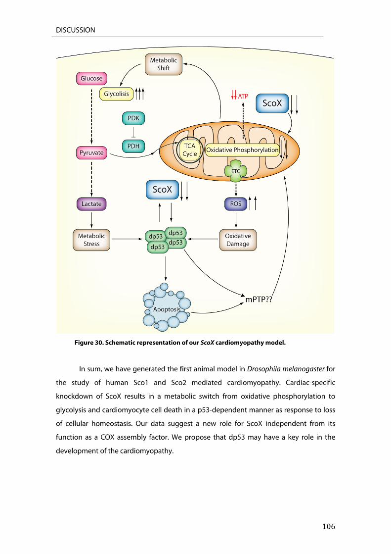
DISCUSSION
106
Figure 30. Schematic representation of our ScoX cardiomyopathy model.
In sum, we have generated the first animal model in Drosophila melanogaster for
the study of human Sco1 and Sco2 mediated cardiomyopathy. Cardiac-specific
knockdown of ScoX results in a metabolic switch from oxidative phosphorylation to
glycolysis and cardiomyocyte cell death in a p53-dependent manner as response to loss
of cellular homeostasis. Our data suggest a new role for ScoX independent from its
function as a COX assembly factor. We propose that dp53 may have a key role in the
development of the cardiomyopathy.

107
CONCLUSIONES

108
CONCLUSIONES
109
1. La interferencia del factor de ensamblaje ScoX de Drosophila melanogaster en el
corazón, provoca una disfunción mitocondrial acompañada de un cambio metabólico
que favorece la glicolisis frente a la fosforilación oxidativa.
2. La interferencia de ScoX en el corazón de Drosophila causa una severa cardiomiopatía
dilatada, además de afectar gravemente a la estructura de las miofibrillas que conforman
los cardiomiocitos. Tanto la afección cardiaca como la degeneración observada son
dependientes del tiempo.
3. Los defectos estructurales observados en los corazones de los adultos no son debidos
a problemas durante el desarrollo.
4. La interferencia de ScoX provoca un incremento de las especies reactivas de oxígeno.
5. dp53 y ScoX interactúan genéticamente.
6. dp53 ejerce un papel esencial en el desarrollo de la cardiomiopatía.
7. La interferencia de ScoX provoca la activación de la apoptosis en los cardiomiocitos.
8. La degeneración estructural que se observa en el corazón de Drosophila es debida a la
activación de la apoptosis y no a la disfunción mitocondrial que causa la interferencia de
ScoX.
9. La falta de función de Sco2 en el ratón Sco2KIKO , provoca la activación de la apoptosis
tanto en el hígado como en el músculo esquelético.
10. La proteína SCOX, además de ser un factor de ensamblaje del complejo IV, podría
desempeñar una función adicional en la célula.

110

111
REFERENCES

112
REFERENCES
113
10.1038/ng.2610, DOI: (2013), 'Identification of heart rate‐associated loci and their effects on cardiac conduction and rhythm disorders', Nat Genet, 45 (6), 621‐31.
Abdelwahid, E., et al. (2007), 'Mitochondrial disruption in Drosophila apoptosis', Dev Cell, 12 (5), 793‐806.
Achanta, G., et al. (2005), 'Novel role of p53 in maintaining mitochondrial genetic stability through interaction with DNA Pol gamma', EMBO J, 24 (19), 3482‐92.
Alayari, N. N., et al. (2009), 'Fluorescent labeling of Drosophila heart structures', J Vis Exp, (32).
Antonicka, H., et al. (2003a), 'Mutations in COX15 produce a defect in the mitochondrial heme biosynthetic pathway, causing early‐onset fatal hypertrophic cardiomyopathy', Am J Hum Genet, 72 (1), 101‐14.
Antonicka, H., et al. (2003b), 'Mutations in COX10 result in a defect in mitochondrial heme A biosynthesis and account for multiple, early‐onset clinical phenotypes associated with isolated COX deficiency', Hum Mol Genet, 12 (20), 2693‐702.
Arama, E., Agapite, J., and Steller, H. (2003), 'Caspase activity and a specific cytochrome C are required for sperm differentiation in Drosophila', Dev Cell, 4 (5), 687‐97.
Arama, E., et al. (2006), 'The two Drosophila cytochrome C proteins can function in both respiration and caspase activation', EMBO J, 25 (1), 232‐43.
Baden, K. N., et al. (2007), 'Early developmental pathology due to cytochrome c oxidase deficiency is revealed by a new zebrafish model', J Biol Chem, 282 (48), 34839‐49.
Banci, L., et al. (2008), 'Mitochondrial copper(I) transfer from Cox17 to Sco1 is coupled to electron transfer', Proc Natl Acad Sci U S A, 105 (19), 6803‐8.
Barros, M. H. and Tzagoloff, A. (2002), 'Regulation of the heme A biosynthetic pathway in Saccharomyces cerevisiae', FEBS Lett, 516 (1‐3), 119‐23.
Barros, M. H., Nobrega, F. G., and Tzagoloff, A. (2002), 'Mitochondrial ferredoxin is required for heme A synthesis in Saccharomyces cerevisiae', J Biol Chem, 277 (12), 9997‐10002.
Barros, M. H., et al. (2001), 'Involvement of mitochondrial ferredoxin and Cox15p in hydroxylation of heme O', FEBS Lett, 492 (1‐2), 133‐8.
Bensaad, K., et al. (2006), 'TIGAR, a p53‐inducible regulator of glycolysis and apoptosis', Cell, 126 (1), 107‐20.
Bereiter‐Hahn, J. and Voth, M. (1994), 'Dynamics of mitochondria in living cells: shape changes, dislocations, fusion, and fission of mitochondria', Microsc Res Tech, 27 (3), 198‐219.
Berkers, C. R., et al. (2013), 'Metabolic regulation by p53 family members', Cell Metab, 18 (5), 617‐33.
Birks, E. J., et al. (2008), 'Elevated p53 expression is associated with dysregulation of the ubiquitin‐proteasome system in dilated cardiomyopathy', Cardiovasc Res, 79 (3), 472‐80.
Birse, R. T., et al. (2010), 'High‐fat‐diet‐induced obesity and heart dysfunction are regulated by the TOR pathway in Drosophila', Cell Metab, 12 (5), 533‐44.
Bodmer, R. (1993), 'The gene tinman is required for specification of the heart and visceral muscles in Drosophila', Development, 118.

REFERENCES
114
‐‐‐ (1995), 'Heart development in Drosophila and its relationship to vertebrates', Trends Cardiovasc Med, 5 (1), 21‐8.
‐‐‐ (2006), 'Development of the Cardiac Musculature', Landes Bioscience. Bodmer, R. and Venkatesh, T. V. (1998), 'Heart development in Drosophila and
vertebrates: conservation of molecular mechanisms', Dev Genet, 22 (3), 181‐6.
Bourdon, A., et al. (2007), 'Mutation of RRM2B, encoding p53‐controlled ribonucleotide reductase (p53R2), causes severe mitochondrial DNA depletion', Nat Genet, 39 (6), 776‐80.
Brady, C. A. and Attardi, L. D. (2010), 'p53 at a glance', J Cell Sci, 123 (Pt 15), 2527‐32.
Brand, A. H. and Perrimon, N. (1993a), 'Targeted gene expression as a means of altering cell fates and generating dominant phenotypes', Development, 118 (2), 401‐15.
Brand, A. H. and Perrimon, N. (1993b), 'Targeted gene expression as a means of altering cell fates and generating dominant phenotypes', Development, 401‐15.
Brodsky, M. H., et al. (2000), 'Drosophila p53 Binds a Damage Response Element at the reaper Locus', Cell, 101 (1), 103‐13.
Brodsky, M. H., et al. (2004), 'Drosophila melanogaster MNK/Chk2 and p53 Regulate Multiple DNA Repair and Apoptotic Pathways following DNA Damage', Molecular and Cellular Biology, 24 (3), 1219‐31.
Brosel, S., et al. (2010), 'Unexpected vascular enrichment of SCO1 over SCO2 in mammalian tissues: implications for human mitochondrial disease', Am J Pathol, 177 (5), 2541‐8.
Bugiani, M., et al. (2005), 'Novel mutations in COX15 in a long surviving Leigh syndrome patient with cytochrome c oxidase deficiency', J Med Genet, 42 (5), e28.
Bundschuh, F. A., et al. (2009), 'Surf1, associated with Leigh syndrome in humans, is a heme‐binding protein in bacterial oxidase biogenesis', J Biol Chem, 284 (38), 25735‐41.
Campello, S. and Scorrano, L. (2010), 'Mitochondrial shape changes: orchestrating cell pathophysiology', EMBO Rep, 11 (9), 678‐84.
Chartier, A., et al. (2002), 'Pericardin, a Drosophila type IV collagen‐like protein is involved in the morphogenesis and maintenance of the heart epithelium during dorsal ectoderm closure', Development, 129.
Chen, P., et al. (1996), 'grim, a novel cell death gene in Drosophila', Genes & Development, 10 (14), 1773‐82.
Chen, P., et al. (1998), 'Dredd, a novel effector of the apoptosis activators reaper, grim, and hid in Drosophila', Dev Biol, 201 (2), 202‐16.
Claveria, C., et al. (2002), 'GH3, a novel proapoptotic domain in Drosophila Grim, promotes a mitochondrial death pathway', EMBO J, 21 (13), 3327‐36.
Cobine, P. A., et al. (2006), 'The P174L mutation in human Sco1 severely compromises Cox17‐dependent metallation but does not impair copper binding', J Biol Chem, 281 (18), 12270‐6.
Coenen, M. J., et al. (2004), 'Cytochrome c oxidase biogenesis in a patient with a mutation in COX10 gene', Ann Neurol, 56 (4), 560‐4.
Contractor, T. and Harris, C. R. (2012), 'p53 negatively regulates transcription of the pyruvate dehydrogenase kinase Pdk2', Cancer Res, 72 (2), 560‐7.
REFERENCES
115
Cripps, R. M. and Olson, E. N. (2002), 'Control of cardiac development by an evolutionarily conserved transcriptional network', Dev Biol, 246 (1), 14‐28.
Curtis, N. J., Ringo, J. M., and Dowse, H. B. (1999), 'Morphology of the pupal heart, adult heart, and associated tissues in the fruit fly, Drosophila melanogaster', J Morphol, 240 (3), 225‐35.
Danial, N. N. and Korsmeyer, S. J. (2004), 'Cell death: critical control points', Cell, 116 (2), 205‐19.
Degterev, A., Boyce, M., and Yuan, J. (2003), 'A decade of caspases', Oncogene, 22 (53), 8543‐67.
Dell'agnello, C., et al. (2007), 'Increased longevity and refractoriness to Ca(2+)‐dependent neurodegeneration in Surf1 knockout mice', Hum Mol Genet, 16 (4), 431‐44.
Diaz, F. and Moraes, C. T. (2008), 'Mitochondrial biogenesis and turnover', Cell Calcium, 44 (1), 24‐35.
Dickinson, E. K., et al. (2000), 'A human SCO2 mutation helps define the role of Sco1p in the cytochrome oxidase assembly pathway', J Biol Chem, 275 (35), 26780‐5.
Dorn, G. W., 2nd, et al. (2011), 'MARF and Opa1 control mitochondrial and cardiac function in Drosophila', Circ Res, 108 (1), 12‐7.
Dorstyn, L., et al. (2004), 'The two cytochrome c species, DC3 and DC4, are not required for caspase activation and apoptosis in Drosophila cells', J Cell Biol, 167 (3), 405‐10.
Dorstyn, L., et al. (1999), 'DRONC, an ecdysone‐inducible Drosophila caspase', Proc Natl Acad Sci U S A, 96 (8), 4307‐12.
Drose, S. and Brandt, U. (2012), 'Molecular mechanisms of superoxide production by the mitochondrial respiratory chain', Adv Exp Med Biol, 748, 145‐69.
Fan, Y., et al. (2010), 'Dual roles of Drosophila p53 in cell death and cell differentiation', Cell Death Differ, 17 (6), 912‐21.
Feng, Z. and Levine, A. J. (2010), 'The regulation of energy metabolism and the IGF‐1/mTOR pathways by the p53 protein', Trends Cell Biol, 20 (7), 427‐34.
Fernandez‐Silva, P., Enriquez, J. A., and Montoya, J. (2003), 'Replication and transcription of mammalian mitochondrial DNA', Exp Physiol, 88 (1), 41‐56.
Fink, M., et al. (2009), 'A new method for detection and quantification of heartbeat parameters in Drosophila, zebrafish, and embryonic mouse hearts', Biotechniques, 46 (2), 101‐13.
Frank, S., et al. (2001), 'The role of dynamin‐related protein 1, a mediator of mitochondrial fission, in apoptosis', Dev Cell, 1 (4), 515‐25.
Fukui, H., et al. (2007), 'Cytochrome c oxidase deficiency in neurons decreases both oxidative stress and amyloid formation in a mouse model of Alzheimer's disease', Proc Natl Acad Sci U S A, 104 (35), 14163‐8.
Ghezzi, D. and Zeviani, M. (2012), 'Assembly factors of human mitochondrial respiratory chain complexes: physiology and pathophysiology', Adv Exp Med Biol, 748, 65‐106.
Glerum, D. M., Shtanko, A., and Tzagoloff, A. (1996), 'SCO1 and SCO2 Act as High Copy Suppressors of a Mitochondrial Copper Recruitment Defect in Saccharomyces cerevisiae', J Biol Chem, 271 (August 23), 20531‐36.
Gloor, G. B., et al. (1993), 'Type I Repressors of P Element Mobility', Genetics. Green, D. R. and Kroemer, G. (2009), 'Cytoplasmic functions of the tumour
suppressor p53', Nature, 458 (7242), 1127‐30.

REFERENCES
116
Greenblatt, M. S., et al. (1994), 'Mutations in the p53 tumor suppressor gene: clues to cancer etiology and molecular pathogenesis', Cancer Res, 54 (18), 4855‐78.
Grether, M. E., et al. (1995a), 'The head involution defective gene of Drosophila melanogaster functions in programmed cell death', Genes & Development, 9 (14), 1694‐708.
‐‐‐ (1995b), 'The head involution defective gene of Drosophila melanogaster functions in programmed cell death', Genes Dev, 9 (14), 1694‐708.
Haining, W. N., et al. (1999), 'The proapoptotic function of Drosophila Hid is conserved in mammalian cells', Proc Natl Acad Sci U S A, 96 (9), 4936‐41.
Hardie, D. G. (2004), 'The AMP‐activated protein kinase pathway‐‐new players upstream and downstream', J Cell Sci, 117 (Pt 23), 5479‐87.
Harris, S. L. and Levine, A. J. (2005), 'The p53 pathway: positive and negative feedback loops', Oncogene, 24 (17), 2899‐908.
Harvey, R. P. (1996), 'NK‐2 homeobox genes and heart development', Dev Biol, 178 (2), 203‐16.
Hay, B. A. and Guo, M. (2006), 'Caspase‐dependent cell death in Drosophila', Annu Rev Cell Dev Biol, 22, 623‐50.
Hay , B. A., Wolff, T., and Rubin, G. M. (1994), 'Expression of baculovirus P35 prevents cell death in Drosophila', Development.
Hay, B. A., Wassarman, D. A., and Rubin, G. M. (1995), 'Drosophila homologs of baculovirus inhibitor of apoptosis proteins function to block cell death', Cell, 83 (7), 1253‐62.
Hoffman, W. H., et al. (2002), 'Transcriptional repression of the anti‐apoptotic survivin gene by wild type p53', J Biol Chem, 277 (5), 3247‐57.
Honzik, T., et al. (2012), 'Neonatal onset of mitochondrial disorders in 129 patients: clinical and laboratory characteristics and a new approach to diagnosis', J Inherit Metab Dis, 35 (5), 749‐59.
Horng, Y. C., et al. (2004), 'Specific copper transfer from the Cox17 metallochaperone to both Sco1 and Cox11 in the assembly of yeast cytochrome C oxidase', J Biol Chem, 279 (34), 35334‐40.
Horng, Y. C., et al. (2005), 'Human Sco1 and Sco2 function as copper‐binding proteins', J Biol Chem, 280 (40), 34113‐22.
Hu, W., et al. (2010), 'Glutaminase 2, a novel p53 target gene regulating energy metabolism and antioxidant function', Proc Natl Acad Sci U S A, 107 (16), 7455‐60.
Hu, W., et al. (2007), 'A single nucleotide polymorphism in the MDM2 gene disrupts the oscillation of p53 and MDM2 levels in cells', Cancer Res, 67 (6), 2757‐65.
Igaki, T. and Miura, M. (2004), 'Role of Bcl‐2 family members in invertebrates', Biochim Biophys Acta, 1644 (2‐3), 73‐81.
Igaki, T., et al. (2002), 'Down‐regulation of DIAP1 triggers a novel Drosophila cell death pathway mediated by Dark and DRONC', J Biol Chem, 277 (26), 23103‐6.
Inoki, K., Zhu, T., and Guan, K. L. (2003), 'TSC2 mediates cellular energy response to control cell growth and survival', Cell, 115 (5), 577‐90.
Irwin, W. A., et al. (2003), 'Mitochondrial dysfunction and apoptosis in myopathic mice with collagen VI deficiency', Nat Genet, 35 (4), 367‐71.
REFERENCES
117
Jackson, M. W. and Berberich, S. J. (2000), 'MdmX protects p53 from Mdm2‐mediated degradation', Mol Cell Biol, 20 (3), 1001‐7.
Jagasia, R., et al. (2005), 'DRP‐1‐mediated mitochondrial fragmentation during EGL‐1‐induced cell death in C. elegans', Nature, 433 (7027), 754‐60.
Jaksch, M., et al. (2000), 'Mutations in SCO2 are associated with a distinct form of hypertrophic cardiomyopathy and cytochrome c oxidase deficiency', Hum Mol Genet, 9 (5), 795‐801.
Jaksch, M., et al. (2001), 'Cytochrome c oxidase deficiency due to mutations in SCO2, encoding a mitochondrial copper‐binding protein, is rescued by copper in human myoblasts', Hum Mol Genet, 10 (26), 3025‐35.
Jenkins, V. K., Timmons, A. K., and McCall, K. (2013), 'Diversity of cell death pathways: insight from the fly ovary', Trends Cell Biol, 23 (11), 567‐74.
Jiang, P., et al. (2011), 'p53 regulates biosynthesis through direct inactivation of glucose‐6‐phosphate dehydrogenase', Nat Cell Biol, 13 (3), 310‐6.
Jin, S., et al. (2000), 'Identification and characterization of a p53 homologue in Drosophila melanogaster', Proc Natl Acad Sci U S A, 97 (13), 7301‐6.
Johnson, T. M., et al. (1996), 'Reactive oxygen species are downstream mediators of p53‐dependent apoptosis', Proc Natl Acad Sci U S A, 93, 11848‐52.
Jones, R. G. and Thompson, C. B. (2009), 'Tumor suppressors and cell metabolism: a recipe for cancer growth', Genes Dev, 23 (5), 537‐48.
Kanda, H. and Miura, M. (2004), 'Regulatory roles of JNK in programmed cell death', J Biochem, 136 (1), 1‐6.
Kanuka, H., et al. (1999), 'Control of the cell death pathway by Dapaf‐1, a Drosophila Apaf‐1/CED‐4‐related caspase activator', Mol Cell, 4 (5), 757‐69.
Kawauchi, K., et al. (2008a), 'Activated p53 induces NF‐kappaB DNA binding but suppresses its transcriptional activation', Biochem Biophys Res Commun, 372 (1), 137‐41.
‐‐‐ (2008b), 'p53 regulates glucose metabolism through an IKK‐NF‐kappaB pathway and inhibits cell transformation', Nat Cell Biol, 10 (5), 611‐8.
Knuf, M., et al. (2007), 'Identification of a novel compound heterozygote SCO2 mutation in cytochrome c oxidase deficient fatal infantile cardioencephalomyopathy', Acta Paediatr, 96 (1), 130‐2.
Kondoh, H., et al. (2005), 'Glycolytic Enzymes Can Modulate Cellular Life Span'. Koopman, W. J., Willems, P. H., and Smeitink, J. A. (2012), 'Monogenic
Mitochondrial Disorders', The New England Journal of Medicine. Koopman, W. J., et al. (2010), 'Mammalian mitochondrial complex I: biogenesis,
regulation, and reactive oxygen species generation', Antioxid Redox Signal, 12 (12), 1431‐70.
Kroemer, G. and Reed, J. C. (2000), 'Mitochondrial control of cell death', Nat Med, 6 (5), 513‐9.
Kruse, J. P. and Gu, W. (2008), 'SnapShot: p53 posttranslational modifications', Cell, 133 (5), 930‐30 e1.
Kulawiec, M., Ayyasamy, V., and Singh, K. K. (2009), 'p53 regulates mtDNA copy number and mitocheckpoint pathway', J Carcinog, 8, 8.
Lakatta, E. G. (2003), 'Arterial and Cardiac Aging: Major Shareholders in Cardiovascular Disease Enterprises: Part II: The Aging Heart in Health: Links to Heart Disease', Circulation, 107 (2), 346‐54.
Lassus, P., et al. (1996), 'Anti‐apoptotic activity of low levels of wild‐type p53', EMBO J, 15 (17), 4566‐73.
REFERENCES
118
Leadsham, J. E., et al. (2013), 'Loss of cytochrome c oxidase promotes RAS‐dependent ROS production from the ER resident NADPH oxidase, Yno1p, in yeast', Cell Metab, 18 (2), 279‐86.
Leary, S. C. (2010), 'Redox regulation of SCO protein function: controlling copper at a mitochondrial crossroad', Antioxid Redox Signal, 13 (9), 1403‐16.
Leary, S. C., Winge, D. R., and Cobine, P. A. (2009a), '"Pulling the plug" on cellular copper: the role of mitochondria in copper export', Biochim Biophys Acta, 1793 (1), 146‐53.
Leary, S. C., et al. (2009b), 'Human SCO2 is required for the synthesis of CO II and as a thiol‐disulphide oxidoreductase for SCO1', Hum Mol Genet, 18 (12), 2230‐40.
Leary, S. C., et al. (2004), 'Human SCO1 and SCO2 have independent, cooperative functions in copper delivery to cytochrome c oxidase', Hum Mol Genet, 13 (17), 1839‐48.
Leary, S. C., et al. (2007), 'The human cytochrome c oxidase assembly factors SCO1 and SCO2 have regulatory roles in the maintenance of cellular copper homeostasis', Cell Metab, 5 (1), 9‐20.
Leary, S. C., et al. (2013), 'Novel Mutations in SCO1 as a Cause of Fatal Infantile Encephalopathy and Lactic Acidosis', Hum Mutat, 34 (10), 1366‐70.
Lebedeva, M. A., Eaton, J. S., and Shadel, G. S. (2009), 'Loss of p53 causes mitochondrial DNA depletion and altered mitochondrial reactive oxygen species homeostasis', Biochim Biophys Acta, 1787 (5), 328‐34.
Lee, S. J., Hwang, A. B., and Kenyon, C. (2010), 'Inhibition of respiration extends C. elegans life span via reactive oxygen species that increase HIF‐1 activity', Curr Biol, 20 (23), 2131‐6.
Lightowlers, R. N., et al. (1997), 'Mammalian mitochondrial genetics: heredity, heteroplasmy and disease', Trends Genet, 13 (11), 450‐5.
Lin, A. L., et al. (2013), 'Decreased in vitro mitochondrial function is associated with enhanced brain metabolism, blood flow, and memory in Surf1‐deficient mice', J Cereb Blood Flow Metab, 33 (10), 1605‐11.
Lin, M. T. and Beal, M. F. (2006), 'Mitochondrial dysfunction and oxidative stress in neurodegenerative diseases', Nature, 443 (7113), 787‐95.
Liu, X., et al. (2005), 'Evolutionary conservation of the clk‐1‐dependent mechanism of longevity: loss of mclk1 increases cellular fitness and lifespan in mice', Genes Dev, 19 (20), 2424‐34.
Lo, P. C. and Frasch, M. (2001), 'A role for the COUP‐TF‐related gene seven‐up in the diversification of cardioblast identities in the dorsal vessel of Drosophila', Mech Dev, 104 (1‐2), 49‐60.
Madan, E., et al. (2013), 'SCO2 induces p53‐mediated apoptosis by Thr845 phosphorylation of ASK‐1 and dissociation of the ASK‐1‐Trx complex', Mol Cell Biol, 33 (7), 1285‐302.
Maddocks, O. D. and Vousden, K. H. (2011), 'Metabolic regulation by p53', J Mol Med (Berl), 89 (3), 237‐45.
Mammucari, C., et al. (2011), 'Molecules and roles of mitochondrial calcium signaling', Biofactors, 37 (3), 219‐27.
Mannella, C. A. (2008), 'Structural diversity of mitochondria: functional implications', Ann N Y Acad Sci, 1147, 171‐9.

REFERENCES
119
Marchenko, N. D. (2000), 'Death Signal‐induced Localization of p53 Protein to Mitochondria. A POTENTIAL ROLE IN APOPTOTIC SIGNALING', Journal of Biological Chemistry, 275 (21), 16202‐12.
Margulis, L. and Bermudes, D. (1985), 'Symbiosis as a mechanism of evolution: status of cell symbiosis theory', Symbiosis, 1, 101‐24.
Margulis, Lynn (1970), Origin of eukaryotic cells; evidence and research implications for a theory of the origin and evolution of microbial, plant, and animal cells on the Precambrian earth (New Haven,: Yale University Press) xxii, 349 p.
Marine, J. C. and Jochemsen, A. G. (2004), 'Mdmx and Mdm2: brothers in arms?', Cell Cycle, 3 (7), 900‐4.
Martin, W., et al. (2001), 'An overview of endosymbiotic models for the origins of eukaryotes, their ATP‐producing organelles (mitochondria and hydrogenosomes), and their heterotrophic lifestyle', Biol Chem, 382 (11), 1521‐39.
Masoud, W. G., et al. (2013), 'Failing mouse hearts utilize energy inefficiently and benefit from improved coupling of glycolysis and glucose oxidation', Cardiovasc Res.
Massa, V., et al. (2008), 'Severe infantile encephalomyopathy caused by a mutation in COX6B1, a nucleus‐encoded subunit of cytochrome c oxidase', Am J Hum Genet, 82 (6), 1281‐9.
Mathupala, S. P., Rempel, A., and Pedersen, P. L. (2001), 'Glucose catabolism in cancer cells: identification and characterization of a marked activation response of the type II hexokinase gene to hypoxic conditions', J Biol Chem, 276 (46), 43407‐12.
Matoba, S., et al. (2006), 'p53 regulates mitochondrial respiration', Science, 312 (5780), 1650‐3.
Means, J. C., Muro, I., and Clem, R. J. (2006), 'Lack of involvement of mitochondrial factors in caspase activation in a Drosophila cell‐free system', Cell Death Differ, 13 (7), 1222‐34.
Melkani, G. C., et al. (2013), 'Huntington's disease induced cardiac amyloidosis is reversed by modulating protein folding and oxidative stress pathways in the Drosophila heart', PLoS Genet, 9 (12), e1004024.
Mihara, Motohiro, et al. (2003), 'p53 Has a Direct Apoptogenic Role at the Mitochondria', Molecular Cell, 11 (3), 577‐90.
Miyashita, T. and Reed, J. C. (1995), 'Tumor Suppressor p53 Is a Direct Transcriptional Activator of the Human bax Gene', Cell, 80.
Mobley, B. C., et al. (2009), 'A novel homozygous SCO2 mutation, p.G193S, causing fatal infantile cardioencephalomyopathy', Clin Neuropathol, 28 (2), 143‐9.
Moll, U. M. and Zaika, A. (2001), 'Nuclear and mitochondrial apoptotic pathways of p53', FEBS Lett, 493 (2‐3), 65‐9.
Momand, J., Wu, H. H., and Dasgupta, G. (2000), 'MDM2‐‐master regulator of the p53 tumor suppressor protein', Gene, 242 (1‐2), 15‐29.
Momota, R., et al. (2013), 'Drosophila type XV/XVIII collagen mutants manifest integrin mediated mitochondrial dysfunction, which is improved by cyclosporin A and losartan', Int J Biochem Cell Biol, 45 (5), 1003‐11.
Monier, B., et al. (2005), 'Steroid‐dependent modification of Hox function drives myocyte reprogramming in the Drosophila heart', Development, 132 (23), 5283‐93.
REFERENCES
120
Mootha, V. K., et al. (2003), 'Identification of a gene causing human cytochrome c oxidase deficiency by integrative genomics', Proc Natl Acad Sci U S A, 100 (2), 605‐10.
Murphy, M. P. (2009), 'How mitochondria produce reactive oxygen species', Biochem J, 417 (1), 1‐13.
Murphy, M. P., et al. (2011), 'Unraveling the biological roles of reactive oxygen species', Cell Metab, 13 (4), 361‐6.
Nakamura, H., et al. (2012), 'p53 promotes cardiac dysfunction in diabetic mellitus caused by excessive mitochondrial respiration‐mediated reactive oxygen species generation and lipid accumulation', Circ Heart Fail, 5 (1), 106‐15.
Nakano, K. and Vousden, K. H. (2001), 'PUMA, a Novel Proapoptotic Gene, Is Induced by p53', Molecular Cell, 7, 683‐94.
Nascimben, L., et al. (2004), 'Mechanisms for increased glycolysis in the hypertrophied rat heart', Hypertension, 44 (5), 662‐7.
Neely, G. G., et al. (2010), 'A global in vivo Drosophila RNAi screen identifies NOT3 as a conserved regulator of heart function', Cell, 141 (1), 142‐53.
Nijtmans, L. G., et al. (1998), 'Assembly of cytochrome‐c oxidase in cultured human cells', Eur J Biochem, 254 (2), 389‐94.
Nikoletopoulou, V., et al. (2013), 'Crosstalk between apoptosis, necrosis and autophagy', Biochim Biophys Acta, 1833 (12), 3448‐59.
Ocorr, K., et al. (2009), 'Semi‐automated Optical Heartbeat Analysis of small hearts', J Vis Exp, (31).
Ocorr, K., et al. (2007a), 'Genetic control of heart function and aging in Drosophila', Trends Cardiovasc Med, 17 (5), 177‐82.
Ocorr, K., et al. (2007b), 'KCNQ potassium channel mutations cause cardiac arrhythmias in Drosophila that mimic the effects of aging', Proc Natl Acad Sci U S A, 104 (10), 3943‐8.
Ollmann, Michael, et al. (2000), 'Drosophila p53 Is a Structural and Functional Homolog of the Tumor Suppressor p53', Cell, 101 (1), 91‐101.
Olovnikov, I. A., Kravchenko, J. E., and Chumakov, P. M. (2009), 'Homeostatic functions of the p53 tumor suppressor: regulation of energy metabolism and antioxidant defense', Semin Cancer Biol, 19 (1), 32‐41.
Olson, E. N. (2006), 'Gene regulatory networks in the evolution and development of the heart', Science, 313 (5795), 1922‐7.
Olson, M. R., et al. (2003), 'A GH3‐like domain in reaper is required for mitochondrial localization and induction of IAP degradation', J Biol Chem, 278 (45), 44758‐68.
Oquendo, C. E. (2004), 'Functional and genetic studies demonstrate that mutation in the COX15 gene can cause Leigh syndrome', Journal of Medical Genetics, 41 (7), 540‐44.
Owusu‐Ansah, E., Song, W., and Perrimon, N. (2013), 'Muscle mitohormesis promotes longevity via systemic repression of insulin signaling', Cell, 155 (3), 699‐712.
Papadopoulou, L. C., et al. (1999), 'Fatal infantile cardioencephalomyopathy with COX deficiency and mutations in SCO2, a COX assembly gene', Nat Genet, 23 (3), 333‐7.
Park, J. Y., et al. (2009), 'p53 improves aerobic exercise capacity and augments skeletal muscle mitochondrial DNA content', Circ Res, 105 (7), 705‐12, 11 p following 12.
REFERENCES
121
Pecina, P., et al. (2004), 'Genetic defects of cytochrome c oxidase assembly', Physiol Res, 53 Suppl 1, S213‐23.
Pequignot, M. O., et al. (2001), 'Mutations in the SURF1 gene associated with Leigh syndrome and cytochrome C oxidase deficiency', Hum Mutat, 17 (5), 374‐81.
Peralta, S., et al. (2012), 'Coiled coil domain‐containing protein 56 (CCDC56) is a novel mitochondrial protein essential for cytochrome c oxidase function', J Biol Chem, 287 (29), 24174‐85.
Perez‐Carreras, M., et al. (2003), 'Defective hepatic mitochondrial respiratory chain in patients with nonalcoholic steatohepatitis', Hepatology, 38 (4), 999‐1007.
Piao, L., et al. (2010), 'The inhibition of pyruvate dehydrogenase kinase improves impaired cardiac function and electrical remodeling in two models of right ventricular hypertrophy: resuscitating the hibernating right ventricle', J Mol Med (Berl), 88 (1), 47‐60.
Porcelli, D., et al. (2010), 'Genetic, functional and evolutionary characterization of scox, the Drosophila melanogaster ortholog of the human SCO1 gene', Mitochondrion, 10 (5), 433‐48.
Ray, V. M. and Dowse, H. B. (2005), 'Mutations in and deletions of the Ca2+ channel‐encoding gene cacophony, which affect courtship song in Drosophila, have novel effects on heartbeating', J Neurogenet, 19 (1), 39‐56.
Rea, S. L., Ventura, N., and Johnson, T. E. (2007), 'Relationship between mitochondrial electron transport chain dysfunction, development, and life extension in Caenorhabditis elegans', PLoS Biol, 5 (10), e259.
Robles, A. I., et al. (2001), 'APAF‐1 Is a Transcriptional Target of p53 in DNA Damage‐induced Apoptosis', Cancer Res.
Rodriguez, A., et al. (1999), 'Dark is a Drosophila homologue of Apaf‐1:CED‐4 and functions in an evolutionarily conserved death pathway', Nat Commun, 1.
Ruiz‐Lozano, P., et al. (1999), 'p53 Is a Transcriptional Activator of the Muscle‐specific Phosphoglycerate Mutase Gene and Contributes in Vivo to the Control of Its Cardiac Expression', Cell Growth and Differentiation, 10, 295‐306.
Sahin, E., et al. (2011), 'Telomere dysfunction induces metabolic and mitochondrial compromise', Nature, 470 (7334), 359‐65.
Salvesen, G. S. and Abrams, J. M. (2004), 'Caspase activation ‐ stepping on the gas or releasing the brakes? Lessons from humans and flies', Oncogene, 23 (16), 2774‐84.
Sanguinetti, M. C. and Tristani‐Firouzi, M. (2006), 'hERG potassium channels and cardiac arrhythmia', Nature, 440 (7083), 463‐9.
Sano, M., et al. (2007), 'p53‐induced inhibition of Hif‐1 causes cardiac dysfunction during pressure overload', Nature, 446 (7134), 444‐8.
Sansome, C., et al. (2001), 'Hypoxia death stimulus induces translocation of p53 protein to mitochondria. Detection by immunofluorescence on whole cells', FEBS Lett, 488 (3), 110‐5.
Schaefer, A. M., et al. (2004), 'The epidemiology of mitochondrial disorders‐‐past, present and future', Biochim Biophys Acta, 1659 (2‐3), 115‐20.
Schapira, Anthony H. V. (2006), 'Mitochondrial disease', The Lancet, 368 (9529), 70‐82.
Schenkel, L. C. and Bakovic, M. (2014), 'Formation and Regulation of Mitochondrial Membranes', Int J Cell Biol, 2014, 709828.
REFERENCES
122
Schiff, M., Ogier de Baulny, H., and Lombes, A. (2011), 'Neonatal cardiomyopathies and metabolic crises due to oxidative phosphorylation defects', Semin Fetal Neonatal Med, 16 (4), 216‐21.
Schmidt, O., Pfanner, N., and Meisinger, C. (2010), 'Mitochondrial protein import: from proteomics to functional mechanisms', Nat Rev Mol Cell Biol, 11 (9), 655‐67.
Schwartzenberg‐Bar‐Yoseph, F., Armoni, M., and Karnieli, E. (2004), 'The tumor suppressor p53 down‐regulates glucose transporters GLUT1 and GLUT4 gene expression', Cancer Res, 64 (7), 2627‐33.
Sciacco, M. and Bonilla, E. (1996), 'Cytochemistry and immunocytochemistry of mitochondria in tissue sections', Methods Enzymol, 264, 509‐21.
Scorrano, L. (2013), 'Keeping mitochondria in shape: a matter of life and death', Eur J Clin Invest, 43 (8), 886‐93.
Shah, A. P., et al. (2011), 'Cardiac remodeling in Drosophila arises from changes in actin gene expression and from a contribution of lymph gland‐like cells to the heart musculature', Mech Dev, 128 (3‐4), 222‐33.
Shen, Y. and Shenk, T. (1994), 'Relief of p53‐mediated transcriptional repression by the adenovirus E1B 19‐kDa protein or the cellular Bcl‐2 protein', Proc Natl Acad Sci U S A, 91 (19), 8940‐4.
Shimizu, I., et al. (2012), 'p53‐induced adipose tissue inflammation is critically involved in the development of insulin resistance in heart failure', Cell Metab, 15 (1), 51‐64.
Shuster, R. C., Rubenstein, A. J., and Wallace, D. C. (1988), 'Mitochondrial DNA in anucleate human blood cells', Biochem Biophys Res Commun, 155 (3), 1360‐5.
Shvarts, A., et al. (1996), 'MDMX: a novel p53‐binding protein with some functional properties of MDM2', EMBO J, 15 (19), 5349‐57.
Siemen, D. and Ziemer, M. (2013), 'What is the nature of the mitochondrial permeability transition pore and what is it not?', IUBMB Life, 65 (3), 255‐62.
Skladal, D., Halliday, J., and Thorburn, D. R. (2003), 'Minimum birth prevalence of mitochondrial respiratory chain disorders in children', Brain, 126 (Pt 8), 1905‐12.
Smeitink, J. A., et al. (2006), 'Mitochondrial medicine: a metabolic perspective on the pathology of oxidative phosphorylation disorders', Cell Metab, 3 (1), 9‐13.
Song, Z., McCall, K., and Steller, H. (1997), 'DCP‐1, a Drosophila Cell Death Protease Essential for Development', Science, 275 (5299), 536‐40.
Spierings, D., et al. (2005), 'Connected to death: the (unexpurgated) mitochondrial pathway of apoptosis', Science, 310 (5745), 66‐7.
St‐Pierre, J., et al. (2002), 'Topology of superoxide production from different sites in the mitochondrial electron transport chain', J Biol Chem, 277 (47), 44784‐90.
Stambolsky, P., et al. (2006), 'Regulation of AIF expression by p53', Cell Death Differ, 13 (12), 2140‐9.
Starkov, A. A. (2008), 'The role of mitochondria in reactive oxygen species metabolism and signaling', Ann N Y Acad Sci, 1147, 37‐52.
Steller, H. (2000), 'Drosophila p53‐ meeting the Grim Reaper', Nat Cell Biol, 2, 100‐02.
‐‐‐ (2008), 'Regulation of apoptosis in Drosophila', Cell Death Differ, 15 (7), 1132‐8.
REFERENCES
123
Stiburek, L., et al. (2009), 'Loss of function of Sco1 and its interaction with cytochrome c oxidase', Am J Physiol Cell Physiol, 296 (5), C1218‐26.
Stiburek, L., et al. (2005), 'Tissue‐specific cytochrome c oxidase assembly defects due to mutations in SCO2 and SURF1', Biochem J, 392 (Pt 3), 625‐32.
Sugden, M. C., et al. (2000), 'Expression and regulation of pyruvate dehydrogenase kinase isoforms in the developing rat heart and in adulthood: role of thyroid hormone status and lipid supply', Biochem J, 352 Pt 3, 731‐8.
Sung, H. J., et al. (2010), 'Mitochondrial respiration protects against oxygen‐associated DNA damage', Nat Commun, 1, 5.
Tait, S. W. and Green, D. R. (2010), 'Mitochondria and cell death: outer membrane permeabilization and beyond', Nat Rev Mol Cell Biol, 11 (9), 621‐32.
Tang, M., et al. (2013), 'Pygopus maintains heart function in aging Drosophila independently of canonical Wnt signaling', Circ Cardiovasc Genet, 6 (5), 472‐80.
Tay, S. K., et al. (2004), 'Association of mutations in SCO2, a cytochrome c oxidase assembly gene, with early fetal lethality', Arch Neurol, 61 (6), 950‐2.
Thompson, C. B. (1995), 'Apoptosis in the pathogenesis and treatment of disease', Science, 267 (5203), 1456‐62.
Tiefenbock, S. K., et al. (2010), 'The Drosophila PGC‐1 homologue Spargel coordinates mitochondrial activity to insulin signalling', EMBO J, 29 (1), 171‐83.
Tiranti, V., et al. (1999), 'Characterization of SURF‐1 expression and Surf‐1p function in normal and disease conditions', Hum Mol Genet, 8 (13), 2533‐40.
Tiranti, V., et al. (1998), 'Mutations of SURF‐1 in Leigh disease associated with cytochrome c oxidase deficiency', Am J Hum Genet, 63 (6), 1609‐21.
Tsukihara, T., et al. (1995), 'Structures of metal sites of oxidized bovine heart cytochrome c oxidase at 2.8 A', Science, 269 (5227), 1069‐74.
‐‐‐ (1996), 'The whole structure of the 13‐subunit oxidized cytochrome c oxidase at 2.8 A', Science, 272 (5265), 1136‐44.
Vahsen, N., et al. (2004), 'AIF deficiency compromises oxidative phosphorylation', EMBO J, 23 (23), 4679‐89.
Valnot, I., et al. (2000), 'Mutations of the SCO1 gene in mitochondrial cytochrome c oxidase deficiency with neonatal‐onset hepatic failure and encephalopathy', Am J Hum Genet, 67 (5), 1104‐9.
Vaseva, A. V. and Moll, U. M. (2009), 'The mitochondrial p53 pathway', Biochim Biophys Acta, 1787 (5), 414‐20.
Vaseva, A. V., et al. (2012), 'p53 opens the mitochondrial permeability transition pore to trigger necrosis', Cell, 149 (7), 1536‐48.
Vaux, D. L. and Korsmeyer, S. J. (1999), 'Cell death in development', Cell, 96 (2), 245‐54.
Villeneuve, C., et al. (2013), 'p53‐PGC‐1alpha pathway mediates oxidative mitochondrial damage and cardiomyocyte necrosis induced by monoamine oxidase‐A upregulation: role in chronic left ventricular dysfunction in mice', Antioxid Redox Signal, 18 (1), 5‐18.
Viscomi, C., et al. (2011), 'In vivo correction of COX deficiency by activation of the AMPK/PGC‐1alpha axis', Cell Metab, 14 (1), 80‐90.
Vogelstein, B., Lane, D., and Levine, A. J. (2000), 'Surfing the p53 network', Nature, 408 (6810), 307‐10.
REFERENCES
124
von Stockum, S., et al. (2011), 'Properties of Ca(2+) transport in mitochondria of Drosophila melanogaster', J Biol Chem, 286 (48), 41163‐70.
Vousden, K. H. and Prives, C. (2009), 'Blinded by the Light: The Growing Complexity of p53', Cell, 137 (3), 413‐31.
Vousden, K. H. and Ryan, K. M. (2009), 'p53 and metabolism', Nat Rev Cancer, 9 (10), 691‐700.
Wallace, D. C. (2007), 'Why do we still have a maternally inherited mitochondrial DNA? Insights from evolutionary medicine', Annu Rev Biochem, 76, 781‐821.
Wang, S. L., et al. (1999), 'The Drosophila caspase inhibitor DIAP1 is essential for cell survival and is negatively regulated by HID', Cell, 98 (4), 453‐63.
Wanka, C., Steinbach, J. P., and Rieger, J. (2012), 'Tp53‐induced glycolysis and apoptosis regulator (TIGAR) protects glioma cells from starvation‐induced cell death by up‐regulating respiration and improving cellular redox homeostasis', J Biol Chem, 287 (40), 33436‐46.
Warburg, O. (1956), 'On respiratory impairment in cancer cells', Science, 124 (3215), 269‐70.
Webster, N. J., et al. (1996), 'Repression of the insulin receptor promoter by the tumor suppressor gene product p53: a possible mechanism for receptor overexpression in breast cancer', Cancer Res, 56 (12), 2781‐8.
White, K., et al. (1994), 'Genetic control of programmed cell death in Drosophila', Science, 264 (5159), 677‐83.
Williams, J. C., et al. (2005), 'Crystal structure of human SCO1: implications for redox signaling by a mitochondrial cytochrome c oxidase "assembly" protein', J Biol Chem, 280 (15), 15202‐11.
Williams, S. L., et al. (2004), 'Cytochrome c oxidase subassemblies in fibroblast cultures from patients carrying mutations in COX10, SCO1, or SURF1', J Biol Chem, 279 (9), 7462‐9.
Wolf, M. J. (2012), 'Modeling dilated cardiomyopathies in Drosophila', Trends Cardiovasc Med, 22 (3), 55‐61.
Wolf, M. J. and Rockman, H. A. (2011), 'Drosophila, genetic screens, and cardiac function', Circ Res, 109 (7), 794‐806.
Xu, F., et al. (2004), 'The role of the LRPPRC (leucine‐rich pentatricopeptide repeat cassette) gene in cytochrome oxidase assembly: mutation causes lowered levels of COX (cytochrome c oxidase) I and COX III mRNA', Biochem J, 382 (Pt 1), 331‐6.
Yang, H., et al. (2010), 'Analysis of mouse models of cytochrome c oxidase deficiency owing to mutations in Sco2', Hum Mol Genet, 19 (1), 170‐80.
Yoshikawa, S., Tsukhihara, T., and Shinzawa‐Itoh, K. (1996), '[Crystal structure of fully oxidized cytochrome c‐oxidase from the bovine heart at 2.8 A resolution]', Biokhimiia, 61 (11), 1931‐40.
Zaffran, S. and Frasch, M. (2002), 'Early signals in cardiac development', Circ Res, 91 (6), 457‐69.
Zeitouni, B., et al. (2007), 'Signalling pathways involved in adult heart formation revealed by gene expression profiling in Drosophila', PLoS Genet, 3 (10), 1907‐21.
Zhang, C., et al. (2011), 'Parkin, a p53 target gene, mediates the role of p53 in glucose metabolism and the Warburg effect', Proc Natl Acad Sci U S A, 108 (39), 16259‐64.
REFERENCES
125
Zhu, Z., et al. (1998), 'SURF1, encoding a factor involved in the biogenesis of cytochrome c oxidase, is mutated in Leigh syndrome', Nat Genet, 20 (4), 337‐43.
Zhuang, J., et al. (2012), 'Metabolic regulation of oxygen and redox homeostasis by p53: lessons from evolutionary biology?', Free Radic Biol Med, 53 (6), 1279‐85.
Zhuang, J., et al. (2013), 'Mitochondrial disulfide relay mediates translocation of p53 and partitions its subcellular activity', Proc Natl Acad Sci U S A, 110 (43), 17356‐61.
Zimmermann, K. C., et al. (2002), 'The role of ARK in stress‐induced apoptosis in Drosophila cells', J Cell Biol, 156 (6), 1077‐87.
Zordan, M. A., et al. (2006), 'Post‐transcriptional silencing and functional characterization of the Drosophila melanogaster homolog of human Surf1', Genetics, 172 (1), 229‐41.

126