Embed Size (px)
Citation preview
IntroductionLiquorice root extract (LE), derived from the plant
Glycyrrhiza glabra has long been used in medicine. Itis used as a flavoring and sweetening agent forAmerican-type tobaccos, chewing gums, candies,etc. (Dehpour et al., 1995; Fuhrman et al., 2002;Nomura et al., 2002; Kitagava, 2002; Paolini et al.,1998). Clinical studies have shown that LE hasspasmolytic and beneficial influence on healingprocess of gastric ulcer (Bennet et al., 1985; Tylor etal., 1988). Also, antiarthritic (Paolini et al., 1998),antiallergic (Kumagai et al., 1967), antiviral (Pompeiet al., 1979), antihepatotoxic (Kiso et al., 1984),anticholinergic, antiestrogenic, anti-inflammatory(Paolini et al., 1998), antileukaemogenic(Logemanna and Lauria, 1960), anticarcinogenic(Mirsalis et al., 1993) and antiatherosclerotic
(Fuhrman et al., 2002) actions are only some of thepossible therapeutic properties of LE and one of itsconstituents, glycyrrhizin (Paolini et al., 1998, 1999).
On the other hand, wound healing involves aseries of well coordinated biochemical and cellularevents leading to the growth and regeneration ofwounded tissue in a specific manner. Participation ofvarious inflammatory cells such as macrophages andneutrophils is extremely crucial to the repair process.These cells also promote the migration andproliferation of endothelial cells, leading toneovascularization of connective tissue cells, whichsynthesize the extracellular matrices includingcollagen; and of keratinocytes leading to re-epithelialization of the wounded tissue (Clark, 1991;Rasik et al., 1999). These intervening events arecontrolled by the coordinated action of certainspecific growth factors and cytokines by acting ontarget cells at the site of injury (Bennet and Schultz,
Healing potential of liquorice root extract on dermal wounds inrats
Oloumi, M. M.1*, Derakhshanfar, A.2, Nikpoor, A.3
1Department of Clinical Studies, Faculty of Veterinary Medicine, Shahid Bahonar University ofKerman, Kerman- Iran.
2Department of Pathobiology, Faculty of Veterinary Medicine, Shahid Bahonar University of Kerman,Kerman-Iran.
3Graduated from the Faculty of Veterinary Medicine, Shahid Bahonar University of Kerman, Kerman-Iran.
(Received 29 September 2005 , Accepted 27 November 2006)
Abstract: Liquorice root extract (LE) has been used from long ago as an effective medication,especially in gastric ulcer. In this study, the healing potential of aqueous LE on dermal woundswas evaluated. The study was carried out on 45 male Spragne-Dawly rats. Two uniform 7mm-diameter skin defects were created on the back of each animal by 7mm skin punch (total of 90wounds). LE was applied once daily on half of the wounds for 7 days, after which the animalswere sacrificed for histopathological, biochemical (hydroxyproline content) and biomechanicalstudies. The ultimate surface area of the wounds was also measured. LE caused a significantincrease in the number of fibroblasts and capillary buds, collagen contents and tensile strengthof the wounds. The wound surface area in the treatment group was also significantly less thanthe control group. It can be concluded that LE is an effective herbal remedy in wound healing.
Key words: liquorice root extract, skin wound healing.
*Corresponding author's email: [email protected],Tel: 0341-3221450-0913-142-0441, Fax: 0341-3222047
Code m2-5
J.Vet.Res. 62,4:147-154,2007
Oloumi, M. M.148
1993). Unfortunately, all these processes involved intissue repair are altered during pathologicalconditions such as diabetes, immune disorders andischemia and in injuries such as burns and gunshotwounds. In such conditions, wound does not healperfectly. It would be interesting to unravel thepossible mechanisms involved in such cases as thiswould help to identify precisely newer agents, whichmay improve the delayed healing process (Rasik etal., 1999).
In an ancient Iranian medical text publishedbetween 1895 and1898 (Dagha'egh Al'alaj; "detailsof treatment", written by Haj Mohammad KarimKermani), powdered liquorice root was administereddirectly on the wounds and believed that it couldexaggerate wound healing. Considering this oldremedy and the ethnopharmacological profile andreputed medicinal use in traditional practice, thepresent study was undertaken for the first time toevaluate the possible dermal wound healing potentialof aqueous LE.
Materials and Methods Preparation of LE: Powdered roots of
Glycyrrhiza glabra were soaked in 80°C distilledwater (1:3 weight:volume) for 40 min. The mixturewas then passed through paper filter and the clearliquid put in 50°C water bath until a honey likeconsistency with 20% moisture was achieved. Thesterile solution was kept in refrigerator at 4°C.
Animal: 45 male Spragne-Dawley rats, weighing180-200 grams were housed under controlledconditions (12 h. light-dark cycle, 22°C, 60%humidity). They were fed rodent chow and had tapwater ad lib. All the procedures were conducted inaccordance with the European community guidelinesfor laboratory animals.
Wound Creation: The back of animals in thethoraco-lumbar region was surgically prepared foraseptic surgery. A circular wound was created oneach side by a 7-mm biopsy punch (total of 90wounds). The surgical procedures were carried outunder general anesthesia (90 mg/kg ketamine + 10mg/kg xylazine).
Treatment Schedule: All the wounds (treatment
and control) were rinsed daily by 10 ml sterile salinesolution. In the treatment group (45 wounds)following rinsing, LE was applied topically by sterilecotton tip soabs once daily for seven days, whereasthe control wounds (45 wounds) were remaineduntreated. Wounds remained uncovered in bothgroups throughout the experiment.
Wound Harvesting: After seven days, theanimals were sacrificed by intracardiac injection of20 mg/kg thiopental sodium. For biochemicalanalysis, 15 wounds from each group were excised bythe same punch used for wound creation and care wastaken to chop off only the newly formed regeneratedtissue without any contamination with normal skin(Rasik et al., 1999). The samples were kept at -70°Cfor biochemical analysis. For histopathologicalstudies (15 wounds from each group), regeneratedtissues were cut in the form of square pieces alongwith normal skin on either side of the wound andpreserved in 10% buffered formalin. Forbiomechanical studies (15 wounds from each group),a strip of skin, 7 cm. long, with the same widths ofwound diameter, in the manner that the wound waslocated at the middle of the strip, was removed by adouble-blade scalpel. The skin was then wrapped inRinger's soaked gauze, aluminum foils and plasticbags and kept in -20°C freezer until tensile testing.
Measurement of Wound Area: Wound area wasmeasured before wound excision in order todetermine unhealed wound area (raw wound) bydrawing wound boundaries around it on transparentpaper and the area within the boundary was calculatedby using graph paper. The values for each treatmentwere averaged and presented in mm2.
Collagen Content: Hydroxyproline, the basicconstituent of collagen was taken as a marker ofcollagen synthesis. Seven days treated tissue wasdried in hot air oven at 60-70°C till consistent weightwas achieved. These dried samples were hydrolyzedwith 6N HCl for 4 h at 130°C. The hydrolyzedsamples were adjusted to pH 7 and subjected tochloramines T oxidation and finally the coloredadduct formed with Ehrllich reagent at 60°C was readat 557 nm after cooling for 5 min. Standardhydroxyproline was also run concurrently and values
Healing Potential of Liquorice Root Extract... 149
reported as mg/g dry wt. of tissue.Histopathological Study: Following routine
preparation of tissues, serial sections of paraffinembedded tissues of 5 μm thickness were cut with amicrotome and stained with hemotoxylin and eosinand studied under light microscope for fibroblasticproliferation, angiogenesis and re-epithelialization.A new method was used to quantify thehistopathological data. For this purpose, the depth ofthe granulation tissue in each slide was measured by
objective micrometry lens under 40× magnification.A field at the middle of this length was thenconsidered and the number of fibroblasts andcapillary buds were counted under 100× and 400×magnifications, respectively and presented innumber per mm2. To evaluate the re-epithelialization, the epithelial gap was measuredunder 200× magnification (fig. 1). The values wereaveraged for each group.
Biomechanical Study: The samples weredefrosted by immersing in 20°C Ringer's solution.The samples were then mounted on a Strographmechanical test frame (Toyoseiky Tensile TestingUnit, model R3, Japan) fitted with appropriateclamps, with the distance between the clamps at thestart of the testing being 4 cm. The strips were loadedwith 0-50 kg load cell, with a strain rate of 1cm/min.and the load-elongation curves were drawn. Thefollowing parameters were measured from the load-elongation curves: yield strength (yield point) (kg),ultimate strength (kg), maximum stored energy(kg.cm) and stiffness (kg.cm).
Statistical Analysis: Student's t-test was used tocompare two means. A value of p<0.05 wasconsidered as significant.
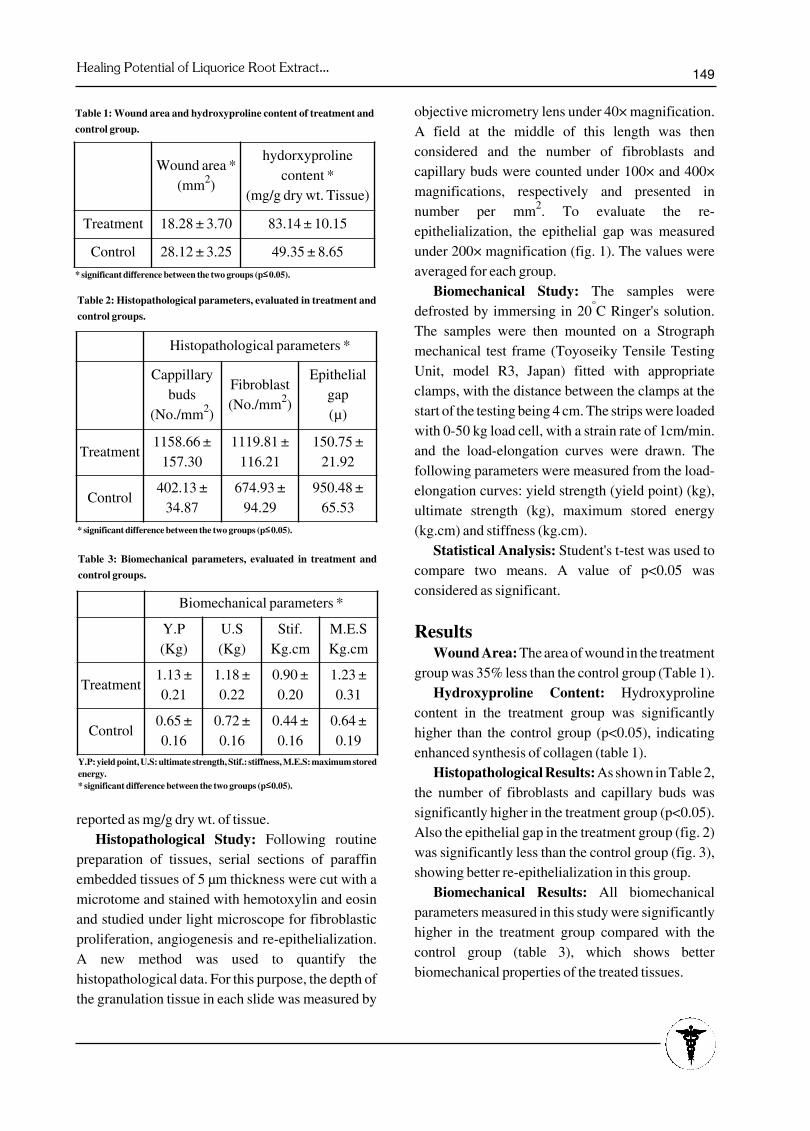
ResultsWound Area: The area of wound in the treatment
group was 35% less than the control group (Table 1). Hydroxyproline Content: Hydroxyproline
content in the treatment group was significantlyhigher than the control group (p<0.05), indicatingenhanced synthesis of collagen (table 1).
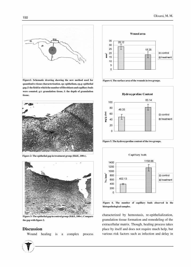
Histopathological Results: As shown in Table 2,the number of fibroblasts and capillary buds wassignificantly higher in the treatment group (p<0.05).Also the epithelial gap in the treatment group (fig. 2)was significantly less than the control group (fig. 3),showing better re-epithelialization in this group.
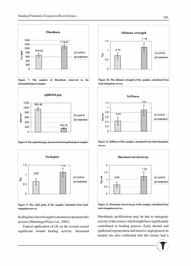
Biomechanical Results: All biomechanicalparameters measured in this study were significantlyhigher in the treatment group compared with thecontrol group (table 3), which shows betterbiomechanical properties of the treated tissues.
Table 1: Wound area and hydroxyproline content of treatment and
control group.
Wound area *(mm2)
hydorxyprolinecontent *
(mg/g dry wt. Tissue)
Treatment 18.28 ± 3.70 83.14 ± 10.15
Control 28.12 ± 3.25 49.35 ± 8.65
* significant difference between the two groups (p££ 0.05).
Histopathological parameters *
Cappillarybuds
(No./mm2)
Fibroblast(No./mm2)
Epithelialgap(μ)
Treatment1158.66 ±
157.301119.81 ±
116.21150.75 ±
21.92
Control402.13 ±
34.87674.93 ±
94.29950.48 ±
65.53
Table 2: Histopathological parameters, evaluated in treatment and
control groups.
* significant difference between the two groups (p££ 0.05).
Biomechanical parameters *
Y.P(Kg)
U.S(Kg)
Stif.Kg.cm
M.E.SKg.cm
Treatment1.13 ±0.21
1.18 ±0.22
0.90 ±0.20
1.23 ±0.31
Control0.65 ±0.16
0.72 ±0.16
0.44 ±0.16
0.64 ±0.19
Y.P: yield point, U.S: ultimate strength, Stif.: stiffness, M.E.S: maximum storedenergy.* significant difference between the two groups (p|££| 0.05).
Table 3: Biomechanical parameters, evaluated in treatment and
control groups.
Oloumi, M. M.150
Discussion Wound healing is a complex process
characterized by hemostasis, re-epithelialization,granulation tissue formation and remodeling of theextracellular matrix. Though, healing process takesplace by itself and does not require much help, butvarious risk factors such as infection and delay in
Figure1: Schematic drawing showing the new method used for
quantitative tissue characterization. ep: epithelium, ep.g: epithelial
gap, f: the field in which the number of fibroblasts and capillary buds
were counted. g.t: granulation tissue, l: the depth of granulation
tissue.
Figure 3: The epithelial gap in control group (H&E, 100×). Compare
the gap with figure 2.
Figure 4. The surface area of the wounds in two groups.
Figure 2: The epithelial gap in treatment group (H&E, 100×).
Figure 5. The hydroxyproline content of the two groups.
Figure 6. The number of capillary buds observed in the
histopathological samples.
Healing Potential of Liquorice Root Extract... 151
healing have been brought to attention to promote thisprocess (Shanmuga Priya et al., 2002).
Topical application of LE on the wound causedsignificant wound healing activity. Increased
fibroblastic proliferation may be due to mitogenicactivity of the extract, which might have significantlycontributed to healing process. Early dermal andepidermal regeneration and massive angiogenesis intreated rats also confirmed that the extract had a
Figure 7. The number of fibroblasts observed in the
histopathological samples.
Figure 8. The epithelial gap, measured in histopathological samples.
Figure 9. The yield point of the samples calculated from load-
elongation curves.
Figure 10. The ultimate strength of the samples, calculated from
load-elongation curves.
Figure 11. Stiffness of the samples, calculated from load-elongation
curves.
Figure 12. Maximum stored energy of the samples, calculated from
load-elongation curves.
Oloumi, M. M.152
positive effect towards cellular proliferation,granulation tissue formation and re-epithelialization(Shanmuga Priya et al., 2002).
Biochemical analysis showed increasedhydroxyproline content, which is a reflection ofincreased fibroblastic proliferation and therebyincreased collagen synthesis (Nayak et al., 1999).Collagen not only confers strength and integrity to thetissue matrix, but also plays an important role inhomeostasis and epithelialization at the later phase ofhealing (Clark, 1996). Collagen is one of the mostdominant extracellular matrix proteins in thegranulation tissue, which appears to be significantlyhigh by the fifth day of wounding and after day seven,collagen production is further advanced (Grillo,1964). Therefore, enhanced synthesis of collagenprovides strength to repaired tissue and also healingpattern (Shanmuga Priya et al., 2002).
Our biomechanical results well correlate withbiochemical and histopathological results. Gain intensile strength correlates with the rate of collagensynthesis through the first 10 weeks of healing(Capperauld, 1989; Freeman et al., 1989; Maddenand Peacock, 1971; Miro et al., 1995; Paul et al.,1997). As shown in Table 1, maximum load andstiffness of the treated tissues were significantlyhigher than the control one. Maximum load, which isthe functionally most important parameter forcharacterizing healing wounds (Quirinia and Viidik,1991), reflects the ultimate tensile strength of thespecimen, at which complete failure occurs rapidly,and load supporting ability of the tissue issubstantially reduced (Carlstedt and Nordin, 1989).This happens as the intermolecular cross links arebroken and the collagen fibrils pass each other or ascollagen fibrils lose contact with ground substance(Freeman et al., 1989). Higher stiffness andmaximum load of the treated tissue confirm ourbiochemical and histopathological observationsregarding more fibroblasts, less epithelial gaps andincreased collagen synthesis in the treatment group.
Size reduction in treatment wounds was alsosignificantly more than the control ones, which can beattributed to prominent re-epithelialization in treatedwounds, which was confirmed microscopically by
measuring epithelial gap. We can conclude that the remedy prescribed by
Kermani in 1890s in the book "Dagha'egh Al'alaj"must have been effective on wound healing, althoughleaving the particles of non-sterile powdered root inan open wound must have had some negative effecton the healing process.
Considering the results of the present study, it canalso be concluded that LE posses some pro-healingactivity, which can affect the process of woundhealing at various phases of tissue repair.
Acknowledgements: The authors would like to thank the Research
Council of the Faculty of Veterinary Medicine,Shahid Bahonar University of Kerman, for fundingthe research. The authors are also in debted to Dr. M.Derakhshan, the head of Razi Institute, Kermanbranch for providing the laboratory animals, and Mr.Moshrefi, general manager of Barez tire company,Kerman and all his kind and cooperative colleagues,who assisted us in the biomechanical testing.
Bennet, N.T. and Schultz, G.S. (1993) Growth
factors and wound healing: Biomechanical properties
of growth factors and their receptors. Am. J. Surg.
165: 728-737.
Bennet, A., Melhuish, P.B., Stamford, I.F. (1985)
Carbenoxolone and deglycyrrhized liquorice have
little or no effect on prostanoid synthesis by rat
gastric mucosa ex vivo. Br. J. Pharmacol. 86: 693-
695.
Capperauld, I. (1989) Suture materials: a review.
Clin. Mater. 4: 3-12.
Carlstedt, C.A., Nordin, M. (1989) Biomechanics of
tendon and ligaments; in Basic Biomechanics of the
Musculoskeletal System, Nordin, M.and Frankel,
V.H. (eds.), 2ndEd., pp. 59-74, Lea and Febiger,
Philadelphia.
Clark R.A.F. (1991) Cutaneous wound repair; in
Physiology, Biochemistry and Molecular Biology of
Skin, Goldsmith, L.A. (ed), pp. 576-592, Oxford
University Press, New York.
References1.
2.
3.
4.
5.
Healing Potential of Liquorice Root Extract... 153
Clark R.A.F.(1996) Wound repair. Overview and
general considerations; in The Molecular and
Cellular biology of Wound Repair, Clark, R.A. and
Henson, P.M. (eds.), pp. 3-27, Plenum Press, New
York.
Dehpour, A.R., Zolfaghari, M.E., Samandian, T.,
Kobarfard, F., Faizi, M., Assari M. (1995) Antiulcer
activities of liquorice and its derivatives in
experimental gastric lesion induced by ibuprofen in
rats. Int. J. Pharm. 119: 133-138.
Freeman, L.J., Hegreberg, G.A., Robinette, J.D.,
Kimbrell, J.T. (1989) Biomechanical properties of
skin and wounds in Ehlers-Danlos syndrome. Vet.
Surg. 18: 97-102.
Fuhrman B., Volkova, N., Kaplan, M., Presser, D.,
Attias, J., Hayek, T., Aviram, M. (2002)
Antiatherosclerotic effects of licorice extract
supplementation on 3 hypercholesterolemic patients:
increased resistance of LDL to atherogenic
modifications, reduced plasma lipid levels, and
decreased systolic blood pressure. Nutrition. 18: 268-
273.
Grillo, H.C. (1964) Aspects of the origin, synthesis,
and evolution of fibrous tissue in repair; in Advances
in Biology of Skin, Montagna, W.and Billingham,
R.E. (eds.), Vol 5, pp. 128-154., Macmillan, New
York.
Kiso, Y., Tohkin, M., Hikino, H., Hattori, M.,
Sakamoto, T., Namba, T. (1984) Mechanism of
antihepatotoxic activity of glycyrrhizin, I. Effect of
free radical generation and lipid peroxidation. Planta
Med. 50: 298-302.
Kitagawa, I.( 2002) Licorice root. A natural
sweetener and an important ingredient in Chinese
medicine. Pure Appl. Chem. 74: 1189-1198.
Kumagai, A., Nanaboshi, M., Asanuma, Y., Yagura,
T., Nishino, K. (1967) Effect of gylcyrrhizin on
thymolytic and immunosuppressive action of
cortisone. Endocrinol. Japon. 14: 39-42.
Logemanna, W., Lauria, F. (1960) Antileukaemic
activity of glycyrrhetinic acid. Nature. 187: 607-608.
Madden, J.W., Peacock Jr., E.E. (1971) Studies on
the biology of collagen during wound healing: III.
Dynamic metabolism of scar collagen and
6.
7.
8.
9.
10.
11.
12.
13.
14.
remodeling of dermal wounds. Ann. Surg. 174: 511-
520.
Miro, D., Julia, M.V., Sitges-Serra, A. (1995) Wound
breaking strength and healing after suturing
noninjured tissues. J. Am. Coll. Surg. 180: 659-665.
Mirsalis, J.C., Hamilton, C.M., Schindler, J.E,
Green, C.E., Dabbs, J.E. (1993) Effects of soy bean
flakes and liquorice root extract on enzyme induction
and toxicity in B6C3F1 mice. Food Chem. Toxicol.
31: 343-350.
Nayak, B.S., Udupu, A.L., Udupa, S.L. (1999) Effect
of Ixora coccinea flowers on dead space wound
healing in rats. Fitoterapia. 70: 233-236.
Nomura, T., Fukai, T., Akiyama, T. (2002)
Chemistry of phenolic compounds of licorice
(Glycyrrhiza species) and their estrogenic and
cytotoxic activities. Pure Appl. Chem. 74: 1199-
1206.
Paolini, M., Barillari, J., Broccoli, M., Pozzetti, L.,
Perocco, P., Cantelli-Forti, G. (1999) Effect of
liquorice and glycyrrhizin on rat liver carcinogen
metabolizing enzymes. Cancer Lett. 145: 35-42.
Paolini, M., Pozzeti, L., Sapone, A., Gantelli-Forti,
G. (1998) Effect of licorice and glycyrrhizin on
murine liver CYP-dependent monooxygenases. Life
Sci. 62: 571-582.
Paul, R.G., Tarlton, J.F., Purslow, P.P., Sims, T.J.,
Watkins, P., Marshall, F., Ferguson, M.J., Bailey,
A.J. (1997) Biomechanical and biochemical study of
a standardized wound healing model. Int. J.
Biochem. Cell B. 29: 211-220.
Pompei, R., Fore, O., Marcialis, M.A. Pani, A.,
Loddo, B. (1979) Glycyrrhizic acid inhibits virus
growth and inactivates virus particles. Nature. 281:
689-690.
Quirinia, A., Viidik, A. (1991) Freezing for
postmortal storage influences the biomechanical
properties of linear skin wounds. J. Biomech. 24:
819-823.
Rasik, A.M., Raghubir, R., Gupta, A., Shukla, A.,
Dubey, M.P., Srivastava, S., Jain, H.K., Kulshrestha,
D.K. (1999) Healing potential of Calotropis procera
on dermal wounds in Guinea pigs. J.
Ethnopharmacol. 68: 261-266.
15.
16.
17.
18.
19.
20.
21.
22.
23.
24.
Oloumi, M. M.154
Shanmuga Priya, K., Gnanamani, A.,
Radhakrishnan, N., Babu, M. (2002) Healing
potential of Datura alba on burn wounds in albino
rats. J. Ethnopharmacol. 83: 193-199.
Tylor, V.E., Brady, L.R., Robbers, J.E. (1988)
Pharmacognosy, 9thEd., pp. 68-72, Lea and Febiger,
Philadelphia.
25.
26.
Abstracts in persian language 173
ìXéú| OdÛýÛBR kAìLryßþ,6831, kôoû 26, yíBoû 4,451-741|
Gpouþ gõAÁ AèPýBìþ|οBoû oüzú âýBû yýpüò GýBó Gp qgî|øBÿ KõuPþ ko ìõ} ¾dpAüþ ||
ìdíl ìùlÿ Îéõìþ 1* Aìýò kogzBó Öp
2ÎHBx ðýà Kõo
3
1âpôû Îéõï koìBðãBøþ kAðzßlû kAìLryßþ kAðzãBû yùýl GBøñp ÞpìBó, ÞpìBó-AüpAó.
|2
âpôû KBOõGýõèõsÿ kAðzßlû kAìLryßþ kAðzãBû yùýl GBøñp ÞpìBó, ÞpìBó-AüpAó.3
kAð{|@ìõgPú kAðzßlû kAìLryßþ kAðzãBû yùýlGBøñp ÞpìBó, ÞpìBó–AüpAó.
|(|||koüBÖQ ìÛBèú:7 ||ìùpìBû 4831 , Knüp} ðùBüþ: 6 ||@mo ìBû 5831)
οBoû yýpüò GýBó Aq qìBó øBÿ kôo Gú ÎñõAó üà kAoôÿ ìõöSp Gh¿õÁ GpAÿ koìBó qgî ìÏlû ìõok AuP×Bkû ÚpAo âpÖPú AuQ. ko Aüò ìÇBèÏú ASp
AèPýBìþ οBoû @Gþ oüzú âýBû yýpüò GýBó Gp qgî øBÿ KõuPþ ìõok Gpouþ ÚpAo âpÖQ. Gú Aüò ìñËõo Aq 54 up ìõ} ¾dpAüþ u×ýl ðp Aq ðtAk
AuLpAâò–kôèþ AuP×Bkû yl. GB AuP×Bkû Aq KBða KõuPþ 7 ìýéþ ìPpÿ kô qgî ìPdl Aèzßê ko ÆpÖýò uPõó ìùpû øB ko KzQ øp cýõAó AüXBk âpkül
(ìXíõÎB« 09 qgî). οBoû @Gþ oüzú yýpüò GýBó Gú ìlR 7 oôq oôqAðú Gp oôÿ ðýíþ Aq qgî øB ÚpAo âpÖQ. Kw Aq @ó cýõAðBR GpAÿ ìÇBèÏBR
øývPõKBOõèõsÿ, GýõyýíýBüþ (ìdPõAÿ øýloôÞvþ Kpôèýò) ô GýõìßBðýà ÚpGBðþ ylðl. uÇe ðùBüþ qgî ðýr AðlAqû âýpÿ âpkül. οBoû @Gþ oüzú
yýpüò GýBó uHI AÖrAü{ ìÏñþ kAoÿ ko OÏlAk ÖýHpôGçuQ øB ô WõAðú øBÿ ìõüpâþ, ìdPõAÿ øýloôÞvþ Kpôèýò ô AuPdßBï Þzzþ qgî øB ylû Gõk.
øí̀ñýò uÇe qgî ko âpôû koìBó GÇõo ìÏñþ kAoÿ ÞíPp Aq âpôû yBøl Gõk. Gp AuBx Aüò ìÇBèÏú ìþ OõAó ̂ñýò ðPýXú âpÖQ Þú οBoû yýpüò GýBó kAoôÿ
âýBøþ ìõöSpÿ ko AèPýBï qgî ìþ GByl.
ôAsû|øBÿ Þéýlÿ:οBoû yýpüò GýBó, AèPýBï qgî KõuQ.
∗) ðõüvñlû ìvõöôë: Oé×ò: 0541223-1430ðíBGp: 7402223-1430 ri.ca.ku.liam@imuolo:liamE