Embed Size (px)
Citation preview

Greater Manchester and Cheshire HPB Unit
Guidelines for the Assessment &
Management of Hepatobiliary and
Pancreatic Disease
Chapter 7

78
Contents
7. Benign liver conditions _____________________________________________________________ 79
7.1. Pyogenic liver abscess ___________________________________________________________________ 80
7.2. Management of hydatid cysts (cystic echinococcosis) _________________________________________ 81
7.3. Solitary & Polycystic liver disease _________________________________________________________ 86
7.4. Hepatocellular adenomas ________________________________________________________________ 88
7.5. Acute Liver Failure _____________________________________________________________________ 90
7.6. Liver Transplantation: UK Selection Criteria _________________________________________________ 92

79
7. Benign liver conditions

80
7.1. Pyogenic liver abscess

81
7.2. Management of hydatid cysts (cystic echinococcosis)
Fig. 1. Diagrammatic representation of structure of the echinococcal cyst.
Fig. 2. WHO-IWGE CLASSIFICATION OF CYSTIC ECHINOCOCCOSIS
The WHO-IWGE standardised classification allows a natural grouping of the cysts into three relevant
groups: active (CE1 and 2), transitional (CE3) and inactive (CE4 and 5). It includes a “cystic lesion” (CL) stage
(undifferentiated).

82
Imaging Features of Hydatid Cysts:
Cystic Lesion
Status: if cystic echinococcosis [CE] active
Unilocular cystic lesion [CL] with uniform anechoic content, not clearly delimited by hyper echoic
rim ( = cyst wall not visible)
Normally round but may be oval
Size variable often small; CLs (< 5.0cm), but may be medium sized (CLm (5-10cm), or large (CLl
>10cm)
Normally these are non-parasitic cystic lesions, but if there is a suspicion of CE, these cysts are
usually at an early stage of development and are not fertile. US does not detect any pathognomonic
signs. Differential diagnosis of these cystic lesions requires the application of the additional
diagnostic techniques.
Cystic Echinococcosis
Active
Unilocular simple cyst with uniform anechoic content. Cyst may exhibit fine echoes due to shifting
of brood capsules which is often called hydatid sand or “snowflake sign”
Cyst wall is visible
Normally round or oval
Size variable: CE1s (<5.0cms), CE1m (5-10cms), CE1l (>10cm)
Usually fertile – pathognomonic signs include visible cyst wall and snowflake sign
CE2
Status: Active
Multivesicular, multiseptated cysts in which the daughter cysts may partly or completely fill with
unilocular mother cysts. Cyst septations may produce “wheel-like” structures or the contained
daughter cysts may produce a “rosette-like” or “honeycomb” structure.
Cyst wall normally visible
Normally round or oval
Size variable: CE2s (<5.0cms), CE2m (5-10cms), CE2l (>10cm)
Usually fertile
US features are pathognomonic

83
CE3
Status: Transitional
Anechoic content with detachment of laminated membrane from, the cyst wall visible as floating
membrane or as “water-lily sign”: which is indicative of wavy membranes floating on top of the
remaining cyst fluid
Unilocular cyst which may contain daughter cysts (anechoic appearance) and echoic areas
(disrupted membranes/degenerating daughter cysts). These cysts appear at US as a “complex
mass”
Cyst form may be less rounded due to decrease of intra-cystic fluid pressure
Size variable: CE3s (<5.0cms), CE3m (5-10cms), CE3l (>10cm)
Transitional stage: Cyst is most usually starting to degenerate. Degenerative signs of US
examination are “detachment and rupture of membranes”. Occasionally may be followed by
daughter cyst production
US features are pathognomonic
CE4
Status: Inactive
Heterogenous hyperechoic or dyshomogenous degenerative contents. No daughter cysts
May show a “ball of wool” sign which is indicative of degenerating membranes
Size variable: CE4s (<5.0cms), CE4m (5-10cms), CE4l (>10cm)
Most cysts of this type are not fertile
US features are usually not pathognomonic and further diagnostic tests are required to confirm the
diagnosis. Differential diagnosis may be possible if there is presence of a cystic wall, lateral cone
shadow, little calcifications, or if an echoic and anechoic spiral “ball of wool” image is clearly seen
within a focal hepatic lesion
CE5
Status: Inactive
Cysts are characterised by thick walled calcified wall which is arch shaped producing a cone shaped
shadow. Degree of calcification varies from partial to complete.
Size variable: CE5s (<5.0cms), CE5m (5-10cms), CE5l (>10cm)
Cysts not fertile in majority of cases
Diagnosis uncertain, features are not pathognomonic, but highly suggestive of E. granulosus.

84
Figure 3. Diagnostic algorithm for hydatid liver cysts
Table 1. WHO-IWGE suggested stage-specific approach to uncomplicated cystic echinococcosis of the liver
WHO classification Surgery Percutaneous treatment Drug therapy Suggested Resources
CE1 <5cm ABZ Optimal
PAIR Minimal
√ √ >5cm PAIR + ABZ Optimal
PAIR Minimal
CE2 √ Other PT + ABZ Optimal
√ √
Other PT Minimal
CE3a <5cm ABZ Optimal
√ √ PAIR Minimal
>5cm PAIR + ABZ Optimal
PAIR Minimal
CE3b Non-PAIR PT + ABZ Optimal
√ √ √
Non-PAIR PT Minimal
CE4 Watch and Wait Optimal
CE5 Watch and Wait Optimal
PAIR; Puncture, aspiration, injection, re-aspiration.

85
Figure 4. Surgical management of active and transitional hydatid cysts

86
7.3. Solitary & Polycystic liver disease
Transverse CT images of polycystic liver disease
a | Gigot type I cystic liver containing a couple of large (>10 cm) cysts, but <10 cysts in total.
b | Gigot type II polycystic liver with diffuse involvement of liver parenchyma by multiple medium-sized
cysts.
c | Gigot type III polycystic liver. The liver is completely occupied with numerous cysts, and only a few
areas of visible liver parenchyma are present.
Reprinted by permission from Macmillan Publishers Ltd: Nature Reviews Gastroenterology & Hepatology,
Gevers et al. Copyright 2013

87
Management pathway for symptomatic non-parasitic benign cystic liver disease
Symptomatic non-parasitic cystic
liver disease
Symptomatic benign
hepatic cysts
Cyst adenoma or cyst
adenocarcinoma
suspected
Solitary cyst
> 5 cm in diameter
Gigot type I or type II Gigot type III
Aspiration
sclerotherapyFailure
Laparscopic
fenestrationPartial liver resection
Consider somatostatin
analoguesSurgical work-up
Liver transplantation
Discontinue
exogenous oestrogens
Surgery

88
7.4. Hepatocellular adenomas
Risk factors, related disorders and molecular classification of hepatocellular adenoma
89
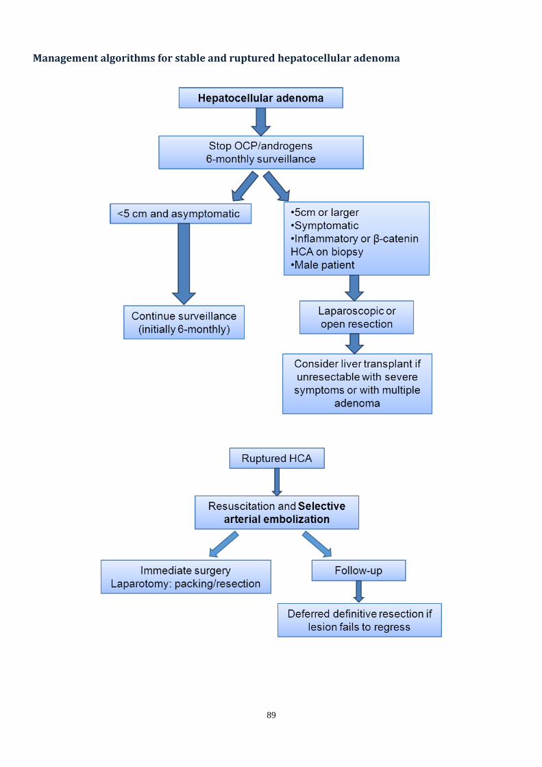
Management algorithms for stable and ruptured hepatocellular adenoma

90
7.5. Acute Liver Failure
Common Management Issues
Organ system and Common Conditions
Assessment Specific Elements of Care
Cardiovascular system
Hypotension Invasive monitoring for all conditions;
echocardiography for low cardiac output and right ventricular failure
Intravascular volume depletion
Correction of volume depletion
Vasodilation Vasopressors
Low cardiac output and right ventricular failure
Inotropic support
Hepatic system
Evolving hepatic dysfunction Serial biochemical and coagulation
testing Intravenous acetylcysteine
Respiratory system
Risk of aspiration pneumonia Neurologic observation to monitor
level of consciousness Early tracheal intubation for
depressed level of consciousness
Metabolic and renal systems
Hypoglycaemia Serial biochemical testing Maintain normoglycaemia
Hyponatraemia Active fluid management
Renal dysfunction, lactic acidosis, hyperammonaemia
Renal-replacement therapy
Impaired drug metabolism Review drug administration
Central nervous system
Progressive encephalopathy
Neurologic observation; monitoring of serum ammonia level; transcranial ultrasonography; consideration of intra-cranial pressure monitoring
Treatment of fever and hyponatraemia; screening for sepsis
High-grade encephalopathy; endotracheal intubation; avoidance of PaCO2 od <30mmHG or >45 mmHg;
target for serum sodium, 145-150 mmol/l; risk assessment for intra-
cranial hypertension
Intracranial hypertension
Interventions for pressure surges; osmotherapy (mannitol, hypertonic saline); temperature control; rescue
therapies (indomethacin, thiopentone)
Haematologic system
Coagulopathy Laboratory coagulation testing No routine correction of coagulation
abnormalities, only for invasive procedures
Immunologic system
High risk of sepsis Clinical evaluation Antibiotic prophylaxis

91
West Haven criteria for grading mental state in hepatic encephalopathy
Grade Features
Grade 0 No signs or symptoms
Grade 1 Euphoria, anxiety, trivial lack of awareness, impaired performance, shortened attention span, mild asterixis
Grade 2 Lethargy, minimal personality changes, subtle personality change, inappropriate behaviour, asterixis
Grade 3 Somnolence, confusion, gross disorientation
Grade 4 Coma
King's College criteria for liver transplantation in Acute Hepatic Failure.
Acetaminophen-associated AHF All other causes of AHF
pH < 7.3 INR >6.5
Or Or
INR > 6.5, serum creatinine >3.4 mg/dl, and grade III–IV encephalopathy
Three of the following variables:
1. Age <10 or >40 years
2. Cause is non-A, non-B hepatitis or idiosyncratic drug reaction
3. Duration of jaundice before encephalopathy >7 days
4. INR > 3.5
5. Serum bilirubin >17.5 mg/dl

92
7.6. Liver Transplantation: UK Selection Criteria
Liver Advisory Group on behalf of NHSBT
http://www.odt.nhs.uk/pdf/liver_selection_policy.pdf
Conditions that are considered for transplantation
Adult patients
Most adult patients with liver disease are not managed in transplant centres. Patients referred for
assessment for liver transplant will include those with the following broad categories of conditions:
Acute liver failure
Multi-system disorder in which severe acute impairment of liver function with encephalopathy
occurs within 8 weeks of the onset of symptoms and no recognised underlying chronic liver disease
Chronic liver disease; any cirrhosis which may be due to:
Alcoholic liver disease
Non-alcoholic fatty liver disease
Chronic viral hepatitis B, C or D
Autoimmune liver diseases: primary biliary cirrhosis, primary sclerosing cholangitis, chronic active
liver disease and overlap syndromes
Genetic haemochromatosis
Wilson’s disease
Alpha-1 antitrypsin deficiency
Congenital hepatic fibrosis and other congenital or hereditary liver diseases
Secondary sclerosing cholangitis
Liver tumours
Hepatocellular carcinoma (See: 6.7 Hepatocellular Cancer - Liver Transplantation)
Variant syndromes
Diuretic resistant ascites
Chronic hepatic encephalopathy
Intractable pruritus
Hepatopulmonary syndrome
Familial amyloid polyneuropathy
Familial hypercholesterolaemia
Polycystic liver disease
Hepatic epithelioid haemangioendothelioma
Sickle cell hepatopathy
Patients not falling within these categories may be considered through the National Appeals Panel route.