Embed Size (px)
Citation preview

Greater Manchester and Cheshire Cancer Network
Signature: ___________________________
Date:____________________________
Operational Policy for the Greater Manchester and Cheshire Cancer Network Specialist Skin Cancer MDT
This Annual Report was agreed by Dr Vindy Ghura, Lead Clinician for the Specialist Skin MDT in April 2017
D Waters/ V Ghura 210416 Date for Review: March 2018

1
Contents Page
Introduction & Purpose of SSMDT (A12/S/b-001) 3
Leadership Arrangements and Responsibilities (A12/S/b-001) 3
Specialist Nurse Arrangements 4
The SSMDT Meeting (A12/S/b-16-002) A12/S/b-16-003) 5.6
Referral Arrangements 8
Skin Pathway – 62 Day Pathway Patients 9
Referrals requiring treatment decision before next scheduled LMDT meeting
10
Attendance at the Network Group 14
Operational Policy for the Key Worker (A12/S/b-002) (9A12/s/b-16-003) 14.15
Communication with Primary Care 15
Patient Information (A12/S/b-16-002) 15.16
Appendices
Appendix 1 Responsibilities of Lead Clinician, Specialist Skin MDT( A12/S/b-16-002)
Appendix 2 Clinical Nurse Specialist timetables
Appendix 3 “Two Week Wait” Referral Form
Suspected Malignant Melanoma or Squamous Cell Carcinoma
Appendix 3.1 Two Week Wait Referral: Patient Information Leaflet
Appendix 4 Clinical Upgrade to Suspected Cancer 62 Day Tracking for Suspected NEW Primary
Cancers Salford Royal NHS Foundation Trust
Appendix 5 MDT: Skin Referral Form
Salford Royal NHS Foundation Trust
Appendix 6 SSMDT Skin Cancer Proforma
Appendix 7 Nomination Process for patient referral to the Skin Multi-Disciplinary Team meeting (MDT)
Appendix 8 Specialist Dermatology MDT Outcomes
Appendix 9 Communication and Referral Proforma
Appendix 10 Clinical Guidelines Peer Review Measure (A12/S/b-16-004)
Greater Manchester & Cheshire Cancer Network
Appendix 11 31 and 62 Day Referral Form (A12/S/b-16-005)
Teenage & Young Adult Referral Documents
TYA MDT Members List
Teenage and Young Adult (TYA) MDT Notification
Teenage and Young Adult (TYA) TYA MDT –OUTCOME FORM

2
Introduction The Lead Clinician for the Specialist Skin Cancer SMDT (SSMDT) is Dr Vindy Ghura (Appendix 1). The deputy lead is Dr Rebecca Brooke (A12/S/b-16-001) Salford Royal NHS Foundation Trust is the Host Organisation for the Greater Manchester and Cheshire Cancer Network Skin Cancer Specialist MDT reflecting Improving Outcome Guidance (IOG). The Specialist Skin Cancer Multidisciplinary Team (SSMDT) commenced in January 2009. The Specialist Skin Cancer Multidisciplinary Team (SSMDT) is a multi-professional group serving the Greater Manchester and Cheshire Cancer Network. The Skin Cancer Multidisciplinary Team serves the population of Greater Manchester approximately 3.5 million. The Specialist Skin Cancer MDT comprises of Salford Royal NHS Foundation Trust, The Christie NHS Foundation Trust, and University Hospitals of South Manchester NHS Trust, Royal Bolton Hospital NHS Foundation Trust, Central Manchester NHS Foundation Trust and Mid-Cheshire NHS Foundation Trust.
This document outlines the Operational Policy for the SSMDT and this policy is reviewed on a yearly basis at an Annual General Meeting. The AGM for this team commenced in April 2011 and has taken place annually thereafter. Purpose of the SSMDT The aim of the SSMDT is to ensure a co-ordinated approach to diagnosis, treatment and care services for all patients diagnosed with Skin Cancers, falling within the Skin Cancer Improving Outcomes Guidance. The SSMDT has the combined function of diagnosis (to rapidly assess and achieve confirmation of skin cancer), treatment (discussing the management of all newly diagnosed skin cancers) and communication (with the appropriate agencies e.g. Plastic Surgeons, Medical/Clinical Oncologists, Primary Care Teams). Furthermore the SSMDT is committed to achieving the highest standards of care and patient outcomes by:
Collection of high quality data
Analysis of such data in audit cycles
Involvement in research
Incorporation of new research and best practice into patient care
Providing comprehensive information to patients and their relatives
Involving patients in assessment and redesign of the services.
Leadership Arrangements and Responsibilities The Lead Clinician for the Specialist Skin Cancer SMDT is Dr Vindy Ghura This is agreed with the Trust Lead Cancer Clinician. (A12/S/b-16-001)
Responsibilities of the Lead Clinician (Appendix 1) Lead the clinical activity of the SSMDT. Working to agreed guidelines, ensuring a high quality
integrated service which meets local, regional and national standards.
Ensure the MDT engages in the relevant Clinical Sub Group (CSG) and contributes to its work.
Ensure that clinical management guidelines are produced and revised regularly (this was usually carried out within the CSG which has now been disbanded).
Organise “management meetings” of the SSMDT and ensure its deliberations are recorded.
Ensure the collection of the appropriate cancer minimum dataset, working with the team’s audit coordinator.
Establish an audit programme and review of outcomes (this will include audits carried out across the Network).
Produce an Annual Report with the support of the Cancer Management Team.

3
The Skin Cancer Specialist MDT is listed in the Local Cancer Services Directory as part of the services of the Salford Royal NHS Foundation Trust with appropriate contact names and numbers.
Specialist Nurse Arrangements (A12/S/b-16-002)
The Core Nurse Members will have a set of agreed responsibilities, which includes completion of specialist study modules and Advanced Communication Skills Training.
Responsibilities of the Core Nurse Members Contributing to the multidisciplinary discussion and patient assessment/care planning decision of the
team at their regular meetings. Providing expert nursing advice and support to other health professionals in the Nurse’s specialist
area of practice. Involvement in Clinical Audit. Leading on patient and carer’s communication issues and coordination of the patient pathway for
patients referred to the team – acting as Key Worker or responsible for nominating the Key Worker for the patient’s dealings with the team.
Facilitating access to members of the MDT where requested by patients or their carer. Contributing to the management of the service. Utilising research in the nurse’s specialist area of practice. Holistic Needs Assessment of patients.
Also, agreed are the following List of Additional Responsibilities Contribute to the management of the service. Evaluate service delivery: identify areas for improvement and initiate change. Utilise current research and evidence based practice to the benefit of users of the service and on-
going service development. Responsible for the development and implementation of policies, procedures and guidelines at local,
network and national level. Address specific health targets related to Skin cancer e.g. NICE, IOG, DOH guidelines relevant to
the SSMDT.
The timetable for Clinical Nurse Specialists (CNS) providing cancer support can be seen in Appendix 2. Qualifications Julie Collins, Macmillan Skin Cancer Clinical Nurse Specialist (UHSM), has completed the following study:
Level 2 Psychological Training, March 2012 (A12/S/b-16-002) Certification available in the evidence file
Psychological Support Monthly with Dr Dorinda (A12/S/b-002) Confirmation available in the evidence file
Diploma in Professional Studies in Nursing March 1998
BSc (Hons) in Nursing Practice June 2006
Oncology Course at the University of Manchester, January 2011
Member of Northwest Skin Cancer Specialist Nurses Forum
Advanced communication Skills training 2009
National Melanoma conference December 2012
Case management and clinical examination (skin Cancer) course (MSc) June 2013 Nicola O’Callaghan, Skin Cancer Clinical Nurse Specialist (SRFT), has completed the following study:
BSc (Hons) Nursing Studies 2011
Advanced Diploma in Nursing Studies 2003
Health Science Access to Higher Education 1999
Metastatic Spread to the Brain study day 2014
TYA consensus event 2014(contributing to the development of TYA nurse competencies for RCN-to be published 2014)
Advanced Communication Skills Training 2014 (Certification available in evidence file)
Psychological support with Frances Collins ( dates in evidence file ) (A12/S/b-16-002)
Member of Northwest Skin Cancer Specialist Nurses Forum since 2014
Member of the BDNG since 2012
Psychological support level 2 Training 2014

4
Dianne Waters, Skin Cancer Clinical Nurse Specialist (SRFT), completed the following study:
RGN 1994
Module from death and dying Diploma level 6 ( 20 cats) open university 2000
Dermatology Module Diploma level 6 ( 20 cats) university of Glamorgan 2008
Lecture in palliative care and neuropathic pain June 2005
Benefits Advice for People Affected by Cancer seminar Dec 2005
Palliative Care employment and training (St Anne’s hospice) 1998-1999
Foundations in Palliative Care course employment and training ( Marie Curie1999 -2000 )
Advanced Communication Skills Training 2014( Certification available in evidence file)
TYA Consensus Event 2014(contributing to the development of TYA nurse competences for RCN- to be published 2014)
Member of British Association of Skin Cancer Nurses
Psychological support Level 2 Training ( certificate in evidence file) 2015
Clinical supervision with Frances Collins monthly (dates in evidence file( ( A12/S/b-16-002)
Mindfulness Training 2015
Member of NICE research community 2015 ongoing
Macmillan development programme
Jackie Hodgetts, Nurse Clinician (The Christie), has completed the following study:
RGN 1981
MSc in Advanced Practice Health and Social Care 2009
Non-Medical Prescriber 2010
Advanced communication skills training 2010 Caroline Owens, Surgical Specialist Skin Cancer Nurse (The Christie), has completed the following study:
RGN
DPSN nursing 2005
Wound Care Course 2007
Advanced Communications Skills Training 2008
Mentorship course 2009
Foundations in cancer care course 2010
BSc Nursing studies 2011
Case Management and Clinical examination (Skin Cancer) course (Masters Level)
Sarah Fulton, plastic surgery CNS at Christies-
Copies of the certificates for the qualifications listed above can be seen within the evidence file.
The SSMDT Chair is Dr Vindy Ghura
Dianne Waters and Nicola O’Callaghan are currently responsible for users’ issues including patient /carer information in collaboration with the relevant CNS from participating organisations.
The SSMDT has agreed one of the members as a lead for integration of research and recruitment in to clinical trials. This is currently Dr J Lear at SRFT.
The SSMDT Meeting
The SSMDT meets on the first and third Thursday of the month from 2 pm to 3.30 pm in Seminar Room 7 of the Mayo Building and via video conferencing with The Christie and Wythenshawe Hospital. Core members will agree an individual who will in, general, be expected to attend the SSMDT meeting in their absence. It was agreed by the North West review Unit that the Clinical Oncologist can cover the Medical Oncologist. (A12/S/b-16-001) (A12/S/b-16-002) (A12/S/b-16-003) In addition, the SSMDT will meet for an AGM to discuss, review; agree operational policies, work plans and an Annual Report. The SSMDT has agreed to have bi-annual audit meetings.
The MDT Co-ordinator will circulate a list of patients to be discussed to the members of the SSMDT.

5
It is the responsibility of the MDT Co-ordinator to ensure that case notes, Histopathology slides and Clinical Photographs are available for the meeting to enable fully informed decision-making. The patient’s consultant/team or the Chair will provide a brief summary of each patient’s treatment to the meeting. The MDT Co-ordinator will record the attendance of the core membership.
The outcomes are recorded by the MDT Co-ordinator and confirmed as correct by the Lead Clinician or CNS before being distributed to the managing Consultant.
If a patient requires referral to another MDT i.e. Lymphoma MDT, Soft Tissue Sarcoma, this will be done via a clinical letter. Membership Arrangements (A12/S/b-001) (A12/S/b-002)
The Specialist Skin Cancer MDT consists of the following Core Members:
Co
re M
em
be
rs
Role Name Cover
Consultant Dermatological Surgeon/Lead Clinician
Dr V Ghura Dr Brookes, Dr Lear, Dr Telfer, Dr
Madan, Dr Williams
Consultant Dermatologist Dr Williams Dr Lear, Dr Madan, Dr Telfer
Consultant Dermatologist Dr J Lear Dr Williams, Dr Telfer, Dr Madan
Consultant Mohs’/Dermatological Surgeon Dr N Telfer Dr Ghura / Dr Madan
Consultant Mohs’/Dermatological Surgeon Dr V Madan Dr Telfer / Dr Ghura
Consultant Dermatopathologist Dr L Jamieson Dr L Motta, Dr R Green
Consultant Dermatopathologist Dr L Motta Dr L Jamieson, Dr R Green
Consultant Dermatopathologist Dr R Green Dr L Jamieson, Dr L Motta,
Consultant Histopathologist Dr P Shenjere Dr D Nonaka
Consultant Histopathologist Dr P Hyder Dr P Shenjere
Consultant Plastic Surgeon (South Manchester)
Mr. C Duff Mr. S Wilson
Consultant Plastic Surgeon (South Manchester)
Mr. S Wilson Mr. C Duff
Consultant Plastic Surgeon (Christie) Mr. Mowatt Mr. Oudit, Mr. Lambe
Consultant Plastic Surgeon (Christie) Mr. D Kosutic Mr. Mowatt, Mr. Lambe, Mr. Oudit
Consultant Plastic Surgeon (Christie) Mr. Lambe Mr. Oudit, Mr. Mowatt,
Specialty Doctor Plastic Surgery (Christie) Mr. Gajanan Mr. Mowatt, Mr. Oudit,
Consultant Radiologist Dr F Wong Dr Barker / Dr H Laasch
Consultant Radiologist Dr C Barker Dr Wong/ Dr H Laasch
Consultant Clinical Oncologist Dr A Sykes Dr A Rembielak

6
Consultant Clinical Oncologist Dr A Rembielak Dr A Sykes
Consultant Medical Oncologist Dr P Lorigan Dr A Sykes Dr S Chow
Consultant Oncologist Dr S Chow Dr A Sykes / Dr P Lorigan
Skin Cancer Clinical Nurse Specialist (Salford)
Sister N O’ Callaghan
D Waters, M Kirk
Skin Cancer Clinical Nurse Specialist (Salford)
Sister D Waters N O’CallaghanMike Kirk
Skin Cancer Clinical Nurse Specialist (Bolton)
Sister C O’Neil J Hodgetts, C Owens, J Collins, N
O’Callaghan, D Waters
Advanced Nurse Practitioner Melanoma (Christie)
Sister J Hodgetts Dr Lorigan
Macmillan Skin Cancer Clinical Nurse Specialist (Wythenshawe)
Sister J Collins C Owen, J Hodgetts,
N O’Callaghan, D Waters
Surgical Skin Cancer Specialist Nurse (Christie)
Sister C Owens
Sister S Fulton J Hodgetts, J Collins,
N O’Callaghan, D Waters
MDT Co-ordinator (Salford) J Buchan Christine Farnworth
MDT Co-ordinator (Wythenshawe) Mike Ransome
MDT Co-ordinator (Christie) Andrea Harrison/
Sarah Ward/ Rosie Tunstall
Andrea Harrison / Sarah Ward / Rosie Tunstall
Dianne Waters and Nicola O’Callaghan are responsible for Patient and Carer Issues. The person responsible for Clinical Trials at SRFT is Dr Jason Williams.
Exte
nd
ed
Mem
be
rs
Designation Name
Palliative Care (Salford) Steve Ingle
Palliative Care – Skin Cancer (UHSM) Julie Collins
Clinical Psychologist (Salford) Francis Collins
Clinical Psychologist (Christie) Tania Hawthorne
Consultant Oculoplastic Surgeon Mr. Saj Ataullah
Consultant ENT Surgeon Mr. Tim Woolford
Clinical Geneticist/Genetic Counsellor Prof Gareth Evans
Occupational Therapist Local Provision will be made at individual Trusts as required. Nominated SRFT individual is Katherine
Atkin
Prosthetic Service Paul MacDonald
Orthotics Service (Christie) Tony Stanton
Orthotics Service (UHSM) Carol Winters
Physiotherapist Local Provision will be made at individual Trusts as
required. Nominated SRFT individual is Victoria Dickens
Lymphoedema Service (Christie) Paula Williams
Lymphoedema Service (UHSM) Karen Maclean
Consultant Max Fax Surgeon Mr. T Blackburn
Dermatology Specialist Practitioner Mr. M Kirk
All the core members of the team with direct patient contact will attend the National Advanced Communication Skills Training.

7
Referral Arrangements The Two Week Wait (TWW) standard for all cancers was introduced in 2000, and guarantees that everyone referred urgently by their GP with suspected cancer will be able to see a specialist within two weeks. It is an important milestone to achieving the 62-day target from urgent referral for suspected cancer to first definitive treatment, set in the NHS Cancer Plan (2000). This is applicable to all suspected Squamous Cell and Malignant Melanomas. As per guidance Basal Cell Carcinoma is not included within the Two Week Wait Standard. The Two Week Wait (TWW) Skin Proforma can be seen in Appendix 3.
The GP determines that TWW Skin appointment is required and completes the proforma, which can be compiled electronically within the practice clinical system and attached via the Choose and Book system (C&B).
The proforma has been produced as per the HSC 2000/13 guidance.
The completed referral template is e-mailed electronically on a work list via Choose & Book or via RBMS to Salford Royal NHS Foundation Trust Cancer Services on 0161 206 1303 within 24 hours of the decision to refer.
The call centre at Salford Royal NHS Foundation Trust either telephones the patient or sends a letter to advise about appointment within 7 days.
TWW is written on the front of all investigation requests by all staff requesting investigations, to initiate investigation within 7 days.
The Two Week Wait Proformas are filed in the back of the medical case notes.
Referral proforma will be kept under review to ensure compliance with guidance and local practice. Consultation and training with GP’s support this process. Triage of referrals enables routine assessment of compliance and feedback to referring GP. Audit reports of proforma uptake, use of proforma and referral outcomes are provided routinely to the SIF and GP lead who report back to PCT and practices. Consultant Upgrade Forms are used if there is a suspicion of cancer at any time during the patient pathway before Decision to Treat. A copy of the Upgrade Proforma can be seen in Appendix 4.
Patients who are upgraded need to be authorised by Consultant or CNS.
All forms are faxed to Cancer Services on 0161 206 1303 and Call Centre Registration on 0161 206 1048.
Day One of 62 commences at upgrade. Patient Pathways (A12/S/b-16-005) The patient pathway below provides evidence to support the processes involved for accessing SRFT MDT. This process describes how patients access the MDT with a Skin Cancer; both from a Two week Wait referral, an upgrade from General clinic, or investigations planned following suspected cancer referral, and are shown on the following page in diagrammatic form. The Operational policy for the Review of Skin Cancer Patients by the Greater Manchester and Cheshire Network Specialist Skin Cancer MDT is documented and includes all New Patients specified at level 4, 5 or 6 care to be reviewed by the MDT.

8
Greater Manchester & Cheshire Cancer Network
Skin Pathway – 62 Day Pathway Patients
End End
Appointment for Excision & Excision by Day 62
End
End
End
End
GP Referral
First Seen Outpatient Appointment – Secondary
Care Cancer Clinic
Referral to Surgical Specialists
(Includes: Plastics, ENT, Maxio-facila, opthal/oculoplastic and
general surgery)
Biopsy (may also be first
treatment)/Minor Ops List Other Treatment without
Biopsy
Pathology Patients diagnosed with SCC insitu or BCC are
stepped off the pathway
Discuss with Patient
Discuss with Patient (& relative if wished)
Referral to Surgical Specialists
Refer to Christie if SCC or MM> 1mm. Send CaRP
Discuss with Patient Discuss with Patient
Agree treatment with Patient
Treatment Options: Radiotherapy PDT
Systemic Therapy
First Definitive Treatment First Definitive Treatment First Definitive Treatment
1) With all cases, all treatments should be able to be provided within the 62 day time scale 2) Many constraints/pressures on the system is the volume of these patients competing with non-cancer
capacity.
14
21
24
28/31
62
0
Day of
Pathway
9
Discussion of all new Cancer Cases (A12/S/b-16-003)
Referrals of cases for discussion to the SSMDT are made on the approved pro-forma for any cases felt appropriate for referral. (Appendix 6) As per the NHS Cancer Plan (2000) and IOG (1999), all new cancer cases as per network guidelines will be discussed at the SSMDT. In order to maintain a compliant SSMDT, all appropriate cases from the contributing Trusts will be referred, presented and discussed at the SMDT. All referrals to the SMDT will be made as per the nomination process. (Appendix 7) The MDT Co-ordinator has responsibility for ensuring that all referrals are added to the SMDT discussion list and all proformas completed fully in advance of the meeting. (Appendix 8) A CaRP form should accompany all referrals requiring treatment at the Centre. This is to be faxed to the MDT Co-ordinator and a copy to the Cancer Services Team, as per the nomination process. (Appendix 9) It is the responsibility of the referring team to inform the patient of their referral to the SSMDT for discussion. A nominated person will relay the outcome of the SSMDT discussion, by the referring team, to the patient. An appropriate appointment will be made with the patient for immediately following the meeting. For the purpose of audit:
The Cancer Tracking Record will be created for each SMDT referral requiring treatment at the centre
An MDT proforma will be created for each case presented to the SMDT Treatment Planning Decision (A12/S/b-16-003) The SSMDT will agree and record on a proforma individual patients’ treatment plans (Appendix 14). This record should include:
The identity of patient discussed
The treatment planning decision
In the case of patients referred to a supra-network team or another SMDT in the network, the team to which they are referred should be named
For Melanoma patients the multidisciplinary treatment planning decision on suitable treatment modalities
Referrals requiring treatment decision before next scheduled SSMDT meeting
Urgent treatment decisions are made following an individual assessment of the patient.
All such decisions are made in the best interest of the patient
All treatment decisions will be confirmed by the Chair of the SSMDT prior to recording to ensure accuracy of documentation
Referrals are made to the Lead Clinician for an urgent decision via, telephone, fax, face to face referral, or referral of the patient direct to the Lead Clinician’s outpatient clinic or ward round
All treatment decisions are documented in the case notes and a SSMDT Proforma completed
For all treatment decisions made the SSMDT Co-ordinator is informed immediately
SSMDT Co-ordinator enters the decision to treat (DTT) on the Tracking Records
Retrospective presentation at the SSMDT will take place on all cases where treatment decision has been made outside of schedule weekly meeting because of clinical urgency

10
Surgical Procedures There is a requirement that the procedures specified together with the acute post-operative care take place at the same hospital and this occurs at The Christie Hospital.
Block lymph node dissections
Sentinel node biopsy
Metastatectomy / debulking for recurrent melanoma
Isolated limb perfusion
Isolated limb infusion
Reconstruction procedures involving microvascular surgical techniques. There is a requirement that each individual surgical member of the SSMDT who performs inguinal or axillary lymph node dissections on the SSMDT’s patients should performed a summed total of 15 or more groin plus axillary lymph node dissections. Diagnostic Services The SSMDT agreed pathology guidelines for the diagnosis and assessment of skin cancer can be seen in (Appendix10). (A12/S/b-16-004) The SSMDT has agreed pathology guidelines for the diagnosis and assessment of malignant melanoma. The core histopathologist member, Dr Lynne Jamieson, (Skin Cancer Dermatopathologist) participates in the National Specialist Dermatopathology EQA scheme and is a member of the steering committee. Dr Jamieson is a Fellow of the Royal College of Pathologists and holds the Diploma in Dermatopathology from the Royal College of Pathologists as recommended in the NICE IOG Guidelines. Dr Jamieson is a member of the British Society of Dermatopathology she participates in the NSDEQA which includes the Melanoma Club. In addition Dr Jamieson is on the steering committee of the NSDEQA and is a Core member of the Supraregional Cutaneous Lymphoma MDT. She is currently an examiner for the Diploma in Dermatopathology. She is the chair of the Specialty sub-committee on Dermatology which advises the Royal College of Pathologists on Subspecialty issues inc tumour datasets and tissue pathways. A copy of the certificate of participation can be seen within the evidence file. (A12/S/b-16-002) The Christie Histopathologist is, Dr P Shenjere is a core member of the SSMDT. He participates in the National Specialist Dermatopathology EQA scheme, North West EQA scheme (general histopathology EQA including skin lesions), National Musculoskeletal Pathology (Sarcoma) EQA scheme, and National Haematopathology EQA scheme. He is also a member of Melanoma Focus, British Bone and Soft Tissue Tumour Panel, International Society for Bone and Soft Tissue Pathology (ISBSTP), UK Cutaneous Lymphoma Group, and British Lymphoma Pathology Group. Dr Shenjere holds the qualifications of MBChB, MMed (Histopathology), FRCPath, and is also European Board of Pathology certified. He is a core member of the Supra-Regional Cutaneous Lymphoma MDT and a core member of the Greater Manchester and Oswestry Sarcoma Service MDT Dr Luisa Motta, Consultant Dermatopathologist participates in the National Specialist Dermatopathology EQA scheme. Dr Motta is double qualified in Histopathology (Fellow member of the Royal College of Pathologist in the UK) and Dermatology (Overseas qualification). Dr Ruth Green, the Skin Cancer Local MDT Dermatopathologist since February 2014 participates in the National Specialist Dermatopathology EQA scheme. Dr Green holds part 1 of the Diploma of Dermatopathology from the Royal College of Pathologists. Dr Daisuke Nonaka holds the qualifications of M.D. and Diplomate of the American Board of Pathology
Provision of Clinics for immunocompromised patients with Skin Cancer
Clinics for immunocompromised patients are held at:
Salford Royal Hospital NHS Foundation Trust: Renal Transplant 3 times a month by Dr Lear
Manchester Royal Infirmary (Central Manchester Hospitals NHS Foundation Trust): Renal Transplant weekly by Dr Lear and Specialist Renal Transplant Nurse
11
When patients with a suspected melanoma are due to attend clinic the Skin Cancer Specialist Nurse participates in the clinic.
Wythenshawe Hospital (University Hospitals of South Manchester NHS Foundation Trust) Dermatology Department holds a monthly clinic. UHSM is the Cardiothoracic Transplant Centre and also hosts a regular transplant clinic.
Patients with Lymphoma
The SSMDT will follow the agreed Regional clinical guidelines on the management of patients with Cutaneous Lymphoma (A12/S/b-16-004)

12
Clinical Guidelines Due to the closure of the Network, the guidelines have been checked and agreement has been given by the team. (A12/S/b-16-004) The following are agreed and can be found in Appendix 10:
Referral
Clinical Management
Imaging
Pathology
Sentinel Lymph Node Biopsy
Surgical Management of Cutaneous Melanoma Data Collection The MDT has agreed the Minimum Dataset (MDS). This includes the data items required for:
The Cancer Waiting Times Monitoring including Going Further on Cancer Waits in accordance with DSCN 20/2008, to the specified timetable as specified in the national contract for acute services.
The Cancer Registration Dataset as specified in the National Contract of Acute Services. The MDS includes all items required for the national contract; any additional items will use definitions and codes taken from the National Dataset and the NHS Data Dictionary. The MDT will record the MDS for each patient on proformas and/or in a retrievable electronic form. Clinical Indicators Review / Audit. (Appendix 5 Annual report)
An Audit of the Mohs patients over the previous 12months was presented at the AGM on 20th April 2017
Clinical Trials
The MDT will produce an annual written report of approved list of trials which fulfils the following:
For each clinical trial and other well designed study the MDT will enter patients or state the reason why it has been unable to do so.
The SSMDT has agreed one of the members as a lead for integration of research and recruitment in to clinical trials. This is currently Dr J Lear at SRFT.
Current trials and recruitment are listed in the annual report. Governance Arrangements for Community Practitioners The following clinical governance policy exists for the arrangements for community practitioners.
Group 3 and skin lesion GPwSIs and model 2 practitioners practicing in the network should each be associated with a named LSMDT and SSMDT.
Community skin cancer practitioners should have their practice included in the network audit.
The MDT lead clinicians should monitor the attendance of any GPwSIs associated with the MDT, at four MDT meetings a year and an annual community practitioner’s educational Network meeting.
At present there are no Community Practitioners in the SSMDT

13
Training Policy for Model 2 Community Practitioners The agreed training policy for the network for level 2 community practitioners includes;
Unless they fulfil the exemption conditions, practitioners should be trained and assessed in an agreed selection of the skin surgery curriculum and competencies as set out in the ‘guidance for the accreditation of General Practitioners with a special interest in Dermatology (GPwSIs) and general practitioners performing skin surgery 2011’.
At present there are no Model 2 Community Practitioners in the SSMDT Attendance at the Pathway Board (A12/S/b-004)
The Manchester Skin Cancer Pathway Board is now in place. The first meeting took place in May 2014.The board meets each quarter). . The SSMDT and the Manchester Skin Cancer Pathway Board follow the British Association Dermatologists guidelines for the treatment of Basal Cell Carcinoma, Squamous Cell Carcinoma and Malignant Melanoma.
Operational Policy for the Key Worker (A12/S/b-16-002) Background to policy It is a requirement of the Manual of Cancer Services 2004 that each SMDT identify a single named Key Worker for each individual patient and the name of the current Key Worker is recorded in the patient’s case notes. The nurse SMDT member(s) are responsible for ensuring that the Key Worker is identified. Scope of policy The purpose of this document is to outline the requirements of the policy. Policy statement It is the policy of this Trust to ensure that there is a “single named Key Worker for each individual patient identified and discussed by the SMDT” (Manual of Cancer Services 2004, Quality Measures 2A 119). The Clinical Nurse Specialist is the identified Key Worker within this Trust and therefore the term “Clinical Nurse Specialist” will be used in place of “Key Worker” throughout this policy. The Clinical Nurse Specialist may decide, in conjunction with the patient and/or carer and the SMDT that another health care professional may be more appropriate for that patient and/or carer at that point in time and therefore delegate the responsibility for that period of time. In such circumstances, the Clinical Nurse Specialist will identify and agree a review date with the patient and/or carer and the appropriate health care professional. The Clinical Nurse Specialist (CNS) will ensure that their contact details are recorded in the patient case notes. This is currently recorded via specialist nurse referral flow sheet on EPR. The doctor in clinic or CNS will provide the patient and/or carer with CNS contact details. If in the opinion of the CNS, the patient and/or carer require further discussion and/or assessment by the SMDT, they will contact the relevant SMDT coordinator and list the patient and/or carer for the following meeting. If the patient and/or carer are referred to the Specialist Palliative Care Team, the CNS and the Specialist Palliative Care Nurse member will discuss and agree who will be the primary Clinical Nurse Specialist for the patient and/or carer. The CNS will provide information to the patient and/or carer in appropriate formats. The CNS will liaise with other health care teams, both within the organization and externally to promote continuity of care. The CNS will ensure continuity of care along the patients’ pathway and all relevant plans are communicated to all members of the SMDT involved in that patients’ care. The CNS will ensure that the next Key Worker has the appropriate information about the patient to fulfil the role.

14
The CNS will assist to empower patients as appropriate. Identification of the Key Worker
The Key Workers for the SSMDT are: CNS Dianne Waters and Nicola O’ Callaghan as cover for (SRFT); Julie Collins (Wythenshawe); Sarah Fulton, Caroline Owens and Jackie Hodgetts (The Christie) The identification of the Key Worker will be the responsibility of the designated Core Nurse Member at the SMDT meeting. The Key Worker will be agreed with the patient and this must be documented by the SMDT. The name of the Key Worker will be clearly documented within the patients’ case notes. It is important to ensure that the patient and carer understand the role of the Key Worker as early as possible on the patient’s pathway of care. The Key Worker is identified in the Cancer nurse and MDT booklet updated July 2015 which is given to patient on diagnosis. It is recognised that the Key Worker may change, as the patient’s needs change during their journey. Any changes will be negotiated with the patient and carer prior to implementation, and a clear handover provided to the next Key Worker. Communication with Primary Care Policy Notification to GP of Diagnosis of Cancer The patient’s GP will be informed of the diagnosis via letter within 24 hours of the patient being informed. Notification of Diagnosis, see Appendix. In patients with a high likelihood of malignancy on a clinical basis, yet without pathological diagnosis, the GP will be notified of the clinical diagnosis using this same Notification Fax. Confirmation of pathology will follow.
The ‘Notification of Diagnosis’ Proforma is completed in clinic following discussion with the patient and written notification is sent GP by Consultant Secretary within 24 hours.
The duplicate proforma is filed within the patient notes and the original filed for purpose of audit.
A detailed letter will be dictated by the appropriate Consultant and sent by post by the Consultant’s Secretary.
Policy Notification to GP of Inadvertent Skin Cancer Excision
The Lead Clinician of the Local MDT writes directly to the Community Clinician who has inadvertently excised a skin cancer. The patient is referred to the MDT via histology. A copy of the letter is sent to the Primary Care Cancer Lead. The Service Improvement Facilitator will audit GP skin cancer excisions on an annual basis. Patient Information (A12/S/b-002)
Members of the MDT can provide written information to patients using the relevant British Association of Dermatologists information sheet for SCC or BCC and for Malignant Melanoma they are given MacMillan Understanding Melanoma booklet. And further information in cancer nurse and MDT booklet. Please see evidence file.
The full range of cancer information leaflets available at the Macmillan Cancer Information Centre at Salford Royal NHS Foundation Trust, The Christie NHS Foundation Trust, and University Hospitals of South Manchester NHS Trust, Royal Bolton Hospital NHS Foundation Trust, Central Manchester NHS Foundation Trust and Mid-Cheshire NHS Foundation Trust.
Patients coming into hospital for surgery will be sent the ward-contact number on their admission letters. On leaving the ward, nursing staff ensure that patients and their families have the contact number for the ward should they have any queries when they are at home.
Patients at SRFT, The Christie and Wythenshawe are offered a permanent record of their consultation at their initial clinic appointment which covers diagnosis, Treatment options and plan and relevant follow up (discharge) arrangements. The policy and anonymous consultation letter can be seen in the evidence file.
The SSMDT will provide patients and carers with written materials which include: (A12/S/b-002)
Information specific to the SSMDT about local service provision for the treatment of skin cancer.
Information about patient involvement groups and patient self-help groups, complying with network quality criteria
Information about services offering psychological, social and spiritual/cultural support, if available.

15
Specific information about skin cancer and the treatment options, which will include the names and functions of the team caring for them. http://intranet/policies-resources/leaflets/de/cs4614/ http://www.srft.nhs.uk/about-us/depts/derm/info/leaflets/
Copies are available in the Specialist Nurse Evidence file Patient Feedback (A12/S/b-002) The SSMDT will obtain feedback from service users on a regular basis. This may be from patient surveys, focus groups or participation in a Patient Partnership Forum. The results of this feedback will be presented and discussed at an MDT and actions arising implemented. The Trust Cancer Plan Implementation Meeting (CPIM) meets once every 3 months and discusses patient care and feedback from patient satisfaction surveys amongst other issues. The CNS will feed information obtained from the meeting back to the rest of the team. A patient questionnaire was circulated in January 2017. (Appendix 13) Results can be seen in annual report.
Education and Service Improvement (A12/S/b-16-006) The SSMDT will meet biannually for educational events/audit. The 2 events for the past 12 months are recorded in the annual report and can be viewed in evidence file) Joint Treatment Planning for Teenagers and Young Adults (TYA’s) (A12/S/b-16-004) All patients aged 16-24 who are discussed at the SMDT will be referred to The Christie for the involvement of the TYA team. TYA Pathway Consultant Medical Oncologist Referral Proforma (Appendix 12) Follow up on Completion of First Line Treatment The pathway was designed by The Christie and implemented by all Trusts (Appendix 12)

16
Appendix 1
Responsibilities of Lead Clinician, Specialist Skin MDT (A12/S/b-16-001) The responsibilities of the SSMDT Lead Clinician are as follows:
Ensure that objectives of SSMDT working (as laid out in Manual of Cancer Service Standards) are met. Specifically, these include:
To ensure that designated Specialists work effectively together in teams such that decisions regarding all aspects of diagnosis, treatment and care of individual patients and decisions regarding the team’s operational policies are multidisciplinary decisions.
To ensure that care is given according to recognised guidelines (including guidelines for onward referrals) with appropriate information being collected to inform clinical decision-making and to support clinical governance/audit.
To ensure mechanisms are in place to support entry of eligible patients into clinical trials, subject to patients giving fully informed consent.
To ensure that the SSMDT meeting and team meet Peer Review Quality Measures.
To help ensure that attendance levels of core members are maintained, in line with Quality Measures with all core members or their nominated cover aiming for 67% attendance. This will be monitored via audit and reported via the Clinical Governance Framework.
To ensure that relevant specialist cancer patients are discussed at the SSMDT. This will be monitored via audit and reported via the Clinical Governance Framework.
To organise and Chair an annual meeting examining the functioning of the team, review operational policies, and collating any activities that are required to ensure optimal function of the team (e.g. training for team members) and report progress to the Cancer Clinical Advisory Group.
To ensure appropriate SSMDT activities are audited and results documented and to report progress to the Cancer Clinical Advisory Group.
To ensure that the outcomes of the SSMDT meeting are clearly recorded and clinically validated and that appropriate data collection is supported and progress is reported to the Cancer Clinical Advisory Group.
17
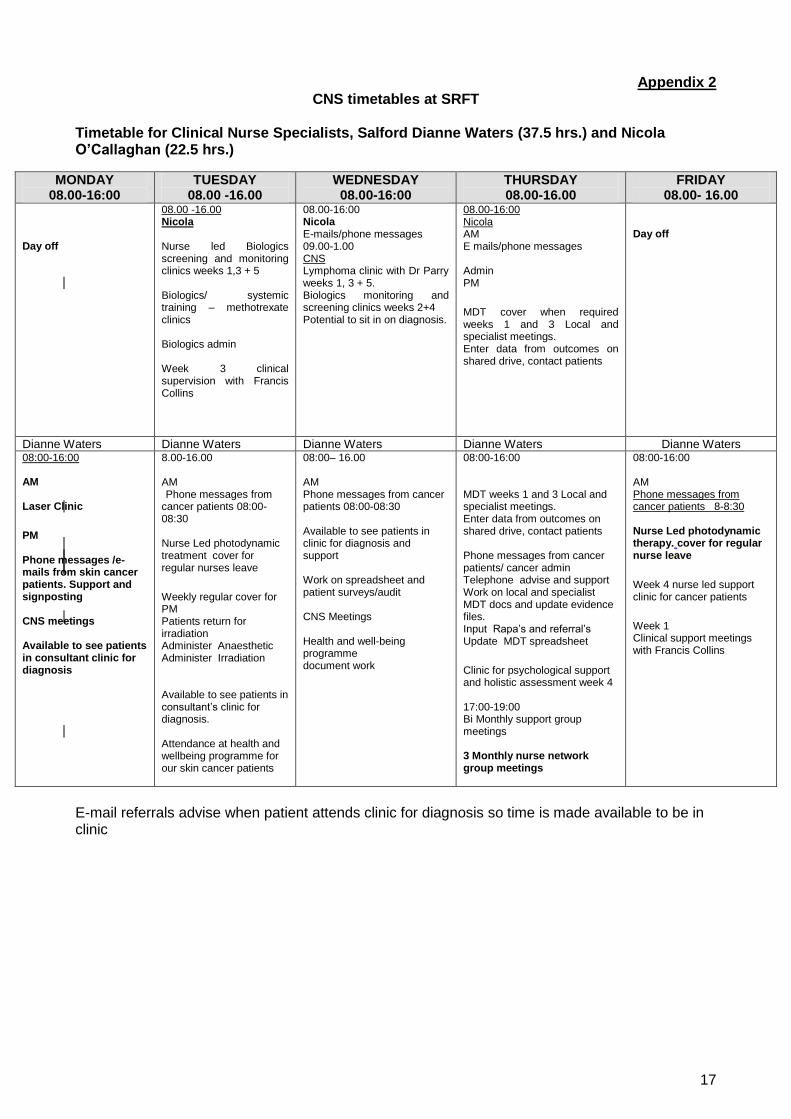
Appendix 2 CNS timetables at SRFT
Timetable for Clinical Nurse Specialists, Salford Dianne Waters (37.5 hrs.) and Nicola O’Callaghan (22.5 hrs.)
E-mail referrals advise when patient attends clinic for diagnosis so time is made available to be in clinic
MONDAY 08.00-16:00
TUESDAY 08.00 -16.00
WEDNESDAY 08.00-16:00
THURSDAY 08.00-16.00
FRIDAY 08.00- 16.00
Day off
08.00 -16.00 Nicola Nurse led Biologics screening and monitoring clinics weeks 1,3 + 5 Biologics/ systemic training – methotrexate clinics Biologics admin Week 3 clinical supervision with Francis Collins
08.00-16:00 Nicola E-mails/phone messages 09.00-1.00 CNS Lymphoma clinic with Dr Parry weeks 1, 3 + 5. Biologics monitoring and screening clinics weeks 2+4 Potential to sit in on diagnosis.
08.00-16:00 Nicola AM E mails/phone messages Admin PM
MDT cover when required weeks 1 and 3 Local and specialist meetings. Enter data from outcomes on shared drive, contact patients
Day off
Dianne Waters Dianne Waters Dianne Waters Dianne Waters Dianne Waters 08:00-16:00 AM Laser Clinic
PM Phone messages /e-mails from skin cancer patients. Support and signposting CNS meetings Available to see patients in consultant clinic for diagnosis
8.00-16.00 AM Phone messages from cancer patients 08:00-08:30 Nurse Led photodynamic treatment cover for regular nurses leave
Weekly regular cover for PM Patients return for irradiation Administer Anaesthetic Administer Irradiation Available to see patients in consultant’s clinic for diagnosis. Attendance at health and wellbeing programme for our skin cancer patients
08:00– 16.00 AM Phone messages from cancer patients 08:00-08:30 Available to see patients in clinic for diagnosis and support Work on spreadsheet and patient surveys/audit CNS Meetings Health and well-being programme document work
08:00-16:00 MDT weeks 1 and 3 Local and specialist meetings. Enter data from outcomes on shared drive, contact patients Phone messages from cancer patients/ cancer admin Telephone advise and support Work on local and specialist MDT docs and update evidence files. Input Rapa’s and referral’s Update MDT spreadsheet
Clinic for psychological support and holistic assessment week 4 17:00-19:00 Bi Monthly support group meetings 3 Monthly nurse network group meetings
08:00-16:00 AM Phone messages from cancer patients 8-8:30 Nurse Led photodynamic therapy. cover for regular nurse leave
Week 4 nurse led support clinic for cancer patients
Week 1 Clinical support meetings with Francis Collins
18
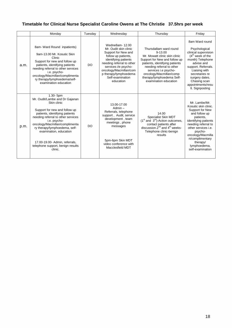
Timetable for Clinical Nurse Specialist Caroline Owens at The Christie 37.5hrs per week
Monday Tuesday Wednesday Thursday Friday
a.m.
8am- Ward Round inpatients)
9am-13.00 Mr. Kosutic Skin Clinic
Support for new and follow up patients, identifying patients
needing referral to other services i.e. psycho-
oncology/Macmillan/complimentary therapy/lymphoedema/self-
examination education
DO
Wedne8am- 12.00 Mr. Oudit skin clinic Support for New and
follow up patients, identifying patients
needing referral to other services i/e psycho-
oncology/Macmillan/comp therapy/lymphoedema
Self-examination education
Thursda8am ward round 9-13.00
Mr. Mowatt clinic skin clinic Support for New and follow up patients, identifying patients
needing referral to other services i.e psycho-
oncology/Macmillan/comp therapy/lymphoedema Self-
examination education
8am Ward round
Psychological clinical supervision
(4th week of the
month) Telephone advise and
support. Referrals. Liaising with
secretaries re surgery dates. Chasing scan
appointments/result. Signposting
p.m.
1.30- 5pm Mr. Oudit/Lambe and Dr Gajanan
Skin clinic
Support for new and follow up patients, identifying patients
needing referral to other services i.e. psycho-
oncology/Macmillan/complimentary therapy/lymphoedema, self-
examination, education
17.00-19.00- Admin, referrals, telephone support, benign results
clinic.
DO
13.00-17.00 Admin –
Referrals, telephone support , Audit, service
development , team meetings , phone
messages
5pm-6pm Skin MDT video conference with
Macclesfield MDT
14.00 Specialist Skin MDT
(1st and 3
rd)-Action outcomes,
contact patients after discussion.2
nd and 4
th weeks-
Telephone clinic-benign results
Mr. Lambe/Mr. Kosutic skin clinic. Support for New
and follow up patients,
identifying patients needing referral to other services i.e.
psycho-oncology/Macmillan/complimentary
therapy/ lymphoedema,
self-examination

19
Timetable for Julie Collins Macmillan skin cancer nurse specialist at Wythenshawe
Monday Tuesday Wednesday Thursday Friday
8.30 -9.00am handover of weekend
caseload to appropriate nurse
9.30-12pm Professional
development time -Research
-Audit Projects -Teaching Preparation -Service Development
Clinical governance monthly
Available for Prof McGrouther’s & Dr Marsland’s clinic
(every wk.) -support and information needs at new diagnosis and
follow up 12md team diary
check
8-9.30am Palliative care team MDT
Telephone advice /support
Mr. Duff’s Clinic (1st &
4th) -support and
information needs at new diagnosis and
follow up
If not in clinic review inpatients for symptom
control Psychological support
Family meetings Informal teaching
Referrals etc.
Telephone
advice/support
Review inpatients – Symptom control
Psychological support
Family meetings Informal teaching
Referrals
Available for Mr. Watson’s & Dr
Ferguson’s clinic (every week) -support and
information needs at new diagnosis and
follow up
Telephone
support/advice Mr. Wilson clinic
(1st & 3rd) support and information needs at new diagnosis and
follow up
Mole Mapping (2nd
, 4
th & 5
th) Buccleuch
Lodge (prevention,
surveillance and patient education)
Telephone advice and
support
Mr. Duff’s clinic (every wk.)
-support and information
needs at new diagnosis and
follow up
L U N C H
Mr. Wilson & Miss Rose Clinic -support
and information needs at new diagnosis and
follow up Clinical admin- Gp
notification, referrals, liaising with waiting list
clerks
Telephone advice and support
Local skin MDT (alt wks.)- pt. advocacy Mr. Kanitkar’s clinic
(alt wks.) -support and information needs at new diagnosis and
follow up
Brokering investigations and
reporting If not inpatient review
Clinical supervision (3
rd Tuesday in month)
Telephone advice and
support
Telephone advice/support
Mr. Babar’s clinic (alt wks.)
-support and information needs at new diagnosis and
follow up
Clinical Admin Rescue work psychological
support, Informal teaching, information giving,
signposting
Telephone advice and support
Specialist Skin MDT (1
st & 3
rd) –pt.
advocacy Mr. Khan clinic
(every wk.) Mr. Wilson (every
wk.) - support and
information needs at new diagnosis
and follow up Inpatient reviews – Symptom control
Psychological support
Family meetings Informal teaching
Referrals Telephone advice
and support
Available for Dr Wong’s clinic (every week) -support and information
needs at new diagnosis and
follow up
Review inpatients – Symptom
control Psychological
support Family meetings
Informal teaching Referrals
Telephone advice and
support
Daily ad hoc telephone rescue work,
case management and specialist symptom control

20
Sarah Fulton Skin Cancer CNS Christies Timetable
Monday Tuesday Wedensday Thursday Friday
TIME Location Location Location Location
8.00-9.00 Ward Round Ward Round Ward Round Ward Round Ward Round
9.00-
13.00 Mr Kosutic Clinic Emails & Catch up Mr Oudit Clinic Mr Mowatt Clinic Emails & Catch up
Suite 3 Suite 2 Suite 4
13.00-
13.30 LUNCH LUNCH LUNCH LUNCH LUNCH
13.30-
17.00 Mr Oudit Clinic Service Admin SSMDT 2pm Mr Lambe
Suite 5 Development 1st & 3rd weeks Suite 2
13.30-
17.00 Mr Lambe Macc MDT Telephone clinic Mr Kosutic
Suite 2 alt weeks HTU 2nd & 4th weeks Suite 4

21
Appendix 3
Salford Royal NHS Foundation Trust
“Two Week Wait” Referral Form Suspected Malignant Melanoma or Squamous Cell Carcinoma
(This form should not be used for suspected basal cell carcinoma, which should be referred in the normal fashion)
To make a referral, FAX this form to the RBMS on 212 4291 Patient Details:
Name: «PATIENT_Forename1» «PATIENT_Surname»
D.O.B: «PATIENT_Date_of_Birth»
Gender M F
Address: «PATIENT_House» «PATIENT_Road» «PATIENT_Locality» «PATIENT_Town»
Tel no (home): «PATIENT_Main_Comm_No»
Tel no (work):
NHS No: «PATIENT_Current_NHS_Number»
Post code: «PATIENT_Postcode» Hospital No:
From:
Address: «PRACTICE_Name» «PRACTICE_Road» «PRACTICE_Town»
GP Name:
«PATIENT_Registered_GP»
Tel no: «PRACTICE_Main_Comm_No»
Post Code: «PRACTICE_Postcode» Fax no:
Date of decision to refer: «SYSTEM_Date»
Interpreter required: First Language:
Does the patient require transport?
In patients with a suspected lesion, an urgent referral should be made and excision in Primary Care should be avoided.
Referral information (please boxes):
Diagnosis suspected Melanoma* Squamous Cell carcinoma**
* Melanoma **Squamous Cell Carcinoma
Characteristics: Characteristics:
Site Site
Size Size
Yes No Yes No
Largest diameter 7mm + Crusting/Keratinizing
Growing in size Indurate base
Changing Shape Documented growth over 8 weeks
Changing Colour
Non-healing lesions >1cm, with significant induration on palpation
Oozing/Ulceration
Inflammation
Change in sensation
Clinical History (current medication/other relevant medical history)
IT IS IMPERATIVE THAT THE PATIENT IS INFORMED OF THE NATURE OF THIS REFERRAL AND POTENTIAL CANCER DIAGNOSIS. This proforma has been produced to enable Salford Royal NHS Foundation Trust to identify Cancer Referrals identified by GP’s as being URGENT in line with the Government undertaking that all patients with suspected Cancer will be able to see a specialist within 2 weeks. URGENT REFERRALS MUST BE RECEIVED BY FAX WITHIN 24 HOURS OF THE GP’S DECISION TO REFER.

22
Appendix 3.1 «PATIENT_Forename1» «PATIENT_Surname» «PATIENT_House» «PATIENT_Road» «PATIENT_Town» «PATIENT_Postcode» ‘Two Week Wait Referral’ Patient Information Leaflet Following the recent visit to your doctor a referral has been arranged for you to go to the local
hospital for some tests. For most patients, the test results will not show signs of major illness, but
it is important that we identify quickly any conditions that could need prompt treatment.
Your doctor has referred you under the ‘Two Week Wait’ system which means that you should be
seen at the hospital or specialist clinic within 14 days.
The hospital clinic staff are likely to contact you by telephone and will offer you an appointment at
short notice. This is quite normal and means you will see a Specialist doctor quickly.
Once you have an appointment it is important that you attend the clinic. If you do find that you are
unable to attend the clinic please contact the hospital staff on 0161 206 4100.
You can bring a family member or friend with you when you attend the clinic if you wish.
If you have not heard from the hospital within 4 days please contact your GP surgery.

23
Appendix 4
Salford Royal NHS Foundation Trust
Clinical Upgrade to Suspected Cancer 62 Day Tracking for Suspected NEW Primary Cancers
Patient Name: ____________________ Consultant: ________________
Hosp Number: ______________________ DOB: ________________
Date of Upgrade: ________________________ Tumour Gp: ___________________
Please indicate the point of upgrade as indicated in the criteria below.
Option 1 Letter Triage indicates that either a Re-referral on a TWW Proforma by the GP is required or the GP will discuss the Upgrade with the patient
The Triage clinician has discussed the upgrade with the GP and the following action has been agreed:
The GP will re-refer the patient on the appropriate TWW Proforma
The GP will inform the patient change in referral status.
Yes / No Yes / No
A referral using the appropriate GP TWW Proforma should be received in Cancer Services within 7 days of the change to referral status. If a TWW referral form has not been received in Cancer Services within the specified time period a member of the team will liaise with the GP (administrative action only).
Please fax this completed form to Cancer Services on 0161 206 1303 and to Call Centre Registration on 0161 206 1048 as a matter of urgency as a matter of urgency. Thank you.
Option 2 Upgrade following First Seen Appointment (select one of the two options)
1. This patient has now been seen and the clinical opinion is Highly suspicious of cancer.
2. This patient has received an appropriate diagnostic test and has a Confirmed cancer diagnosis.
Signature of Upgrading Clinician ______________________________ Printed Name of Upgrading Clinician ______________________________ Target: From January 2009 all patients upgraded will be monitored under the cancer waiting time’s standard of 62 days
from the consultant’s decision to upgrade the urgency of a suspected cancer patients to first treatment. Day 1 of 62 commences at decision to upgrade.
Form Completion: Form must be authorised by Clinician/CNS. Please fax this completed form to Cancer Services on 0161 206 1303 as a matter of urgency. Thank you.
24
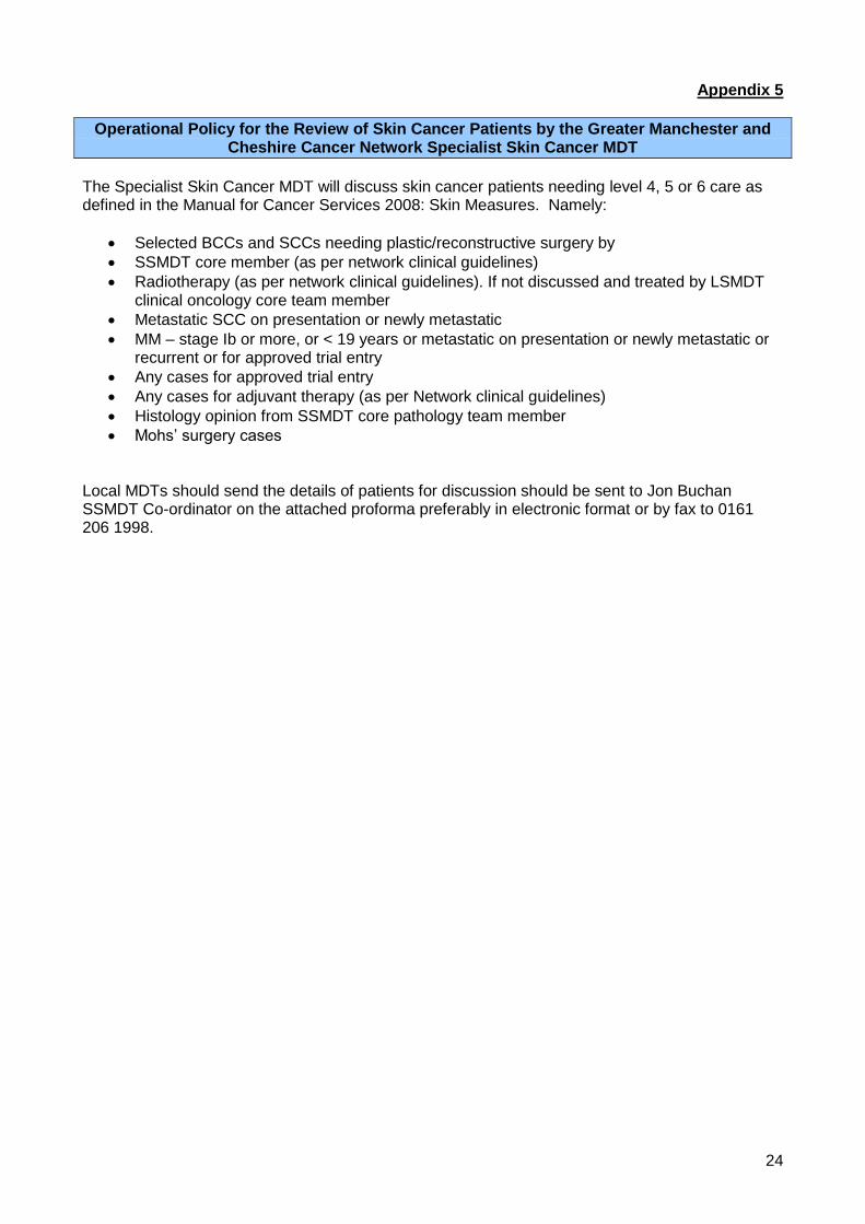
Appendix 5
Operational Policy for the Review of Skin Cancer Patients by the Greater Manchester and Cheshire Cancer Network Specialist Skin Cancer MDT
The Specialist Skin Cancer MDT will discuss skin cancer patients needing level 4, 5 or 6 care as defined in the Manual for Cancer Services 2008: Skin Measures. Namely:
Selected BCCs and SCCs needing plastic/reconstructive surgery by
SSMDT core member (as per network clinical guidelines)
Radiotherapy (as per network clinical guidelines). If not discussed and treated by LSMDT clinical oncology core team member
Metastatic SCC on presentation or newly metastatic
MM – stage Ib or more, or < 19 years or metastatic on presentation or newly metastatic or recurrent or for approved trial entry
Any cases for approved trial entry
Any cases for adjuvant therapy (as per Network clinical guidelines)
Histology opinion from SSMDT core pathology team member
Mohs’ surgery cases Local MDTs should send the details of patients for discussion should be sent to Jon Buchan SSMDT Co-ordinator on the attached proforma preferably in electronic format or by fax to 0161 206 1998.

25
Appendix 6
Patient Details Date of SSMDT
Name
DOB
NHS No.
Original Hospital
Histology Yes No
Yes No
Stage of Disease
Performance Score:
4 (completely disabled,
cannot carry out any self
care, to tally confined to
If review is required please state the question to be answered (without this a
histology review will not occur)
2 (able to walk and carry
out all self care, but unable
to work, mobile more than
5 (not recorded)
Review required:
If Radiology review is required please state the question to be answered (without this a
radiololgy review will not occur)
0 (able to carry out all normal
activities, without restriction)
1 (restricted in physically
strenuous activity, but able
to walk and do light work)
3 (limited self care, confined
to bed/chair more than 50%
waking hours)
Relevant Co-morbidities
Scans to be reviewed:
Hospital number:
GP/Consultant
Clinical information - please give specific details
SSMDT
Skin Cancer Proforma
Type of scan, date & hospital site:
Review required:
Reason for referral or question to be answered
Histology number:
Specimen type and site (e.g skin, right cheek) or specimen part (eg part A.)
Hospital site of pathology:
Radiology

26
Appendix 7
Nomination Process for patient referral to the Skin Multi-Disciplinary Team meeting (MDT) Nomination process for patient referral to the Skin Specialist Multi-Disciplinary Team Meeting (SMDT) The Specialist Skin SMDT is held in Seminar Room 7 of the Mayo Building on the first and third Thursday afternoon of every month from 2 pm to 3.30 pm. It is accepted that precise timings are not possible as the number and complexity of cases will vary.
1. Nominations to be submitted to the MDT Co-ordinator. The submission deadline for
nominations is 2 pm on the Friday prior to the meeting.
Email: [email protected] OR Fax: 0161 206 1998
The deadline for receipt of pathology and radiology reports is Friday at 10am. 2. Any patient on the cancer access database referred as a TWW who at day 21 has not been
previously discussed at an SSMDT, consideration should be made and put forward by the SSMDT Co-ordinator for discussion at the next SSMDT.
3. On receipt of the MDT Referral Proforma, the MDT Co-ordinator will list on the next MDT
Meeting Patient Review List for MDT discussion. 4. The MDT Co-ordinator will collate the SSMDT list by Thursday 10am and notify the referring
clinician of any outstanding information required for the SSMDT.
5. This list of patients for the discussion at the SSMDT will be sent electronically to core SSMDT members by Friday at 3 pm. An anonymised copy of the MDT list can be seen in Appendix 8.
Post MDT 1. Patients who are discussed at the local Skin MDT which require consideration by the Specialist
MDT will generally be discussed in the Specialist MDT meeting immediately after the LMDT.
2. Patients who require referral to another Specialist SMDT/or Consultant at another Trust will require a clinical referral letter with CaRP form sending to the Specialist SMDT Co-ordinator accompanied by the Cancer Referral Fax Cover Sheet. The responsible Consultant, if not at the meeting, will be invited to write a comprehensive referral letter at their own discretion. Such referrals are to be faxed to the Specialist Skin Cancer MDT hosted at Salford Royal prior to day 31 of the pathway. This CaRP and referral letter is sent to the Cancer Services Team, Salford Royal NHS Foundation Trust.
3. The MDT Referral Proforma (Appendix 6) will be completed and a copy placed in patients
clinical notes.
4. A record of the decision for each individual patient is kept on the recorded outcomes sheet and is circulated to all members post MDT meeting. SMDT outcome decisions are sent to Lead Clinicians, all members of SMDT and GPs.
5. A copy of the SSMDT Proforma is completed and placed in the patient’s case notes.
6. A dictated letter will be sent to the GP and used as an onward referring letter.

27
Appendix 8
Thursday, 2nd February 2017 2 PM
Salford Royal NHS Foundation Trust
Local Dermatology MDT
Patient Name D.O.B. Local Patient
No. NHS No.
Consultant /
Dermatologist Diagnosis
Ph
oto
s
Y/N
His
to
Y/N
Sc
an
s
Y/N
Dia
gn
osti
c
dis
cre
pan
cie
s
Min
or/
Sig
nif
ican
t
Comments / Outcome
06.10.32 Mr. D Mowatt
Christie-Plastic-Surgeon
MM on scalp Y Y For Palliative Radiotherapy.
20.11.43
Mr D Oudit / Dr J Byrne
Christie Plastic Surgeon / Stockport
Dermatologist
LMM on L cheek
Y For wide excision.
29.11.47 Mr S Hargreaves Bolton ENT Surgeon
Sino nasal MM Y Y Y
Needs CT of TAP and head. Awaiting BRAF status.
Already provisionally listed for XRT.
18.11.38 Dr P Lorigan
Christie Medical Oncologist
MM on R post auricular region
Aim to find out why pt. not
listed for completion lymphadenectomy previously
13.04.40
Mr D Oudit/Dr H Young
Christie Plastic surgeon/Salford Dermatologist
MM on upper back
Y
For PET/CT+/-guided biopsy. For referral to the
Wythenshawe Lung MDT-?excision/SRS
29.09.62 Mr D Mowatt Christie Plastic Surgeon
MM For MR of lumber spine then consider left axillary clearance and refer to medical oncology
05.04.48 Mr D Oudit Christie Plastic Surgeon
MM on Scalp To be considered for XRT

28
Appendix 9
Communication and Referral Proforma
Part A 17 August 2011
From: J. Buchan Referring Trust: RM301
Tel: .....0161 206 0527.............
Patient Details:
62 Day Pathway
PPI: X09__-
Hospital Number: Target Date: 06 Oct 2011
NHS Number: Day Of Journey: 12
Date Of Birth: Days Until Breach: 50
Urgent Referral Type: 08 – Dermatology
Consultant: Rhodes
Referral Details:
Trust First Seen: Trust Organisation Code: RM301 - Salford Royal
Urgent GP Suspected Cancer Referral (if applicable)
No GP Referral Received Date:
Date First Seen: 26 Jul 2011 Referral Priority Type: 01 – Routine
Consultant Upgrade Date: 05 Aug 2011 Place of Diagnosis: RM301 - Salford Royal
Screening Referral Date: Date of Diagnosis: 26 Jul 2011
Treatment Cycle: 01 – Initial ICD10 Code: C43.7
18 week start date 27 May 2011
Referral to Treat Status Existing
Date of Decision to Refer (to a treating trust)
First Seen WTA Reason: 09 - No adjustment to waiting time
First Seen WTA: 0
Actual Tumour Group: 08 – Dermatology
Part B Date of Decision to Treat
Clinician (Referring): Prof L Rhodes Clinician Referring Trust: RM301
Delay Reason (DTT):
Clinician Referred To: Plastic Surgeon Clinician Referred To Trust: Christie
Trust Organisation Code: RBV01 - Christie Hospital
Has a referral letter (with reports) been sent to clinician?
Yes
Referred for Treatment: Yes
Referred for Diagnosis: No
Planned Treatment Type:
Important – The information contained in this document is private & confidential and intended for the exclusive use of the addressee. If you are not the addressee, any disclosure, reproduction, distribution or other dissemination or use of this communication is strictly prohibited. If you have received this transmission in error, please contact the sender immediately by telephone so that we can arrange its return. Thank you

29
Appendix 10
Greater Manchester & Cheshire Cancer Network
Clinical Guidelines Peer Review Measure (A12/S/b-16-004)
The following guidelines have been adopted by the GMCCN Skin NSSG:
1. Guidelines for the management of basal cell carcinoma. British Association of Dermatologists, 2008
2. Multiprofessional guidelines for the management of the patient with primary cutaneous
squamous cell carcinoma. British Association of Dermatologists, 2009
3. Revised UK guidelines for the management of cutaneous melanoma. British Association of Dermatologists, 2010
4. Joint British Association of Dermatologists and UK Cutaneous Lymphoma Group guidelines
for the management of primary cutaneous T-cell lymphomas, 2006 Internet links to these documents can be found below: http://www.bad.org.uk/site/622/default.aspx The NSSG will review the above guidelines at least annually or as and when new guidance is published.
30
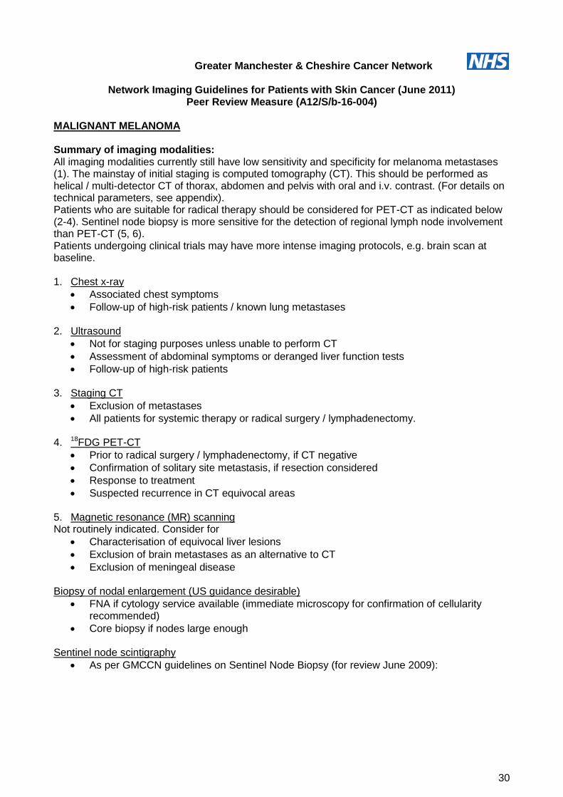
Greater Manchester & Cheshire Cancer Network
Network Imaging Guidelines for Patients with Skin Cancer (June 2011) Peer Review Measure (A12/S/b-16-004)
MALIGNANT MELANOMA Summary of imaging modalities: All imaging modalities currently still have low sensitivity and specificity for melanoma metastases (1). The mainstay of initial staging is computed tomography (CT). This should be performed as helical / multi-detector CT of thorax, abdomen and pelvis with oral and i.v. contrast. (For details on technical parameters, see appendix). Patients who are suitable for radical therapy should be considered for PET-CT as indicated below (2-4). Sentinel node biopsy is more sensitive for the detection of regional lymph node involvement than PET-CT (5, 6). Patients undergoing clinical trials may have more intense imaging protocols, e.g. brain scan at baseline. 1. Chest x-ray
Associated chest symptoms
Follow-up of high-risk patients / known lung metastases 2. Ultrasound
Not for staging purposes unless unable to perform CT
Assessment of abdominal symptoms or deranged liver function tests
Follow-up of high-risk patients 3. Staging CT
Exclusion of metastases
All patients for systemic therapy or radical surgery / lymphadenectomy. 4. 18FDG PET-CT
Prior to radical surgery / lymphadenectomy, if CT negative
Confirmation of solitary site metastasis, if resection considered
Response to treatment
Suspected recurrence in CT equivocal areas 5. Magnetic resonance (MR) scanning Not routinely indicated. Consider for
Characterisation of equivocal liver lesions
Exclusion of brain metastases as an alternative to CT
Exclusion of meningeal disease
Biopsy of nodal enlargement (US guidance desirable)
FNA if cytology service available (immediate microscopy for confirmation of cellularity recommended)
Core biopsy if nodes large enough Sentinel node scintigraphy
As per GMCCN guidelines on Sentinel Node Biopsy (for review June 2009):

31

32
Initial imaging by pathological tumour stage Stage I & IIA
Imaging not routinely indicated, to evaluate specific signs or symptoms only
Chest x-ray or CT for suspected metastases, according to symptoms
PET-CT and MRI only by discussion within the context of the MDT Stage IIB and above
CT thorax, abdomen and pelvis
Consider PET-CT if CT negative where management affected
MR for equivocal CT / organ-specific assessment (e.g. liver, brain) Follow-up imaging Stage I
Routine imaging not indicated Stage IIA
Annual chest x-ray Stage IIB and above Patient on observation:
As dictated by symptoms Patients receiving treatment:
CT @ Baseline, mid- and end-of-treatment (3 & 6 months)
6-monthly CT thereafter Nodal recurrence
Staging CT +/- PET-CT if treatment appropriate (as above) Distant metastatic disease
CT if impact on management NON-MELANOMA SKIN CANCER Squamous Cell Carcinoma and Basal Cell Carcinoma Baseline investigations including CXR as clinically indicated. In advanced local disease consider MR for assessment of local invasion. Distant staging: CT of chest and abdo +/- pelvis to including loco-regional nodes (e.g. neck). Consider PET-CT in:
Advanced disease suitable for radical treatment, if negative CT
Equivocal nodal disease Merkel Cell Tumour Stage 1 Chest x-ray only. Discuss other imaging with radiologist at Skin MDT. Consider sentinel node biopsy Stage 2 As for Stage IIb Melanoma.
33
References 1. Veit-Haibach P, Vogt FM, Jablonka R, et al. Diagnostic accuracy of contrast-enhanced FDG-PET/CT in primary staging of cutaneous malignant melanoma. Eur J Nucl Med Mol Imaging. 2009;36(6):910-918. 2. Krug B, Crott R, Lonneux M, Baurain JF, Pirson AS, Vander Borght T. Role of PET in the initial staging of cutaneous malignant melanoma: systematic review. Radiology. 2008; 249(3):836-844. 3. Pfannenberg C, Aschoff P, Schanz S, et al. Prospective comparison of 18F-fluorodeoxyglucose positron emission tomography/computed tomography and whole-body magnetic resonance imaging in staging of advanced malignant melanoma. Eur J Cancer. 2007;43(3):557-564. 4. Reinhardt MJ, Joe AY, Jaeger U, et al. Diagnostic performance of whole body dual modality 18F-FDG PET/CT imaging for N- and M-staging of malignant melanoma: experience with 250 consecutive patients. J Clin Oncol. 2006;24(7):1178-1187. 5. El-Maraghi RH, Kielar AZ. PET vs sentinel lymph node biopsy for staging melanoma: a patient intervention, comparison, outcome analysis. J Am Coll Radiol. 2008;5(8):924-931. 6. Aloia TA, Gershenwald JE, Andtbacka RH, et al. Utility of computed tomography and magnetic resonance imaging staging before completion lymphadenectomy in patients with sentinel lymph node-positive melanoma. J Clin Oncol. 2006;24(18):2858-2865.

34
Sample protocol for melanoma staging using MD-CT (March 2009) Preparation: 900mls Gastrografin 3% or 800mls EZ-CAT Positioning: Supine, feet first Centering Point: Xiphisternum iv Contrast: Iohexol 300: 100mls at 3 ml/sec Smart Prep ROI: Aortic Arch Scan Region, Parameters and Reconstructions:
Region Slice thickness
Slice interval
Pitch Detectors kV Reconstructions
Lung apex – top of liver
5mm 5mm 1.5 0.75mm 120 Axial: 1. 3mm/3mm B31f kernel, Abdo windows
Axial: 3mm/3mm B80f kernel, Lung windows
Sag & Coronal: 5mm/5mm B41 kernel, Abdo windows
Top of liver – symphysis
5mm 5mm 1.5 0.75mm 120 Axial: 1. 3mm/3mm B31f kernel, Abdo windows
Axial: 3mm/3mm B80f kernel, Lung windows
Sag & Coronal: 5mm/5mm B41 kernel, Abdo windows

35
Christie Melanoma Disease Group Recommendations for the Surgical Management of Cutaneous Melanoma
Investigations Imaging CT scans may be considered for all patients with stage IIB disease or greater. Strong consideration to be given to patients with tumours ≥T4, unless a positive scan would not alter management. Standard staging CT scan should include imaging of the thorax, abdomen and pelvis. In addition all patients should have an informed discussion on the pros and cons of including the brain in staging scan and their verbal consent to imaging sought. For patients with a primary tumour of the head and neck undergoing imaging, this should also include a CT of the head and neck. USS examination and USS assisted FNA of the nodal basin may be considered for assessment of suspected nodal disease. PETCT should be considered for carcinoma of unknown primary or for patients with stage III/IV melanoma where a positive result would change the extent of surgical resection. Blood tests The role of blood tests (full blood count, biochemistry) as a staging investigation is unclear. They are routinely required for clinical trials, and were previously done for all patients with tumour ≥T4. It is proposed that this continues. Blood tests may be requires as part of the pre-op assessment or in patients with significant comorbidity. Surgical Margins The recommended clinical margins for wide local excision are Tis 5mm clinical margin, no re-excision required if complete histological clearance achieved T1 10mm margin T2 10-20mm margin T3/4 20-30mm margin Sentinel node biopsy All patients with melanomas stage 1B should be referred for consideration of sentinel node biopsy. The inclusion of patients with tumours less than 1mm needs to be determined based on the potential risk of occult disease versus the morbidity associated with the procedure”. For tumours over 4mm the benefits of detecting occult disease need to be weighed up against the increased risks of false negativity, the morbidity of the procedure and the increased difficulty of monitoring the nodal basin following a negative sentinel node biopsy Lymphadenectomy Microscopic nodal disease:
There is no clear benefit to completion lymphadenectomy in patients with sentinel nodal disease based on the MSLT-1, study, though the results of the MSLT-2 study are awaited. However, it is still considered an appropriate treatment for the majority of patients.
The risks of developing metastatic disease are lower in patients with small volume disease in the lymph nodes, and data from a large series indicates that for tumour deposits <0.1 mm, this is equivalent to node negative patients. Accuracy of pickup is heavily dependent on the protocol used to process the node. The majority of patients with tumour deposits > 0.1m will go on to completion lymphadenectomy
Where nodal disease is detected out with the normal drainage basin or where equivocal disease is present either clinically or radiologically management should be determined on an individual patient basis. Where disease encroaches into an unusual site, opinions regarding resectability and extent of surgery may require discussion with other disease orientated groups.

36
Macroscopic nodal disease: Therapeutic lymphadenectomy is recommended for all patients with macroscopic nodal disease. Pelvic Node Dissection Pelvic node dissection should be considered for:-
1. Patients with >1 clinically palpable inguinal / femoral triangle node
2. CT / Ultrasound evidence of > 1 involved node evidence of pelvic nodes
3. Patients with > 1 microscopically involved Sentinel node
4. Patients with Conglomerate of inguinal / femoral triangle nodes
5. Microscopic / macroscopic involvement of Cloquet’s node
These recommendations need to be discussed with the patient regarding the pros and cons of the technique. Oncological Referral Referral is recommended for all Stage 2B patients and above.
Indications for radiotherapy for melanoma
Adjuvant local radiotherapy Post-operative radiotherapy can be considered in order to maximise local control when either inadequate margins or macroscopic disease remains in situ and further surgery is not feasible for medical reasons, for unacceptable morbidity, or for cosmetic limitations. For Sino nasal melanoma, because of the difficulty in achieving a R0 resection, and the significant morbidity associated with local recurrence adjuvant post-operative radiotherapy should be considered. Where surgical resection, following discussion at the head and neck and skull based MDT’s, is deemed inappropriate radical radiotherapy may be considered. The role of adjuvant radiotherapy in patients with rectal or anal canal melanoma who have undergone a narrow margin excision is unclear, but is used in a number of centres and should be referred to the colorectal/anal MDT.

37
Adjuvant nodal radiotherapy This is controversial and potential improvements in local control need to be balanced with treatment related morbidity. International trials are on-going (TROG). Retrospective series suggest improved local control when radiotherapy is used after lymph node dissection in high-risk patients. It should be considered if:
More than 4 nodes, or more than 50% of the nodes sampled are involved. Assuming an adequate number of nodes dissected (the number can be discussed).
N2a disease or greater in the neck. Significant extra capsular spread. Known residual disease.
Palliative radiotherapy Painful/bleeding or troublesome lesions in bone and soft tissue may be palliated with short courses of conventionally fractionated radiotherapy. Cranial radiotherapy may benefit patients with brain metastases who are able to maintain reasonable performance status with or without steroids. Follow up The melanoma follow up as per the BAD guidelines is to be discussed at the skin CSG for potential adoption. Stage 1A follow up for 12 months (2-4 times) locally. 5 year follow up may be considered. Stage 1B – 111A 3 monthly for 3 years and 6 monthly for 5 years. 10 year follow up may be considered. Stage 111B - 111C 3 monthly for 3 years and 6 monthly for 5 years and yearly for a further 5 years Stage IV – as per patient requirements It is suggested that patients with Stage 1/11 melanoma be offered follow up locally including patients with multiple naevi. Patients staged SNB –ve should be followed up jointly by plastics/locally. Stage 111/IV patients should be followed up surgically/oncologically in multidisciplinary clinics and have yearly dermatology follow up locally due to increased risk of second primaries.

38
Network guidelines for sentinel node biopsy in melanoma - Development of network guidelines and validation for the network
The recent development of BAD guidelines in the management of malignant melanoma has highlighted the importance of sentinel node biopsy as a staging tool1. Investigation and management of lymph node basins in melanoma patients should be carried out by the network SSMDTs. There is no place for elective lymph node dissection in the management of primary melanoma unless this is unavoidable because the primary melanoma lies over the lymph node basin (Level Ib, Grade A). Sentinel lymph node biopsy (SLNB) was developed as a means of identifying the first lymph node draining the skin in which the melanoma arises2. The procedure is carried out at the same time as definitive wider excision of the primary melanoma3. SLNB gives information about prognosis, and is therefore increasingly a requirement for randomisation into clinical trials. Patients with melanoma of Breslow thickness1.2-3.5 mm and a positive SLNB have a 75% five-year survival compared with 90% if the SLNB is negative.83 SLNB is normally considered for patients with melanoma ≥ 1 mm, when about 20% are positive; however the risk of a positive SLNB in a melanoma < 1.0 mm is still 5%4,5. The procedure is associated with a 5% morbidity, which is less than that seen with complete nodal dissection. In patients with a positive SLNB, 20% have pathological evidence of metastases in additional regional nodes2. Patients with a positive SLNB therefore proceed to completion lymphadenectomy in the majority of cases. In about 5% it is not possible to identify the sentinel node either on lymphoscintigraphy, at surgery, or both. Patients should be aware of this limitation. A false-positive SLNB is possible6. The relevance of increasingly detailed evaluation of the sentinel node and its correlation with prognosis remains to be defined. MSLT-1 showed no overall five-year survival benefit following SLNB and completion lymphadenectomy7 The BAD guidelines have recommended that sentinel node biopsy be offered to all patients staged 1B or higher. The Christie currently provides the sentinel node biopsy service for melanoma for the greater Manchester network and these guidelines document the current treatment pathway and guidelines for sentinel node biopsy within the network. This documents details the guidelines used in sentinel node biopsy for nuclear medicine, surgery and pathology. 1 J.R. Marsden, J.A. Newton-Bishop, L. Burrows, M. Cook,P.G. Corrie, N.H. Cox,M.E. Gore, P. Lorigan, R. Mackie, P. Nathan, H. Peach, B. Powell,C. Walker. British Association of Dermatologists Guidelines for the Management of Cutaneous Melanoma 2009. Br J Dermatology (in print) 2 Morton DL, Wen DR, Wong JH et al. Technical details of intraoperative lymphatic mapping for early stage Melanoma. Arch Surg 1992; 127:392-9. 3. Morton D, Thompson J, Cochrane A et al. Sentinel node biopsy or nodal observation in melanoma. N Eng. J Med 2006; 355:1307-17. 4. Morton DL, Cochran AJ, Thompson JF et al. Sentinel node biopsy for early stage melanoma - Accuracy and Morbidity in MSLT-1, an international multicentre trial. Ann Surg 2005; 242:302-13. 5. Wright BE, Scheri RP, Ye X et al. Importance of sentinel lymph node biopsy in patients with thin melanoma. Arch Surg 2008; 143:892-9. 6. Weiguo L, Stall A, Shivers SC et al. Clinical relevance of molecular staging for melanoma: comparison of RTPCR and immunohistochemistry staining in sentinel lymph nodes of patients with melanoma. Ann Surg 2000; 231:795-803. 7. Karakousis CP, Emrich LJ, Rao U et al. Groin dissection In malignant melanoma. Am J Surg 1986; 152:491-5.

39
NETWORK NUCLEAR MEDICINE GUIDELINES FOR SENTINEL NODE LOCALISATION
Patient preparation none normally. Exceptionally it may be helpful to have Emla cream applied to the primary site at least one hour before injection, for very sensitive locations. To prevent contamination, use an Incopad with a hole cut in the centre, and taped in place, to isolate injection sites.
Radiopharmaceutical Tc-99m HSA Nano colloid (Nanocoll™) Activity 40 MBq in 0.2 - 0.4 ml for study on day before surgery 20 MBq in 0.2 - 0.4 ml for study on same day as surgery
Small air bubble behind liquid may help to ensure complete delivery. Measure activity in syringe before injection, using Syrad.
ARSAC DRL 40 MBq Route of administration Intradermal via a fresh orange needle
Injection split between four locations as close as possible to, and surrounding the primary site. Note – gentle massage of the injection sites stimulates lymphatic flow. Direct static pressure inhibits flow.
Imaging delay immediate dynamic for up to 30 mins, followed by ant and lateral
statics, if delayed images not necessary Delayed 2-3 hours statics if necessary Delayed 4-6 hours statics if necessary
Collimators Low energy high resolution IMAGES Protocols User / Sentinel Node /…. Sentinel node dynamic for initial dynamic and static Sentinel node statics for delayed statics only Summary
Type View Pre-set
Dynamic Nodal basin most likely to contain sentinel node(s) – image may or may not include the injection site
60 x 30 seconds
Static Nodal basin as above. Other areas which may contain sentinel node(s)
5 minutes each
Static Lateral view(s) for depth measurement, or for approximate A-P position of nodes, or for orthogonal view to clarify node locations
5 minutes

40
Where to image
Primary location
Dynamic images Static images
Head or neck Ant or post, and lateral head, neck and upper chest (L-mode)
As dynamic. + Laterals of head and neck
Anterior trunk * Ant and post axillae and shoulders, arms down
Ant and post axillae and shoulders, with arm(s) abducted laterally to 90 degrees. + Lateral(s) of axillae with arm(s) extended over head, for approximate A-P position of node(s) + Ant pelvis (and lateral pelvis for node depth measurement if nodes seen – see “node depth measurement”)
Posterior trunk * Ant and post axillae and shoulders, arms down (patient supine)
Ant and post axillae and shoulders, with arm(s) abducted laterally to 90 degrees. + Lateral(s) of axilla(e) with arm(s) extended over head, for approximate A-P position of node(s) + Ant pelvis (and lateral pelvis for node depth measurement if nodes seen – see “node depth measurement”)
Upper arm Ant and post axilla, shoulders and upper arm, with the arm abducted laterally at 90 degrees
As dynamic - anterior to include upper arm (“interval” nodes are sometimes seen at mid-upper arm) + Lateral of axilla with arm extended over head, for approximate A-P position of node(s)
Forearm / hand Ant and post axilla, shoulders and upper arm, with the arm abducted laterally at 90 degrees
As dynamic. + Arm image to include elbow and upper arm (“interval” nodes are sometimes seen at mid-upper arm, and epitrochlear nodes are sometimes seen at the elbow) + Lateral of axilla with arm extended over head, for approximate A-P position of node(s)
Upper leg Ant pelvis As dynamic, to include ant and post upper leg (though “interval” nodes within upper leg are rare) + Lateral pelvis for node depth measurement - see “node depth measurement”
Lower leg Ant pelvis
As dynamic, to include ant and post upper leg (though “interval” nodes within upper leg are rare) + Lateral pelvis for node depth measurement – see “node depth measurement” + Ant and post knees for popliteal nodes behind knees. To mark these – patient prone for posterior view.
* It cannot be assumed that sentinel node(s) for primaries on the trunk will only be seen on the same side of midline as the primary tumour, even when the primary is well away from midline. Node(s) may be seen on both sides, though not necessarily at the same time. When to image
Start dynamic imaging as soon as possible after injection. Continue for 20-30 minutes.
At the end of this time, it is necessary to make a judgement about the need for delayed imaging at 2-3 hours, and even again at 4-6 hours if these are inconclusive. The dynamic

41
frames can be displayed in “snake” format during the acquisition by selecting to display “acquired” images on the View control box.
Delayed imaging is necessary in those cases where sentinel nodes are not clearly seen in the early images, in all regions that could potentially drain the primary site. As a guide :-
Primary location Delayed imaging
Head or neck Possibly
Anterior trunk Possibly
Posterior trunk Possibly
Arm Not needed if sentinel nodes seen in ipsilateral shoulder and axilla. Epitrochlear nodes, if present, are seen before axillary nodes.
Leg Not needed if sentinel nodes seen in ipsilateral groin and higher in the pelvis. Popliteal nodes, if present, are seen before groin nodes.
If delayed imaging is not needed, proceed straight to static images after the dynamic. Which node(s) to mark
Sentinel nodes are defined as the first node or nodes that receive drainage directly from the tumour. It may be necessary to view the initial dynamic image to identify the sentinel nodes.
If drainage to the node basin is along a single channel, then usually one node will be seen first. This is the sentinel node, and should be marked. Nodes further along the channel may become visible later, but these should not be marked.
If there is drainage along more than one channel, then there may be a sentinel node in each channel, and these may be seen at similar or different times. They should all be marked.
If it is not clear whether or not to mark an additional node, then you should mark it.
Sentinel nodes known as “interval” nodes are quite common. These are nodes situated away from the standard nodal fields in the axillae and groins etc. If found, they should be marked.
Note that it is possible for pooling of lymphatic fluid (sometimes called “lakes”), to occur in channels which are dilated, partially blocked, or temporarily trapped. The pooling may appear like a hot node. If the activity within the area varies up and down during the dynamic sequence, it is not a node. Usually the pooling will clear with exercise, or before delayed images, and node(s) beyond the pool will become evident. If the node locations or appearances are suspicious, then limited exercise (e.g. patient off couch and walking) may be helpful in clarification. Note that the dilated or restricted channel may become hot again during subsequent imaging.
How to mark the node(s) after static images have been acquired
Make a Tc-99m point source, as follows. Draw up 0.2 ml of the Nanocoll into a 1 ml syringe, and re-sheath the needle. Holding the syringe vertically downwards, carefully press the plunger until a few drops of solution are in the end of the needle sheath. Then replace the syringe and needle with clean ones, keeping the original sheath vertical so that the only activity is at the tip of the sheath. Keep the syringe and sheath vertical from then on, in a suitable labelled lead pot.
Acquire the 5-minute static image first, at each orientation. Use Copy to add further 5-minute statics, so that the persistence image is available for marking.
Leave the camera in persistence mode.
Position the patient and camera as appropriate for the node location. It is recommended that the patient should be supine, except for marking popliteal nodes, or interval nodes on the posterior trunk. For marking axillary nodes on the anterior skin surface, the patient’s arm should be abducted laterally at 90 degrees, as at surgery. For marking axillary nodes from a lateral view, the patient’s arm should be raised over their head. It is recommended that all
42
marking should be done from ant, post or lateral projections, rather than oblique’s which are difficult to replicate in theatre.
Set the persistence refresh rate to “None” to accumulate the persistence image, so that nodes are clearly seen.
In the Image Settings control box, click on “Show ROI”. Then click on the ROI edge on the image required (usually detector 1).
Drag the ROI across the image until the central cross overlies the centre of the node to be marked.
Set the persistence refresh rate to “High”.
Without moving the patient, one operator should move the marker source over the patient’s skin, guided by instruction from the other operator looking at the persistence image. It may be necessary to refresh the persistence image periodically by pressing “Clear”.
When the source position overlies the cross on the display screen, the position of the source on the patient should be marked on the patient’s skin with an indelible marker.
When the mark has been made, check the position by reapplying the marker source to the skin and checking the image.
Repeat as necessary for each further node. Repeat as necessary for each further node. Before acquiring further images it is necessary to reset the position (“pan”) of the image back to zero in X and Y directions.
When complete, move the camera head and couch to improve access to the patient, and clarify and enhance the skin marks with the indelible marker. Explain to the patient that these must not be scrubbed off before the operation.
Node depth measurement: this is helpful for groin nodes in particular, to establish whether a particular node is inguinal or pelvic. A lateral image can be used to measure the node depth. During acquisition of a lateral image, mark the anterior skin surface on the image by slowly drawing the Tc-99m point source across the skin, along a sup-inf line through the position of the skin mark(s).
Body outlining: this is not normally necessary – it can help with interpretation of the images by inexperienced surgeons or for demonstration to other staff. During acquisition of the final anterior image, the patient’s body outline may be marked on the image by slowly drawing the Tc-99m point source along each lateral edge of the body – or by positioning a Co-57 flood source behind the patient.
Hardcopy and communication with the surgeons
View the relevant initial dynamic image using general image display to establish the order in which nodes appeared, and identify sentinel nodes.
Use Analysis / Image processing / reframe / composite to sum the dynamic dataset, and save it with the name “summed”.
See the archive file for examples of suitable image displays and annotations, and follow these guidelines :- o Display the summed image, and relevant static images of the nodes, setting the
colour scale to reverse hot iron. o Label each image with orientation and approximate time since injection. Mark
Right and Left clearly. If there are multiple skin marks, these must be explained by annotating the images. Add sufficient annotation to the display to make image orientations clear.
o If there are important observations from the viewing of the dynamic sequence -for instance a hotspot which looks like a node, but which varied up and down in intensity during the acquisition, and is therefore not a node – annotate the images accordingly.
o Do not hardcopy any images that may have been acquiring whilst marker sources were being moved across the patient to locate the nodes, other than any laterals used for node depth measurement. These images would be very confusing.
o If a lateral view has been taken for inguinal nodes, use the electronic cursors on the Annotation card to measure the depth of the node(s) from the anterior skin surface.
43
Then delete the cursor line (and with it the depth value), and manually annotate the image with the measured depth value.
Save the final display as a Screen Capture (1024 colour) called Screen Cap.
Hardcopy on paper (2 copies) on the Windows printer – one for the patient’s notes the other for the archive file.
The hard copied images and their annotations form a preliminary report of the study, which will be seen and used by the surgeons before a formal report is produced. It is essential that the final display is double-checked by another experienced member of staff.
Send one of the hardcopies to the ward as soon as possible, so that it can be seen by the surgeons when they see the patient prior to the operation.
The study will be reported either by a suitably experienced member of staff, as arranged with the ARSAC certificate holder, or by the one of the certificate holders. If sending a hardcopy away for final reporting, add any other information which may be helpful to the radiologist, for example further detail on the location of the primary.
SURGERY FOR SENTINEL NODE BIOPSY “All patients with stage 1B or higher should be referred to the Christie for consideration of sentinel node biopsy” Inclusion Criteria The most important inclusion criterion for SNB is a clinically negative nodal basin, as defined by physical examination. Although clinical imaging has been used to stage the nodal basin such as computed tomography (CT), contrast-enhanced magnetic resonance imaging (MRI), ultrasound-guided fine-needle aspiration cytology (USg-FNAC) and/or 18F-fluoro-deoxyglucose (FDG) positron emission tomography (PET) with or without low dose CT (PET/CT) there remains considerable debate on the preferred imaging modality, and to date none have the ability to detect small or micro metastatic tumour deposits. Gross lymphatic involvement can lead to distortion of the normal architecture, leading to aberrant drainage patterns and biopsy of false sentinel nodes. SNB is therefore contraindicated to stage clinically positive nodal basins and where gross disease is apparent FNAC with or without radiological control is recommended.
All patients with stage 1B or higher should be referred for consideration of sentinel node biopsy.
Patients who have received prior radiation or surgical treatment to the nodal basin are routinely excluded from SNB protocols, since the previous intervention can distort the normal lymphatic pathways and give rise to unexpected patterns of metastasis. It is possible that lymphatic mapping and SNB may yield potentially useful information in these patients. Similarly, patients with small recurrent or second primary tumours may also benefit from lymphatic mapping to guide surgical intervention. However, these applications of the SNB technique, whilst clinically attractive, remain largely unexplored.
Exclusion Criteria
In pregnant women, the urgency and the necessity of staging the nodal basin should be discussed. Lymphoscintigraphy is specifically contraindicated in the pelvis of pregnant women. SNB protocols should be modified in pregnant patients to minimize the risks of radiation exposure and blue-dye injections. SNB can be performed in lactating women but it is advised that breastfeeding be discontinued following the procedure. In the UK, Nanocolloid is approved for use in children, though licensing varies between countries. The potential benefits of sentinel node biopsy are not as well delineated in the paediatric population and, in practice; most SNB trial protocols generally exclude these patients from participation. Off-label use of radiopharmaceuticals should be considered with caution and respect to an individual risk-benefit analysis. Other relative

44
contraindications include a known allergy to albumin colloid, and primary tumour treatment by external beam radiotherapy.
The use of blue dye is a useful adjunct to aid SLN localization and harvest. Blue dye is contraindicated in children, pregnant women, lactating women and those who have a history of allergy to the blue dye or any of the products.
Procedure
At the time of lymphoscintigraphy, SLNs are marked on the skin surface under scintigraphic guidance of a Co57-labeled pen marker (held 90 degrees to the skin surface) and controlled transcutaneously by the nuclear medicine physician
Following radiotracer injection and LSG, patients undergo general anaesthesia and preparation for operation.
One vial of dye is injected slowly into the tissues surrounding and deep to the tumour/scar, to minimize leakage from ulcerated tumours. Gauze swabs may be used to protect normal tissue and mop up excess dye. The number of injections is usually between two and four, but is as many as is necessary to completely surround the tumour on its deep and lateral surfaces.
In the operating room, the gamma probe is covered with sterile latex and applied transcutaneously to confirm the accuracy of the skin markings, which may have changed due to changes in patient positioning between LSG and surgery. The theoretically optimal search pattern is to start closest to the injection site, with the probe perpendicular to the skin, using a raster pattern of 2 cm-spaced parallel lines at right angles to the direction of the injection site. A rise in activity is then confirmed by scanning in the other direction. Scanning should be no faster than a few cm/s. However, excessively slow scanning can lead to loss of information from the change in pitch as the probe passes over a hot node. The drop in counts as the probe is angulated whilst over a hot spot can confirm location. Location The lymphoscintigraphy images and skin markings guide the site of incision, which is placed along the relaxed skin tension lines and positioned to facilitate excision of the scar should subsequent neck dissection be required. Sentinel nodes are reached using one or more small incisions, and removed. The gamma probe is introduced into the space along the plane of dissection and angled in various directions to guide the surgeon to the sentinel node which is then excised. Blue-stained lymphatics may be followed to the sentinel nodes, which may be hot, blue, or both. The anatomical location of the sentinel nodes should be noted, as should its colour and radioactivity ex vivo in the operating theatre because both blue dye and radioactivity will dissipate before the pathological examination. Following excision of SLNs, repeated readings are taken of the excision bed to ensure that there are no adjacent hot nodes that also need to be removed.
While preoperative imaging should detect the majority of grossly involved nodes, clinical staging remains imperfect. Any suspicious lymph nodes observed during SNB must be excised, even in the absence of radioactivity, since gross lymphatic involvement may block the flow of radiotracer to these nodes.
45
Activity Counting
Following excision, SLN radioactivity is confirmed ex-vivo using the gamma probe, and must be above background activity to be classified as hot. The SLN should be placed on a surface away from the patient, or on the upturned probe tip (facing the ceiling). Anatomic location and radioactivity levels (cps) are recorded for all excised nodes. All radioactive nodes should be considered SLNs because, while there exists some confusion over the exact definition of a SLN, it is best to err on the side of patient safety. The lymphatic basin should be rechecked for reduced radioactivity after SLN excision. Bed counts in the after removal of SLN almost never exceed 8 - 10 cps (with the head of the probe slightly turned away from the injection site).
Risk
Sentinel node biopsy is not without risks such as wound infection, haematoma, seroma, lymphocele and nerve/vascular damage. Although complications rates of less than 1% are reported.
Experience
It is clear that experience is needed before a surgeon starts performing sentinel node biopsy, as it carries a steep learning curve. The technique should only be performed by surgeons who have had adequate training and validated their results. It is recommended that all surgeons be evaluated following their first 30 cases to ensure safety of the technique. Surgeons should be mentored during their validation phase. In addition, it is necessary for practitioners of SNB to understand the theoretical aspects including handling of radioactivity and optimal use of gamma probes. The same elements of training are required for nuclear medicine staff and pathologists.

46
Pathology Evaluation of Sentinel Node
Gross Sectioning
Sentinel nodes are transported to the laboratory unfixed or in formalin. The surgeon should, as evidence to support the sentinel status of the submitted node, record detected radioactivity in the requisition. Sentinel node specimens should have neither crushed nor cautery artefacts. Sentinel node may be partially dissected free of associated fat, but leaving some so that the lymphatics can be assessed for the presence of tumour. During dissection, the Sentinel node is examined for blue coloration and measured (maximum length x width in mm). The node is bisected through its longest meridian. Cut surfaces are carefully examined for metastases or collections of melanin. SN halves are placed in a single cassette.
Step Sectioning
Six pairs of sections are cut at 50-μm intervals and stained respectively with H&E, S100, and Melan A as proposed by EORTC clinical trials group. Spare sections are made available at each level to help resolve difficult cases by additional immunohistochemistry. (See below) SENTINEL NODE MELANOMA- PROTOCOL Initially:
1. H&E only 2. Spare 3. Spare
If H& E negative then: 2. S100 on spare 3. Melan A on spare 50 micron gap 4. H&E 5. S100 6. Melan A 7. Spare 50 micron gap 8. H&E 9. S100 10. Melan A 11. Spare 50 micron gap 12. H&E 13. S100 14. Melan A 15. Spare 50 micron gap 16. H&E 17. S100 18. Melan A 19. Spare 50 micron gap 20. H&E

47
21. S100 22. Melan A 23. Spare Microscopy Report The following information is included in the histopathology report: Number of tumour foci; Location of tumour (subcapsular, parenchymal); Presence of extracapsular spread; Diameter of the largest deposit; Tumour immunophenotype (S100, Melan A, HMB-45, others); Naevus (capsule, trabeculae). For measurements: • Only measure what you can see. • The most reproducible measurement is a single measurement of the maximum diameter of the largest lesion in any direction (1-D). N.B. Isolated Tumor Cells, Micrometastasis, and Sentinel Lymph Nodes The 7th edition TNM staging system defines nodal involvement by the presence of any tumor cells regardless of quantity, size, or mode of detection as nodal involvement (i.e., >pN0). Macrometastasis is defined histologically as when nodal metastasis exhibits gross Extracapsular extension Validation Assessment
The success of this multidisciplinary staging technique depends on good communication between all of the individual components; imaging, surgery, and histopathology. The multidisciplinary team (SSMDT) setting should be utilised for the discussion of every patient, and regular audit of patient outcomes against published data should be carried out. The accuracy of the technique can be assessed by the proportions of patients whose SLN contains metastases, which should match that of END (20-30%, depending on patient population and tumour size. Lastly, the rate of false negatives (SLN-negative patients who develop early recurrent disease) should be < 5-10%.
Further studies are required to determine the full clinical significance of micrometastasis and individual tumour cells to determine the need for block dissection in patients with isolated tumour cells and small micrometastasis.
48
Appendix 10
Greater Manchester & Cheshire Cancer Network
Agreed Network Pathology Guidelines Peer Review Measure (14-2J-210)
Dr L A Jamieson, Consultant Dermatopathologist at Salford Royal is the Lead pathologist for the SSMDT. Dr L Motta, Consultant Dermatopathologist at Salford Royal is the deputy lead. Dr Lia Menasce, Consultant Histopathologist at Christie Hospital NHS Trust, is the Lead Pathologist in the network for cutaneous lymphomas. Double reporting of Malignant Melanomas and Severely Dysplastic Naevi was established at the end of 2010. The Skin NSSG has adopted the Royal College of Pathologist’s Guidelines for histological reporting of Basal Cell Carcinoma, Squamous Cell Carcinoma, Malignant Melanoma and Lymphoma. Internet links to these documents can be found below: http://www.rcpath.org/resources/pdf/skincancers2802.pdf Basal Cell Carcinoma – page 5 Squamous Cell Carcinoma – page 11 Malignant Melanoma – page 17 Lymphoma http://www.rcpath.org/resources/pdf/lymphomaminimumdatasetCORRECTED.pdf *The NSSG will review the above guidelines at least annually or as and when new guidance is published.
49
Greater Manchester & Cheshire Cancer Network
Referral for Extra Corporeal Photophoresis (ECP) Peer Review Measure (A12/S/b-16-005))
The Greater Manchester & Cheshire Cancer Network Board, North West Specialist Commissioning Group, and the Skin Cancer Network Site Specific Group have agreed that: Cases of erythrodermic cutaneous T-cell lymphoma, stages III & IVa, are suitable for consideration of ECP. All referrals should come via the STLMDT from Salford or Christie, and should be discussed with the clinician in charge of the ECP facility. The named facility for extra corporeal Photophoresis which the GMCCN will use is:
Manchester Blood Centre, Plymouth Grove, Manchester, M13 9LL. The clinician in charge is:
Dr Deepak Sadani

50
Greater Manchester & Cheshire Cancer Network
Pathway for Skin Lymphomas (A/S/b-16-005) Cases of lymphoma presenting in the skin should be investigated locally and discussed at the local skin MDT and local Haemato-oncology MDT, in order to determine the presence or absence of systemic involvement. LYMPHOMA
Primary investigation and discussion
Investigations to determine presence or absence
of systemic involvement (May also be done at LSMDT or Haemato-
Oncology MDT level)
Local Skin MDT (LSMDT) (Wigan; Bolton, Salford, Trafford, Central Manchester, Tameside, Stockport, South
Manchester, East Cheshire, Mid Cheshire)
Systemic / nodal lymphomas identified
Haemato-Oncology MDT MMM
Supra Network T-cell lymphoma MDT (STLMDT)
(Christie, Salford)
Cutaneous lymphomas identified
Supra Network T-cell lymphoma MDT
(STLMDT)

51
PRIMARY CUTANEOUS B CELL LYMPHOMA
PRIMARY CUTANEOUS T CELL LYMPHOMA – MYCOSIS FUNGOIDES Primary Cutaneous T cell lymphoma should be referred to the Supra-network T-cell Lymphoma MDT (STLMDT) as below:
* Refer to Haemato-Oncology MDT when STLMDT has requested radiotherapy / chemotherapy or further / repeat investigation to be administered locally as shared care or where patient is too ill to travel to Manchester
Primary investigation and discussion
Supra Network T-cell lymphoma MDT (STLMDT) (Christie)
Primary investigation & discussion
Local Skin MDT * (LSMDT) (Wigan; Bolton, Salford, Trafford, Central Manchester, Tameside, Stockport, South
Manchester, East Cheshire, Mid Cheshire)
Supra Network T-cell lymphoma MDT* (STLMDT) (As below)
Stage IA, IB, IIA, and III: Refer to Dr Eileen Parry
Salford Royal
Stage IIB and IV: Refer to Dr Richard Cowan
Christie Hospital

52
CUTANEOUS LYMPHOMAS – CD30+ve Spectrum
OTHER CUTANEOUS T CELL LYMPHOMAS NB: Patients from Wigan Local Skin MDT are referred to the Specialist Skin MDT and plastic surgeons at Whiston Hospital, St Helens & Knowsley NHS Trust, part of the Mersey & Cheshire Cancer Network. GMCCN Patients may be referred to Mr Telfer, Mr Ghura, or Mr Madan, Consultant Mohs Surgeons based at Salford.
Primary discussion and treatment
Local Skin MDT (LSMDT) (Wigan; Bolton, Salford, Trafford, Central Manchester, Tameside, Stockport, South
Manchester, East Cheshire, Mid Cheshire)
Supra Network T-cell lymphoma MDT (STLMDT) (Christie)
LYMPHOMATOID PAPULOSIS Management by Local Skin MDT
(LSMDT) (Wigan; Bolton, Salford, Trafford, Central Manchester, Tameside,
Stockport, South Manchester, East Cheshire, Mid Cheshire)
CD30+ve CUTANEOUS LYMPHOMA
Supra Network T-cell lymphoma MDT (STLMDT)
(Christie)
Review by Supra-network T-cell Lymphoma MDT

53
NB: Patients from Wigan Local Skin MDT are referred to the Specialist Skin MDT and plastic surgeons at Whiston Hospital, St Helens & Knowsley NHS Trust, part of the Mersey & Cheshire Cancer Network. GMCCN Patients may be referred to Mr Telfer, Mr Ghura, or Mr Madan, Consultant Mohs Surgeons based at Salford.
Primary investigation and discussion
Local Skin MDT (Wigan; Bolton; Salford; Tameside; Stockport; Trafford, Central &
South Manchester; East Cheshire; Mid Cheshire)
Refer to Supra-Regional Sarcoma Meeting (Christie)
all deep soft tissue sarcomas secondarily
involving skin
Refer all cases of incompletely excised or recurrent dermatofibrosarcoma
protuberans, all cases of angisarcoma, cutaneous leiomyosarcoma and any other
cutaneous sarcomas to the Supra-Regional Sarcoma Meeting
(Christie)
Specialist Skin MDT (Salford)

54
Patient Pathway for Basal Cell Carcinoma
CLINICALLY OR HISTOLOGICALLY DIAGNOSED
BASAL CELL CARCINOMA
Low Risk Tumour Based on Clinical features, Histology,
Body Site (non-head / neck)
Surgical Intervention (in some cases this may be undertaken
by appropriately accredited GPSIs
Non Surgical Intervention. Topical Therapy: 5FU,
Imiquimod, Photodynamic Therapy, Cryosurgery.
Follow Up depending on Treatment Modality, Local Policy and patient history. Also discuss -Self monitoring. Signs and symptoms. Re-accessing service. Support (including
psychological). Risk reduction/prevention
High Risk Tumour Based on Clinical features, Histology,
Body Site, Recurrence, Immuno-suppression, Size, Genetic Factors.
Surgical Intervention. Excision with predetermined
margins either by Plastic Surgeon or Dermatologist
Referral for Mohs opinion +/- Plastics, ENT Oculoplastic
Surgery. SSMDT discussion if required
Radiotherapy e.g. if patient is poor surgical risk or
unwilling to undergo surgery.
LSMDT discussion if necessary e.g. incompletely excised

55
Patient Pathway for Squamous Cell Carcinoma
CLINICALLY DIAGNOSED OR BIOPSY PROVEN SCC
Low Risk Tumour (Based on Clinical features, Histology, Body Site, Recurrence, Immuno-suppression, Size, Genetic
Factors).
Surgical Management within Dermatology Centre or referral to Plastics depending on size and site.
Follow up depending on-site, histological staging. Also discuss -Self monitoring. Signs and
symptoms. Re-accessing service. Support (inc psychological). Risk reduction/prevention as
appropriate.
High Risk Tumour (Based on Clinical features, Histology,
Body Site, Recurrence, Immuno-suppression, Size, Genetic Factors).
Treatment options: Excisional surgery, Radiotherapy or Mohs opinion.
SSMDT discussion if required
Follow up depending on-site, immune state & histological staging. Also
discuss – Self monitoring. Signs and symptoms. Re-accessing service.
Support (including psychological). Risk reduction/prevention as appropriate
Treatment options: Excisional surgery, Radiotherapy or Mohs opinion
LSMDT discussion if required e.g.
incompletely excised

56
Patient Pathway for Malignant Melanoma
Primary excision with 2-5mm lateral margin
If < 1mm re-excision with 1cm Margin either at SRFT or refer to Plastics depending on body site.
If > than 1mm refer to Plastics for further management Check Vit D level
Nodular Melanoma or +ve Nodes refer to Plastics for Further Management.
In Situ Melanoma No further surgical intervention required. No follow up required.
3 monthly follow up for 3 years then discharge. Also discuss -Self monitoring. Signs and symptoms. Re-accessing service. Support (including psychological). Risk reduction/prevention
3 monthly follow up for 3 years then 6 monthly for further 2 years. Either by Dermatologist or Plastics, depending on tumour depth and staging.
Clinical Diagnosis / Suspicion of Malignant Melanoma

57
31 & 62 Day Referral
CONFIDENTIAL FACSIMILIE TRANSMISSION
To: __________________________________________________ From : __________________________________________________ Date: __________________________________________________ Recipients Fax No: ___________________________________________ Safe Haven fax number No. of pages (including cover sheet) ____________________________
A copy of this fax has also been sent to : (tick to confirm)
For patients transferred to the Christie Hospital Carole Hughes Fax No 446 3352 For patients transferred to South Manchester (Wythenshawe) Hospital Helen Burgess Fax No 291 2612 For patients transferred to Central Manchester (MRI) Hospital Liz Guilfoyle Fax No 276 8756 For patients transferred to North Manchester (NMGH) Hospital Juliette Mottram Fax No 778 8718
Appendix 11
58
For TYA Service Use Only
59
Date Received: ……………………………………….. Screened by: ……………………………………….. Date: …………………………………… Outcome Following Screening
o Not for Appointment
o Appointment with Nurse Consultant for assessment
o Telephone advice to referrer
o Referral to TYA medical team
o Referral to Youth Support Coordinator
o Referral to TYA Community Liaison
o Referral to Social Work Team
o Referral to TYAMDT
First Appointment Date
Outcome of Advice
Name: …………………………………………..
Date: …………………………………………..

60
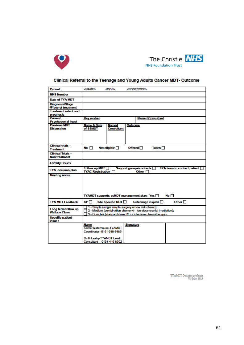
61