Embed Size (px)
Citation preview
CASE REPORTHip Pelvis 28(4): 249-253, 2016http://dx.doi.org/10.5371/hp.2016.28.4.249
Copyright ⓒ 2016 by Korean Hip Society 249
Print ISSN 2287-3260Online ISSN 2287-3279
One of the catastrophic complications of total hiparthroplasty (THA) is wear of articulating surfaces,leading to local metallosis. Metallosis has been reportedwith some designs of metal-on-metal articulations or afterthe exchange of fractured ceramic bearings with a metalhead and a polyethylene acetabular liner1). Metal ions andnanoparticles can disseminate through blood and lymphaticvessels and cause systemic complications1,2). We present a
case of pulmonary granulomatous disease secondary torevision of a fractured ceramic THA head.
CASE REPORT
In 2005, an otherwise healthy 55 year-old femalepatient, with a primary diagnosis of hip osteoarthritisand otherwise unremarkable history, underwent bilateralstaged THA at another institution. A cementless systemwith a ceramic-on-ceramic articulation was used on bothoccasions (R3 acetabulum, Synergy femoral stem; Smith& Nephew, Memphis, TN, USA). Commencing earlypostoperatively, the patient had recurrent dislocation ofher right arthroplasty (Fig. 1A) as often as 3-4 times ayear, which she managed to reduce on her own. Six yearspostoperatively, the patient sustained a fracture of theright ceramic head (Fig. 1B). The acetabular prosthesiswas exchanged with a tantalum shell and an ultra-highmolecular weight polyethylene liner. The existing stemwas well fixed and therefore was left in situ. The
Granulomatous Lung Disease:A Novel Complication followingMetallosis from Hip Arthroplasty
Theodoros Balbouzis, MD, Thomas Georgiadis, MD*, Peter Grigoris, FRCS, FACSDepartment of Orthopedics, Iaso General Hospital, Athens, Greece
Laboratory of Pathology, Hygeia General Hospital, Athens, Greece*
A case of a female patient with local and systemic complications of metallosis, following catastrophic wear of arevised hip arthroplasty, is presented. The patient had a history of a fractured ceramic-on-ceramic implant,exchanged with a metal-on-polyethylene prosthesis. Systemic complications included sarcoidosis-like reactions,presenting as granulomatous lung disease, along with chorioretinitis, erythema nodosum, and cardiomyopathy.High local and circulating cobalt and chromium levels established the diagnosis. The patient underwentextensive debridement and implant revision. One year postoperatively, she had no respiratory symptoms orfunctional impairment. Local and systemic complications of metallosis after hip arthroplasty should be promptlyrecognized and treated operatively.
Key Words: Hip replacement arthroplasty, Pulmonary sarcoidosis, Heavy metal toxicity, Ceramics, Reoperation
Submitted: October 14, 2016 1st revision: November 30, 2016Final acceptance: December 9, 2016Address reprint request toTheodoros Balbouzis, MDDepartment of Orthopedics, Iaso General Hospital, Office 1.17,Mesogeion 264, Holargos 15562, GreeceTEL: +30-210-7290959 FAX: +82-53-250-7205E-mail: [email protected]
This is an Open Access article distributed under the terms of the CreativeCommons Attribution Non-Commercial License (http://creativecommons.org/licenses/by-nc/4.0) which permits unrestricted non-commercial use,distribution, and reproduction in any medium, provided the original work isproperly cited.

www.hipandpelvis.or.kr250
Hip Pelvis 28(4): 249-253, 2016
fractured ceramic head was exchanged with one made ofcobalt-chromium alloy (Continuum acetabulum, Versyshead; Zimmer, Warsaw, IN, USA) (Fig. 1C).
In the following months, the patient gradually developedsevere activity related right hip pain. Six months post-revision, the patient developed erythema nodosum of theleft leg and bilateral chorioretinitis and papilledema. A
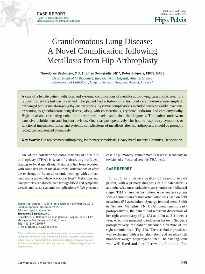
chest computed tomography (CT) revealed diffuseradiopaque nodules in both lungs and enlargement of theright hilum (Fig. 2). Biopsy of the right lung showedfibrosis and extensive inflammation with multiple non-necrotizing granulomata, consisting of histiocytes,multinucleated giant cells, and lymphocytes (Fig. 3).Special stains for mycobacteria and fungi were negative
FFiigg.. 11.. (AA) Posterior dislocation of the original arthroplasty. (BB) Multiple episodes of dislocation ultimately led to a fracture ofthe ceramic head. (CC) At the first revision, a metal head and a new metal shell with a polyethylene liner were inserted. (DD)Absorption of the greater trochanter occurred within three years. (EE) A second revision with a long stem and ceramicbearings was undertaken (radiograph at 12 months postoperatively).
A B
C D E
FFiigg.. 22.. Multiple granulomata were present in both lungs (arrows).
A B

www.hipandpelvis.or.kr 251
Theodoros Balbouzis et al. Granulomatous Lung Disease following Metallosis from Hip Arthroplasty
and no atypical cells were found. A diagnosis ofsarcoidosis was made. The patient received corticosteroidsand azathioprine for six months, which led to clinical andradiological improvement. Between 2013 and 2015,echocardiograms and chest magnetic resonance imagingshowed severe cardiomyopathy with the ejection fractiondecreasing from 70% to 35% and evidence of non-ischemic type fibrosis of the lower and posterior ventricularwall, as well as moderate dilation of the left ventricle.
The patient sought advice from our team in February2015. Physical examination revealed a 76 kg, 159 cmtall woman, with a severely antalgic gait and painful,
decreased range of motion of the right hip. Radiographicimaging showed severe osteolysis of the proximal femur(Fig. 1D). A CT of pelvis revealed an iliac pseudotumor(Fig. 4). The presence of a pseudotumor, extensiveosteolysis, and the history of a fractured ceramic implantexchanged with a metal alloy head and a polyethyleneliner, aroused suspicion for metallosis. A hip aspirationproduced black fluid with high concentrations ofchromium and cobalt, 25,400μg/L and 26,500μg/Lrespectively (Fig. 5). Whole blood chromium and cobaltconcentrations were also significantly elevated, 31.8μg/Land 22.2μg/L respectively.
It was therefore decided to proceed with a secondrevision operation. Intraoperatively, gross localmetallosis was found and large volumes of black fluidpoured out from a trochanteric pseudotumor. There wasextensive osteolysis of the proximal femur but the stemremained fixed (Fig. 6A). Multiple ceramic fragmentswere still present in the periprosthetic tissues. Ceramicdebris was also embedded in the articular surface of thepolyethylene liner (Fig. 6B). The metallic head wasdistorted due to massive wear (Fig. 6C). The Morsetaper of the stem was also damaged, allowing freerotation of the prosthetic head (Fig. 6D).
Meticulous soft tissue debridement was carried out.
FFiigg.. 33.. Resection biopsy from the upper right lung revealedextensive, non-necrotizing inflammation, with multiplegranulomata (white arrows), consisting of histiocytes,lymphocytes and multinucleated giant cells (black arrows),arranged around vessels and bronchial walls (H&E stain,××100).
FFiigg.. 44.. A large mass (arrow) in close contact with the iliacbone represented a pseudotumor.
FFiigg.. 55.. Hip aspiration produced abundant black-stainedmetallic fluid.

www.hipandpelvis.or.kr252
Hip Pelvis 28(4): 249-253, 2016
The acetabular shell, which remained well fixed, wasleft in place but the worn polyethylene liner was removed.The femoral stem was exchanged with a long revisionimplant (Wagner SL revision uncemented stem; ZimmerGmbH, Winterthur, Switzerland). To prevent recurrenceof the problem, a ceramic-on-ceramic articulation wasinserted (Biolox Delta; Zimmer GmbH) (Fig. 1E).
Recovery was uneventful. At three months, bloodchromium and cobalt levels had returned to normal, 1.17μg/L and 0.19μg/L respectively. One year later, thepatient was able to mobilize painlessly, using onecrutch. She had no respiratory symptoms and herpulmonary function tests were normal. Her heartfunction had stabilized to an ejection fraction of 50%.
DISCUSSION
To our knowledge, this is the first report, associating
granulomatous lung disease with wear of metalimplants, following the revision of a failed ceramic-on-ceramic THA. Granulomatous lung disease has beenattributed to exposure to a variety of environmental andoccupational agents, including mycobacteria, fungi,pollen, building materials and metallic dust and fumes3,4).Sarcoidosis is a diagnosis of exclusion, suggesting anunrecognized causal factor, causing non-necrotic, non-infectious granulomas that are clinically and histologicallyindistinguishable from secondary granulomas3). Sinceinitially no causative agent had been identified, theoverall presentation of the patient had been characterizedas sarcoidosis. The identification of the hip arthroplastyas a source of heavy metal ions and microparticlesoffered a reasonable specific etiology.
Inhalation of metallic fumes and dust has beenassociated with pulmonary fibrosis and giant cellinfiltration of pulmonary interstitial tissue4). Metal
FFiigg.. 66.. (AA) At second revision, metallosis in the periprosthetic region was evident, necessitating generous debridement. (BB)Ceramic debris was found embedded in the polyethylene liner, causing wear of the metallic head. (CC) Excessive wearresulted in loss of the spherical shape of the prosthetic femoral head. (DD) The Morse taper of the femoral prosthesisappeared scratched and distorted.
A B
C D

www.hipandpelvis.or.kr 253
Theodoros Balbouzis et al. Granulomatous Lung Disease following Metallosis from Hip Arthroplasty
nanoparticles are internalized by distant tissue cells,inducing chromosomal damage and oxidative stress2).Macrophage and monocyte activation, followingexposure to metal nanoparticles and ions, has beenproven in vitro5). This event could be the first step in acascade leading to granuloma formation6). It can beexpected that similar tissue reactions occur withhematogenous spread of heavy metal particles. Péoc’h etal.7) have reported granulomatous lesions in the spleenand liver after a metal-on-polythylene THA, withhistological evidence of titanium and polyethyleneinfiltration of macrophages.
In young patients, ceramic implants are consideredmore appropriate than the usual, metal-on-polyethylenebearings, because they produce less wear debris,allowing for increased longevity of the reconstruction.However, ceramic materials are associated with an up to1.5% risk of fracture8). Revision of a fractured ceramicimplant, using a metal head and a polyethyleneacetabular liner, may result in catastrophic wear of thenew head1). Since complete removal of the ceramicdebris from the periprosthetic tissues is impossible,embedding of remaining debris in the polyethylene lineracts as sandpaper, damaging the relatively softer newmetallic head and producing metal nanoparticles andions, causing local metallosis and systemic toxicity.
Gessner et al.9) identified twenty five cases of systemictoxicity following metallosis from chrome/cobalt hiparthroplasty, with thyroid dysfunction, psychosocialmanifestations, sensory and motor neuropathy, visualand auditory disturbances, or cardiomyopathy. However,since five of these cases have been identified by thesame team in Alaska only, it is probable that systemiccomplications following metallosis from hip replacementare underreported9). It is therefore important, for bothphysicians and orthopedic surgeons, to suspect metallosisin patients who present with systemic disorders andhave a history of a fractured ceramic bearing exchangedwith a metal-on-polyethylene combination. Diagnosticdifficulties arise with elderly patients, who, due to alower activity level, have only mild symptoms from apotentially malfunctioning prosthetic joint, and whosemedical problems can be easily attributed to comorbiditiesor medications. Guidelines, such as those of the UKMedicines and Health-related Products Regulatory
Agency10), imposing frequent monitoring of blood ionlevels and repeated imaging in arthroplasties with highrisk for metal release, can lead to earlier diagnosis. Withprompt synovectomy, lavage and revision to a ceramic-on-ceramic bearing, systemic complications of metallosiscan be avoided.
CONFLICT OF INTEREST
The authors declare that there is no potential conflictof interest relevant to this article.
REFERENCES
01.Oldenburg M, Wegner R, Baur X. Severe cobalt intoxicationdue to prosthesis wear in repeated total hip arthroplasty. JArthroplasty. 2009;24:825.e15-20.
02.Keegan GM, Learmonth ID, Case CP. Orthopaedic metalsand their potential toxicity in the arthroplasty patient: Areview of current knowledge and future strategies. J BoneJoint Surg Br. 2007;89:567-73.
03. Iannuzzi MC, Rybicki BA, Teirstein AS. Sarcoidosis. NEngl J Med. 2007;357:2153-65.
04.Nemery B. Metal toxicity and the respiratory tract. EurRespir J. 1990;3:202-19.
05.Newman KL, Newman LS. Occupational causes ofsarcoidosis. Curr Opin Allergy Clin Immunol. 2012;12:145-50.
06.Caicedo MS, Desai R, McAllister K, Reddy A, Jacobs JJ,Hallab NJ. Soluble and particulate Co-Cr-Mo alloyimplant metals activate the inflammasome dangersignaling pathway in human macrophages: a novelmechanism for implant debris reactivity. J Orthop Res.2009;27:847-54.
07.Péoc’h M, Moulin C, Pasquier B. Systemic granulomatousreaction to a foreign body after hip replacement. N Engl JMed. 1996;335:133-4.
08.Koo KH, Ha YC, Jung WH, Kim SR, Yoo JJ, Kim HJ.Isolated fracture of the ceramic head after third-generation alumina-on-alumina total hip arthroplasty. JBone Joint Surg Am. 2008;90:329-36.
09.Gessner BD, Steck T, Woelber E, Tower SS. A systematicreview of systemic cobaltism after wear or corrosion ofchrome-cobalt hip implants. J Patient Saf. Publishedonline June 12, 2015; doi: 10.2106/JBJS.F.01489.
10.Medicines and Healthcare Products Regulatory Agency.Appendix. Management recommendations for patients withmetal-on-metal hip replacement implants [Internet].Medicines and Healthcare Products Regulatory Agency;2012 Jun 25. Available from: https://assets.digital.cabinet-office.gov.uk/media/5485abf640f0b6024400027d/con155766.pdf.