Embed Size (px)
Citation preview
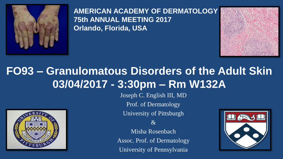
FO93 – Granulomatous Disorders of the Adult Skin
03/04/2017 - 3:30pm – Rm W132A
Joseph C. English III, MD
Prof. of Dermatology
University of Pittsburgh
&
Misha Rosenbach
Assoc. Prof. of Dermatology
University of Pennsylvania
AMERICAN ACADEMY OF DERMATOLOGY
75th ANNUAL MEETING 2017
Orlando, Florida, USA
Disclaimer:
• Conflict of Interest
– None
• Financial Associations
– None
• Off-label drug usage may be
discussed

Practice Gap: Granulomatous disorders represent a unique group of diseases, both
noninfectious and infectious, that require the utmost clinical pathologic
correlation combined with a keen sense of inquiry for underlying systemic
disease and immunosuppression.
Dermatologists need to be able to differentiate these entities, evaluate patients
for specific underlying systemic disease (i.e. from diabetes to cancer), and treat
them with a wide range of immunosuppressant to anti-infectious medications.
By being aware of the skin manifestations of these abnormal physiologic
responses dermatologists can improve patient safety, outcome and health care
costs.
Objectives:
• Differentiate the Non-infectious granulomatous diseases
(NIGD) and Infectious granulomatous diseases (IGD) of adult
skin.
• Recognize the cutaneous clinical, histological and systemic
manifestations of NIGD and IGD.
• Determine the best evidence based therapeutic modalities for
the treatment of NIGD and IGD.
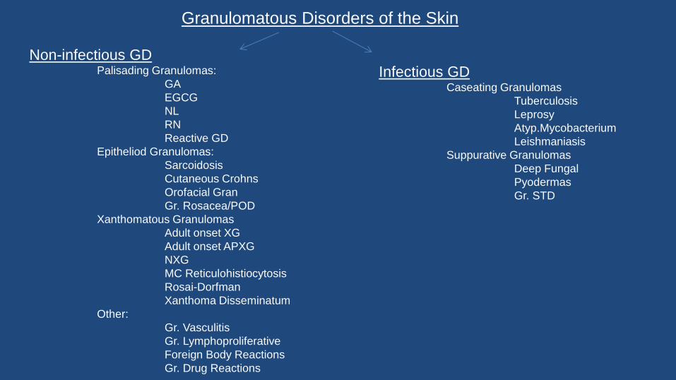
Granulomatous Disorders of the Skin
Non-infectious GD Palisading Granulomas:
GA
EGCG
NL
RN
Reactive GD
Epitheliod Granulomas:
Sarcoidosis
Cutaneous Crohns
Orofacial Gran
Gr. Rosacea/POD
Xanthomatous Granulomas
Adult onset XG
Adult onset APXG
NXG
MC Reticulohistiocytosis
Rosai-Dorfman
Xanthoma Disseminatum
Other:
Gr. Vasculitis
Gr. Lymphoproliferative
Foreign Body Reactions
Gr. Drug Reactions
Infectious GD Caseating Granulomas
Tuberculosis
Leprosy
Atyp.Mycobacterium
Leishmaniasis
Suppurative Granulomas
Deep Fungal
Pyodermas
Gr. STD
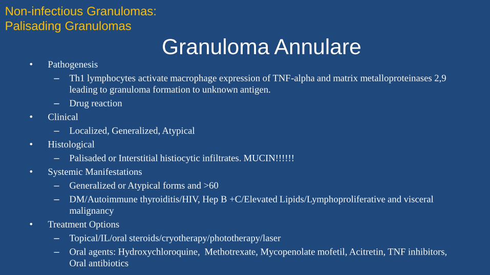
Granuloma Annulare • Pathogenesis
– Th1 lymphocytes activate macrophage expression of TNF-alpha and matrix metalloproteinases 2,9
leading to granuloma formation to unknown antigen.
– Drug reaction
• Clinical
– Localized, Generalized, Atypical
• Histological
– Palisaded or Interstitial histiocytic infiltrates. MUCIN!!!!!!
• Systemic Manifestations
– Generalized or Atypical forms and >60
– DM/Autoimmune thyroiditis/HIV, Hep B +C/Elevated Lipids/Lymphoproliferative and visceral
malignancy
• Treatment Options
– Topical/IL/oral steroids/cryotherapy/phototherapy/laser
– Oral agents: Hydroxychloroquine, Methotrexate, Mycopenolate mofetil, Acitretin, TNF inhibitors,
Oral antibiotics
Non-infectious Granulomas:
Palisading Granulomas
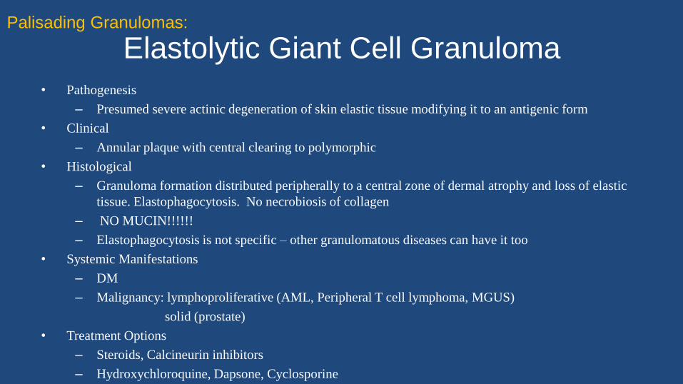
Elastolytic Giant Cell Granuloma
• Pathogenesis
– Presumed severe actinic degeneration of skin elastic tissue modifying it to an antigenic form
• Clinical
– Annular plaque with central clearing to polymorphic
• Histological
– Granuloma formation distributed peripherally to a central zone of dermal atrophy and loss of elastic
tissue. Elastophagocytosis. No necrobiosis of collagen
– NO MUCIN!!!!!!
– Elastophagocytosis is not specific – other granulomatous diseases can have it too
• Systemic Manifestations
– DM
– Malignancy: lymphoproliferative (AML, Peripheral T cell lymphoma, MGUS)
solid (prostate)
• Treatment Options
– Steroids, Calcineurin inhibitors
– Hydroxychloroquine, Dapsone, Cyclosporine
Palisading Granulomas:

Necrobiosis Lipoidica • Pathogenesis
– T-cell mediated hypersensitivity reaction to altered collagen production with immunologically
mediated vascular disease and trauma
• Clinical
– Early/Middle/Late Stages (85% on the lower legs)
• Histological
– Multiple layers of hyalinized and necrotic collagen, surround by histiocytes and lymphocytes.
– NO MUCIN
• Systemic Manifestations
– DM/Retinopathy and nephropathy/Ocular inflammation (Retinal vasculitis in Non-DM) /Joint
immobility
• Treatment Options:
– Steroids
– Hydroxychloroquine, Pentoxyfylline, Cyclosporine, Mycophenolate mofetil, Thalidomide, Biologics
(IL-infliximab), Retinoids
– Pulse Dye, PDT, Hyperbaric oxygen, topical PUVA , UVA-1
Palisading Granulomas:
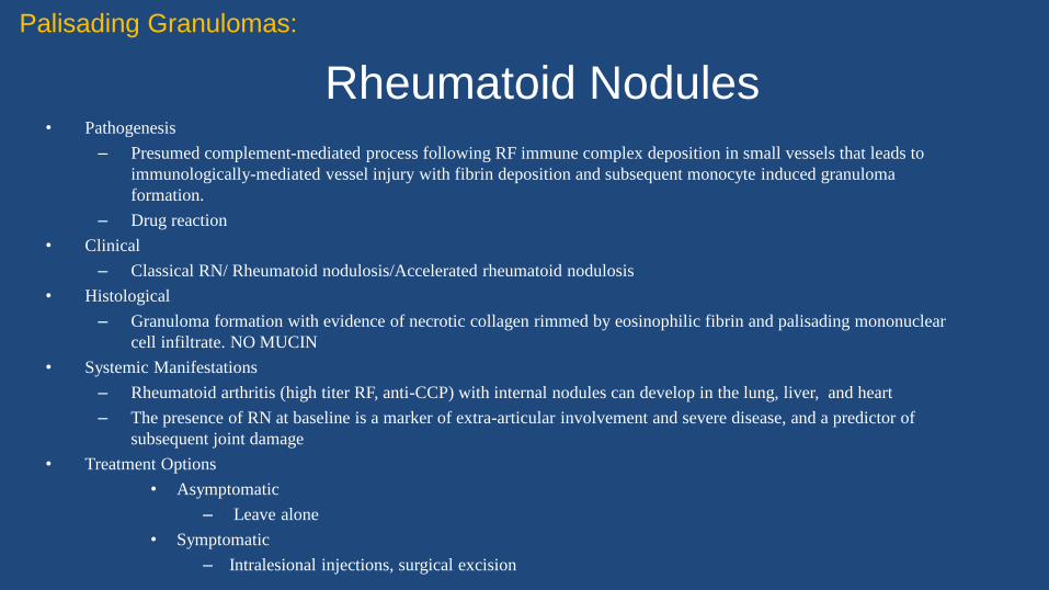
Rheumatoid Nodules
• Pathogenesis
– Presumed complement-mediated process following RF immune complex deposition in small vessels that leads to
immunologically-mediated vessel injury with fibrin deposition and subsequent monocyte induced granuloma
formation.
– Drug reaction
• Clinical
– Classical RN/ Rheumatoid nodulosis/Accelerated rheumatoid nodulosis
• Histological
– Granuloma formation with evidence of necrotic collagen rimmed by eosinophilic fibrin and palisading mononuclear
cell infiltrate. NO MUCIN
• Systemic Manifestations
– Rheumatoid arthritis (high titer RF, anti-CCP) with internal nodules can develop in the lung, liver, and heart
– The presence of RN at baseline is a marker of extra-articular involvement and severe disease, and a predictor of
subsequent joint damage
• Treatment Options
• Asymptomatic
– Leave alone
• Symptomatic
– Intralesional injections, surgical excision
Palisading Granulomas:
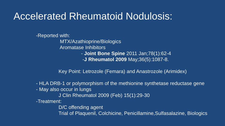
Accelerated Rheumatoid Nodulosis: -Reported with:
MTX/Azathioprine/Biologics
Aromatase Inhibitors
- Joint Bone Spine 2011 Jan;78(1):62-4
-J Rheumatol 2009 May;36(5):1087-8.
Key Point: Letrozole (Femara) and Anastrozole (Arimidex)
- HLA DRB-1 or polymorphism of the methionine synthetase reductase gene
- May also occur in lungs
J Clin Rheumatol 2009 (Feb) 15(1):29-30
-Treatment:
D/C offending agent
Trial of Plaquenil, Colchicine, Penicillamine,Sulfasalazine, Biologics
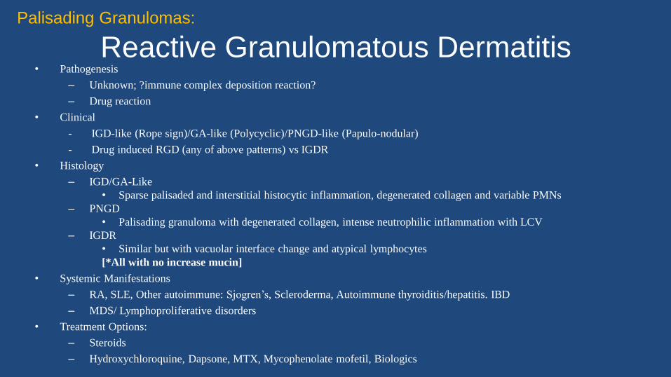
Reactive Granulomatous Dermatitis • Pathogenesis
– Unknown; ?immune complex deposition reaction?
– Drug reaction
• Clinical
- IGD-like (Rope sign)/GA-like (Polycyclic)/PNGD-like (Papulo-nodular)
- Drug induced RGD (any of above patterns) vs IGDR
• Histology
– IGD/GA-Like
• Sparse palisaded and interstitial histocytic inflammation, degenerated collagen and variable PMNs
– PNGD
• Palisading granuloma with degenerated collagen, intense neutrophilic inflammation with LCV
– IGDR
• Similar but with vacuolar interface change and atypical lymphocytes
[*All with no increase mucin]
• Systemic Manifestations
– RA, SLE, Other autoimmune: Sjogren’s, Scleroderma, Autoimmune thyroiditis/hepatitis. IBD
– MDS/ Lymphoproliferative disorders
• Treatment Options:
– Steroids
– Hydroxychloroquine, Dapsone, MTX, Mycophenolate mofetil, Biologics
Palisading Granulomas:
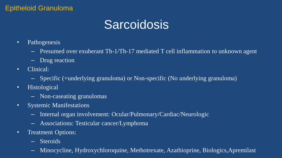
Sarcoidosis
• Pathogenesis
– Presumed over exuberant Th-1/Th-17 mediated T cell inflammation to unknown agent
– Drug reaction
• Clinical:
– Specific (+underlying granuloma) or Non-specific (No underlying granuloma)
• Histological
– Non-caseating granulomas
• Systemic Manifestations
– Internal organ involvement: Ocular/Pulmonary/Cardiac/Neurologic
– Associations: Testicular cancer/Lymphoma
• Treatment Options:
– Steroids
– Minocycline, Hydroxychloroquine, Methotrexate, Azathioprine, Biologics,Apremilast
Epitheloid Granuloma
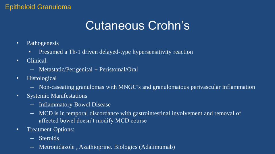
Cutaneous Crohn’s
• Pathogenesis
• Presumed a Th-1 driven delayed-type hypersensitivity reaction
• Clinical:
– Metastatic/Perigenital + Peristomal/Oral
• Histological
– Non-caseating granulomas with MNGC’s and granulomatous perivascular inflammation
• Systemic Manifestations
– Inflammatory Bowel Disease
– MCD is in temporal discordance with gastrointestinal involvement and removal of
affected bowel doesn’t modify MCD course
• Treatment Options:
– Steroids
– Metronidazole , Azathioprine. Biologics (Adalimumab)
Epitheloid Granuloma
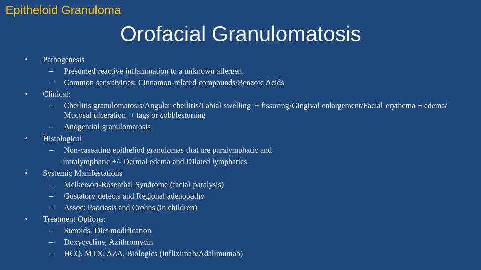
Orofacial Granulomatosis
• Pathogenesis
– Presumed reactive inflammation to a unknown allergen.
– Common sensitivities: Cinnamon-related compounds/Benzoic Acids
• Clinical:
– Cheilitis granulomatosis/Angular cheilitis/Labial swelling + fissuring/Gingival enlargement/Facial erythema + edema/
Mucosal ulceration + tags or cobblestoning
– Anogential granulomatosis
• Histological
– Non-caseating epitheliod granulomas that are paralymphatic and
intralymphatic +/- Dermal edema and Dilated lymphatics
• Systemic Manifestations
– Melkerson-Rosenthal Syndrome (facial paralysis)
– Gustatory defects and Regional adenopathy
– Assoc: Psoriasis and Crohns (in children)
• Treatment Options:
– Steroids, Diet modification
– Doxycycline, Azithromycin
– HCQ, MTX, AZA, Biologics (Infliximab/Adalimumab)
Epitheloid Granuloma
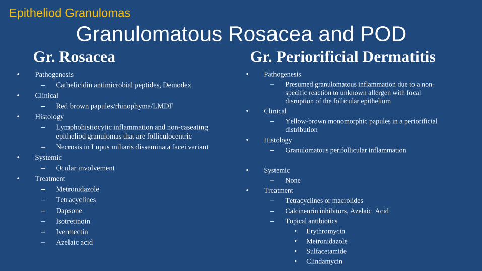
Granulomatous Rosacea and POD Gr. Rosacea
• Pathogenesis
– Cathelicidin antimicrobial peptides, Demodex
• Clinical
– Red brown papules/rhinophyma/LMDF
• Histology
– Lymphohistiocytic inflammation and non-caseating
epitheliod granulomas that are folliculocentric
– Necrosis in Lupus miliaris disseminata facei variant
• Systemic
– Ocular involvement
• Treatment
– Metronidazole
– Tetracyclines
– Dapsone
– Isotretinoin
– Ivermectin
– Azelaic acid
Gr. Periorificial Dermatitis • Pathogenesis
– Presumed granulomatous inflammation due to a non-
specific reaction to unknown allergen with focal
disruption of the follicular epithelium
• Clinical
– Yellow-brown monomorphic papules in a periorificial
distribution
• Histology
– Granulomatous perifollicular inflammation
• Systemic
– None
• Treatment
– Tetracyclines or macrolides
– Calcineurin inhibitors, Azelaic Acid
– Topical antibiotics
• Erythromycin
• Metronidazole
• Sulfacetamide
• Clindamycin
Epitheliod Granulomas
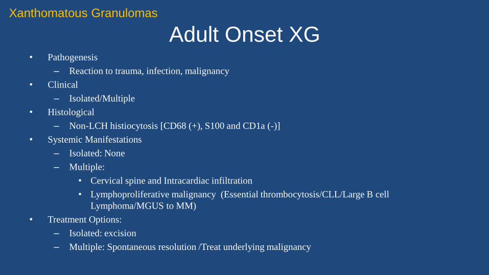
Adult Onset XG • Pathogenesis
– Reaction to trauma, infection, malignancy
• Clinical
– Isolated/Multiple
• Histological
– Non-LCH histiocytosis [CD68 (+), S100 and CD1a (-)]
• Systemic Manifestations
– Isolated: None
– Multiple:
• Cervical spine and Intracardiac infiltration
• Lymphoproliferative malignancy (Essential thrombocytosis/CLL/Large B cell
Lymphoma/MGUS to MM)
• Treatment Options:
– Isolated: excision
– Multiple: Spontaneous resolution /Treat underlying malignancy
Xanthomatous Granulomas
Adult Onset Asthma with Periocular XG
• Pathogenesis
– Systemic immunologic derangement with concurrent bronchiolar and ocular adnexal dysfunction.
• Clinical
– Periorbital yellow-orange plaques, nodules to masses (bilateral)
– Extends to anterior orbital fat, extraocular muscles and/or the lacrimal gland(s)
– No optic nerve damage may cause obstruction
• Histological
– Non-LCH histiocytosis [CD68 (+) S100 and CD1a (-)]
• Systemic Manifestations:
– Adult onset asthma /Lymphadenopathy
– Hematologic malignancies reported :CLL/SLL, multiple myeloma, non-Hodgkin’s lymphoma.
– Associations: Diabetes /Lymphoplasmacytic sclerosing pancreatitis/Rosai- Dorfman
• Treatment Options:
– Surgery
– Corticosteroids and radiotherapy
Xanthomatous Granulomas
Necrobiotic xanthogranuloma • Pathogenesis
– Paraprotein triggering an immune complex formation and inflammation with subsequent granuloma formation
– Reactive inflammation No presence of monoclonal plasma cells
– TCR-PCR clonality can distinguish reactive NXG from malignant granulomatous MF
• Clinical
– Erythematous yellow-orange plaques that develop telangiectasia and may ulcerate
– Periorbital location common with secondary eye inflammation (episcleritis)
• Histologic
- Non-LCH histiocytosis [CD68 (+), S100 and CD1a (-)]
– Prominent palisading granulomas with bizarre-appearing foreign body giant cells around cholesterol cleft
• Systemic
– Multiple myelomaIgG kappa > IgG lambda >IgA
– CTCL, Lymphoma, Leukemia
– Infiltration of internal organs} heart, lungs, eyes
• Treatment Options:
– No curative therapy – consider treating paraprotein
– Topical / intralesional corticosteroids, IFN, nitrogen mustard
– Chlorambucil, Melphalan, Steriods, Interferon alpha 2b, Azathioprine, Cyclophosphamide, Methotrexate therapy,
Plasmapheresis, IVIG, Thalidomide/Lenalidomide
– Surgical excision, radiation
Xanthomatous Granulomas

Multicentric Reticulohistiocytosis • Pathogenesis
– Unknown
• Clinical
– Solitary/Multicentric (Periungal Coral beading)
• Histologic
– Non-LCH histiocytosis [CD68 (+) S100 and CD1a (-)]
– Histiocytes demonstrate a “ground glass appearance” - i.e. copious eosinophilic, granular cytoplasm.
• Systemic
– Solitary: None
– Multicentric:
• Weight loss/anorexia / dysphagia/ pruritus/weakness /myalgia /fever/malaise
• Systemic erosive arthritis and lymphadenopathy
• Associations:
– Autoimmunity (PBC, SS, SLE, DMM, Sjorgens)
– Malignancy (solid/lymphoproliferative)
– Pregnancy (solitary and multicentric)
• Treatment
– hydroxychloroquine, methotrexate, leflunomide, mycophenolate mofetil, azathioprine, cyclophosphamide,
cyclosporine, chlorambucil, dapsone, TNF-alpha inhibitors (etanercept, infliximab, adalimumab), bisphosphonates
Xanthomatous Granulomas
Rosai Dorfman * Pathogenesis
- Unknown reactive process
- Mutations in SLC29A3, the gene that encodes the equilibrative nucleoside transporter
hENT3, have been found in 2 families with familial Rosai-Dorfman disease.
• Clinical
– Painless bilateral cervical adenopathy and red to red-brown papules or nodules in skin
– Nodular inflammatory infiltrate by large foamy histiocytes (dermis and lymphatics)
• Histological
– S100 (+) no beirbeck granules with nodular inflammatory infiltrate by large foamy histiocytes (dermis
and lymphatics) + Emperipolesis [CD68 (+) S100 (+) and CD1a (-)]
• Systemic:
– Lymphadenopathy, fever, elevated ESR, leukocytosis, anemia, MGUS, organ infiltration
– Associations: Crohns/Bloodline malignancies
• Treatment Options:
– Surgery or Radiation for vital organ involvement/ Corticosteroids/ MTX/Chemotherapy
Interferon
Xanthomatous Granulomas
Xanthoma Disseminatum • Pathogenesis:
– Remains elusive
• Clinical:
– Orange-red papulo-nodules to plaques (inverse location)
• Histological:
– Non-LCH, xanthoma cells, Touton GC, mild inflammatory infiltrate
[CD68 (+) S100 and CD1a (-)]
• Systemic Manifestations:
– Diabetes insipidus, other hypopituitarism, dysphagia, dysphonea, ocular
(corneal/conjunctiva), lipid abnl
• Treatment options
– Cyclophosphamide, Azathioprine, Vinblastine, 2-chlorodeoxyadenosine
Xanthomatous Granulomas
Other Granulomas
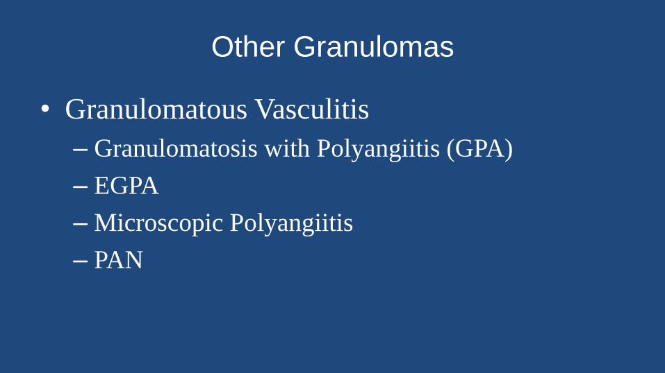
• Granulomatous Vasculitis
– Granulomatosis with Polyangiitis (GPA)
– EGPA
– Microscopic Polyangiitis
– PAN
Other Granulomas
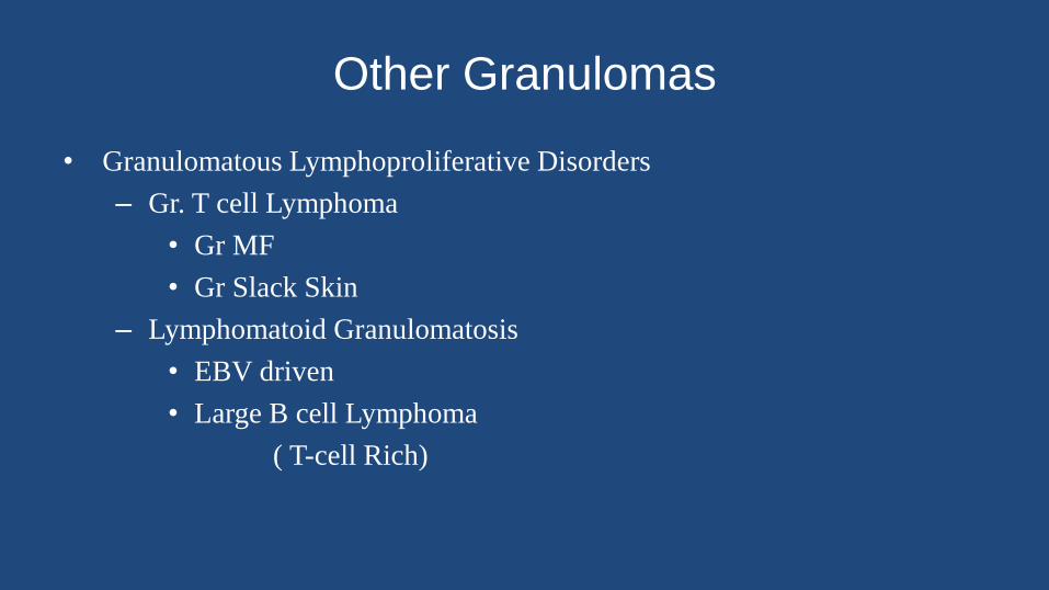
• Granulomatous Lymphoproliferative Disorders
– Gr. T cell Lymphoma
• Gr MF
• Gr Slack Skin
– Lymphomatoid Granulomatosis
• EBV driven
• Large B cell Lymphoma
( T-cell Rich)
Other Granulomas
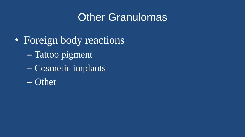
• Foreign body reactions
– Tattoo pigment
– Cosmetic implants
– Other
Other Granulomas
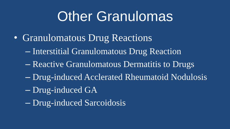
• Granulomatous Drug Reactions
– Interstitial Granulomatous Drug Reaction
– Reactive Granulomatous Dermatitis to Drugs
– Drug-induced Acclerated Rheumatoid Nodulosis
– Drug-induced GA
– Drug-induced Sarcoidosis
.
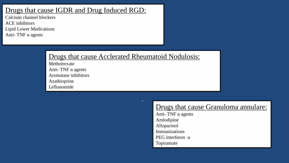
Drugs that cause IGDR and Drug Induced RGD: Calcium channel blockers
ACE inhibitors
Lipid Lower Medications
Anti- TNF α agents
Drugs that cause Acclerated Rheumatoid Nodulosis: Methotrexate
Anti- TNF α agents
Aromatase inhibitors
Azathioprine
Leflunomide
Drugs that cause Granuloma annulare: Anti- TNF α agents
Amlodipine
Allopurinol
Immunizations
PEG interferon α
Topiramate
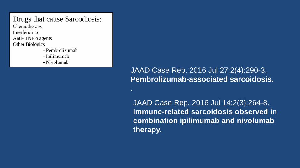
JAAD Case Rep. 2016 Jul 27;2(4):290-3.
Pembrolizumab-associated sarcoidosis.
.
JAAD Case Rep. 2016 Jul 14;2(3):264-8.
Immune-related sarcoidosis observed in
combination ipilimumab and nivolumab
therapy.
Drugs that cause Sarcodiosis: Chemotherapy
Interferon α
Anti- TNF α agents
Other Biologics
- Pembrolizumab
- Ipilimumab
- Nivolumab
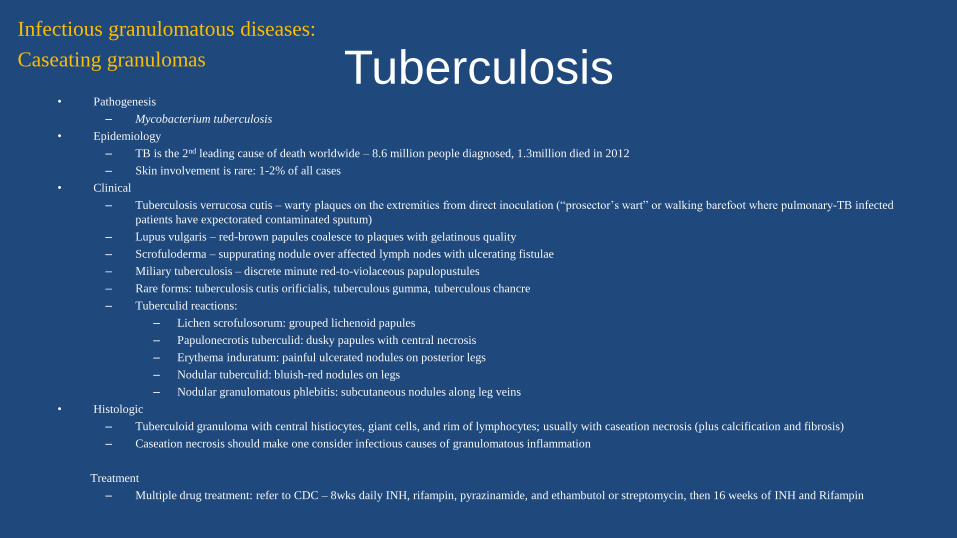
Tuberculosis • Pathogenesis
– Mycobacterium tuberculosis
• Epidemiology
– TB is the 2nd leading cause of death worldwide – 8.6 million people diagnosed, 1.3million died in 2012
– Skin involvement is rare: 1-2% of all cases
• Clinical
– Tuberculosis verrucosa cutis – warty plaques on the extremities from direct inoculation (“prosector’s wart” or walking barefoot where pulmonary-TB infected
patients have expectorated contaminated sputum)
– Lupus vulgaris – red-brown papules coalesce to plaques with gelatinous quality
– Scrofuloderma – suppurating nodule over affected lymph nodes with ulcerating fistulae
– Miliary tuberculosis – discrete minute red-to-violaceous papulopustules
– Rare forms: tuberculosis cutis orificialis, tuberculous gumma, tuberculous chancre
– Tuberculid reactions:
– Lichen scrofulosorum: grouped lichenoid papules
– Papulonecrotis tuberculid: dusky papules with central necrosis
– Erythema induratum: painful ulcerated nodules on posterior legs
– Nodular tuberculid: bluish-red nodules on legs
– Nodular granulomatous phlebitis: subcutaneous nodules along leg veins
• Histologic
– Tuberculoid granuloma with central histiocytes, giant cells, and rim of lymphocytes; usually with caseation necrosis (plus calcification and fibrosis)
– Caseation necrosis should make one consider infectious causes of granulomatous inflammation
Treatment
– Multiple drug treatment: refer to CDC – 8wks daily INH, rifampin, pyrazinamide, and ethambutol or streptomycin, then 16 weeks of INH and Rifampin
Infectious granulomatous diseases:
Caseating granulomas
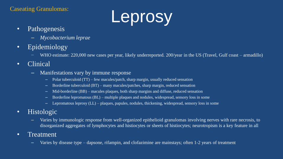
Leprosy • Pathogenesis
– Mycobacterium leprae
• Epidemiology − WHO estimate: 220,000 new cases per year, likely underreported. 200/year in the US (Travel, Gulf coast – armadillo)
• Clinical
– Manifestations vary by immune response
– Polar tuberculoid (TT) – few macules/patch, sharp margin, usually reduced sensation
– Borderline tuberculoid (BT) – many macules/patches, sharp margin, reduced sensation
– Mid-borderline (BB) – macules plaques, both sharp margins and diffuse, reduced sensation
– Borderline lepromatous (BL) – multiple plaques and nodules, widespread, sensory loss in some
– Lepromatous leprosy (LL) – plaques, papules, nodules, thickening, widespread, sensory loss in some
• Histologic – Varies by immunologic response from well-organized epithelioid granulomas involving nerves with rare necrosis, to
disorganized aggregates of lymphocytes and histiocytes or sheets of histiocytes; neurotropism is a key feature in all
• Treatment – Varies by disease type – dapsone, rifampin, and clofazimine are mainstays; often 1-2 years of treatment
Caseating Granulomas:

Atypical mycobacteria • Pathogenesis
– Nontuberculous mycobacteria (NTM) include over 170 species, can often cause skin and soft tissue infections (SSTI)
– Divided into rapidly growing and slow growing mycobacteria
– RGM: M. fortuitum, M. chelonae/absecssus, M. mucogenicum, M. smegmatis, and early pigmenting RGM
– SGM: M. marinum, M. ulcerans, M. kansasii, M. haemophlium, M. avium complex and more
• Epidemiology
– NTM are ubiquitous – water, soil, plants, animals; tap water is a major reservoir, can contaminate hospital equipment – pedicure/nail salon
outbreaks
• Clinical
– Varies by species – generally for dermatologists can be disseminated disease (skin seeding in severely immunocompromised patients) or
primary skin and soft tissue infection (in suppressed or normal hosts)
– Disseminated disease (HIV, organ transplant, iatrogenic suppression, leukemia): red, draining nodules, ulcerations, abscesses
– SSTI: sporotrichoid nodules, “fish-tank” granuloma, Buruli ulcer, abscesses, cellulitis, sinus tracts, panniculitis, etc
• Histologic
– May vary somewhat by organism and clinical morphology; generally intense granulomatous inflammation with neutrophils and necrosis;
organisms may be seen on special stains, but cultures and sometimes PCR is necessary to ID
• Treatment
– Varies by organism type; most require multidrug therapy and can develop resistance rapidly to single agents.
– Cultures may be slow to grow but should be performed for antibiotic sensitivities (although educated guesses can be made once speciation is
available)
– Most cases should be co-managed with an infectious disease doctor
– Local surgical treatment is indicated in some cases
Infectious granulomatous diseases
Leishmaniasis • Pathogenesis
– Leishmania infection – multiple species, divided into “New World” (Western) and “Old World” (Eastern Hemisphere)
– Transmited through bite of female sandfly
– 4 types: cutaneous leish, diffuse cutaneous leish, mucocutaneous leish, and visceral leish
• Epidemiology
– 12 million infected, 2 million new cases per year, 20-30,000 deaths annually
• Clinical
– Subclinical, self-healing disease is common
– Cutaneous lesions: solitary papules at bite site, enlarge into nodules/plaques, often ulcerate (painless); may have satellite lesions, multiple
primary lesions, or sporotrichoid spread
– Patients can have persistent leish in healed scars and lymph nodes, and can get delayed mucocutaneous disease depending on the organism and
immune response
• Histologic
– Ulceration, intense dermal inflammation with variable granulomas and histiocytes; histiocytes with small organisms
– Confirmatory PCR testing is available through the CDC and speciation can impact follow-up and treatment
• Treatment
− High cost, toxicity, drug resistance, access issues, paucity of high quality data all complicate treatment
– Drugs are available through the CDC and patients should be treated in conjunction with an ID doctor
– Stibogluconate, amphotericin, liposomal ampho, or miltefosine
– Pentamodine, other agents pending intolerance/response
– Need long-term follow up of all patients
Caseating Granulomas

Deep fungal infections • Pathogenesis
– Generally due to Blastomycosis, coccidioidomycosis, Cryptococcus, histoplasmosis, and sporotrichosis
• Epidemiology
− Most are acquired through inhalation and secondary spread to the skin; direct inoculation can occur
− Blastonyosis: Ohio/Mississippi river valleys
− Coccidioidomycosis: San Joaquin Valley, SW US (incidence and area increasing due to climate change)
− Cryptococcus: Pigeon droppings/soil, widespread, seen usually in immunocompromised hosts
− Histoplasmosis: Contaminated soil /bat-bird droppings, caves, often seen in immunocompromised hosts
− Sporotrichosis: Rose thorns, moss, other contaminants, causes infection from direct innoculation
• Clinical
– Varies by infectious agent; many can cause nodules, papules, ulcerated, and/or verrucous/crusted lesions
– Characteristic lymphocutaneous spread seen in sporotrichosis
• Histologic
– Often a dense mixed infiltrate with neutrophils, histiocytes, giant cells and acute granulomas with overlying
pseudoepitheliomatous hyperplasia
– Blastomycosis – large yeast with broad based, single bud
– Coccidioidomycosis – thick-walled spherule with endospores
– Cryptococcus – small narrow-based budding yeast forms
– Histoplasmosis – very small narrow-based budding, intracellular
– Sporotrichosis – round/cigar-shaped yeast, rarely visualized (may need EM)
Suppurative Granulomas
Deep fungal infections • Treatment
– Varies by organism
– All patients should be evaluated for potential immunosuppression, and in most cases skin findings represent likely
secondary seeding from a systemic process (particularly for cryptococcus)
– Treatment should be conducted in consultation with an infectious disease physician and tailored towards the individual
infection
– Itraconazole, amphotericin, voriconazole, posaconazole, debridement all have a role
Suppurative Granulomas
Blastomycoses like-pyoderma • Pathogenesis: Abnormal rxn to bacteria, often Staph, in
immunosuppressed pts
*Must exclude: SCC, Deep fungal, Atypical Myco
bromoderma, iododerma
• Clinical: Verrucous to vegetative plaques with pustules and elevated border
• Histology: PEH and abscesses
• Treatment:
1. Cultures to determine cause & guide antibiotic Tx
2. Other: acitretin, CO2 laser, cryotherapy, curettage
Bacterial causes:
S. aureus/Beta-hemolytic streptococci
E.coli/Proteus/Pseudmonas
Clostridium perfringens/Prevotella
Suppurative Granulomas
Granuloma inguinale Suppurative Granulomas
• Pathogenesis: STI from Klebsiella granulomatis
• Epidemiology: Rare in US , more Tropical areas
• Clinical: Beefy to velvety red papules/plaques
• Histology: Dense infiltrate of lymphocytes, neutrophils
plasma cells and histiocytes (with Donovan bodies)
• Treatment:
– Doxycyline
– Azithromycin
– Erythromycin
Lymphogranuloma venereum • Pathogenesis: STI from Chlamydia trachomatis (serovars L1,2,3)
• Epidemiology: Tropical areas
• Clinical: Rarely seen painless ulceration, f/b painful inquinal adenopathy
• Histology: Lymph node – stellate central necrosis with neuts and palisading granulomatou srxn.
+ MNGC
• Treatment:
– Doxycyline
– Azithromycin
– Erythromycin
Suppurative Granulomas
Reference of Choice:
Granulomatous Disorders of the Skin
Editor: Joseph C English III
Consulting Editor: Bruce THiers
Dermatology Clinics July 2015 (pgs 315-639)
Selected Other References: • GA
– Thornsberry LA et al. Etiology, diagnosis and therapeutic management of granuloma annulare: an update. Am J Clin
Dermatol 2013;14:279.
– Lukacs J et al. Treatment of generalized granuloma annulare – a systematic review. J Eur Acad Dermatol Venereol
2015 Feb 4 [Epub ahead of print]
• EGCG
– Gutierrez-Gonzalez E et al. Elastolytic giant cell granuloma: a clinic-pathologic review of twenty cases. Dermatol
Online 2013 Oct 16;19:20019
– El-Khoury J. Elastophagocytosis: underlying mechanisms and associated cutaneous entities. J Am Acad Dermatol
2014;70:934.
• NL
– Reid SD et al. Update on necrobiosis lipoidica: a review of etiology, diagnosis and treatment options. J Am Acad
Dermatol 2013;69:783.
– Erfurt-Berge C et al. Update on clinical and laboratory features in necrobiosis lipoidica: a retrospective multi-centre
study of 52 patient. Eur J Dermatol 2012;22:770.
• RN
– Yamamoto T. Cutaneous necrobiotic conditions associated with rheumatoid arhtritis: important extra-articular
involvement. Mod Rheumatol 2013;23:617.
– Sayah et al. Rheumatoid arthritis: a review of the cutaneous manifestations. J Am Acad Dermatol 2005:53:191.
• RGD
– Coutinho I et al. Interstitial granulomatous dermatitis: a Clincopathological study. Am J Dermatopathol 2015 Mar 31
[Epub ahead of print]
– Hawryluk EB, et al. Non-infectious granulomatous diseases of the skin and their associated systemic diseases: an
evidence-based update to important clinical questions. Am J Clin Dermatol 2010;11:171.
• Sarcodiosis
– Hamiovic A et al. Sarcoidosis: a comprehensive review and update for the dermatologist: part I. cutaneous disease. J
Am Acad Dermatol 2012;66:699.
– Hamiovic A et al. Sarcoidosis: a comprehensive review and update for the dermatologist: part II. extra cutaneous
disease. J Am Acad Dermatol 2012;66:6719.
• Cutaneous Crohn’s
– Kurtzman DJ et al. Metastatic Crohn’s disease: a review and approach to therapy. J Am Acad Dermatol 2014;71:804.
– Marzano AV et al. Cutaneous manifestations in patients with inflammatory bowel disease: pathophysiology, clinical
features and therapy. Inflamm Bowel Dis 2014;20:213.
• OFG
– Campbell et al. Distinguishing orofacial granulomatosis from crohn’s disease: two separate disease entities? Inflamm
Bowel Dis 2011:17:2109.
– Al Johani KA et al. Orofacial granulomatosis: clinical features and long-term outcome of therapy. J AM Acad
Dermatol 2010;62:611.
• Gr R and POD
– Vemuri RC et al. Major pathophysiologic correlations of rosacea: a complete clinical appraisal. Int J Med Sci
2015:12;387.
– Tarm etal. Granulomatosis periorifical dermatitis. Cutis 2004;73:399.
• XG
– Shoo BA et al. Xanthogranulomas associated with hematologic malignancy in adulthood. J Am Acad Dermatol
2008;59;488.
• AOAPXG
– London J et al. Adult-onset asthma associated with periocular xanthogranuloma: new diagnostic and therapeutic
approaches in a very rare systemic disease. Ophthal Plast Reconstr Surg 2013:29:104.
– Cavallazzi R et al. Clinical manifestations and treatment of adult asthma and periocular xanthogranuloma. Can Respir
J 2009;16:159.
• NXG
– Wood AJ et al. Necrobiotic xanthogranuloma: a review of 17 cases with emphasis on clinical and pathologic
correlation. Arch Dermatol 2009;145:279.
– Abdul-Hay M. Immunomodulatory drugs for the treatment of periorbital necrobiotic xanthogranuloma. Clin Adv
Hematol Oncol 2013;11:680.
• MR
– Selmi C et al. Mutlicentric reticulohistiocytosis: a critical review Curr Rheumatol Rep 2013;17:511.
– Islam AD et al. Mutlicentric reticulohistiocytosis: a rare yet challenging disease. Clin Rev Allergy Immunol
2013;45:281.
• RD
– Maia RC et al. Rosai-Dorfamn disease: a report of eight cases in a tertiary care center and a review of the literature.
Braz J Med Biol Res 2015;48:6.
– Dalia S et al. Rosai-Dorfam disease: tumor biology, clinical feature, pathology and treatment. Cancer Control
2014;21;322
• XD
– Park M et al. Xanthoma disseminatum: a case report and mini-review of the literature. Acta Dermatovenerol Croat
2014;22:150.
– Khezri F et al. Xanthoma disseminatum: effective therapy with 2-chlorodeoxyadenosine in a case series
• Atypical Mycobacteria
– Grossman, Fox, Kovarik, Rosenbach. Cutaneous manifestations of infection in the immunocompromised host, 2nd Ed.
Eds:
• Leishmaniasis
– Schwartz et al. New world leishmaniasis in travellers. Lancet Infect Dis 2006;6:342