Embed Size (px)
Citation preview

Gold Nanoparticles as Drug Delivery
Agents. Detoxifying the
Chemotherapeutic Drug Cisplatin
(copia xifrada)
Joan Comenge Farre
Maig 2013

Universitat Autònoma de Barcelona
Departament de Bioquímica i Biologia Molecular
Gold Nanoparticles as Drug Delivery
Agents. Detoxifying the
Chemotherapeutic Drug Cisplatin
Joan Comenge Farré
Inorganic Nanoparticles Group
Institut Català de Nanotecnologia
Maig 2013

Gold Nanoparticles as Drug Delivery
Agents. Detoxifying the
Chemotherapeutic Drug Cisplatin
Memòria presentada per
Joan Comenge Farré
Per optar al grau de
Doctor per la Universitat Autònoma de Barcelona
Programa de Doctorat en Bioquímica, Biologia Molecular i
Biomedicina
Tesi realitzada sota la direcció del Dr. Víctor Franco Puntes,
Inorganic Nanoparticles Group, Institut Català de Nanotecnologia,
i amb la tutoria de la Dra. Ester Boix Borràs,
Departament de Bioquímica i Biologia Molecular, Universitat Autònoma
de Barcelona.
Cerdanyola del Vallès, Maig 2013


i
Prologue
In this thesis dissertation, the use of gold nanoparticles as agents for delivery of cisplatin is
discussed. The book has been organized following a logic sequence that starts by the
synthesis of gold nanoparticles, followed by their functionalization, first with surfactants
and then with the drug to finally analyze the performance of these drug delivery vehicles
in in vitro and in in vivo experiments. Every chapter has been structured so to allow its
individual lecture.
Although aspects applicable to other drug delivery systems such as the colloidal and
chemical stability in physiological media, the drug release profile, etc. are discussed here
using our system as an example, this work does not pretend to be a general discussion
about the state of the art in nanomedicine and/or drug delivery, but a contribution to
these growing areas. However, I encourage the lecture of some of the very interesting
references provided here to achieve a deeper understanding in specific aspects.
This multidisciplinary work could have not been possible without the contribution and
advice of a great number of people. Thus, I feel the obligation to acknowledge all those that
directly contribute to this work especially all the members of the Inorganic Nanoparticles
Group, led by Prof. Víctor Puntes, for their invaluable help and knowledge, but also Prof.
Fernando Domínguez and Carmen Sotelo from school of medicine at Santiago de
Compostela, prof. Franciso Romero from Molecular Science Institute at València and Dr.
Óscar Gallego and Prof. Agustí Barnadas from the Santa Creu I Sant Pau Hospital at
Barcelona for their active contribution and discussions about this work. Last but not least,
my gratitude to all the members of the Radiation Biology Group lead by Prof. Kevin Prise
in the Centre of Cancer Research and Cell Biology at Belfast for receiving me and teaching
me the little I know about this field.
Also, a special mention goes to Nanotargeting S.L. for trusting in this project.
It is impossible to extend here my gratitude to all of those that indirectly contribute to the
work so I will let you know one by one personally. Just be patient.

1
Chapter 1
Introduction
Many of the conventional therapies can be improved through the use of drug delivery
systems (DDS). They are designed mainly to modify the pharmacokinetics and
biodistribution of small molecular drugs. This is of special importance in the case of
anticancer therapies in which a widespread distribution of small molecular
chemotherapeutic drugs is often limiting treatments due to severe side effects that make
impossible to reach the full benefits of the therapy. In this context, nanotechnology
emerges as a disruptive technology to design carriers that improve the delivery of drugs to
their target organs.
1.1 Engineering AuNPs as scaffolds for drug delivery
Although a plethora of nanoparticles (NPs) including polymeric NPs, liposomes,
dendrimers, metallic NPs, etc. have been used to design nanocarriers for drug delivery,1, 2
we will focus the discussion on the use of gold nanoparticles (AuNPs). The synthesis of
AuNPs have been first described by J. Turkevich at 1954,3 but it was not until last years
that advances on the engineering of these NPs allowed researchers to control the size,
shape, and functionalization of these nanocarriers and thus starting using them as
scaffolds for drug delivery. Thus, new synthetic protocols make possible to achieve
biocompatible spherical AuNPs from 5 to 200 nm with a monodispersity lower than 10 %.4
The control over the size is a key point in the use of AuNPs in biomedicine since it
influences important biological properties such as interaction with proteins,
biodistribution and clearance rate. The variety of sizes of AuNPs that can be easily
obtained facilitated a better understanding of the role played by this property in

Chapter 1
2
important parameters for cancer treatment with NPs such as accumulation in tumors,
blood half-life, or penetration. From a simplistic point of view, one could say that the final
fate of the NPs is strongly influenced by their size since smallest AuNPs (<5 nm, core +
surfactant) are rapidly cleared up by the kidneys and the the largest ones (>100 nm) are
removed easily from the circulation by the immune system.5 Sizes in between showed the
best accumulation and penetration in tumors; at this range of sizes other parameters such
as the presence of stealthing agents or different surface charge should be tuned case by
case to achieve the desired behavior. In addition, shape of the AuNPs is also important.
Spherical NPs are usually the chosen option when they wanted to be used just as carriers
due to the simple synthesis and easy functionalization. However, other shapes such as
nanorods or nanoboxes have been used too. These advanced AuNPs have special
physicochemical properties that make them appealing to be used not only as vehicles but
also as effectors by themselves. They present a surface plasmon resonance (SPR) in the
near infrared (NIR) region, which make them suitable to be used in photothermal therapy
in which NIR light is absorbed by NPs, delivering toxic amounts of heat.6 In this type of
therapy, working with wavelengths that can penetrate deep into tissue such as IR is
required and only non-isotropic AuNPs present a SPR at this region.
Regarding functionality, chemistry on gold planar surfaces has been extensively studied to
obtain molecular Self-Assembled Monolayers (SAMs).7 Thus, this technology has been
easily adapted to functionalize AuNPs in solution. However, some considerations need to
be taken into account for NPs: the curvature radius may play a role in the determination of
important properties of the molecules conjugated to small NPs such as apparent pka 8 and
the coverage density9 of the attached ligands amongst others. Also, AuNPs need to be
stabilized during the synthesis to avoid aggregation by means of conjugation to a
surfactant. Thus synthetic protocols in which the surfactant is weakly bound to the NPs
(e.g. sodium citrate), and therefore can be easily exchanged by other ligands, are
recommended for further functionalization of these AuNPs.4 Note that physiological
conditions (high ionic strength) are harsh for citrate-capped AuNPs. Conjugation may also
result in destabilization of NPs if special care is not taken.10
AuNPs show affinity to different chemical groups such as phosphates, carboxylic acids,
ammines and specially to thiols. Thus, one can found in the literature AuNPs conjugated to
a great variety of molecules including alkanethiols, polymers, proteins, amino acids, and
drugs amongst others.11 Interestingly, the control over the conjugation facilitates
engineering multifunctional AuNPs. Hence, it would be possible to create a nanocarrier
that contains, for example, a molecule to stealth the DDS from the immune system, a ligand

Introduction
3
that promotes uptake by specific cells, and the drug which in its turn can be attached via a
responsive link which release the drug only after a specific response such as a pH drop.
Last but not least, the greater monodispersity of AuNPs compared to other metallic or
polymeric NPs is another advantage of using this type of NPs
1.2 Nanoparticles for anticancer therapy: Advances and challenges of a new
therapy
NPs are perfect candidates to be used in anticancer therapy since they showed passive
accumulation in tumors due to the Enhanced Permeation and Retention effect (EPR). This
effect first reported by Matsumura and Maeda12 exploits the ability of NPs to permeate
through the leaky tumor vessels (with fenestrations between 100 and 800 nm 13, 14) and to
be accumulated into the tumor due to the lack of a functional lymphatic system (Figure
1.1). Long-time circulating NPs maximize the EPR effect since the changes to permeate
through the tumor vessels are greater. Related to this, the size of NPs plays an important
role in determine tumor accumulation and distribution: 60nm AuNPs showed a greater
accumulation than 20 nm AuNPs likely due to a slower clearance. However, larger AuNPs
are accumulated in the perivascular region of the tumor failing to penetrate deeper, whilst
20 nm AuNPs were still found significantly at 50 m from the blood vessel.14
Figure 1.1. Scheme showing the differences of the vascular system of normal tissue (A) and
tumor (B). Leaky vessels and non-functional lymphatic system are responsible for the passive
accumulation of NPs into tumors. Adapted from reference 15.15
The intrinsic property of NPs to be passively accumulated into tumors has been exploited
to deliver a great variety of drugs.1 Some groups proposed to improve further this
accumulation by attaching a ligand recognized by receptors overexpressed in tumor cells.

Chapter 1
4
These include epidermal growth factor (EGF)16, folate17 and transferrin18 amongst other.
However, there is some controversy regarding the efficiency of this approach.19 It is not
clear that active targeting favors the accumulation in tumor, since physicochemical
properties of NPs govern the pharmacokinetics properties of DDS. Thus, size and surface
charge and composition (e.g. presence of stealthing agents or not) of NPs seems to play a
more important role in determining final distribution of DDS20 and ligands influence only
in interactions directly onto the cell once the cell is reached (e.g. a greater uptake)
Once the NPs are localized in the tumor, different strategies have been used to release the
drug. Normally, the strategies are based on the different physicochemical properties found
in the cytoplasm compared to the extracellular environment (e.g. blood). For example, NPs
are known to be internalized via endocytic pathways, in which a pH drop is produced.21
Thus, a pH-sensitive link between drug and NPs ensures non-specific release of drug
during the circulation through the body.22 A controlled release can be also achieved by the
reduction of an oxidized inactive prodrug in the cytoplasm, releasing the drug in its active
form.23 Also one can take advantage of specific enzymes to cleave the link between NP and
drug.24
The metallic core of AuNPs conjugates can also act as an effector in thermotherapy as
explained above. In addition, AuNPs have been recently proposed as radiosensitizers due
to the induction of an Auger cascade after an ionizing event localized in the vicinity of the
AuNP surface. These special properties open up the possibility to strategies that use
AuNPs as carrier and effector.
Thus the use of AuNPs as carriers for drug delivery might provide several advantages
including:
Increased blood half-life of the small drug
Accumulation in the tumor via the EPR effect
Reduced toxicity in normal tissue
Controlled release of the drug, delivering high payloads directly to the site
of action
Possibility of multiple functionalization which allow combined therapy
and/or tuning blood half-life, immune responses toward the vehicle, tumor
uptake, etc.
Increase the solubility of poorly soluble drugs in plasma
Protection of the drug from immune system and from agents present in the
plasma that might deactivate the drug

Introduction
5
Promote a double effect by acting as carriers and as effectors by
themselves
However, the use of AuNPs for drug delivery is not without problems and some aspects
should be considered:
Classical syntheses of AuNPs render concentrations of AuNPs in the range of 1012-1013
NP/mL,25 thus AuNPs are typically synthesized at the nanomolar scale. Even though high
densities of loading have been achieved,26, 27 concentrations of drugs onto NPs as
synthesized will rarely be higher than 10 micromolar. Taking into account that there is a
limitation of volume that can be injected into the body (e.g. 10 mL/Kg for intravenous
injection in mice), the therapeutic dose might not be reached unless NPs had been
previously concentrated. This concentration step is not always straightforward because
concentration of AuNPs could lead to aggregation if special care is not taken (e.g.
concentration of AuNPs without any surfactant increase the chances of aggregation). Note
that colloids are intrinsically out of equilibrium and they tend to minimize surface energy
when this is possible either via functionalization or aggregation.
The need of concentration of AuNPs to reach therapeutic doses implies that a single lab-
scale synthesis might not be enough to achieve the amount of NPs needed to perform in
vivo experiments. Thus, not only monodispersity should be guaranteed to control size
effects, but also reproducibility batch to batch. In addition, robust synthesis protocols will
also facilitate the scale up of the production, which is essential for the transference of the
technology to the industry.28
The special properties of AuNPs are lost if they aggregate. In too many cases the loss of
colloidal stability is behind the lack of (or unexpected) biological behaviors.20 Therefore,
one has to ensure the maintenance of colloidal stability along the process (synthesis –
storage – exposure/treatment). It is common to assay the stability of AuNPs in the storage
conditions (e.g. in aquose media) even if they are different from the working conditions.
However, biological fluids such as cell culture media or blood are complex mixtures of
salts, proteins, sugars, etc. that may promote destabilization of AuNPs, and therefore
stability should be assayed in this media. The same consideration is valid for the stability
of the link with the drug. For example, cisplatin adsorbed on AuNPs is not released in
aquose media, whilst it is unspecifically quickly released in cell culture media. On the other
hand, cisplatin linked via coordination bond is stable in both media and only released after
a pH drop.26
Chapter 1
6
The choose of appropriate models to determine the potential therapeutic benefits is also
not trivial, specially for new technologies in which the amount of literature is limited. In
vitro simple test such as those assessed in monolayer cell cultures do not consider
important factors such as tumor vascularity, penetration and other differential properties
given by the tumor microenvironment.15 Consequently, in vitro results do not necessarily
reflect the behavior of the assayed DDS in a real case. In this context, 3D cell cultures have
been proposed as models to study the behavior of drugs in the particular environment of
solid tumors due to the possibility to have an extracellular matrix and different regions of
tumor (e.g. hypoxic cells in the intern areas). Since the advantages of using DDS are
achieved mainly by modification on the pharmacokinetic properties of the drug, in vivo
models are preferred. However, there is a great variety of tumor models including
xenografted, orthotopic, and spontaneously and induced autochthonous tumor models.29
This models show variations between them such as different size of porous in vessels or
different immunological responses which may result in different efficiency of the same
DDS depending on the model that has been used.
Finally, treatments with AuNPs are still very few and not beyond clinical trials. Thus, there
is a lack of knowledge about regulatory aspects. Possible adverse effects of NPs such as
acute toxicity and longtime accumulation should not be underestimated and should be
studied case by case. There is a controversy regarding the in vivo toxicity of NPs and the
parameters that play a role in the NPs-induced toxicity.30 Some works have found no toxic
signals after AuNPs administration31 or small alterations in the biochemical markers due
to metabolization of the AuNPs or indicating a temporal inflammation.32 On the other
hand, it is believed that the disfunction of major organs can be related to the presence of
NPs at the site of abnormalities. For that reason, there are a lot of studies regarding
toxicity in spleen and liver, which are generally accepted to be the organs with the highest
accumulation of NPs. The liver toxicity, when found, seems to be associated to an
hyperplasia of Kupffer cells that induce an acute inflammation with neutrophils influx.33
This acute inflammation is a transient response due to the insult of AuNPs, however
apoptosis and necrosis of hepatocytes as well as accumulation of AuNPs could be related
to toxic effects.34 Regarding the spleen, macrophages of the periphery seem to be involved
in the uptake of NPs by this organ. Thus, leading to a temporal inflammation of the spleen,
however in other cases a loss of weight has been observed after intravenous
administration.35 White pulp aberration has been also observed.36 Any general trend
cannot be extracted from the current literature, but it is clear that there are some
parameters that have to be taken into account before using AuNPs as a vehicle for medical
applications: i. Size of the vehicle, since it will determine the clearance rate and the

Introduction
7
biodistribution 36; ii. Surface composition, it has been seen that by changing the surface
composition of AuNPs the toxicological profile might be different37; iii. Dose and
administration route obviously will play a role in the potential toxicity. Therefore for
every AuNP-conjugate, a toxicology study must be done to prove their usability in medical
applications.
1.3 Cisplatin as anticancer drug
A paradigmatic case of anticancer drugs are platinum compounds. Cisplatin or cis-
diamminedichloroplatinum(II), [PtCl2(NH3)2], was originally synthesized in 1845, but not
until 1970 was its antitumor activity established.38 Today cisplatin is used to treat various
types of cancers (i.e., non-small-cell lung cancer, ovarian cancer, germ cell tumors,
osteosarcomas, etc), with a cure rate as high as 90% in testicular cancer.39 After both
passive and active cellular uptake, the platinum complex reacts in vivo to form adducts
with DNA, which ultimately trigger apoptosis.40 However, chronic cisplatin usage results in
resistance by several possible mechanisms including increased interactions with
metallothioneins and glutathione, which deactivate the drug, as well as increased DNA
repair and/or cisplatin efflux.41 To counteract resistance and its rapid renal clearance,
which lowers the efficiency of cisplatin significantly, very high systemic doses of cisplatin
should be administered. Unfortunately, such high dose of cisplatin results in severe
systemic toxicity and poor patient wellfare, including nausea/vomiting, renal toxicity,
gastrointestinal toxicity, peripheral neuropathy, asthenia, and ototoxicity, thus limiting its
clinical use.41, 42 Of all the toxicities induced by cisplatin, nephrotoxicity is considered to be
the dose-limiting factor.40 Such side effects make it impossible to achieve the full benefit of
the treatment in a large number of patients.43 In humans, the cisplatin treatment generally
involves series of intravenous injections administered every 3-4 weeks at a dose of 50-120
mg/m2 (1.2 – 2.7 mg/Kg). In addition to the undesired side effects, there is also a loss of
drug activity in the body associated with poor circulation and poor delivery to the tumor,
as well as deactivation mechanisms that irreversibly alter the chemistry of these
molecules before reaching the tumor cells.42 Since its discovery, many attempts to find
derivatives has been carried out looking for both reduced side effects and modified body
distribution (in order to target different organs), rather than improving cisplatin
efficacy.41, 44 Here, second-generation platinum drugs as carboplatin and oxaliplatin
represent an improvement in some cancer treatments, as lung and colorectal respectively,
although the limitations observed for cisplatin have not been entirely overcome40, 41.

Chapter 1
8
1.4 Carrying cisplatin
Hence, recent efforts have been focused on targeting the tumor by using drug delivery
systems to avoid the organs to which cisplatin is toxic. As the kidney is responsible for
filtration and removal from the blood of molecules smaller than 50 KDa, which
corresponds to molecular sizes of around 6 nm in diameter, any larger delivery vehicle
will divert the drug away from the kidney.45 In addition, more than 90 % of the
administrated cisplatin is known to bind irreversible to albumin, which deactivates the
drug.46 Thus, a properly designed nanocarrier will not only transport the cisplatin to the
tumor, but also protect it against plasma deactivation. In this context, approaches based
on the encapsulation and transport of cisplatin have emerged recently. Sterically stabilized
polymeric nanoparticles, having excellent stability in plasma, a much longer circulation
time, better efficacy, and lower toxicity than free cisplatin have been reported.47-50 As the
case of lipid capsules51 or polymers, as in Prolindac®, a 22 kDa
hydroxypropylmethacrilamide copolymer as a backbone and then a glycine chelator linker
which is pH sensitive.52 Other examples include soluble CNTs,53 carbon nanohorns,54 and
Fe3O4 NPs.55 Similarly, CytImmune corp. is developing AuNPs as carrier for TNF-a and
doxorubicin. In a close related work, Ren et al.56-58 reported the adsorption of commercial
cisplatin to gold colloids via ionic interactions. In the case of adsorption via ionic
interactions on the surface of the nanomaterials, a non-controlled rapid liberation of the
drug is observed as soon as the conjugates are dispersed in highly ionic media as serum.26
1.5 References
(1) Davis, M. E.; Chen, Z.; Shin, D. M., Nanoparticle therapeutics: an emerging treatment modality for cancer. Nat Rev Drug Discov 2008, 7, 771-782. (2) Allen, T. M.; Cullis, P. R., Drug Delivery Systems: Entering the Mainstream. Science 2004, 303, 1818-1822. (3) Turkevich, J.; Garton, G.; Stevenson, P. C., The Color of Colloidal Gold. Journal of Colloid Science 1954, 9, S26-S35. (4) Synthesis of Citrate-Stabilized Gold Nanoparticles of up to 200 nm: Size Focusing versus Ostwald Ripening. Langmuir 2011, 27, 11098-11105. (5) Sperling, R. A.; Casals, E.; Comenge, J.; Bastus, N. G.; Puntes, V. F., Inorganic Engineered Nanoparticles and Their Impact on the Immune Response. Current Drug Metabolism 2009, 10, 895-904. (6) Jain, P. K.; El-Sayed, I. H.; El-Sayed, M. A., Au nanoparticles target cancer. Nano Today 2007, 2, 18-29. (7) Love, J. C.; Estroff, L. A.; Kriebel, J. K.; Nuzzo, R. G.; Whitesides, G. M., Self-Assembled Monolayers of Thiolates on Metals as a Form of Nanotechnology. Chemical Reviews 2005, 105, 1103-1170.

Introduction
9
(8) Wang, D.; Nap, R. J.; Lagzi, I. n.; Kowalczyk, B.; Han, S.; Grzybowski, B. A.; Szleifer, I., H W p ’ v R Apparent pKa of the Coating Ligands. J. Am. Chem. Soc. 2011, 133, 2192-2197. (9) Xia, X.; Yang, M.; Wang, Y.; Zheng, Y.; Li, Q.; Chen, J.; Xia, Y., Quantifying the Coverage Density of Poly(ethylene glycol) Chains on the Surface of Gold Nanostructures. ACS Nano 2011. (10) Ojea-JimeÌ•nez, I.; Puntes, V., Instability of Cationic Gold Nanoparticle Bioconjugates: The Role of Citrate Ions. J. Am. Chem. Soc. 2009, 131, 13320-13327. (11) Eugenii, K.; Itamar, W., Integrated Nanoparticle-Biomolecule Hybrid Systems: Synthesis, Properties, and Applications. Angewandte Chemie International Edition 2004, 43, 6042-6108. (12) Matsumura, Y.; Maeda, H., A New Concept for Macromolecular Therapeutics in Cancer Chemotherapy: Mechanism of Tumoritropic Accumulation of Proteins and the Antitumor Agent Smancs. Cancer Res 1986, 46, 6387-6392. (13) Vasir, J. K.; Reddy, M. K.; Labhasetwar, V. D., Nanosystems in drug targeting: Opportunities and challenges. Current Nanoscience 2005, 1, 47-64. (14) Perrault, S. D.; Walkey, C.; Jennings, T.; Fischer, H. C.; Chan, W. C. W., Mediating Tumor Targeting Efficiency of Nanoparticles Through Design. Nano Letters 2009, 9, 1909-1915. (15) Tredan, O.; Galmarini, C. M.; Patel, K.; Tannock, I. F., Drug Resistance and the Solid Tumor Microenvironment. J. Natl. Cancer Inst. 2007, 99, 1441-1454. (16) Tseng, C.-L.; Su, W.-Y.; Yen, K.-C.; Yang, K.-C.; Lin, F.-H., The use of biotinylated-EGF-modified gelatin nanoparticle carrier to enhance cisplatin accumulation in cancerous lungs via inhalation. Biomaterials 2009, 30, 3476-3485. (17) Hilgenbrink, A. R.; Low, P. S., Folate receptor-mediated drug targeting: From therapeutics to diagnostics. Journal of Pharmaceutical Sciences 2005, 94, 2135-2146. (18) Choi, C. H. J.; Alabi, C. A.; Webster, P.; Davis, M. E., Mechanism of active targeting in solid tumors with transferrin-containing gold nanoparticles. Proceedings of the National Academy of Sciences 107, 1235-1240. (19) Pirollo, K. F.; Chang, E. H., Does a targeting ligand influence nanoparticle tumor localization or uptake? Trends in Biotechnology 2008, 26, 552-558. (20) Ruenraroengsak, P.; Cook, J. M.; Florence, A. T., Nanosystem drug targeting: Facing up to complex realities. Journal of Controlled Release 2010, 141, 265-276. (21) Nel, A. E.; Madler, L.; Velegol, D.; Xia, T.; Hoek, E. M. V.; Somasundaran, P.; Klaessig, F.; Castranova, V.; Thompson, M., Understanding biophysicochemical interactions at the nano-bio interface. Nat Mater 2009, 8, 543-557. (22) Comenge, J.; Sotelo, C.; Romero, F.; Gallego, O.; Barnadas, A.; Parada, T. G.-C.; Domínguez, F.; Puntes, V. F., Detoxifying Antitumoral Drugs via Nanoconjugation: The Case of Gold Nanoparticles and Cisplatin. PLoS ONE 2012, 7, e47562. (23) Dhar, S.; Gu, F. X.; Langer, R.; Farokhzad, O. C.; Lippard, S. J., Targeted delivery of cisplatin to prostate cancer cells by aptamer functionalized Pt(IV) prodrug- L A– E nanoparticles. Proceedings of the National Academy of Sciences 2008, 105, 17356-17361. (24) de la Rica, R.; Aili, D.; Stevens, M. M., Enzyme-responsive nanoparticles for drug release and diagnostics. Advanced Drug Delivery Reviews 2012, 64, 967-978. (25) Daniel, M. C.; Astruc, D., Gold nanoparticles: Assembly, supramolecular chemistry, quantum-size-related properties, and applications toward biology, catalysis, and nanotechnology. Chemical Reviews 2004, 104, 293-346. (26) Comenge, J.; Romero, F. M.; Sotelo, C.; Dominguez, F.; Puntes, V., Exploring the binding of Pt drugs to gold nanoparticles for controlled passive release of cisplatin. Journal of controlled release : official journal of the Controlled Release Society 2010, 148, e31-2. (27) Craig, G. E.; Brown, S. D.; Lamprou, D. A.; Graham, D.; Wheate, N. J., Cisplatin-Tethered Gold Nanoparticles That Exhibit Enhanced Reproducibility, Drug Loading, and Stability: a Step Closer to Pharmaceutical Approval? Inorganic Chemistry 2012, 51, 3490-3497.

Chapter 1
10
(28) Wheate, N. J., Nanoparticles: the future for platinum drugs or a research red herring? Nanomedicine 2012, 7, 1285-1287. (29) Talmadge, J. E.; Singh, R. K.; Fidler, I. J.; Raz, A., Murine Models to Evaluate Novel and Conventional Therapeutic Strategies for Cancer. The American Journal of Pathology 2007, 170, 793-804. (30) Khlebtsov, N.; Dykman, L., Biodistribution and toxicity of engineered gold nanoparticles: a review of in vitro and in vivo studies. Chemical Society Reviews 40, 1647-1671. (31) Lasagna-Reeves, C.; Gonzalez-Romero, D.; Barria, M. A.; Olmedo, I.; Clos, A.; Sadagopa Ramanujam, V. M.; Urayama, A.; Vergara, L.; Kogan, M. J.; Soto, C., Bioaccumulation and toxicity of gold nanoparticles after repeated administration in mice. Biochemical and Biophysical Research Communications 2010, 393, 649-655. (32) Size-dependent in vivo toxicity of PEG-coated gold nanoparticles Journal: International Journal of Nanomedicine Volume: 6 Issue: 1 Pages: 2071-2081 Date: 20 September 2011 Short Title: Size-dependent in vivo toxicity of PEG coated gold nanoparticles. (33) Cho, W.-S.; Cho, M.; Jeong, J.; Choi, M.; Cho, H.-Y.; Han, B. S.; Kim, S. H.; Kim, H. O.; Lim, Y. T.; Chung, B. H.; Jeong, J., Acute toxicity and pharmacokinetics of 13 nm-sized PEG-coated gold nanoparticles. Toxicology and Applied Pharmacology 2009, 236, 16-24. (34) Abdelhalim, M.; Jarrar, B., Gold nanoparticles administration induced prominent inflammatory, central vein intima disruption, fatty change and Kupffer cells hyperplasia. Lipids in Health and Disease 2011, 10, 133. (35) Zhang, X.-D.; Wu, H.-Y.; Wu, D.; Wang, Y.-Y.; Chang, J.-H.; Zhai, Z.-B.; Meng, A.-M.; Liu, P.-X.; Zhang, L.-A.; Fan, F.-Y., Toxicologic effects of gold nanoparticles in vivo by different administration routes. International Journal of Nanomedicine 2010, 5, 771-781. (36) Chen, Y.-S.; Hung, Y.-C.; Liau, I.; Huang, G., Assessment of the In Vivo Toxicity of Gold Nanoparticles. Nanoscale Research Letters 2009, 4, 858 - 864. (37) Simpson, C. A.; Huffman, B. J.; Gerdon, A. E.; Cliffel, D. E., Unexpected Toxicity of Monolayer Protected Gold Clusters Eliminated by PEG-Thiol Place Exchange Reactions. Chemical Research in Toxicology 2010, 23, 1608-1616. (38) Rosenberg, B.; Vancamp, L.; Trosko, J. E.; Mansour, V. H., Platinum Compounds: a New Class of Potent Antitumour Agents. Nature 1969, 222, 385-386. (39) Wang, D.; Lippard, S. J., Cellular processing of platinum anticancer drugs. Nat Rev Drug Discov 2005, 4, 307-320. (40) Jung, Y.; Lippard, S. J., Direct Cellular Responses to Platinum-Induced DNA Damage. Chemical Reviews 2007, 107, 1387-1407. (41) Kelland, L., The resurgence of platinum-based cancer chemotherapy. Nat Rev Cancer 2007, 7, 573-584. (42) Reedijk, J., New clues for platinum antitumor chemistry: Kinetically controlled metal binding to DNA. Proceedings of the National Academy of Sciences of the United States of America 2003, 100, 3611-3616. (43) Oliver, T. G.; Mercer, K. L.; Sayles, L. C.; Burke, J. R.; Mendus, D.; Lovejoy, K. S.; Cheng, M.-H.; Subramanian, A.; Mu, D.; Powers, S.; Crowley, D.; Bronson, R. T.; Whittaker, C. A.; Bhutkar, A.; Lippard, S. J.; Golub, T.; Thomale, J.; Jacks, T.; Sweet-Cordero, E. A., Chronic cisplatin treatment promotes enhanced damage repair and tumor progression in a mouse model of lung cancer. Genes & Development 24, 837-852. (44) Graham, M. A.; Lockwood, G. F.; Greenslade, D.; Brienza, S.; Bayssas, M.; Gamelin, E., Clinical Pharmacokinetics of Oxaliplatin: A Critical Review. Clinical Cancer Research 2000, 6, 1205-1218. (45) Soo Choi, H.; Liu, W.; Misra, P.; Tanaka, E.; Zimmer, J. P.; Itty Ipe, B.; Bawendi, M. G.; Frangioni, J. V., Renal clearance of quantum dots. Nat Biotech 2007, 25, 1165-1170. (46) DeConti, R. C.; Toftness, B. R.; Lange, R. C.; Creasey, W. A., Clinical and Pharmacological Studies with cis-Diamminedichloroplatinum(II). Cancer Research 1973, 33, 1310-1315.

Introduction
11
(47) Dhar, S.; Kolishetti, N.; Lippard, S. J.; Farokhzad, O. C., Targeted delivery of a cisplatin prodrug for safer and more effective prostate cancer therapy in vivo. Proceedings of the National Academy of Sciences 2011, 108, 1850-1855. (48) Farokhzad, O. C.; Cheng, J.; Teply, B. A.; Sherifi, I.; Jon, S.; Kantoff, P. W.; Richie, J. P.; Langer, R., Targeted nanoparticle-aptamer bioconjugates for cancer chemotherapy in vivo. Proceedings of the National Academy of Sciences 2006, 103, 6315-6320. (49) Moreno, D.; Zalba, S.; Navarro, I.; Tros de Ilarduya, C.; Garrido, M. J., Pharmacodynamics of cisplatin-loaded PLGA nanoparticles administered to tumor-bearing mice. European journal of pharmaceutics and biopharmaceutics : official journal of Arbeitsgemeinschaft fur Pharmazeutische Verfahrenstechnik e.V 2010, 74, 265-74. (50) Cheng, L.; Jin, C.; Lv, W.; Ding, Q.; Han, X., Developing a Highly Stable PLGA-mPEG Nanoparticle Loaded with Cisplatin for Chemotherapy of Ovarian Cancer. PLoS ONE 2011, 6, e25433. (51) Burger, K. N. J.; Staffhorst, R. W. H. M.; de Vijlder, H. C.; Velinova, M. J.; Bomans, P. H.; Frederik, P. M.; de Kruijff, B., Nanocapsules: lipid-coated aggregates of cisplatin with high cytotoxicity. Nat Med 2002, 8, 81-84. (52) [http://www.accesspharma.com/prolindac-content.shtml. (accessed on 2013-04-25) (53) Feazell, R. P.; Nakayama-Ratchford, N.; Dai, H.; Lippard, S. J., Soluble Single-Walled Carbon Nanotubes as Longboat Delivery Systems for Platinum(IV) Anticancer Drug Design. J. Am. Chem. Soc. 2007, 129, 8438-8439. (54) Ajima, K.; Murakami, T.; Mizoguchi, Y.; Tsuchida, K.; Ichihashi, T.; Iijima, S.; Yudasaka, M., Enhancement of In Vivo Anticancer Effects of Cisplatin by Incorporation Inside Single-Wall Carbon Nanohorns. ACS Nano 2008, 2, 2057-2064. (55) Cheng, K.; Peng, S.; Xu, C.; Sun, S., Porous Hollow Fe3O4 Nanoparticles for Targeted Delivery and Controlled Release of Cisplatin. J. Am. Chem. Soc. 2009, 131, 10637-10644. (56) Huang, X.-L.; Zhang, B.; Ren, L.; Ye, S.-F.; Sun, L.-P.; Zhang, Q.-Q.; Tan, M.-C.; Chow, G.-M., In vivo toxic studies and biodistribution of near infrared sensitive Au-Au2S nanoparticles as potential drug delivery carriers. Journal of Materials Science-Materials in Medicine 2008, 19, 2581-2588. (57) Ren, L.; Chow, G. M., Synthesis of nir-sensitive Au-Au2S nanocolloids for drug delivery. Materials Science and Engineering: C 2003, 23, 113-116. (58) Ren, L.; Huang, X.-L.; Zhang, B.; Sun, L.-P.; Zhang, Q.-Q.; Tan, M.-C.; Chow, G.-M., Cisplatin-loaded Au-Au2S nanoparticles for potential cancer therapy: Cytotoxicity, in vitro carcinogenicity, and cellular uptake. Journal of Biomedical Materials Research Part A 2008, 85A, 787-796.

Chapter 1
12

Introduction
13
13
Chapter 2
Synthesis of Biocompatible Gold
Nanoparticles with Control of the
Size
2.1 Introduction
When using AuNPs for biological applications, special care has to be taken in the synthesis
step since the size and size distribution play an important role in some biological
responses such as biodistribution, time of circulation, and immune response among other.
Moreover, the nanoparticles surface should be ready for further chemical modifications in
order to finally link the drug.
The most common approach in the synthesis of AuNPs was introduced by Turkevich in
1951 and involved citrate reduction of a gold salt to produce 20 nm Au NPs with a
relatively narrow size distribution.1 ,2 This methodology, as well as subsequent
modifications,3 is based on supersaturation of the monomer in solution which induces
homogenous nucleation followed by growth of these nuclei without additional nucleation
events.4 This “burst nucleation” is a necessary condition to obtain highly uniform
nanoparticles.5 Nevertheless, the range of Au NPs sizes available to obtain using these
methods is small and goes from 7 nm to 25 nm. Frens proposed to decrease the citrate /
HAuCl4 ratio in order to increase the nanoparticle size up to 150 nm.6 However, it has been
recently demonstrated that this approach do not follow the “burst nucleation” mechanism
and consequently monodispersity becomes poor, and also the spherical shape of the Au
NPs is partially lost.7 To overcome these limitations, a great number of synthetic protocols
to achieve better control over the size and shape of nanoparticles have been developed
during the last years.5 Amongst them, the seeding-mediated strategies based on the
temporal separation of nucleation and growth processes are considered very efficient

Chapter 2
14
methods to obtain monodisperse NPs.8-11 In this strategy, small particles are synthesized
first and later used as seeds (nucleation centers) to grow larger NPs. For example, Murphy
and coworkers proposed the use of a weak reducing agent (ascorbic acid), which reduces
Au3+ to Au+ and prevented the total reduction to Au0 unless seeds are present.12 By using
this methodology additional nucleation that would lead to polidispersity is avoided.
However, the use of CTAB as capping agent restricts the possibilities of further
functionalization since their replacement by thiols is difficult to achieve.13 This condition is
especially important in nanomedicine, where the ability to render a biological
functionality to inorganic nanostructures is one of the cornerstones of this emerging
field.14 In this context, citrate-stabilized AuNPs appear as unique candidates since the
loosely bound capping layer provided by the sodium citrate can easily be exchanged by
thiolated molecules that pseudo-covalently bind (~45 kcal/mol) to the gold surface.15
However, the control of the size is complicated since the energy needed to form Au nuclei
is not much higher than the needed to grow the NPs and therefore to separate nucleation
from growth is not trivial. Obviously, the formation of new nuclei during growing stages
would lead to a broadening of the size distribution and should be avoided in any seed
growth approach.
In this chapter, a kinetically controlled seeded growth methodology to synthesize
monodisperse citrate-stabilized gold nanoparticles from 8 to 200 nm will be discussed. By
using this methodology, AuNPs of a desired size can be obtained with excellent control of
the size (the relative standard deviation is not higher than 15 %) and secondary
populations of particles are avoided.
2.2 Experiminetal
Synthesis of Gold Nanoparticles.
Synthesis of Au seeds. A solution of 2.2 mM sodium citrate in Milli-Q water (150 mL) was
heated with a heating mantle in a 250 mL three-necked round-bottomed flask for 15 min
under vigorous stirring. A condenser was utilized to prevent the evaporation of the
solvent. 5 minutes after boiling had commenced, 1 mL of HAuCl4 (25 mM) was injected.
The color of the solution changed from yellow to bluish gray and then to red. The resulting
particles (∼10 nm, ∼3 x 10¹² NPs/mL) are coated with negatively charged citrate ions and
hence are well suspended in H2O.
Seeded growth of AuNPs of up to 30 nm in diameter. Immediately after the synthesis of
the Au seeds and in the same vessel, the reaction was cooled until the temperature of the

Synthesis of Biocompatible AuNPs
15
solution reached 90 ⁰C. Then, 1 mL of sodium citrate (60 mM) and 1 mL of a HAuCl4
solution (25 mM) were sequentially injected (time delay ∼2 min). After 30 min, aliquots of
2 mL were extracted for further characterization by transmission electron microscopy
(TEM) and UV-Vis spectroscopy. By repeating this process (sequential addition of 1 mL of
60 mM sodium citrate and 1 mL of 25 mM HAuCl4), up to 14 generations of gold particles
of progressively larger sizes were grown (further details in Scheme 2.1). The
concentration of each generation of NPs was approximately the same as the original seed
particles (∼3 x 10¹² NPs/mL).
Seeded growth of AuNPs of up to 180 nm in diameter. Immediately after the synthesis
of the Au seeds and in the same reaction vessel, the reaction was cooled until the
temperature of the solution reached 90 ⁰C. Then, 1 mL of a HAuCl4 solution (25 mM) was
injected. After 30 min, the reaction was finished. This process was repeated twice. After
that, the sample was diluted by extracting 55 mL of sample and adding 53 mL of MQwater
and 2 mL of 60mM sodium citrate (further details in Scheme 2.2). This solution was then
used as a seed solution, and the process was repeated again. By changing the volume
extracted in each growth step, it is possible to tune the seed particle concentration.
UV-Vis Spectroscopy. UV-visible spectra were acquired with a Shimadzu UV-2400
spectrophotometer. AuNPs solution (1 mL) was placed in a cell, and spectral analysis was
performed in the 300 to 800 nm range at room temperature.
Transmission Electron Microscopy. Gold nanoparticles were visualized using 80 keV
TEM (JEOL 1010, Japan). Ten microliter droplets of the sample were drop casted onto a
piece of ultrathin Formvar-coated 200-mesh copper grid (Ted-pella, Inc.) and left to dry in
air. TEM images of the prepared colloidal Au NPs were used for the size distribution
measurements. For each sample, the size of at least 100 particles was measured and the
average size and the standard distribution were obtained. ImageJ was used to perform the
image analysis. ImageJ is open source free software, and the macro used here was
developed by Dr. Ralph Sperling and can be found in Google docs free of charge at:
https://docs.google.com/document/edit?id=1zNkzP5Ntan62CeRhABFzc2ZMcH8ZsUsC64
3M4f_YjDg

Chapter 2
16
2.3 Results and discussion
2.3.1 Step by step seeding growth approach
It is important to understand the mechanism of AuNPs synthesis before introducing any
variation of traditional methodologies to develop improved synthetic routes. Although
several methods have been proposed for the bottom-up synthesis, the most popular one is
the reduction of gold tetrachloroaurate (HAuCl4) by sodium citrate.1, 3 When reaction
conditions are properly adjusted, a narrow size distribution is achieved. In those cases the
size of AuNPs is controlled in a way similar to the concept introduced by LaMer of “burst
nucleation”,16 in which many nuclei are generated at the same time, concentration of
monomer dramatically decreases, and then they start to grow without any additional
nucleation.4 Because all particles nucleate almost simultaneously, their growth histories
are nearly the same, resulting in monodisperse nanoparticles. Thus, the mechanism can be
divided into two main stages: nucleation and growth of these nuclei. Hence, it can be
assumed that to prepare highly uniform nanoparticles it is necessary to induce a single
nucleation event and to prevent additional nucleation during subsequent growth steps.
In our first approach, we used previously synthesized AuNPs as nucleation centres for
further growth after successive additions of gold salt (see scheme 2.1 and experimental
section). The growth of the seeds is thermodynamically favored respect to the formation
of new nuclei; additionally in previous results we observed a decrease of the nucleation
rate when decreasing the temperature reaction. For those reasons, we expected that by
decreasing the temperature 10 ⁰C, the nucleation rate will be drastically decreased and
hence the growth will be even more favored minimizing the possibility of new nucleation
events.
Scheme 2.1: Seeded Growth synthesis of Au NPs by subsequent injections of Au salt.

Synthesis of Biocompatible AuNPs
17
The success in the growth of the AuNPs by this methodology is observed in the
Transmission Electron Microscopy (TEM) images of relevant samples (figure 2.1). The
initial seeds were 12.4 ± 1.7 nm, and they were growth up to 30.5 ± 3.9 nm after 13
generations (a generation is the AuNPs resulting from a growth step). No secondary
populations are observed in any case, which indicates that no additional nucleation events
were occurred. Moreover, a narrow size distribution was maintained all over the
subsequent generations.

Chapter 2
18
Figure 2.1. TEM characterization of relevant samples. (a) Representative TEM images of seeds,
and AuNPs after 3, 6, and 13 growing steps. (b) The histograms showed the size distribution for
these AuNPs. Mean sizes values are 12.4 ± 1.7 nm for the seeds, 18.7 ± 1.8 nm for the 3rd generation
of AuNPs, 23.5 ± 2.5 nm for the 6th, and finally 30.5 ± 3.9 for the 13th .

Synthesis of Biocompatible AuNPs
19
Additionally, the growth process was monitored by UV-VIS spectroscopy (Figure 2.2). The
6.3 nm red-shift of the SPR peak as well as the increase in the intensity (from 0.336 to
4.52) is consistent with the Mie theory in which the max as well as the intensity of the SPR
increase with the volume of the AuNP.3, 17 This is related to an increase of the extinction
coefficient as AuNPs size increases.18 Note that the first generations are not very sensitive
to changes on the max. This deviation for smaller NPs has been reported elsewhere18.
Figure 2.2. UV-VIS monitorization of the growing steps. A red-shift of the SPR peak and a
progressive increase of the Absorbance were observed. This is in agreement with the Mie theory in
which the extinction coefficient increases with the volume of the metallic NP.
The pH control is an important point here since each growth step involves the reduction of
HAuCl4 by citrate.7 Protons are formed in the first stage of this reaction19,20:
C6H5O7 3- C5H4O52- + CO2 + 2e- + H+
AuCl4- + 2e- AuCl2- + 2 Cl-
This step is followed by the disproportionation of aurous species to gold atoms. This later
step does not affect the pH values. To compensate the decrease of pH, an extra amount of
sodium citrate is needed in every growth step in order to buffer the solution. In fact, a
parallel experiment was carried out without the addition of this extra-amount of sodium
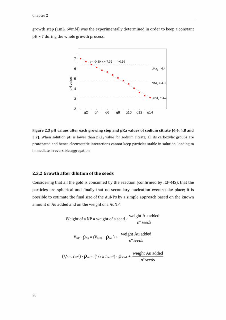
citrate and the pH after each generation was measured (Figure 2.3). It is observed a
constant decrease of 0.3 units of pH after each generation of NPs achieving a value of 3.14
after 14 generations. At this point, the AuNPs precipitated. This can be explained by
considering the pKa values of sodium citrate (6.4, 4.8 and 3.2). Since at pH 3.14 all the
carboxylic groups from citrate are protonated, the electrostatic interactions cannot
maintain particles stable in solution. Thus, the amount of sodium citrate added in each
Chapter 2
20
growth step (1mL, 60mM) was the experimentally determined in order to keep a constant
pH ~7 during the whole growth process.
g2 g4 g6 g8 g10 g12 g142
3
4
5
6
7
pKa3 = 6.4
pKa2 = 4.8
pH
va
lue
pKa1 = 3.2
y= -0.30 x + 7.39 r2=0.99
Figure 2.3 pH values after each growing step and pKa values of sodium citrate (6.4, 4.8 and
3.2). When solution pH is lower than pKa1 value for sodium citrate, all its carboxylic groups are
protonated and hence electrostatic interactions cannot keep particles stable in solution, leading to
immediate irreversible aggregation.
2.3.2 Growth after dilution of the seeds
Considering that all the gold is consumed by the reaction (confirmed by ICP-MS), that the
particles are spherical and finally that no secondary nucleation events take place; it is
possible to estimate the final size of the AuNPs by a simple approach based on the known
amount of Au added and on the weight of a AuNP.
Weight of a NP = weight of a seed +weight Au added
ºn seeds
VNP · ρAu = (Vseed · ρAu ) + weight Au added
ºn seeds
(4/3 rNP3) · ρAu= (4/3 rseed3) · ρseed +
weight Au added
ºn seeds

Synthesis of Biocompatible AuNPs
21
rNP3 = 3/4
weight Au added
º n seedsAu
+ rseed3 (equation 2.1)
Where VNP is the volume of a NP, is ρAu gold density, rNP is the radius of the NP.
According to this theoretical approach, the amount of gold needed to obtain 50 nm AuNPs
from 10 nm seeds at 3.2 x 10¹² NP/mL would be the equivalent of 125 consecutive
injections. One could think on the addition of a higher amount of gold in each injection.
Unfortunately, this would not be possible because the ratio seeds to gold monomer should
be kept between a certain range to avoid secondary nucleation (vide infra). Thus, the
previous growth protocol was modified by the addition of a dilution step every three
additions of HAuCl4 as shown in scheme 2.2. The advantage of this approach is that one
can synthesize large AuNPs in a few steps and without compromising the stability and the
monodispersity of the NPs. AuNPs were grown up to 14 growing steps and all the
generations analyzed by TEM and UV-VIS. Representative images of every step are shown
in figure 2.4. It is observed that the monodispersity is maintained in all the samples as well
as the absence of secondary populations. AuNPs were grown from the initial seeds (8.4 ±
1.0 nm) to the 14th generation (180.5 ± 10.7 nm). We decided to stop the synthesis here,
but, in principle, there is no reason against keep growing these AuNPs. Although, the NPs
are rather faceted and thus not perfectly spherical, the growth is very uniform, avoiding
formation of elongated NPs such as the ones obtained by the Frens approach.6

Chapter 2
22
Scheme 2.2 Seeded growth synthesis of AuNPs up to 180 nm. Dilution steps were introduced in
this approach to facilitate the growth to larger sizes.
Figure 2.4 Representative TEM images of every growth step. Monodispersity and absence of
secondary populations are maintained all over the process. The morphology of the AuNPs is quasi-
spherical in all cases avoiding the formation of elongated AuNPs.

Synthesis of Biocompatible AuNPs
23
A summary of sizes and size distributions, as well as SPR peak position is provided in table
2.1. Obviously, the final concentration of AuNPs depends on the dilution factor applied
(95/150 in our case). As expected, a detailed analysis of the SPR position revealed a linear
behavior due to the dependence of the peak position on the volume of the NPs.18 This
tendency is however disrupted for AuNPs larger than 100 nm, in which appears a
quadrupolar component of the SPR. Note that standard deviation is never higher than 10
% of mean value except for the seeds. Note also that the actual size of the AuNPs is quite
similar to the theoretical size calculated as explained above (equation 2.1). Thus the
presumptions made to calculate the formula (sphericity, all the added gold is consumed,
and no secondary populations) are likely accomplished.
Growth step
Diameter ± sd (nm)
sd (%)
Concentration (NPs/mL)
SPR peak (nm)
Theoretical Diameter
Seeds 8.4 ± 1.0 11.9 3.0 x 10¹² 518
1st 17.6 ± 1.2 6.8 1.9 x 10¹² 521.5 18.2
2nd 22.3 ± 2.2 9.8 1.2 x 10¹² 523.5 24.5
3rd 31.1 ± 2.8 9.0 7.6 x 10¹¹ 525.5 30.4
4th 36.0 ± 2.4 6.6 4.8 x 10¹¹ 527.5 36.7
5th 42.2 ± 2.3 7.8 3.1 x 10¹¹ 530.5 43.7
6th 54.4 ± 3.3 6.1 1.9 x 10¹¹ 535 51.5
7th 64.8 ± 3.4 5.2 1.2 x 10¹² 540 60.5
8th 69.3 ± 4.5 6.5 7.8 x 10¹⁰ 542.5 70.7
9th 80.1 ± 5.4 6.7 4.9 x 10¹⁰ 546.5 82.6
10th 96.1 ± 5.6 5.8 3.1 x 10¹⁰ 555.5 96.4
11th 109.2 ± 7.6 6.9 2.0 x 10¹⁰ 574.5 112.4
12th 123.6 ± 10.6 8.6 1.2 x 10¹⁰ 606 131.0
13th 150.0 ± 9.9 6.6 7.9 x 10⁹ 649 152.6
14th 180.5 ± 10.7 5.9 5.0 x 10⁹ 720 177.8
Table 2.1 Summary of sizes, concentration and SPR peak position of AuNPs after 14
consecutive growing steps. The theoretical size is also shown.

Chapter 2
24
2.3.3 Variation of seed and gold salt concentrations
From the results above, it is observed that the final size of the nanoparticles will depend
on the amount of seeds and gold added in solution. Taking into account these two
parameters, it seems possible to obtain AuNPs of a desired size. In this section, we explore
how these parameters affect the resultant AuNPs.
Firstly, the effect of seed concentration on the size of AuNPs after a growing step was
studied using 17.0 ± 1.7 nm seeds at concentrations ranging from 4.18 x 10¹¹ NP/mL to
8.36 x 10¹⁰ NP/mL. The final volume (75 mL) and the amount of gold precursor added (0.5
mL, 25 mM HAuCl4) were the same in all cases. Size and size distribution were analyzed by
TEM imaging and they are showed in figure 2.5 (black squares). As expected, the higher is
the concentration of seeds, the smaller is the growth of the resultant NPs. Hence, 23.3 ± 1.7
nm AuNPs were obtained at the highest concentration (4.18 x 10¹¹ NP/mL), whilst 36.6 ±
4.4 nm AuNPs were obtained when 5 times less seeds were added. It is also observed the
fitting of the empirical values with the ones predicted using the equation 2.1. Also note the
greater increase in the size for high dilutions of the seeds. Thus, one could think that a high
dilution of seeds could lead to large AuNPs. However, it should be taken into account that
new nucleation events must be avoided to get a single population of AuNPs with narrow
size distribution. This cannot be ensured when very few AuNPs are present in solution. In
fact, at the lower concentration assayed here a small percentage of a secondary population
of AuNPs appeared. Therefore the addition of subsequent growth steps seems to be the
appropriate strategy to get larger AuNPs. Additionally, it is shown here the great control of
the process and the ability to get AuNPs of a desired size. Obviously, it is necessary to
know previously both the size and the concentration of the seeds to make the required
calculations.

Synthesis of Biocompatible AuNPs
25
1 2 3 4 5 6 7 8
20
30
40
50
60
AuN
Ps
Dia
met
er
Seed Concentration x 1011
(NP/mL)
Theoretical diameter
seed size
Figure 2.5 Theoretical and empirical AuNPs diameter in function of the concentration of
seeds. The final size depends on the concentration of seeds added. It is observed here how the
empirical values match satisfactorily the theoretical value.
The other component of this system that can be modified is the amount of HAuCl4 added in
a growth step. We followed a similar strategy to analyze the role played by the precursor:
The concentration of 17.0 ± 1.7 nm seeds was kept constant (1.71 x 10¹¹ NP/mL), whilst
the concentration of HAuCl4 was varied from 0.022 mM to 0.334 mM. The size of the
resultant AuNPs, as measured by TEM, as well as the predicted value are shown in table
2.2. Obviously, the size of the AuNPs increased as the amount of gold precursor increased.
Here, the predicted size calculated from equation 2.1 also matches the empirical value.
However, a secondary population of small AuNPs was clearly observed when the highest
concentration of HAuCl4 was assayed. Thus, new nucleation of NPs was not avoided in the
case of the highest HAuCl4 concentration and consequently the monodispersity of this
sample was lost. This can be also observed analyzing the SPR peak shift: The peak shifts to
higher values as long as the amount of gold added increases, but at 0.334 mM the peak
shifts to a smaller wavelength indicating the deficient growth of the AuNPs. Since part of
the Au added was consumed by the formation of new AuNPs, which in addition competed
with the initial seeds for the remaining Au, the resultant AuNPs are polydisperse with an
unpredictable size. Hence, it is important to add the precursor in successive steps instead
of a single one which would increase the concentration of precursor too much causing new
nucleation.

Chapter 2
26
HAuCl4
(mM) Size
(nm) Theoretical
size (nm) SPR
peak (nm)
0.022 19.5 ± 2.1 19.3 519.9
0.083 24.7 ± 2.1 23.9 522.1
0.167 28.6 ± 2.7 28.2 524.1
0.334 xxx 34.2 522.2
Table 2.2 Size of AuNPs depending on the amount of gold added.
Summarizing, it has been demonstrated here the possibility to obtain AuNPs of a desired
size just by adjusting the amounts of seeds and HAuCl4 according to a simple theoretical
approach. The less seeds and the higher amount of gold added, the larger are the resultant
AuNPs. However, the variation of both components should be in a certain range to avoid
new nucleation events and therefore the formation of a secondary population of AuNPs.
Thus, a range of sizes can be obtained in a single step by adjusting these two parameters,
but a subsequent synthesis step should be introduced if larger AuNPs are desired. For
example, starting with 17 nm seeds it is easy to obtain AuNPs à la carte from 18 to 35 nm,
but if larger AuNPs are desired, an additional synthesis step (normally implying dilution of
the AuNPs) will be required.
2.4 Conclusions
The control of the size and morphology of NPs are key points in the synthesis of inorganic
NPs. In the field of biomedicine, and specifically in the use of AuNPs as scaffolds for drug
delivery, achieving a great control over the synthesis is recommended since some
important properties such as biodistribution are strongly influenced by the size of the
scaffold rather than by the composition. In fact, size effects are paramount at the
nanoscale.
Here it has been demonstrated that the synthesis of AuNPs is very well controlled in terms
of morphology, size and size distribution. Our approach is based on a seeding growth
synthesis due to the fact that the growth of AuNPs is to some extend thermodynamically
favored respect to the formation of Au nuclei. To even favor more the growth, the
temperature was decreased to 90 ⁰C instead of the 100 ⁰C required for the synthesis of
new AuNPs. AuNPs up to 30 nm can be synthesized by successive growing steps of HAuCl4.

Synthesis of Biocompatible AuNPs
27
The pH control has been shown to be crucial to maintain colloidal stability, since protons
are formed in every growth step. To obtain AuNPs up to 180 nm, dilution steps were
introduced to minimize the consumption of Au. In addition, it has been shown the
possibility to nicely control the final size of AuNPs by adjusting the amounts of either
seeds or gold precursor. The separation of nucleation and growth is the main factor that
determines the success of the synthesis and therefore must be always accomplished if
monodispersity is wanted.
2.5 References
(1) Turkevich, J.; Garton, G.; Stevenson, P. C., The Color of Colloidal Gold. Journal of Colloid Science 1954, 9, S26-S35. (2) Daniel, M. C.; Astruc, D., Gold nanoparticles: Assembly, supramolecular chemistry, quantum-size-related properties, and applications toward biology, catalysis, and nanotechnology. Chemical Reviews 2004, 104, 293-346. (3) Kimling, J.; Maier, M.; Okenve, B.; Kotaidis, V.; Ballot, H.; Plech, A., Turkevich Method for Gold Nanoparticle Synthesis Revisited. The Journal of Physical Chemistry B 2006, 110, 15700-15707. (4) Polte, J.; Ahner, T. T.; Delissen, F.; Sokolov, S.; Emmerling, F.; Thünemann, A. F.; Kraehnert, R., Mechanism of Gold Nanoparticle Formation in the Classical Citrate Synthesis Method Derived from Coupled In Situ XANES and SAXS Evaluation. J. Am. Chem. Soc. 2010, 132, 1296-1301. (5) Park, J.; Joo, J.; Kwon, S. G.; Jang, Y.; Hyeon, T., Synthesis of monodisperse spherical nanocrystals. Angewandte Chemie-International Edition 2007, 46, 4630-4660. (6) Frens, G., Controlled Nucleation for Regulation of Particle-Size in Monodisperse Gold Suspensions. Nature-Physical Science 1973, 241, 20-22. (7) Ji, X. H.; Song, X. N.; Li, J.; Bai, Y. B.; Yang, W. S.; Peng, X. G., Size control of gold nanocrystals in citrate reduction: The third role of citrate. J. Am. Chem. Soc. 2007, 129, 13939-13948. (8) Perrault, S. D.; Chan, W. C. W., Synthesis and Surface Modification of Highly Monodispersed, Spherical Gold Nanoparticles of 50−200 nm. Journal of the American Chemical Society 2009, 131, 17042-17043. (9) Bigall, N. C.; Halrtling, T.; Klose, M.; Simon, P.; Eng, L. M.; Eychmuller, A., Monodisperse Platinum Nanospheres with Adjustable Diameters from 10 to 100 nm: Synthesis and Distinct Optical Properties. Nano Letters 2008, 8, 4588-4592. (10) Jana, N. R.; Gearheart, L.; Murphy, C. J., Evidence for seed-mediated nucleation in the chemical reduction of gold salts to gold nanoparticles. Chemistry of Materials 2001, 13, 2313-2322. (11) astu s, N. G. Comenge, . untes, . c., inetically Controlled Seeded Growth Synthesis of Citrate-Stabilized Gold Nanoparticles of up to 200 nm: Size Focusing versus Ostwald Ripening. Langmuir 2011, 27, 11098-11105. (12) Jana, N. R.; Gearheart, L.; Murphy, C. J., Seeding growth for size control of 5-40 nm diameter gold nanoparticles. Langmuir 2001, 17, 6782-6786. (13) Leonov, A. P.; Zheng, J.; Clogston, J. D.; Stern, S. T.; Patri, A. K.; Wei, A., Detoxification of Gold Nanorods by Treatment with Polystyrenesulfonate. Acs Nano 2008, 2, 2481-2488. (14) Eugenii, K.; Itamar, W., Integrated Nanoparticle-Biomolecule Hybrid Systems: Synthesis, Properties, and Applications. Angewandte Chemie International Edition 2004, 43, 6042-6108.

Chapter 2
28
(15) Sellers, H.; Ulman, A.; Shnidman, Y.; Eilers, J. E., Structure and binding of alkanethiolates on gold and silver surfaces: implications for self-assembled monolayers. J. Am. Chem. Soc. 1993, 115, 9389-9401. (16) LaMer, V. K.; Dinegar, R. H., Theory, Production and Mechanism of Formation of Monodispersed Hydrosols. Journal of the American Chemical Society 1950, 72, 4847-4854. (17) Haiss, W.; Thanh, N. T. K.; Aveyard, J.; Fernig, D. G., Determination of Size and Concentration of Gold Nanoparticles from U − is Spectra. Analytical Chemistry 2007, 79, 4215-4221. (18) Njoki, P. N.; Lim, I. I. S.; Mott, D.; Park, H.-Y.; Khan, B.; Mishra, S.; Sujakumar, R.; Luo, J.; Zhong, C.-J., Size Correlation of Optical and Spectroscopic Properties for Gold Nanoparticles. The Journal of Physical Chemistry C 2007, 111, 14664-14669. (19) Kumar, S.; Gandhi, K. S.; Kumar, R., Modeling of Formation of Gold Nanoparticles by Citrate Method†. Industrial & Engineering Chemistry Research 2007, 46, 3128-3136. (20) Young, J.; Lewinski, N.; Langsner, R.; Kennedy, L.; Satyanarayan, A.; Nammalvar, V.; Lin, A.; Drezek, R., Size-controlled synthesis of monodispersed gold nanoparticles via carbon monoxide gas reduction. Nanoscale Research Letters 2011, 6, 1-11.
29
Chapter 3
Conjugation of AuNPs to Achieve
Stable and Functional Scaffolds for
Drug Delivery
3.1 Introduction
The world of NPs and the world of cells and biomolecules are quite different and often
incompatible. Functionalization of NPs surface is our tool to interact at the nano-bio
interface.1 Thus, engineering properly the AuNPs surface is a crucial step to determine the
final fate of the AuNPs since it will provide the functionality of interest and it will
determine important properties such as surface charge and stability.
A great variety of chemical groups such as thiols, amines, phosphates or carboxylic acids
have affinity to the AuNPs surface.2 This versatility allows the functionalization with
different molecules such as organic molecules, proteins, DNA, polymers, etc.3 Among all
the possible links, the SH-Au bond is the most widely exploited due to its pseudo-covalent
character (≈45 Kcal/mol)4, this high bond strength allow the exchange with other ligands
which bind Au more weakly such as citrate, amines, or phosphates. In addition, the high
strength makes the bond very stable over the time. Thus, the use of thiolated molecules is
a common strategy to functionalize AuNPs. Examples of this strategy includes conjugation
with alkanethiols,5 thiolated DNA,6 proteins or peptides containing a superficial cysteine
residue,7 and polymers with a terminal SH group.8 Alkanethiolates, such as 11-
mercaptoundecanoic acid (MUA), have been largely used to obtain self-assembled
monolayers (SAM) onto Au surfaces9 due to the presence of a thiol and a hydrophobic
chain which allow a very compact packing of the layer. In addition, a terminal group gives
the desired functionality and/or charge to the AuNPs. This is of special importance since

Chapter 3
30
some of their biological properties such as potential toxicity or cell uptake are
dramatically influenced by the surface charge given by this layer.10 Moreover, the presence
of terminal groups allows further conjugation steps. For example, EDC coupling or other
click chemistry processes between the monolayer and a molecule of interest have been
largely studied.11 Other strategies of functionalization are based on a non-specific
absorption of molecules onto the AuNP surface. These include unspecific absorption of
protein as in the case of the well-known protein corona12, 13, or weak interactions with
chemical groups such as phosphates from a DNA strand or OH groups from polymers such
as PEG, amongst others.1 In this case the link with the AuNPs is weaker than it was for
thiolated molecules.
Interestingly, by mixing AuNPs and surfactant molecules, AuNPs conjugates with a specific
functionality can be properly inserted in the biological machinery. However, it is worth
noting here that minute differences on the resulting conjugate, often ignored, might have a
dramatic impact in the biological response of the system exposed to some determined
AuNPs.14, 15 Taking into account that nanoparticles for biomedical applications are
reaching a high level of complexity, the fine control and understanding of the conjugate
composition is required to not halt the development of new products. Thus, factors such as
conformational changes of the coating molecules or small variations of classic behavior
induced by the nanoscale have to be considered before using the nanoparticle: For
example, the curvature radius of the AuNPs modifies the pKa of attached alkanethiolates,16
thus two AuNPs with the very same composition might be stable or not at a specific pH
depending on the size of the AuNPs. Also, a change on the conformation of components of
the monolayer (e.g. PEG), rather than in the composition, modifies dramatically the
biological properties of the conjugate.15
In this chapter, we carefully examine the conjugation of two widely used molecules (MUA
and SH-PEG) to the surface of AuNPs and the design of mixed layers whose properties can
be tuned depending on the composition of the layer and the conjugate architecture (global
conformation of the ligands). We also propose methods and principles to control the
conjugation and to characterize (physically, chemically and biologically) the resulting
conjugates and their evolution in physiological conditions. Also, a study to determine how
the layer composition influences the protein binding is showed. Note that the first step on
tuning NP-cell interactions is to control this protein binding. This is of special relevance,
since these are the conjugates that will be used as scaffolds for drug delivery in following
chapters.

Conjugation of AuNPs to Achieve Functional Scaffolds for Drug Delivery
31
3.2 Experimental
MUA Self-Assembled Monolayer on AuNPs. Different concentrations of MUA from 0 to
60 M were added to 20 nm AuNPs (21.3 ± 3.3 nm, 5.0 x10¹¹ NP/mL) and 40 nm AuNPs
(39.3 ± 4.3 nm, 9.0 x10¹⁰ NP/mL) solution by adding corresponding volumes of 1mM or 5
mM MUA aqueous solution. Differences of volume were compensated by addition of miliQ
water. The conjugation was gently stirred for 21 hours (an aliquote was taken and
immediately measured at 30 minutes as well). UV-VIS spectra from 300 to 800 nm were
taken for every sample at 30 minutes and 21 hours.
SH-PEG Self-Assembled Monolayer on AuNPs: Different concentrations of SH-PEG (3.4
KDa) from 0 to 15 mM were added to 20 nm AuNPs (21.3 ± 3.3 nm, 5.0 x10¹¹ NP/mL) by
adding corresponding volumes of 1 mM SH-PEG aqueous solution. Differences of volume
were compensated by addition of miliQ water. The conjugation was gently stirred
overnight. UV-VIS spectra from 300 to 800 nm were taken for every sample.
Mixed layer-AuNPs obtained by co-conjugation. The different SH-PEG / MUA ratios
were prepared by mixing the corresponding volumes of 1mM (or 0.3 mM, or 0.1 mM)
solution of each component. 500 L of these mixtures were subsequently added to 4 mL of
AuNPs as synthesized (14.6 ± 1.5 nm, 4.2 x 10¹² NP/mL) and gently stirred overnight. The
SPR bands of the resulting conjugates were analyzed by UV-VIS spectroscopy at the range
of 300 to 800 nm. The hydrodynamic diameter was analyzed by DLS.
Mixed layer-AuNPs obtained by ligand exchange. An aqueous solution of SH-PEG (125
L, 0.3 mM) was added to 1 mL citrate-stabilized AuNPs (15.2 ± 2.0 nm, 3.7 x 10¹² NP/mL).
The conjugation was allowed to run for 6 hours. Then, the excess of SH-PEG was removed
by 2 centrifugation cycles (18000 rcf, 30 min) and resuspension of AuNPs in fresh water.
Finally, different amounts of MUA (0 to 80 M) were added to the solution and the
agitation was maintained overnight. The resulting AuNPs were analyzed by UV-VIS
spectroscopy and DLS.
Quantification of the SH-PEG-FITC loaded on the AuNPs by fluorescence. The excess
of SH-PEG-FITC and MUA was removed by 3 centrifugation steps (30000 rcf, 30 min).
After the last washing step, AuNPs were suspended in the same volume of an aqueous
NaCN solution (100 mM) and stirred for 6h at 37 ºC. The total dissolution of AuNPs was
proved by the disappearance of the SPR. All the samples were buffered using CAPS (25
mM, pH 10.6). Known concentrations of SH-PEG-FITC under the same conditions were
used to do the calibration curve. 100 L of standards and samples were loaded in a 96-

Chapter 3
32
well plate and the fluorescence spectra (ex=494, em=521) was taken. The concentration
of SH-PEG-FITC in the samples was calculated from the calibration curve.
Quantification of the SH-PEG-FITC by absorbance. AuNPs were removed from the
sample by 3 centrifugation steps (30000 g, 30 min). Centrifugation conditions were more
aggressive here to ensure that all the NPs were removed. 25 L of CAPS buffer (200 M,
pH 10.6) were aded to 975 L of the resulting supernatant. To prepare the standards the
same procedure than the explained above to functionalize the AuNPs was followed but
using sodium citrate 2.2 mM instead of the AuNPs solution. The absorbance of standards
and samples were measured under the same conditions. The amount of SH-PEG-FITC
loaded on the AuNPs was calculated substracting the concentration of the sample from the
concentration of the standard.
Stability in physiological and acidic media. AuNPs functionalized with MUA and SH-PEG
were concentrated to 2.4 x 10¹⁴ NP/mL by centrifugation (18000 rcf, 14 min.) of 1 mL of
AuNPs and resuspension of the pellet in a final volume of 20 l. Then these 20 l were
added to 180 l of DMEM supplemented with 10 % FBS and softly mixed during 30
minutes. To assay the stability in acidic media, 1 mL of previously functionalized AuNPs
were brought to pH 2.6 by adding 2.5 L of a 0.2 M glycine/HCl buffer. The stability in both
cases was evaluated by analyzing the broadening and the red-shift of the SPR band.
Protein adsorption. 1.3 mM solution of bovine serum albumin was prepared by diluting
182 mg in 2.1 mL of 5 mM Hepes buffer at pH 7.5. Then 7.7 L of this solution was added
to 1 mL of previously functionalized AuNPs and softly mixed overnight. The changes of the
hydrodynamic diameter were analyzed by DLS and the shift of the SPR band by UV-VIS.
Gel electrophoresis.AuNPs were functionalized with relevant ratios of SH-PEG / MUA (0,
0.3, 0.6, 0.74, 0.88, 0.96, and 1) as explained above. Then the samples were splitted in two
parts. 1 mL was incubated with 7.7 L of a 1.3 mM BSA solution in 5 mM Hepes buffer (pH
7.5) overnight and the other part was kept without any modification. Samples were loaded
into the wells of a 1% agarose gel.The electrophoresis was run under 60 V in a sodium
borate buffer (30 mM, pH 8.5).

Conjugation of AuNPs to Achieve Functional Scaffolds for Drug Delivery
33
3.3 Results and discussion
3.3.1 MUA Self-Assembled Monolayers onto AuNPs.
The choice of MUA to study the formation of SAM onto AuNPs is not trivial. The presence
of a terminal thiol group, the relatively long (11 C) hydrocarbon chain, and the carboxylic
acid at the other side of the molecule make it a model candidate to form highly stable
SAMs. In addition, the presence of the carboxylic groups will allow a further step of
cisplatin loading via a coordination bond (see chapter 4).
To understand the formation of MUA SAMs onto AuNPs, different amounts of MUA were
added to a solution of AuNPs (21.3 ± 3.3 nm, 5.0 x10¹¹ NP/mL) and the shift of the SPR
analyzed by UV-VIS. The position of the SPR of the AuNPs after MUA addition is
determined by changes in the local refractive index around the NP due to changes in the
density of the monolayer which is determined by the concentration of ligand on the
surface.17, 18 Thus, the denser is the layer, the higher is the shift. This is observed in figure
3.1 in which the position of the peak is represented in function of the concentration of
MUA added. The SPR varies following a sigmoidal curve, typical of processes involving
adsorption on a surface at constant temperatures (e.g. Langmuir isotherms).19 From this
curve, the saturation point, or the point at which adding more MUA causes no change in
the SPR peak is determined at approximately 15 M for the analyzed AuNPs. Taking into
account the footprint of MUA in gold, which is reported to be 0.21 nm2,4 it can be easily
calculated that the amount of MUA needed to completely cover these AuNPs would be 7.25
M. Thus, at least a 2-fold excess of MUA is needed to be added in this case to ensure a
saturation of the monolayer. This excess of ligand is low compared to unspecific and
weaker bindings such as the proteins absorbed onto AuNPs, in which excess as high as 5 to
10 times the theoretical amount are required to saturate the surface.20, 21 Interestingly,
there is a slight increase of the peak observed after 21h. This fact, which has been also
described in SAMs on Au planar surfaces,22 it is attributed to an internal slow
reorganization of the monolayer, to maximize the surface coverage, and consequently
minimize the free surface energy. The differences between 30 minutes and 21 hours are
greater for higher MUA excess than for low concentrations. At concentrations much lower
than saturation, most of the added MUA is rapidly attached to the highly available surface
and therefore the final state (or a very close state) is rapidly reached. On the other side,
since the surface is densely occupied at high concentrations of MUA at short times, there
is a need of reorganization of the layer before more MUA could be attached. This
reorganization is a slow process which is not completed after the first 30 minutes, but

Chapter 3
34
hours. From a practical point of view, this suggests that conjugation of MUA on AuNPs
should be carried out overnight to ensure a complete formation of the monolayer.
0 5 10 15 20 25 30 35
521
522
523
524
SP
R p
eak
(nm
)
MUA concentration (M)
peak 30 min
peak 21 h
Figure 3.1 Shift of SPR in function of MUA added in solution. The formation of the monolayer
was monitored by UV-VIS 30 minutes and 21 hours after MUA addition. The common saturation of
a surface behavior is observed. The slight differences between times are due to a slow
reorganization of the monolayer.
To evaluate whether the size of AuNPs has an influence on the formation of the MUA SAM,
40 nm AuNPs (39.3 ± 4.3 nm, 9.0 x10¹⁰ NP/mL) were also functionalized with MUA. The
conjugation was run 21 hours under the same conditions than assayed before. In figure 3.2
the variation of peak in function of concentration of MUA added is represented for both 20
nm and 40 nm AuNPs. Note that the concentration is normalized by the surface area of the
different AuNPs and the peak shift is normalized from 0 to 1 in order to compare both
samples. It is observed that the conjugation of MUA is rather independent of the AuNPs
size in this range of sizes, showing an almost identical behavior.
The shift of the SPR as a function of the concentration of added MUA fits quite well the
Langmuir isotherm (Figure 3.2 b). This simple approach for adsorption onto surfaces is
based on four assumptions:
i. All the surface sites have the same adsorption energy for the adsorbate.
ii. The adsorption at one site does not affect the energy of adsorption of neighboring sites.
iii. All adsorption occurs through the same mechanism.
iv. At the maximum adsorption, only a monolayer is formed.
The experimental points fit quite well this theoretical approach. The constant of Langmuir

Conjugation of AuNPs to Achieve Functional Scaffolds for Drug Delivery
35
(K) can be extracted from this fitting. This corresponds to the constant of adsorption (Kabs)
assuming that the shift of the SPR is proportional to the coverage of the surface, and that
the maximum coverage is 0.21nm2/molecule.4 In the case of 20 nm, this is 0.179 M-1. This
constant represents the inverse of the bulk solution concentration required to reach half
of the maximum surface coverage. From this constant the free energy of adsorption (Gads)
can be also determined.23
AGads= -RT ln K/Vm
where R is the gas constant, T is the temperature (297 K), K is the previously obtained
constant (1.79 x 10⁵ M-1) and Vm the molar volume of the solution. As concentrations of
MUA were low, molar volume of water is used here (18.0 x 10⁻³ M-1). Gads is calculated to
be -39.8 kJ mol-1. Despite several approximations have been done to obtain this value, the
value is similar to other examples found in the literature in which the loaded molecule was
directly measured:-38.7 kJ mol-1 for SH-PEG 5 KDa,24 -35.1 kJ mol-1 for SH-DNA6 or-52.7 kJ
mol-1 for a short-chain thiolated PEG linked to an antiparkinsonian molecule.23 Thus, the
simple monitorization of the process by UV-Vis spectroscopy gave us information about
the thermodynamics and kinetics of the process. Nevertheless, it is worth mentioning here
that this simple approach is applicable only in one-component monolayer in which the
SPR shift only depends on the number of surfactant molecules per unit of surface (i.e. the
density of the layer) and where the Langmuir assumptions are valid. This is not the case of
SH-PEG, in which the SPR is also influenced by the conformational changes of the ligand,18
or proteins where the energy of adsorption at one site is different whether the
neighboring sites are occupied or vacant and also depends on the protein orientation.20
Figure 3.2. MUA monolayer characterization. (a) Shift of the SPR in function of the concentration
of MUA and size of AuNPs. Both the shift and the concentration were normalized for comparison.
(b) Fitting using the Langmuir approach for 20 nm AuNPs. The value of k can be used to further
calculate Gads.

Chapter 3
36
3.3.2 Pegylation of AuNPs.
Polyethylene glycol (PEG) is a linear synthetic polymer, build up by repeating [-CH2CH2-O]
units. It is uncharged, water soluble, inert and considered as non-toxic and non-
immunogenic. It has been extensively used to stealth liposomes and nanostructures from
being rapidly cleared by the immune system.25, 26 To obtain a stable monolayer, SH-PEG-
CM (3.4 KDa) was used onto 20 nm AuNPs. Similar as in the case of MUA, the saturation of
surface was followed by analyzing the shift of the SPR by UV-VIS spectroscopy 21 hours
after the addition of different amounts of SH-PEG (figure 3.3). After normalizing the
concentration with the surface area of NPs, it is observed that a saturation point was
reached at ca. 2 x 10-15 nM/nm2, which is 10-fold less than in the case of MUA. Thus, the
concentration to reach the saturation point is 10 times smaller. This indicates that the
density of SH-PEG onto the AuNPs was likely about 10 times smaller, since the bond with
Au is also via a SH group and therefore a similar affinity was expected. As the surfactant is
stable, what we are looking is at the bonding preference on the NP surface.
0 2 4 6 80.0
0.2
0.4
0.6
0.8
1.0
No
rma
lize
d S
PR
concentration SH-PEG (nM/nm2) x 10
-15
Figure 3.3 SH-PEG monolayer characterization. Shift of the SPR in function of the concentration
of SH-PEG added. Both shift and concentration are normalized for comparison.
3.3.3 Mixed layers
Formation of mixed layers on AuNPs via a surfactant layer formed by various components
makes possible the achievement of multifunctional AuNPs. The control of this process
allows tuning the proportion of every component on the monolayer. This is very important
since small variations on the composition or the conformation of the monolayer may

Conjugation of AuNPs to Achieve Functional Scaffolds for Drug Delivery
37
account for great changes in the physicochemical properties of the conjugates. In our case,
we were interested on mixed layers formed by MUA to have a highly packed functional
layer as chemical substrate to further attach cisplatin, and SH-PEG to confer additional
colloidal stability. Mixed layers can be obtained via a co-conjugation of the different
components (section 3.3.3.1) or by conjugation of one component and a subsequent
exchange of ligands by the other component (section 3.3.3.2). Interestingly, the individual
behavior of each compound can help to predict the final population on a mixed layer. Thus,
one could use simple analysis of a single component to make a first approach in the design
of multicomponent conjugates. However, it is important to determine the impact of the
competence for the NPs surface when both components are present in solution and see if
the affinities of the individual components for the NP are maintained.
3.3.3.1 Formation of mixed layers by co-conjugation of the components.
14.6 ± 1.5 nm AuNPs were used for these experiments since this is the chosen size for our
biological assays (see chapter 5).
Firstly, we analyzed how the addition of different ratios of SH-PEG / MUA affects the
loading of these molecules (Figure 3.4). Since both molecules compete for the AuNP
surface, it is important to assay whether the proportion of both components in solution is
maintained or not after conjugation to AuNPs. The final concentration of the sum of both
species was kept constant (30 M) and only the proportion of both components was
modified. SH-PEG of the same molecular weight (3.4 KDa), but modified with a terminal
fluorescein (SH-PEG-FITC) was also used to quantify the amount of this molecule on the
surface by measuring the absorbance of the supernatants after removing all AuNPs by
centrifugation, or by measuring the fluorescence of the attached SH-PEG-FITC after
digestion of AuNPs with sodium cyanide of the previously washed AuNPs.27 In both cases,
the amount of SH-PEG on the NPs followed a linear trend reaching a final concentration on
the NPs of aproximately 2.3 M (Figure 3.4).

Chapter 3
38
Figure 3.4. Loading of SH-PEG in function of the ratio SH-PEG / MUA added. (a) The loading on
the AuNPs was quantified with 2 different methodologies (absorbance and fluorescence after
digestion of AuNPs) giving similar results. Note that in the case of the fluorescence-based measures,
the lower ratios could not be measured due to the impossibility of digesting completely the AuNPs,
likely due to the compactness of the MUA shell. The standard curves are also showed (b and c).
This value corresponds to a final coverage of 334 molecules per NP, representing a
footprint of 2.0 nm2 which is also in agreement with the 10-fold lower saturation point
than MUA found for SH-PEG in figure 3.3. Note that in the case of lower ratios (high
[MUA]), this could not be easily measured due to the difficulty to totally digest the NPs,
likely due to the compactness of the layer when MUA is the highly predominant
component of the SAM.28 Although the amount of MUA has not been directly measured,
one could assume that the surface which is not occupied by SH-PEG will be occupied by
MUA. The proportion of MUA that is loaded on the NP will be always higher than the
added. For example, in the case of the ratio 0.5, the amount of SH-PEG loaded is ca. 0.9 M,
which corresponds to 131 molecules per NP and a 39% of surface coverage. Assuming that
the remaining 61% is covered by MUA, and taking into account that the footprint for
alkanethiols on gold is reported to be 0.21 nm2,4 the number of MUA per NP would be ca.
1945, which is 15 times higher than the amount of SH-PEG. Therefore, even if both attach
with a thiol group, the resulting interactions and packing between neighbouring molecules
favors MUA with respect to the SH-PEG molecules. This can be explained by the fact that
MUA is a smaller molecule than SH-PEG with a hydrophobic chain and a hydrophilic head
that favors the formation of dense SAMs on AuNPs9 readily than SH-PEG does.
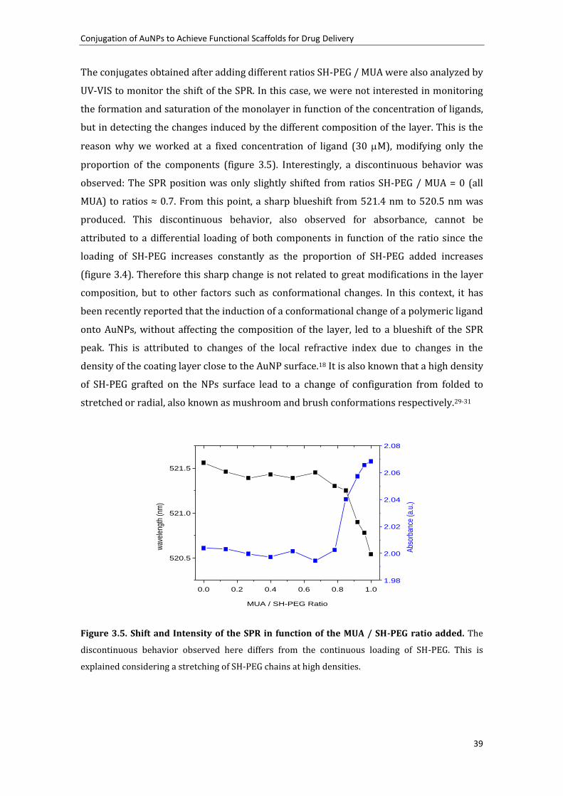
Conjugation of AuNPs to Achieve Functional Scaffolds for Drug Delivery
39
The conjugates obtained after adding different ratios SH-PEG / MUA were also analyzed by
UV-VIS to monitor the shift of the SPR. In this case, we were not interested in monitoring
the formation and saturation of the monolayer in function of the concentration of ligands,
but in detecting the changes induced by the different composition of the layer. This is the
reason why we worked at a fixed concentration of ligand (30 M), modifying only the
proportion of the components (figure 3.5). Interestingly, a discontinuous behavior was
observed: The SPR position was only slightly shifted from ratios SH-PEG / MUA = 0 (all
MUA) to ratios ≈ 0.7. From this point, a sharp blueshift from 521.4 nm to 520.5 nm was
produced. This discontinuous behavior, also observed for absorbance, cannot be
attributed to a differential loading of both components in function of the ratio since the
loading of SH-PEG increases constantly as the proportion of SH-PEG added increases
(figure 3.4). Therefore this sharp change is not related to great modifications in the layer
composition, but to other factors such as conformational changes. In this context, it has
been recently reported that the induction of a conformational change of a polymeric ligand
onto AuNPs, without affecting the composition of the layer, led to a blueshift of the SPR
peak. This is attributed to changes of the local refractive index due to changes in the
density of the coating layer close to the AuNP surface.18 It is also known that a high density
of SH-PEG grafted on the NPs surface lead to a change of configuration from folded to
stretched or radial, also known as mushroom and brush conformations respectively.29-31
0.0 0.2 0.4 0.6 0.8 1.0
520.5
521.0
521.5
MUA / SH-PEG Ratio
wav
elen
gth
(nm
)
1.98
2.00
2.02
2.04
2.06
2.08
Abs
orba
nce
(a.u
.)
Figure 3.5. Shift and Intensity of the SPR in function of the MUA / SH-PEG ratio added. The
discontinuous behavior observed here differs from the continuous loading of SH-PEG. This is
explained considering a stretching of SH-PEG chains at high densities.

Chapter 3
40
Thus, the observed blueshift is attributed to a stretching of the SH-PEG chains induced at
high grafting densities of SH-PEG. To corroborate this point, the hydrodynamic diameter
of the conjugates, measured by Dynamic Light Scattering (DLS), was also monitored in
function of the SH-PEG / MUA ratios (Figure 3.6a). DLS gives the measure of the diameter
that a sphere with the same translational diffusion speed than the corresponding
nanoparticle would have32 and its value is influenced by the length of the ligands as well as
by the compactness of the layer.20 Interestingly, a similar behavior than the previously
shown for UV-VIS was observed here: A slight increase of the hydrodynamic diameter was
produced until ratios ≈ 0.7 in which the hydrodynamic diameter raised rapidly from 14.9
nm to 27.6 nm. This is in agreement with the predicted stretching of the SH-PEG chains.
This is even more clearly observed when the hydrodynamic diameter is plotted in function
of the SH-PEG loaded onto AuNPs measured by fluorescence as explained above (Figure
3.6 b). The initial loading of SH-PEG did not cause a significant change on the
hydrodynamic diameter. On the contrary, when the amount of SH-PEG loaded on the
AuNPs reached values around 1 M, the hydrodynamic diameter dramatically increased.
Thus, the change on SH-PEG conformation is produced here at loadings around the 45 %
of the maximum amount of SH-PEG able to load onto the AuNPs (2.3 M).
We have demonstrated here the importance of not only analyze the mixed layer in terms
of composition, but also taking into account conformational changes of SH-PEG. It is
relevant to detect this point, since it will determine the change in the stabilization
mechanism as well as the protein adsorption profile of this kind of conjugates (see section
3.3.5).
Figure 3.6. Hydrodynamic diameters in function of the composition of the mixed layer. (a)
Measure of the hydrodynamic diameter depending on the ratio SH-PEG / MUA added. (b) The
diameter is shown in function of the loading of SH-PEG-FITC. Note that b shows the amount of SH-
PEG loaded onto NPs whilst a shows the amount of SH-PEG added in solution.

Conjugation of AuNPs to Achieve Functional Scaffolds for Drug Delivery
41
3.3.3.2 Formation of mixed layers by ligand exchange
It has been observed in previous sections that MUA is an excellent SAM-forming agent
because thiol group has a great affinity for Au, the hydrophobic chain favors the formation
of a compact shell, and the negatively charged carboxylate groups confer electrostatic
stability to the AuNPs. Those reasons give more affinity for AuNPs surface to MUA than to
SH-PEG and consequently, MUA is supposed to be able to displace the SH-PEG, and to
completely wash away SH-PEG from the surface when enough excess is added. Formation
of mixed layer by a partial replacement of one of the components of the monolayer has
been also reported in the literature.24, 33 In this section a methodology to replace SH-PEG
with MUA to obtain mixed layers will be described. However, we strongly recommend the
use of co-conjugation (explained in section 3.3.3.1) to obtain this kind of mixed layers due
to its simplicity and greater reproducibility.
First, AuNPs were functionalized with SH-PEG (3.4 KDa) as explained in section 3.3.2.
Then, the excess of SH-PEG was removed and different amounts of MUA were added to the
AuNPs solutions and softly stirred overnight. The resulting AuNPs were analyzed by UV-
VIS spectroscopy and DLS (Figure 3.7). The general trend was that as the concentration of
added MUA increased, the hydrodynamic diameter decreased (MUA is shorter than SH-
PEG), and SPR-band was red-shifted (the coating layer is more compact when MUA is
predominant). However, some deviations from the general trend are also observed and
are difficult to explain due to the complexity of the process in which substitution of the
components takes place together with conformational changes of the components on the
monolayer. Moreover, the substituted ligands (SH-PEG) are ready to compete again for the
NP surface with the substituting ligands (MUA). Hence, different stages were observed: In
the first points, changes in the hydrodynamic diameter and SPR peak were small, which
would indicate that MUA started to exchange with SH-PEG without causing any
conformational change in the later. Then, more remarkable changes are observed from 15
to 30 M of MUA, which could be attributed to a loss of SH-PEG density and therefore to a
change of the conformation from brush to mushroom. From that point until 60 M
changes are less evident. This is consequent with a substitution of the SH-PEG in the
mushroom conformation by MUA. It is worth remembering that it has been observed in
previous section how a progressive loading of SH-PEG resulted in very little variation on
UV-VIS and DLS measures as long as SH-PEG conformation had not been changed to brush.

Chapter 3
42
0 10 20 30 40 50 60 70 80
522.3
522.6
522.9
523.2
MUA added (M)
SP
R p
eak
(nm
)
15
20
25
30
35
Hyd
rody
nam
ic d
iam
eter
(nm
)
Figure 3.7. Monitorization of SH-PEG substitution by addition of MUA. The changes produced
in the hydrodynamic diameter (blue squares), SPR peak (black circles) and Zeta-potential (red
triangles) after adding differents amounts of MUA are indicative of the ligand exchange.
3.3.4 Stability in function of the layer composition at physiological conditions
3.3.4.1 MUA-capped AuNPs
As it is stated at the beginning of this chapter, surfactant layer determines several
physicochemical properties of the NPs which obviously have an influence on the stability
of NPs in different media.34 For example, the complex mixture of electrolites present in
physiological media would induce the compression of the Stem layer leading to
aggregation of electrostatically stabilized AuNPs if they had not been previously protected
with steric repulsor. This phenomenon is often unnoticed since the protein, present also in
physiological media, covers the NP via the well-known protein corona, conferring steric
stability to the NP. However, at high concentration of NPs this protein corona might be
insufficient to guarantee the colloidal stability of NPs. Unfortunately, high concentrations
of AuNPs are often required to reach doses of interest in in vivo and in vitro studies, in
which there is a limitation on the volume of AuNPs solution that can be added to the cell
culture or the animal.
To illustrate that aggregation in physiological media depends on the concentration of
AuNPs, 10 % v/v of MUA capped AuNPs solution at different concentrations was added to

Conjugation of AuNPs to Achieve Functional Scaffolds for Drug Delivery
43
cell culture media (CCM, DMEM supplemented with 10 % Foetal Bovine Serum) and the
peak position of SPR was measured by UV-VIS. Aggregation of AuNPs is known to broaden
and shift to longer wavelengths the SPR band as well as to increase the absorption at the
600 – 800 nm range.35 First, the stabilizing role of the protein corona was confirmed since
all the concentrations aggregated when assayed in DMEM not supplemented with Fetal
Bovine Serum (FBS). Then, as expected, it was confirmed that the stability of AuNPs in
DMEM supplemented with FBS is concentration dependent (figure 3.8). At low
concentrations of AuNPs in CCM (≤ 4.75 x 10¹² NP/mL) are very stable due to the
spontaneous formation of the widely described protein corona36, 37: Briefly, proteins are
adsorbed on the nanoparticles surface, in this case on the MUA layer, conferring stability
against aggregation by steric repulsion. In the case of higher concentrations (≥ 9.5 x 10¹²
NP/mL), a red-shift and a broadening of the SPR band were observed even in the presence
of 10% FBS. This indicates that proteins in the media were not capable of stabilizing high
concentrations of MUA-capped AuNPs. Protein in cell culture media is approximately at
0.1 mM while the highest concentration of AuNPs used here is 2.5 x 10¹³ NP/mL (41.5 nM).
Since the protein in the media is not limiting, the aggregation is likely due to a kinetic
issue: highly concentrated nanoparticles contact between them before proteins have the
chance to be adsorbed conferring stability. It must be pointed out that the methodology
followed in all the experiments here was making a dilution of the NPs and then adding 10
% v/v of this solution to the CCM. Previous observations showed that if a low volume -
even a small drop- of a very high concentration of NPs is added to a large volume of CCM,
AuNPs aggregate rapidly, even though the resultant mixture has a low NP concentration.
On the other hand, if one dilutes first the NP solution in water and then uses this solution
to reach the same final concentration in CCM, the stability is higher.

Chapter 3
44
Figure 3.8. Stability of AuNPs in cell culture media in function of the AuNPs concentration.
The broadening and great redshift of SPR at high concentration of AuNPs in cell culture media show
the aggregation of AuNPs (a). The different behavior in function of the concentration is observed in
the plotting of the peaks (b). Low concentrations were stabilized by the rapid formation of a protein
corona. However, this mechanism was unable to stabilize high concentrations.
Therefore, the spontaneous formation of the protein corona in CCM is not rapid enough to
avoid aggregation of the nanoparticles when high concentrations of AuNPs are assayed. An
easy alternative to circumvent this problem is to preincubate AuNPs with BSA in a low
ionic strength buffer before the addition of these nanoparticles to CCM. Albumin is the
major protein in serum and the most abundantly adsorbed onto hydrophilic surfaces,38
while in the case of hydrophobic surfaces, fibrinogen and immunoglobulins play an
important role and share the surface of the NP with albumin.13 To evaluate the
concentration of BSA needed to stabilize AuNPs, different amounts of BSA in HEPES (pH
7.5, 5mM) were added to a 1.0 x 10¹⁴ NP/mL solution of MUA-capped Au NPs. The
formation of the protein corona can be followed either by Dynamic Light Scattering (DLS)
or UV-VIS spectroscopy (Figure 3.9 a). In DLS measurements, it is observed a gradual
increase in the hydrodynamic diameter with higher concentrations related to the
progressive formation of a more compact BSA shell,20 reaching an 8 nm increase at the
saturation point, which is in agreement with a BSA layer of 4 nm thickness (BSA has an
equivalent diameter of 3.6 nm). The same behavior is observed when analyzing the red-
shift of the SPR band by UV-VIS spectroscopy confirming the formation of the protein
corona12 (Figure 3.9 b).
Further analysis of the stability of these preparations at the highest concentration assayed
in the previous stability test (2.5 x 10¹³ NP/mL) confirmed that preincubation of the
AuNPs with concentrations of BSA ≥ 10 nM was enough to stabilize these NPs in CCM since

Conjugation of AuNPs to Achieve Functional Scaffolds for Drug Delivery
45
the SPR band was not shifted after adding the AuNPs in CCM (Figure 3.10). On the
contrary, AuNPs which had not been previously incubated with BSA showed a great red
shift as well as a broadening of the band indicative of aggregation. Thus, protein corona is
able to stabilize the AuNPs at higher concentrations if they have been preincubated with
albumin in a low ionic strength media (as 2.2 mM sodium citrate) before adding them to
physiological media. Protein corona formation is spontaneous in cell culture media,12
hence the preincubation with BSA should not show any different biological effect if the
conjugation has been performed properly to avoid any loss of NP stability during the
process. It should be noted however, that protein corona spontaneously formed is much
more complex, with other proteins such as immunoglobulins and fibrinogen forming part
of the corona. Note also that protein corona, specially in the early stages, is dynamic.
Hence, proteins which have greater affinity to NP surface, even if they are at low
concentration, slowly replace abundant proteins.13, 38 Thus it is likely that this process may
still take place even after the preincubation with albumin which does not show a high
affinity for NPs surface.
Figure 3.9. Monitoring the protein corona formation. The protein shell increase its density of
packing as more protein is added until a full monolayer is achieved which is translated to an
increase in the hydrodynamic diameter (a) and to a red shift of the SPR band (b). This is tracked by
Dynamic Light Scattering and UV-VIS spectroscopy respectively.

Chapter 3
46
Figure 3.10. Stabilization of AuNPs by protein corona. AuNPs aggregated in cell culture media if
they had not been previously stabilized. Aggregation caused a great shift of the SPR peak as well as
a broadening of the band. The inset shows the difference of the SPR peaks before and after adding
the conjugates to CCM in function of the BSA added. The minimum amount of BSA that shows no
difference is 10 nM.
3.3.4.2 Mixed layers. Stability Dependence on the layer composition.
The presence of SH-PEG on the surfactant layer is known to confer stability to the AuNPs.39
Here, the stability is tested in function of the mixed layer composition. Different AuNPs
with varying compositions of the layer were obtained as explained in section 3.3.4. After
the removal of the excess of ligand, 20 L of AuNPs (2.4 x 10¹⁴ NP/mL) were added to 180
L of 10 % FBS-supplemented DMEM in all cases. The stability was assayed by measuring
the SPR band by UV-VIS spectroscopy after 30 minutes (figure 3.11 a). As expected, AuNPs
which have MUA predominance over SH-PEG aggregate in physiologic conditions. On the
contrary, AuNPs become stable from ratios SH-PEG / MUA higher than 0.7 which coincides
with the point where the rise in the hydrodynamic diameter, associated to a
conformational change of the SH-PEG.
The stability at acidic pH was also tested. MUA-capped AuNPs are elecrostatically
stabilized due to the negative charge given by the deprotonated carboxylic groups.
Therefore, MUA-capped AuNPs become unstable once the pH is decreased below its pKa,
and the carboxylic groups subsequently protonated (indeed, an effective surface charge of
> |30| mV is needed to keep NPs stable in solution following the DVLO theory40). On the
other hand, the sterically stabilized NPs should be stable at any pH. To prove the stability

Conjugation of AuNPs to Achieve Functional Scaffolds for Drug Delivery
47
of bifunctionalized AuNPs in a wide range of pH, the pH of the AuNPs solution was brought
to 2.6 by using a Glycine / HCl buffer and the SPR band measured by UV-VIS spectroscopy
(figure 3.11 b). Similarly as it was observed in the stability at physiologic conditions, the
aggregation of nanoparticles was evident for ratios of added SH-PEG / MUA lower than
0.7. Thus, here again the change in the mechanism of stabilization from electrostatic to
steric coincides with the change in the conformation of SH-PEG. Otherwise the non-
stretched PEG is unable to stabilize sterically the AuNPs.
From these experiments the importance of not only the composition of the layer, but on
the conformation is proved. It seems clear, that the additional stability conferred by SH-
PEG here is due to a change on the stabilization mechanism. MUA stabilizes the AuNPs via
electrostatic repulsions, however when SH-PEG is stretched an additional steric
stabilization is gained. From this moment, the steric stabilization is the main mechanism
against aggregation since they stabilize the NPs under a wider range of conditions
(including high ionic strength and low pH). Thus, the identification of the point at which
the conformational change takes place is crucial in terms of stability. Note also that
functionality given by MUA or any other alkanethiols is maintained since at ratios 0.7, the
occupation of the surface by SH-PEG is just above the 50 %, leaving plenty of space for
MUA. In fact, there would be space for ca. 1590 molecules of MUA per NP according to
calculations based on the theoretical footprint of MUA on Au (0.21 nm2). Therefore, by
tuning the mixed layer, one can guarantee both functionality given by MUA and stability
given by SH-PEG. This extra stability is based on a steric hindrance instead of on an
electrostatic repulsion. Thus, the stability is not affected by the composition of the media
and/or the concentration of NPs.

Chapter 3
48
Figure 3.11. Stability in physiologic and acidic media of concentrated AuNPs. (a) The stability
of AuNPs functionalized with different SH-PEG / MUA ratios was assayed by adding 10 % of 2.4 x
1014 NP/mL solution in DMEM supplemented with FBS. Destabilization is traduced to a broadening
and redshift of the SPR band. At ratios higher than 0.7 the stability is recovered due to the steric
stabilization given by SH-PEG in the brush conformation (b) To assay the stability in acidic media,
the pH was decreased to 2.6 using a Gly/HCl buffer. The protonation of the carboxylic groups
causes the loss of colloidal stability when the stability of the NPs is governed by MUA. On the other
hand, AuNPs remain stable when they are sterically stabilized (PEG in the brush conformation).

Conjugation of AuNPs to Achieve Functional Scaffolds for Drug Delivery
49
3.3.5 Influence of the layer composition and conformation on the protein
adsorption
We have discussed the importance of the mixed layer composition in terms of stability.
However, this is not the only goal pursued when a nanocarrier is pegylated. The role of
PEG as stealthing agent from the immune system for drug delivery systems has been
widely exploited. Hence, this methodology has been used to modify objects asliposomes,
interferon- and viruses for biomedical applications.41, 42 Although, the mechanism of
stealthing is not totally unveiled, it is accepted that avoiding the adsorption of opsonins
plays a central role. Opsonins are proteins of the immune system, such as
immunoglobulins and proteins of the complement, which binds an antigen, ultimately
triggering an immune response. In addition, the absortion of proteins from the sera has
been reported to influence the uptake of AuNPs by cells. Therefore, the inhibition of
protein adsorption needs to be studied when designing the mixed layer since it likely
affects some biological properties of the carrier. To study the adsorption of proteins onto
the different conjugates, they were prepared as explained in section 3.3.4.1, purified from
the excess SH-PEG and MUA and then incubated with 10 M BSA overnight. Albumin was
chosen as model protein to simplify the system and because it has been identified as the
main, if not the only, serum protein absorbed to these types of NPs.38 The adsorption of
protein was then analyzed by DLS, UV-VIS and gel electrophoresis. In figure 3.11, it is
shown that when MUA was the only component of monolayer, there was an increase in the
hydrodynamic diameter after adding BSA. The size increase was 8.1 nm and it is explained
by the adsorption of a monolayer of BSA on the MUA layer.2 This size increase became
only 5.2 and 3.7 nm at ratios 0.70 and 0.74 respectively. The lower increase of size is
attributed to less protein adsorption.20 When the ratios were higher than 0.78 the size
before and after addition of BSA was the same indicating that under this conditions the
protein adsorption was completely inhibited as expected. The same tendency was
observed in the SPR peak analysis (Figure 3.11b). The increase in the peak wavelength is
attributed to the change in the surrounding dielectric environment of the Au surface due
to the adsorption of protein.19 This wavelength shift was up to 4.55 nm for MUA-capped
AuNPs incubated with BSA. The difference became smaller from the ratio 0.70 (3.75 nm)
here too. This difference decreased to 2.83, 2.17, and 1.34 nm at ratios 0.74, 0.78, and 0.8
respectively. At higher ratios, the difference was not significant. Thus, the point where PEG
adopts the stretched conformation is related to a change on the phyisicochemical
properties of the conjugate once again. In this case, it is clearly seen how the adsorption of
protein is precisely tunned in function of the composition and conformation of the mixed
layer. This indicates that steric hindrance allows a better control of NP-protein

Chapter 3
50
interactions than modifications of the surface charge. In fact, differences on total surface
charge seem to not influence the degree of protein adsorption as the change on
conformation of SH-PEG does. Obviously, changes from negative to positive values would
have induced a different behavior.
Figure 3.11. Tunning the protein adsorption. (a) Hydrodinamic Diameter before and after
incubation with BSA. The formation of a protein corona at lower ratios is clearly related to the
increase of the hydrodinamic diameter. The increase of size is from ratio 0.70. From ratios 0.78 to 1
this difference on size is negligible and therefore protein adsorption is totally inhibited. (b) SPR
peak before and after incubation with albumin. The same behavior was found by analysizing the
peak of the SPR band. The differences between before and after addition of albumin are reduced
from ratios 0.7 to 0.8. From 0.84 to 1 the diferences are negligible.
Gel electrophoresis was performed to further confirm the different interaction with
proteins when changing the composition of the monolayer (figure 3.12). Relevant ratios of
SH-PEG / MUA –AuNPs previously incubated overnight with albumin and without
incubation were loaded into the wells of a 1 % agarose gel. The electrophoresis was run 1
h at 60 V in a 30 mM sodium borate buffer at pH 8.5. At ratios < 0.7, it was observed a
decrease of the electrophoretic mobility mainly due to an increase of the size after
albumin adsorption, but also to a decrease of the surface charge. On the other hand, at
ratios > 0.8, the mobility was the same after incubation with albumin than without
previous incubation indicating that no modification of the size and/or surface charge were
induced by albumin incubation. At ratio 0.74, an intermediate behavior was observed,
there was a difference between both conditions, but it was minimal. This latter case can be
explained because the adsorption of albumin was not fully inhibited, but minimized. It was
also observed that when adsorption of protein was not diminished, the electrophoretic
mobility after incubation of BSA was the same, independently of the mixed layer

Conjugation of AuNPs to Achieve Functional Scaffolds for Drug Delivery
51
composition. Thus, it is likely that MUA-capped AuNPs and mixed layer-capped AuNPs in
which the conformational change have not taken place, would show a similar fate on the
organism since the surface would be equally covered by protein corona.
As a side result of this experiment, one can notice the different electrophoretic mobility of
the mixed layer-coated AuNPs before incubating with BSA. From ratios 0 to 0.6, the
mobility was reduced likely due to a lower negative charge. As expected, the
electrophoretic mobility was dramatically reduced due to steric hindrance when the PEG
conformational change of SH-PEG was produced, this happens for ratios > 0.74. However,
as the proportion of SH-PEG kept increasing, a slight recovery of electrophoretic mobility
seems to be produced. This effect is beyond the scope of this work, but one hypothetical
explanation could be given by the higher compaction of the stretched SH-PEG shell when
the density of this component on the surface is increased. The compaction of the SH-PEG
chains may result in a more rigid shell, decreasing the mobility of SH-PEG chains, which
would make the conjugate to interact less with agarose and consequently slightly increase
its mobility.
Figure 3.12. Agarose gel electrophoresis of relevant ratios with (+) and without (-) albumin
preincubation. The difference between the same AuNPs before and after the addition of albumin
indicates the formation of a protein corona. This is inhibited at ratios = 0.88, 0.96 and 1. The
purplish color of the ratio = 0 and 0.3 appeared only after drying the gel.

Chapter 3
52
The reduction or total inhibition of protein adsorption has strong consequences in the
design of NPs for medicine. For example, a different degree and type of complement
activation, which ultimately depends on the recognition of the conjugates by specific
proteins, was directly correlated with the conformation of the PEO monolayer on
polymeric nanoparticles in a recent work.15 Briefly, Hamad et al showed that the
complement activation was dramatically reduced for the nanospheres which have PEO
chains in the brush conformation. The changes in the physicochemical properties of the
nanoparticles induced by the change of the PEG conformation, especially the different
protein adsorption, can account for this different immunological behavior.
From these results, one can distinguish two different regimes:
i. At added ratios SH-PEG /MUA from 0 to 0.7, the SH-PEG has a low influence on the
hydrodynamic diameter, since it is in the mushroom conformation. Here, the
physicochemical properties are governed by MUA and PEG does not show a major
influence. Therefore these AuNPs are not stable in physiologic media when working at
concentrations higher than 5 x 10¹² NP/mL, aggregate at acidic pH due to the protonation
of carboxylic groups of MUA and the inability of PEG to confer steric stabilization in this
mushroom conformation
ii. At added ratios from 0.7 to 1, the SH-PEG changes the conformation from mushroom to
brush and new physicochemical properties were aquired subsequently. These AuNPs are
stable in physiological media at concentrations as high as 2.4 x 10¹³ NP/mL CCM.
Moreover, they are stable at the whole ranges of pH. The protein adsorption is here
minimized or even fully inhibited.
3.4 Conclusions
The importance of the surfactant layer for AuNPs that are going to be used as drug
delivery agents is demonstrated here since it determines not only the chemical
functionality, but also it plays a key role in the stability at physiological conditions. We
were interested on two different ligands: MUA to form highly-packed SAM with carboxylic
acids to further link cisplatin and SH-PEG to confer additional stability to the vehicle.
MUA showed a high affinity for AuNPs leading to highly compact SAMs onto AuNPs. The
formation of MUA SAMs was monitored by the analysis of the SPR shift, which is assumed
to be proportional to the density of molecules on the surface. This process fits quite well
with the simple Langmuir approach for adsorption onto surfaces, independently of AuNP
size in the studied ranges. Therefore, thermodynamic parameters such as free energy of

Conjugation of AuNPs to Achieve Functional Scaffolds for Drug Delivery
53
adsorption were calculated to be quite similar to other examples found on the literature
for thiolated molecules.6, 24 Similarly as in the case of MUA, saturation point was
determined for SH-PEG. Although, the link with Au was also via the thiol group, the
saturation was reached at 10 times less amount than the amount found for MUA. Thus, the
monolayer formed by SH-PEG was not as dense as the one formed by MUA. The lack of
hydrophobic chain and the length of the chain accounts for that low density packing. This
was corroborated after quantification of SH-PEG on the surface of AuNPs. Footprint of SH-
PEG was calculated to be 2.0 nm2, which is 10-fold higher as the widely reported 0.21 nm2
for alkanethiols.
Multifunctional NPs were obtained by the formation of mixed layers of MUA and SH-PEG.
This can be achieved by ligand exchange or by co-conjugation of both components, being
the later, the one that showed a greater reproducibility in our case. The loading of SH-PEG
onto the AuNPs was proportional to the amount added. However, the analysis of the SPR
shift and the hydrodynamic diameter revealed a discontinuous behavior. This was
attributed to conformational changes of SH-PEG: At high densities of SH-PEG (higher than
45 % of coverage) SH-PEG was stretched adopting the brush configuration. It is very
important to detect this point since it determines physicochemical changes of the
conjugates.
MUA stabilized AuNPs electrostatically; thus MUA-capped (and low SH-PEG density mixed
layers-capped) AuNPs were not stable under acidic conditions, in which carboxylic acids
are protonated, and when they were exposed to high ionic strength media due to the
compression of the Stern layer. On the contrary, SH-PEG in the brush conformation
stabilized AuNPs via steric hindrance which turned the NPs to be stable even in acidic and
physiological conditions at high concentration. AuNPs can be also stabilized via the
formation of a protein corona. At low concentrations, the spontaneously formed corona in
CCM was enough to stabilize AuNPs. However, at concentrations above 4 x 10¹² NP/mL
protein in CCM was not able to stabilize them and an additional preincubation with BSA
was required.
The protein adsorption profiles were also dependent on the mixed layer composition and
configuration. As seen for the stability, the stretching of SH-PEG resulted in a progressive
inhibition of protein adsorption. This have implication on the fate of nanocarriers in the
organism since adsorption of protein has been described to influence the immune
response and the cell uptake. In fact, it has been recently reported that increasing the

Chapter 3
54
density of a SH-PEG monolayer (and thus stretching the SH-PEG) resulted in a different
degree and type of complement activation.15
3.5 References
(1) Eugenii, K.; Itamar, W., Integrated Nanoparticle-Biomolecule Hybrid Systems: Synthesis, Properties, and Applications. Angewandte Chemie International Edition 2004, 43, 6042-6108. (2) Bastús, N. G.; Casals, E.; Vázquez-Campos, S.; Puntes, V., Reactivity of engineered inorganic nanoparticles and carbon nanostructures in biological media. Nanotoxicology 2008, 2, 99 - 112. (3) Daniel, M. C.; Astruc, D., Gold nanoparticles: Assembly, supramolecular chemistry, quantum-size-related properties, and applications toward biology, catalysis, and nanotechnology. Chemical Reviews 2004, 104, 293-346. (4) Sellers, H.; Ulman, A.; Shnidman, Y.; Eilers, J. E., Structure and binding of alkanethiolates on gold and silver surfaces: implications for self-assembled monolayers. J. Am. Chem. Soc. 1993, 115, 9389-9401. (5) Wang, Y.; Zeiri, O.; Neyman, A.; Stellacci, F.; Weinstock, I. A., Nucleation and Island Growth of Alkanethiolate Ligand Domains on Gold Nanoparticles. ACS Nano 2011, 6, 629-640. (6) Yang, M.; Yau, H. C. M.; Chan, H. L., Adsorption Kinetics and Ligand-Binding Properties of Thiol-Modified Double-Stranded DNA on a Gold Surface. Langmuir 1998, 14, 6121-6129. (7) astu s, N. . a nche - illo , . Pu als, . Farrera, C. o pe , C. iralt, . Celada, A. Lloberas, J.; Puntes, V., Homogeneous Conjugation of Peptides onto Gold Nanoparticles Enhances Macrophage Response. ACS Nano 2009, 3, 1335-1344. (8) Xia, X.; Yang, M.; Wang, Y.; Zheng, Y.; Li, Q.; Chen, J.; Xia, Y., Quantifying the Coverage Density of Poly(ethylene glycol) Chains on the Surface of Gold Nanostructures. ACS Nano 2011. (9) Love, J. C.; Estroff, L. A.; Kriebel, J. K.; Nuzzo, R. G.; Whitesides, G. M., Self-Assembled Monolayers of Thiolates on Metals as a Form of Nanotechnology. Chemical Reviews 2005, 105, 1103-1170. (10) Nel, A. E.; Madler, L.; Velegol, D.; Xia, T.; Hoek, E. M. V.; Somasundaran, P.; Klaessig, F.; Castranova, V.; Thompson, M., Understanding biophysicochemical interactions at the nano-bio interface. Nat Mater 2009, 8, 543-557. (11) Bartczak, D.; Kanaras, A. G., Preparation of Peptide-Functionalized Gold Nanoparticles Using One Pot EDC/Sulfo-NHS Coupling. Langmuir 2011, 27, 10119-10123. (12) Casals, E.; Pfaller, T.; Duschl, A.; Oostingh, G. J.; Puntes, V., Time Evolution of the Nanoparticle Protein Corona. ACS Nano 2010, 4, 3623-3632. (13) Lundqvist, M.; Stigler, J.; Elia, G.; Lynch, I.; Cedervall, T.; Dawson, K. A., Nanoparticle size and surface properties determine the protein corona with possible implications for biological impacts. Proceedings of the National Academy of Sciences 2008, 105, 14265-14270. (14) Walkey, C. D.; Olsen, J. B.; Guo, H.; Emili, A.; Chan, W. C. W., Nanoparticle Size and Surface Chemistry Determine Serum Protein Adsorption and Macrophage Uptake. J. Am. Chem. Soc. 2011, 134, 2139-2147. (15) Hamad, I.; Al-Hanbali, O.; Hunter, A. C.; Rutt, K. J.; Andresen, T. L.; Moghimi, S. M., Distinct Polymer Architecture Mediates Switching of Complement Activation Pathways at the Nanosphere− erum Interface: Implications for tealth Nanoparticle ngineering. ACS Nano 2010, 4, 6629-6638.

Conjugation of AuNPs to Achieve Functional Scaffolds for Drug Delivery
55
(16) Wang, D.; Nap, R. J.; Lagzi, I. n.; Kowalczyk, B.; Han, S.; Grzybowski, B. A.; Szleifer, I., How and Why Nanoparticles Curvature Regulates the Apparent pKa of the Coating Ligands. J. Am. Chem. Soc. 2011, 133, 2192-2197. (17) Ghosh, S. K.; Nath, S.; Kundu, S.; Esumi, K.; Pal, T., Solvent and Ligand Effects on the Localized Surface Plasmon Resonance (LSPR) of Gold Colloids. The Journal of Physical Chemistry B 2004, 108, 13963-13971. (18) Tagliazucchi, M.; Blaber, M. G.; Schatz, G. C.; Weiss, E. A.; Szleifer, I., Optical Properties of Responsive Hybrid Au@Polymer Nanoparticles. ACS Nano 2012, 6, 8397-8406. (19) Eck, D.; Helm, C. A.; Wagner, N. J.; Vaynberg, K. A., Plasmon Resonance Measurements of the Adsorption and Adsorption Kinetics of a Biopolymer onto Gold Nanocolloids. Langmuir 2001, 17, 957-960. (20) Ardao, I.; Comenge, J.; Benaiges, M. D.; Álvaro, G.; Puntes, V. F., Rational Nanoconjugation Improves Biocatalytic Performance of Enzymes: Aldol Addition Catalyzed by Immobilized Rhamnulose-1-Phosphate Aldolase. Langmuir 2012, 28, 6461-6467. (21) Tsai, D. H.; Zangmeister, R. A.; Pease Iii, L. F.; Tarlov, M. J.; Zachariah, M. R., Gas-Phase Ion-Mobility Characterization of SAM-Functionalized Au Nanoparticles. Langmuir 2008, 24, 8483-8490. (22) Bain, C. D.; Troughton, E. B.; Tao, Y. T.; Evall, J.; Whitesides, G. M.; Nuzzo, R. G., Formation of monolayer films by the spontaneous assembly of organic thiols from solution onto gold. J. Am. Chem. Soc. 1989, 111, 321-335. (23) Amigo, J. M.; Bastús, N. G.; Hoen, R.; Vázquez-Campos, S.; Varón, M.; Royo, M.; Puntes, V., Analysis of time-dependent conjugation of gold nanoparticles with an antiparkinsonian molecule by using curve resolution methods. Analytica Chimica Acta 2011, 683, 170-177. (24) Tsai, D.-H.; DelRio, F. W.; MacCuspie, R. I.; Cho, T. J.; Zachariah, M. R.; Hackley, V. A., Competitive Adsorption of Thiolated Polyethylene Glycol and Mercaptopropionic Acid on Gold Nanoparticles Measured by Physical Characterization Methods. Langmuir 26, 10325-10333. (25) Owens Iii, D. E.; Peppas, N. A., Opsonization, biodistribution, and pharmacokinetics of polymeric nanoparticles. International Journal of Pharmaceutics 2006, 307, 93-102. (26) Vonarbourg, A.; Passirani, C.; Saulnier, P.; Benoit, J.-P., Parameters influencing the stealthiness of colloidal drug delivery systems. Biomaterials 2006, 27, 4356-4373. (27) Xia, X.; Yang, M.; Wang, Y.; Zheng, Y.; Li, Q.; Chen, J.; Xia, Y., Quantifying the Coverage Density of Poly(ethylene glycol) Chains on the Surface of Gold Nanostructures. ACS Nano 2011, 6, 512-522. (28) Mei, B. C.; Oh, E.; Susumu, K.; Farrell, D.; Mountziaris, T. J.; Mattoussi, H., Effects of Ligand Coordination Number and Surface Curvature on the Stability of Gold Nanoparticles in Aqueous Solutions. Langmuir 2009, 25, 10604-10611. (29) Simpson, C. A.; Agrawal, A. C.; Balinski, A.; Harkness, K. M.; Cliffel, D. E., Short-Chain PEG Mixed Monolayer Protected Gold Clusters Increase Clearance and Red Blood Cell Counts. ACS Nano 2011, 5, 3577-3584. (30) Tsai, D.-H.; DelRio, F. W.; MacCuspie, R. I.; Cho, T. J.; Zachariah, M. R.; Hackley, V. A., Competitive Adsorption of Thiolated Polyethylene Glycol and Mercaptopropionic Acid on Gold Nanoparticles Measured by Physical Characterization Methods. Langmuir 2010, 26, 10325-10333. (31) Louguet, S.; Kumar, A. C.; Guidolin, N.; Sigaud, G.; Duguet, E.; Lecommandoux, S.; Schatz, C., Control of the PEO Chain Conformation on Nanoparticles by Adsorption of PEO-block-Poly(l-lysine) Copolymers and Its Significance on Colloidal Stability and Protein Repellency. Langmuir 2011, 27, 12891-12901. (32) Ipe, B. I.; Shukla, A.; Lu, H.; Zou, B.; Rehage, H.; Niemeyer, C. M., Dynamic Light-Scattering Analysis of the Electrostatic Interaction of Hexahistidine-Tagged Cytochrome P450 Enzyme with Semiconductor Quantum Dots. ChemPhysChem 2006, 7, 1112-1118.

Chapter 3
56
(33) Hong, R.; Han, G.; Fernández, J. M.; Kim, B.-j.; Forbes, N. S.; Rotello, V. M., Glutathione-Mediated Delivery and Release Using Monolayer Protected Nanoparticle Carriers. J. Am. Chem. Soc. 2006, 128, 1078-1079. (34) Ojea-JimeÌ•nez, I.; Puntes, V., Instability of Cationic Gold Nanoparticle Bioconjugates: The Role of Citrate Ions. J. Am. Chem. Soc. 2009, 131, 13320-13327. (35) Saha, K.; Agasti, S. S.; Kim, C.; Li, X.; Rotello, V. M., Gold Nanoparticles in Chemical and Biological Sensing. Chemical Reviews 2012, 112, 2739-2779. (36) Cedervall, T.; Lynch, I.; Lindman, S.; BerggÃ¥rd, T.; Thulin, E.; Nilsson, H.; Dawson, K. A. inse, ., Understanding the nanoparticle–protein corona using methods to quantify exchange rates and affinities of proteins for nanoparticles. Proceedings of the National Academy of Sciences 2007, 104, 2050-2055. (37) Casals, E.; Pfaller, T.; Duschl, A.; Oostingh, G. J.; Puntes, V., Time Evolution of the Nanoparticle Protein Corona. ACS Nano 4, 3623-3632. (38) Casals, E.; Pfaller, T.; Duschl, A.; Oostingh, G. J.; Puntes, V. F., Hardening of the Nanoparticle–Protein Corona in Metal (Au, Ag) and Oxide (Fe3O4, CoO, and CeO2) Nanoparticles. Small 2011, 7, 3479-3486. (39) Lipka, J.; Semmler-Behnke, M.; Sperling, R. A.; Wenk, A.; Takenaka, S.; Schleh, C.; Kissel, T.; Parak, W. J.; Kreyling, W. G., Biodistribution of PEG-modified gold nanoparticles following intratracheal instillation and intravenous injection. Biomaterials 2010, 31, 6574-81. (40) Sonavane, G.; Tomoda, K.; Makino, K., Biodistribution of colloidal gold nanoparticles after intravenous administration: Effect of particle size. Colloids and Surfaces B: Biointerfaces 2008, 66, 274-280. (41) Kreppel, F.; Kochanek, S., Modification of Adenovirus Gene Transfer Vectors With Synthetic Polymers: A Scientific Review and Technical Guide. Mol Ther 2007, 16, 16-29. (42) Immordino, M. L.; Dosio, F.; Cattel, L., Stealth liposomes: review of the basic science, rationale, and clinical applications, existing and potential. International Journal of Nanomedicine 2006, 1, 297-315.

57
Chapter 4:
Functionalization of AuNPs with
Cisplatin
4.1 Introduction
Cisplatin or cis-diamminedichloroplatinum (II), [PtCl2(NH3)2], was originally synthesized
in 1845, but not until 1970 was its antitumor activity established.1 FDA approved its use
for testicular and ovarian cancer in 1978. Just 5 years later, cisplatin was already the
leading anticancer drug in USA. Today cisplatin is used worldwide to treat various types of
cancers (i.e., non-small-cell lung cancer, ovarian cancer, germ cell tumors, osteosarcomas,
etc), with a cure rate as high as 90% in testicular cancer.2 However, its use is limited by
severe side effects which result in systemic toxicity and poor patient compliance including
renal toxicity, nausea, gastrointestinal toxicity, periphereal neuropathy, asthenia, and
ototoxicity.3, 4 This high toxicity profile, specially nephrotoxicity, make it impossible to
achieve the full benefit of the treatment in a large number of patients.5 In addition,
acquired resistance and deactivation of the drug by plasma proteins are also limiting
factors. Although second-generation platinum drugs (oxaliplatin and carboplatin)
represent an improvement in some cancer treatments, the limitations due to toxicity have
not been entirely overcome (Figure 4.1).3, 6
Recent efforts have been focused on targeting the tumor by using drug delivery systems to
avoid organs where cisplatin is toxic. Nanoparticles are good candidates as carriers of
cisplatin due to its ability to be accumulated in tumors and to protect cisplatin from
plasma deactivation. However, the use of NPs to deliver drugs to the target is not without
problems and some considerations should be taken into account before using NPs for drug
delivery purposes. Skipping the biological issues, which will be discussed on subsequent

Chapter 4
58
chapters, the typical synthesis of NPs give low concentration of NPs which results in low
doses of drug, which might be insufficient to reach therapeutic effects. The attempt to
concentrate NPs could lead to a lack of stability if special care is not taken. Not only
colloidal stability, but stability of the link should also be guaranteed. Otherwise, unspecific
release of the drug would lead to an impairment of the targeting strategy. In this context,
responsive links, such as pH- or redox-sensitive links are attracting efforts.7, 8 Moreover,
this strategy ensures that the drug will be protected against being deactivated by plasma
proteins which represent as much as the 90 % of the initial dose in classic clinical
treatments.
In addition, cisplatin reactivity should be considered to determine a proper
functionalization strategy. It is known that cisplatin derivatives have to distinct sides
(Figure 4.1): The active one (e.g. the chlorides in cisplatin or water in aq. cisplatin) and the
inert one (e.g. the ammonia groups in cisplatin). In the active part, there are the reactive
groups (the leaving groups), whilst the inert part influence the biodistribution and tunes
the reactivity of the metal center. At low chloride concentration, cisplatin undergoes
exchange of chlorides by water ligands (in a process known as aquation). These aqua
complexes are much more labile than their chloride counterparts. In fact, this substitution
occurs naturally in cytoplasms, where chloride concentration is much lower than in the
extracellular environment, and the resulting specie is the active specie that ultimately
binds DNA.9 Also, it is know that aqua species can undergo hydroxylation at high pH,
which obviously implies a decrease of reactivity.10
The methodology to bind a cisplatin derivative via a pH-sensitive bond to MUA-capped
AuNPs is presented here. The conjugation of cisplatin should be strictly controlled to avoid
loss of colloidal stability: Concentration of cisplatin, time of reaction and pH influence both
stability and loading. A fine control of these 3 parameters, allowed the loading of a great
density of drug onto the surface of highly concentrated AuNPs (≈3 x 10¹⁴ NP/mL) which
make possible to reach therapeutic doses. Stability issues in physiological media are also
discussed here. Finally this study is extended to other sizes of AuNPs and to mixed layer-
capped AuNPs.

Functionalization of AuNPs with cisplatin
59
Figure 4.1. Different platinum anticancer drugs approved by the FDA. The active part is drawn
in black, in all the cases is characterized by the presence of good leaving groups that will allow the
Pt atom to bind the target. On the other side, the amines play a role in the modulation of the activity
and in the distribution of the drug. In the case of cisplatin, the equilibrium that spontaneously
occurs inside the cell (where the chloride concentration drops from 100 mM to 4 mM) is also
shown.
4.2 Experimental
Aquation of cisplatin. A solution of AgNO3 (169 mg, 1 mmol) in H2O (2.5 ml) was added
dropwise to a suspension of cisplatin (150 mg, 0.5 mmol) in H2O (2.5 ml). A white solid
(AgCl) precipitated and the yellow color of the initial mixture vanishes after completing
the addition. The resulting suspension was heated to 50 ºC for 1 h and AgCl was then
separated by centrifugation. The supernatant solution was evaporated to dryness and the
residue recrystallized from an ethanol/water mixture.
Synthesis of MUA-capped AuNPs. AuNPs (13 nm) were synthesized following a seeding
growth mechanism based on the standard method of gold salt reduction by citrate11-13.
Specifically, Au seeds (7.5 nm, 5.5 x 10¹² NP ml–1) were synthesized by adding an aqueous
solution of HAuCl4 (1ml, 25 mM) to a boiling sodium citrate solution (150 ml, 2.2 mM).
When the reaction was completed, the temperature was decreased to 90 ºC and HAuCl4
(1ml, 25 mM) was added to the previously synthesized AuNPs. This step is repeated 2
more times in order to get the final AuNPs (13.3 nm, 5.5 x 10¹² NP ml–1). The conjugation
of MUA was carried out by adding 2.2 ml of a 10 mM MUA basic solution to 50 ml of 13 nm
AuNPs solution and stirred softly overnight.
AuNPs of different sizes (from 9 nm to 60 nm) were obtained after a seeding growth
synthesis as explained in chapter 2. The 4 nm AuNPs were obtained by reduction of gold
salt by NaBH4. Briefly, a 0.25 mM solution of HAuCl4 was prepared in a 0.25 mM solution

Chapter 4
60
of sodium citrate (150 mL). Then, 1.5 mL of an ice cold solution of NaBH4 (0.1 M) was
rapidly added under vigorous stirring. The solution turned to orange-red immediately.
Concentration of MUA-capped AuNPs. The conjugates were concentrated up to 2.75 x
1014 NP ml–1 by a destabilization-resuspension step. 2.5 ml of glycine / HCl buffer (200
mM, pH=2.6) was added to 50 mL of MUA capped AuNPs to destabilize them due to the
protonation of MUA SAM. The solution was then centrifuged (2 min, 2500 rcf) and the
supernatant removed. The pellet was resuspended in 1 ml tricine buffer (50 mM, pH=8).
The excess of MUA was removed by a dialysis step (x1000, overnight). Surface Plasmon
Resonance of AuNPs was determined at wavelengths from 300 to 800 nm.
Conjugation of [Pt (H2O)2(NH3)2]2+ to MUA capped AuNPs. 5 μL of [Pt
(H2O)2(NH3)2](NO3)2 aqueous solution (16.9 mg ml–1) were added to 1 ml of MUA capped
AuNPs (13 nm, 2,75 x 10¹⁴ NP ml–1) at pH 8.3. The solution was softly mixed during 25
minutes and the reaction was stopped by removing the excess of cisplatin derivative by
dialysis (x1000, overnight). Concentrations of aq. cisplatin and reaction times were
modified in the concentration and time monitoring experiments. Actual conditions are
indicated in the results section. Also, the concentrations used in the other sized AuNPs are
indicated in the corresponding table.
Colloidal stability in blood. 17 µL of AuNP-cisplatin (2.75 x 10¹⁴ NP ml –1) were added to
500 µL of human blood and gently mixed over 24 h. Colloidal stability was assayed by
using DLS and UV-Vis spectroscopy.
pH-dependent release. The release of cisplatin in physiological conditions was
performed in solutions consisting of 50 mM buffer species, 120 mM NaCl, and 20 % Foetal
Bovine Serum (FBS). Buffer species were HEPES for pH 7.6, MES for pH 6.2 and 4.4,
Glycine/HCl for pH 4.4 and 3.8 and Acetate for pH 4.4. 100 µL of AuNP-cisplatin were
added to 900 µL of the corresponding buffered solution and mixed over differing lengths
of time (2, 8, 24, and 144 hours). AuNPs were removed by means of two centrifugation
steps (30 minutes, 35000 rcf). The amount of Pt in the supernatant was analyzed by using
ICP-MS.
Stability of the link in physiologic conditions. 10 % of AuNPs-cisplatin (aquated and
non-aquated) were added to cell culture media (DMEM with 4500 mg glucose/L, sodium
bicarbonate and pyridoxine hydrochloride without L-Glutamine and phenol red) and
incubated for 50 hours. AuNPs were removed by two centrifugation steps (30 minutes,
35000 rcf) and the amount of Pt in the subsequent supernatants analyzed by ICP-MS.

Functionalization of AuNPs with cisplatin
61
pH-dependent release in mixed layer-capped AuNPs. 50 L of [Pt(H2O)2(NH3)2](NO3)2
(0.34 mg/mL) were added to 1 mL of mixed-layer AuNPs. The conjugates were gently
stirred for 30 minutes and dialyzed overnight against a 1000-fold excess of deionized
water.The buffers were prepared by adding 30 mM buffering specie ( Hepes for pH 7.5,
MES for pH 5.4, Glycine for pH 3.8 ), 120 mM NaCl, and 10 % FBS. 10 % (v/v) cisplatin
conjugates were incubated with corresponding buffers during 148 hours. Then, AuNPs
were removed by 2 centrifugation steps (35000 rcf, 30 min) and the amount of Pt in the
supernatant analyzed by Inductively Plasma Coupled Mass Spectroscopy (ICP-MS). The
loading of cisplatin on the AuNPs is also determined in every ratio assayed here.
4.3 Results and discussion
4.3.1 Modification (aquation) of cisplatin to promote coordination bond.
Cisplatin undergoes the substitution of Cl by H2O in media which have low concentration
of Cl- (Figure 4.1). This occurs naturally inside the cell where the concentration of Cl- drops
from ≈100 mM to ≈4 mM. The aqua ligands in the resulting [PtCl(H2O)(NH3)2]+ and
[Pt(H2O)2(NH3)2]2+ are more labile than Cl-, activating the drug and allowing the platinum
atom to bind to bases on DNA.14 We were interested in this specie instead of commercial
cisplatin since H2O is a better leaving group than Cl-, allowing the formation of
coordination bonds between the Pt molecule and the deprotonated MUA carboxylic
groups on the NP. Otherwise, the commercial cisplatin molecule would be electrostatically
attached to MUA.15 The [Pt(H2O)2(NH3)2]2+ solution was obtained by adding a solution of
AgNO3 to commercial cisplatin [Pt(Cl)2(NH3)2] to promote the exchange of Cl- by H2O
ligands. After purification by recrystallization-washing steps, the solid [Pt(H2O)2(NH3)2]
(NO3)2 was obtained and dissolved in water before use. Final yield was 89 %. The
substitution of ligands was proved by mass spectroscopy (ES-MS) with the appearance of
aquated cisplatin (aq. cisplatin) peaks at m/z = 263, 264, 265 and 267 corresponding to
the 4 Pt isotopes (194Pt, 195Pt, 196Pt and 198Pt) (Figure 4.2). No peaks at m/z=300 were
observed, proving that no commercial cisplatin was present.
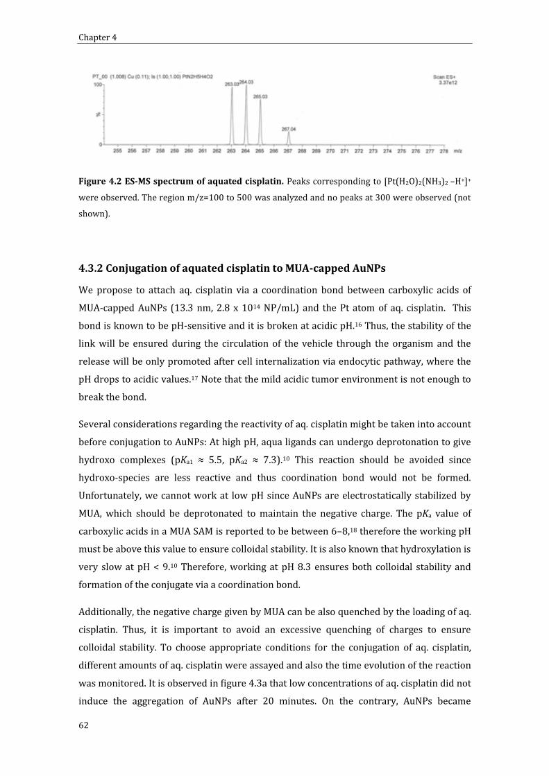
Chapter 4
62
Figure 4.2 ES-MS spectrum of aquated cisplatin. Peaks corresponding to [Pt(H2O)2(NH3)2 –H+]+
were observed. The region m/z=100 to 500 was analyzed and no peaks at 300 were observed (not
shown).
4.3.2 Conjugation of aquated cisplatin to MUA-capped AuNPs
We propose to attach aq. cisplatin via a coordination bond between carboxylic acids of
MUA-capped AuNPs (13.3 nm, 2.8 x 10¹⁴ NP/mL) and the Pt atom of aq. cisplatin. This
bond is known to be pH-sensitive and it is broken at acidic pH.16 Thus, the stability of the
link will be ensured during the circulation of the vehicle through the organism and the
release will be only promoted after cell internalization via endocytic pathway, where the
pH drops to acidic values.17 Note that the mild acidic tumor environment is not enough to
break the bond.
Several considerations regarding the reactivity of aq. cisplatin might be taken into account
before conjugation to AuNPs: At high pH, aqua ligands can undergo deprotonation to give
hydroxo complexes (pKa1 ≈ 5.5, pKa2 ≈ 7.3).10 This reaction should be avoided since
hydroxo-species are less reactive and thus coordination bond would not be formed.
Unfortunately, we cannot work at low pH since AuNPs are electrostatically stabilized by
MUA, which should be deprotonated to maintain the negative charge. The pKa value of
carboxylic acids in a MUA SAM is reported to be between 6–8,18 therefore the working pH
must be above this value to ensure colloidal stability. It is also known that hydroxylation is
very slow at pH < 9.10 Therefore, working at pH 8.3 ensures both colloidal stability and
formation of the conjugate via a coordination bond.
Additionally, the negative charge given by MUA can be also quenched by the loading of aq.
cisplatin. Thus, it is important to avoid an excessive quenching of charges to ensure
colloidal stability. To choose appropriate conditions for the conjugation of aq. cisplatin,
different amounts of aq. cisplatin were assayed and also the time evolution of the reaction
was monitored. It is observed in figure 4.3a that low concentrations of aq. cisplatin did not
induce the aggregation of AuNPs after 20 minutes. On the contrary, AuNPs became

Functionalization of AuNPs with cisplatin
63
unstable for concentrations of aq. cisplatin ≥ 0.386 mM. Regarding the time evolution, it is
shown in figure 4.3b the spectra of AuNPs took before and every 5 minutes after addition
of aq. cisplatin (0.480 mM). AuNPs were progressively losing their stability, as it is
observed by the broadening of the peak; finally leading to aggregation and precipitation,
as observed by the decrease in the absorbance.
Figure 4.3 Stability of conjugates depending on concentration of aq cisplatin and time of reaction. (a)
The SPR band was just slightly shifted for [aq. cisPt] < 0.386 which indicate the stability of these AuNPs.
On the other side, concentrations higher than 0.386 mM led to aggregation of AuNPs. (b) The time
evolution spectra showed a progressive aggregation of AuNPs at higher concentrations.
To achieve the maximum loading of aq. cisplatin without compromising colloidal stability,
aq. cisplatin was added in a slight excess (0.165 mM) to highly concentrated AuNPs after
adjusting the pH at 8.3. The reaction was monitored by zeta potential to avoid an excessive
quenching of charge (Figure 4.4). The reaction was stopped by removing the excess of aq.
cisplatin after 25 minutes by dialysis, when surface charge was -30.8 mV. It is commonly
accepted that values above +/- 30 are needed to achieve colloidal stability.19 This fact is in
agreement with our observations that ζ-potential values that are less negative than -25 mV
led to destabilization of the AuNPs and precipitation due to excessive quenching of the
negatively charged carboxylic acid groups by aq. cisplatin.

Chapter 4
64
Figure 4.4 Time evolution of zeta-potential after addition of [Pt (H2O)2(NH3)2]2+ . It is shown
the quenching of negative charge on the MUA due to the formation of a coordination bond between
the carboxylic acid group and aq. cisplatin. Conjugates remained stable at ζ-potential values more
negative than –25 mV.
At the end of the process, the loading of aq. cisplatin on the NP as measured by inductively
coupled plasma mass spectrometry (ICP-MS) was 42.3 ± 0.8 mg Pt L–1, which represents
an approximate loading of 470 cisplatin molecules per NP. Taking into account the
footprint of MUA on Au, which has been reported to be 0.21 nm2, the number of MUA
molecules per AuNP is calculated to be 2646. Assuming that every cisplatin binds to two
carboxylic groups of MUA, 940 ligands out of 2646 will be occupied. Thus, only 35 % of
MUA is quenched by cisplatin according to these theoretical calculations, leaving the other
65 % free to confer electrostatic stability to the NP. This correlates quite well with the loss
of charge from -48.5 mV to -30.8 mV (36.5 % of charge is lost). Despite this need of leaving
unoccupied MUA groups, the loading density of cisplatin per surface unit is the highest
reported ever as far as our knowledge is concerned.
It is important to remark here that high concentration of AuNPs is required to reach
significant doses of cisplatin even with this high loading density. The most used syntheses
of AuNPs in aquose media give concentrations on the order of 1012 NP/mL,11, 12, 20 which,
considering the grafting density of aq. cisplatin, would yield concentration below 1 mg Pt
L–1. This extremely low concentration would make very difficult to reach therapeutic
benefits since there is a limitation of volume that can be added to animals. For example,
not more than 10 mL/kg should be injected to mice in a single bolus injection (i.e. 200 L
for 20 g mouse).21 In order to overcome the dose limitation, we concentrated our AuNPs

Functionalization of AuNPs with cisplatin
65
50 times respect to the as synthesized samples. Firstly, we functionalized the AuNPs with
MUA as explained in chapter 3. Then, we destabilized the MUA-capped AuNPs by addition
of 2.5 ml of glycine / HCl buffer (200 mM, pH=2.6) to 50 mL of NPs. This caused the
protonation of carboxylic groups from MUA and consequently the destabilization of the
AuNPs. These AuNPs were easily concentrated by centrifugation (2 min, 2500 rcf) and
removal of the supernatant. Finally, the pellet was resuspended in 1 mL tricine buffer (50
mM, pH 8.3) in which MUA is deprotonated again. Obviously, the excess of MUA as well as
the buffer are removed by dialysis. For a typical in vivo experiment, AuNPs coming from
several syntheses are required. Thus, not only narrow size distribution is required but also
batch to batch reproducibility.
Figure 4.5. Synthesis and concentration of MUA-capped AuNPs. Representative TEM images of
MUA-capped AuNPs from 3 different syntheses. Size distribution is 13.3 ± 1.9 nm as calculated
measuring the size of > 2000 NPs by ImageJ software. In the inset, it is observed the appearance of
AuNPs before and after concentration. Scale bars are 1 m (a) and 200 nm (b).
4.3.3 Stability in physiological conditions
4.3.3.1 Colloidal Stability
Aggregation of AuNPs would modify the special properties achieved at the nanoscale and
would result in a rapid clearance of the carriers by the immune system.22 In addition, it
could lead to misinterpret the in vitro results due to a non-homogenous distribution of
NPs on the cell culture.23 Thus, colloidal stability should be ensured not only during the
synthesis step but also in working conditions for success of the in vivo targeting.24 Since
physiologic fluids have high ionic strength, aggregation of AuNPs could occur due to

Chapter 4
66
compression of the Stern layer. Our system was stabilized by the formation of a protein
corona by incubation with BSA as described in chapter 3. Briefly, 60 L of 1.3 mM BSA
solution was added to 1 mL of concentrated AuNPs-cisplatin and incubated for 30 minutes.
The stability in human blood was then assayed by DLS and UV-VIS spectroscopy (figure
4.6). In DLS measurements, no peaks indicating the presence of aggregates in blood were
observed (the 12-nm shift of the AuNPs-cisplatin peak is due to the formation of a
transient soft protein corona).25 Moreover, the aggregation of AuNPs is known to induce a
broadening and shift of the SPR band. 26, 27 The spectrum of the AuNPs-cisplatin in blood is
the same than the sum of blood and AuNPs-cisplatin spectra. Hence, no modification of the
SPR was induced by blood. Therefore it is concluded that there were no aggregation of
AuNPs in blood. Similar results were obtained when assayed in cell culture media (90 %
DMEM w/o phenol red + 10 % FBS).
Figure 4.6 AuNPs-cisplatin stability in blood after 24 hours. (a) DLS measure of AuNPs-cisplatin. The 12
nm increase of the hydrodynamic diameter is due to the formation of a protein corona with serum
proteins. (b) UV-VIS spectra of AuNPs-cisplatin, blood, and the mixture of both. The superposition of the
spectrum obtained empirically and the one that resulted from summing the individual spectra indicates
that AuNPs did not change their SPR band.
4.3.3.2 Stability of the link. The importance of coordination bond
The different reactivity of aq. cisplatin and cisplatin (see section 4.3.1) is translated to a
different type of link. Briefly, while coordination bond with MUA is formed in the aqueous
case, cisplatin is unable to form the coordination bond and it is therefore absorbed more
weakly onto the MUA layer. This has implications in the stability of the link between the
drug and the NPs. Thus, we tested the release profile of both conjugates in physiological

Functionalization of AuNPs with cisplatin
67
conditions. 10 % v/v of conjugate were added to cell culture media (DMEM + 10 % FBS)
and the amount of Pt after removing AuNPs by centrifugation analyzed by ICP-MS. Both
conjugates were stable in the conjugation vial (they are suspended in water). However,
when assayed in cell culture media, the release after 55 hours was only 15 % in the
coordination bond case whilst it was up to 60 %, with a burst release higher than 40 %, for
cisplatin electrostatically linked (Figure 4.7). Cell culture media (as well as body fluids)
has high ionic strength and consequently weak absorption is not efficient there. This
experiment shows the importance of assaying the stability in working conditions since
physiological media are complex media with a mixture of proteins and specially ions that
can affect the properties of the conjugates.
0 5 10 15 20 25 30 35 40 45 50 550
10
20
30
40
50
60
70
% r
ele
ased
cis
pla
tin
time (h)
coordinated cisplatin case
adsorbed cisplatin case
Figure 4.7. Release of cisplatin in cell culture media. Analysis of the amount of Pt in the
supernatant after removal of AuNPs was performed by ICP-MS. Cisplatin weakly adsorbed was
released in a higher degree than the linked via a coordination bond.
4.3.4 pH dependent release of cisplatin
The strength of the coordination bond has been proved above. Therefore, the release of
cisplatin should be triggered in the site of action to have biological effect since cisplatin
attached to the AuNP is not active. It is known that NPs are internalized by the endocytic
pathway, in which the pH is progressively decreased. Fortunately, the coordination bond
between Pt and carboxylic acid is broken under acidic conditions releasing the drug in its
active form.16 To prove the pH sensitivity of our link, AuNPs-cisplatin were incubated at
different pH in physiologic conditions (50 mM buffer specie, 120 mM NaCl, 20% FBS)
during different times. The amount of Pt was measured by ICP-MS after removing AuNPs

Chapter 4
68
by 2 centrifugation steps. Note that the high affinity of cisplatin for proteins make
impossible to assess the pH dependent release by performing a simple dialysis
experiment. Only 5 % of the Pt was released from the AuNPs at pH 7.6 after 144 h, while
values as high as 40 % of the Pt were reached at pH 4.4, and 67 % at pH 3.8 (Figure 4.8).
One could expect that the presence of different nucleophilic species, as the presence of
proteins, would alter the AuNP-MUA-CisPt ↔ AuNP-MUA + CisPt equilibrium. However,
release seems to be independent of the buffering species, as demonstrated by observation
of similar behaviors in three different pH 4.4 buffers. Note that the release at neutral pH
here is lower than the release produced in CCM (figure 4.8). This might be attributed to a
more complex composition of DMEM compared to the buffering species or to a higher
unspecific release for the AuNPs-cisplatin used for the link stability experiment since the
release at 1 h is as high as 7 %.
It is shown here that AuNPs-cisplatin linked via a coordination bond will likely travel
through the body with no significant release of drug. On the contrary, once the pH is
decreased in the endosomes, cisplatin will be released from the NPs. Then, it can exit the
endosome by diffusion and enter the nucleus where it finally binds the DNA.
Figure 4.8 Cisplatin release vs. pH in physiological conditions (high-ionic-strength media
with 20% BSA). At neutral pH the release was almost negligible but it increased at acidic pH
values. The different buffering species used are indicated in the legend.
4.3.6 Conjugation of aq. cisplatin to different sizes of AuNPs
The chemistry and the stability issues discussed in the previous sections are very robust
and they can be applied to different sizes of AuNPs. The only consideration that needs to
be taken into account is the different surface area and consequently the different

Functionalization of AuNPs with cisplatin
69
availability of carboxylic groups. Thus the amounts of aq. cisplatin added should be
adjusted for every size to avoid an excessive quenching of charge that would lead to
aggregation of AuNPs.
MUA and subsequently aq. cisplatin were conjugated to different sizes of AuNPs ranging
from 4 nm to 59 nm as explained above for 13 nm AuNPs (table 4.1). The amounts of MUA
and obviously aq. cisplatin were proportional to the calculated surface area for every size.
For example, surface area is 5.6 x10¹⁶ nm2/mL for 9.1 nm AuNPs and it is 3.8 x 10¹⁶
nm2/mL for 16.8 nm AuNPs. Therefore the amounts of MUA and cisplatin added were 1.5
times greater in the case of 9.1 nm AuNPs than for 16.8 nm. The same protocol was
applied for the other sizes.
The SPR of the different species and the loading of cisplatin were analyzed for every size.
The analysis of the SPR revealed that no aggregation was produced in any case (Table 1).
The expected red-shift after MUA conjugation was produced and it was followed by a
slight red-shift due to cisplatin loading. Note also that these shifts are somehow size
dependent since greater shifts were observed for smaller sizes. More important, peak
analysis revealed that colloidal stability was maintained for every size and therefore that
the amounts of aq. cisplatin added were never above the limit of tolerable charge
quenching.
Size (nm) Conc. (NP/mL)
SPR peak AuNPs (nm)
SPR peak AuNPs-MUA
(nm)
SPR peak AuNPs-cisPt (nm)
3.8 ± 0.7 4.09E+15 505.88 519.25 522.50 9.1 ± 1.4 2.13E+14 514.68 521.69 525.85 16.8 ± 1.9 4.25E+13 518.79 522.6 524.84 26.5 ± 3.8 8.50E+12 521.75 524.07 525.04 39.6 ± 4.2 2.85E+12 526.73 528.58 528.68 58.6 ± 7.2 9.45E+11 534.91 536.64 536.49
Table 4.1. Analysis of the SPR shift after MUA and aq. cisplatin conjugation in different sizes of
AuNPs. No broadening and large shift was observed. Therefore no aggregation occurred in any case.
The loading of cisplatin was quantified by ICP-MS. Results are summarized in table 4.2. As
expected, the total loading (mg/L) is dependent on the total available surface. However,
the footprint of cisplatin on the different sizes is quite similar (except for the 39.6 nm

Chapter 4
70
AuNPs). Note that optimization of the loading of cisplatin onto 13 nm AuNPs had led to a
density of cisplatin ca. 0.8 molecules/nm2 which is the highest loading density achieved in
similar NPs as far as we know. A simple translation of the protocol for different sizes and
concentrations without any optimization led to comparable densities for most of the sizes
assayed here. Thus, the strategy optimized for 13 nm AuNPs is straightforwardly
translated to other sizes just by taking into account the total surface area of the AuNPs of
interest. It is worth to mention, however, that different sizes have different fates inside the
body.
Size (nm)
Conc. (NP/mL)
Total Surface
Ratio
cisPt added
(L) (conc, mg/mL)
cisPt loading
(mg Pt/L)
nº cisPt per NP
density (molec/nm2)
3.8 ± 0.7 4.09E+15 1 7.5 (16.9) 46 34.8 0.76
9.1 ± 1.4 2.13E+14 0.30 22.6 (1.69) 10.9 158.3 0.60 16.8 ± 1.9 4.25E+13 0.20 15.2 (1.69) 6.3 457.5 0.52 26.5 ± 3.8 8.50E+12 0.10 7.5 (1.69) 3.9 1416.2 0.64 39.6 ± 4.2 2.85E+12 0.07 5.6 (1.69) 1.7 1841.1 0.37 58.6 ± 7.2 9.45E+11 0.05 4.1 (1.69) 2.1 6859.2 0.64
Table 4.2. Loadings of cisplatin to different sizes of AuNPs. The amounts of cisplatin added are
proportional to the total surface (i.e. the surface of a NP multiplied by the number of NPs). The ratio
of the total surface divided by the surface of the 3.8 nm case is provided for comparison. Total
loadings were measured by ICP-MS and from this data were obtained the loading per NP and the
footprint of cisplatin.
4.3.7 Conjugation of aq.cisplatin to mixed layer (MUA + SH-PEG) AuNPs
Mixed layers allow tuning the properties conferred by monolayers to the AuNPs. In the
case of monolayers composed of MUA and SH-PEG, functionality and stability are tuned.
The highly packed MUA layer provides plenty of carboxylic acids to further attach
cisplatin, whilst SH-PEG is known to confer steric stabilization to the system. Details on
the characterization and behavior of these conjugates can be found in section 3.3.3 of the
present work. Here the functionalization of these conjugates with cisplatin and their
different behaviors are discussed.
The strategy followed to conjugate cisplatin to mixed layer AuNPs was similar to the
functionalization of MUA-capped AuNPs. 50 L of aq. cisplatin (0.34 mg/mL) was added to
1 mL of mixed layer-capped AuNPs. Excess of aq. cisplatin was removed by dyalisis. The

Functionalization of AuNPs with cisplatin
71
loading of cisplatin in the different conjugates was analyzed by ICP-MS. In these
conditions, Pt atom forms a coordination bond with carboxylic acid whilst its affinity with
ether groups from SH-PEG is lower.10 Consequently, a different drug release behavior is
expected depending on the composition of the layer. The release at different pHs at
physiological conditions (10 % FBS+ buffer + 120 mM NaCl) after 148 hours is assayed
here (Figure 4.9). Note that concentrations of cisplatin were always low to avoid total
quenching of carboxylic groups from MUA; this would have led to a loss of colloidal
stability. Thus, it is not surprising that at lower ratios, in which there is much more MUA
loaded than SH-PEG, the behavior of the conjugates was quite similar since all cisplatin
was expected to be linked to MUA. It is shown how cisplatin was only released
significantly at acidic pH for ratios < 0.6. On the other hand, at higher ratios, in which the
amount of SH-PEG loaded onto the NPs was greater, the unspecific release increased even
at neutral pH. Thus, part of the specificity of the link for MUA is lost here. This is explained
by the fact that MUA was not in such a great excess and some cisplatin was linked to SH-
PEG. Cisplatin bond with SH-PEG is not a coordination, pH-sensitive, bond and therefore
the release profile is different. Surprisingly, there is a decrease of the release when SH-PEG
is the predominant component of the system (ratios > 0.88). This behavior is repeated in
all the conditions and reproduced in different experiments. One possible explanation
would be that a highly compact SH-PEG layer could bind cisplatin stronger, due to the
crosslinking of a Pt atom by two stretched PEG chains. This strongly bond cisplatin was
not significantly released even at low pH. Whichever the reason, it is clear that the pH-
sensitivity, which controls the endosomal release of cisplatin in cell but not in blood, is lost
in the SH-PEG layer, showing the importance of having mixed layers to tune the responses,
in this case, stability (PEG) and functionality (MUA).

Chapter 4
72
Ratio
SH-PEG/
MUA
cisPt loading
(mg Pt/L)
0 1.013
0.3 1.186
0.6 1.053
0.74 0.971
0.8 0.832
0.84 0.921
0.88 0.680
0.96 0.802
1 1.058
Figure 4.9 Cisplatin loading and pH-dependent release of cisplatin at different ratios of SH-
PEG/MUA. The loading of cisplatin depending on the ratio SH-PEG / MUA is shown in the table. The
release profile of cisplatin at 3 different pHs was analyzed for the different conjugates. Unspecific
release increased as the amount of SH-PEG increased in the mixed layer.
4.3.7 Conjugation of other Pt derivative based on oxaliplatin
The strength of the coordination bond makes the link between AuNPs and aq.cisplatin
very stable which is desired to avoid unspecific release of AuNPs. Aq. cisplatin is released
at acidic pH. However, this release is slow with consequent low release at short times.
Thus, one could think in increasing the efficiency of the triggering by making the link
between Pt and AuNP more liable. To make the coordination bond between Pt and
carboxylic acids weaker, we tested an aquated derivative of oxaliplatin (figure 4.10) in
which the oxalate group was exchanged to water groups to facilitate the formation of the
coordination bond. Interestingly, the ammines in the remaining ciclohexane-1,2-diamine
group are greater electron donors than ammonia and therefore satisfy better the need of
electroneutrality at the metal, polarizing the metal centre. The trans Pt-NHx and Pt-COOR
bond of cisplatin share a metal orbital, consequently the polarization induced by the
ammine, which is greater than the one induced by ammonia, makes the bond Pt-COOR
more labile. This phenomenon is known as the trans effect and it is widely exploited in
coordination chemistry, for example to control the specificity in the substitution of ligands.

Functionalization of AuNPs with cisplatin
73
Figure 4.10. Chemical structure of cisplatin, oxaliplatin and their corresponding aquated
derivatives.
We tested the conjugation of [Pt(cyclohexane-1,2-diamine)(H2O)2] (from now on referred
as aq. oxaliplatin despite oxalate group is not present) onto 13 nm MUA-capped AuNPs.
The conjugation was performed in a similar way that the one optimized for aq. cisplatin.
Briefly, 20 L of aq. oxaliplatin (4.22 mg/mL) was added to 1 mL of AuNPs (2.8 x10¹⁴
NP/mL). After 25 minutes, the reaction was stopped by removing the excess of aq.
oxaliplatin by dialysis against water. The loading of Pt onto the NPs was 39.2 mg/L as
assayed by ICP-MS. This corresponds to a loading of 432 molecules/NP, very similar to the
loading of aq. cisplatin (470 molec./NP). UV-VIS spectra showed a red-shift after
conjugation of MUA and aq. oxaliplatin, but no broadening of the band (Figure 4.10). Thus,
no aggregation was produced in this functionalization as well.
Figure 4.10 UV-VIS spectra of AuNPs after MUA and aq. oxaliplatin conjugation. The slight
shifts of the SPR are consequent with conjugation and absence of aggregation.

Chapter 4
74
The release of drug was studied also at pH 7.7 and 5.5 after 144 hours. Interestingly, the
release at acidic pH was higher than the one previously observed for aq. cisplatin. Hence,
the release of aq. oxaliplatin after 144 hours at pH 5.5 was as high as 72 %, far beyond
from the 37 % found in the case of aq. cisplatin at even more acidic pH (4.4).
Unfortunately, the unspecific release also increased: 29 % of aq. oxaliplatin at pH 7.7 was
released after 144 h, whilst only 5 % was released in the case of aq. cisplatin.
Thus, it is clear that the link between Pt and MUA from AuNPs is more labile in the case of
aq. oxaliplatin. However, this causes a drop in the strength of the link and consequently
unspecific release at neutral pH is increased.
4.4 Conclusions
Here it has been presented the use of AuNPs as drug delivery scaffolds for cisplatin from
the chemical point of view but bearing in mind the biological application. Thus, special
attention was paid in some points which cannot be underestimated, namely the
concentration of NPs to reach therapeutic doses, the maintenance of both colloidal and
link stability in physiological conditions and the triggering of the release by a pH drop.
To achieve a strong bond, aquated cisplatin ([Pt(H2O)2(NH3)2]2+) was used instead of
cisplatin ([Pt(Cl)2(NH3)2]). The aqua complexes are much more reactive than cisplatin and
therefore the formation of a coordination bond with MUA was ensured. This confers high
stability to the link in physiological conditions, which guaranteed the avoidance of
unspecific release of drug. In addition, this link is pH-sensitive and therefore a pH-drop,
typically produced in the endosomes after AuNPs internalization,17 triggers the controlled
release of cisplatin.
Also colloidal stability was assessed during the conjugation and in physiological
environments. This is not trivial since the formation of the coordination bond quenches
the negative charge given by carboxylic groups which is responsible of maintaining
colloidal stability. Thus, concentration of aq. cisplatin and time of reaction should be
strictly controlled during the functionalization to avoid excessive quenching. Also the pH
working window is narrow and needs to be controlled since cisplatin would be
hydroxylated at basic pH, whilst MUA would be protonated leading to a loss of colloidal
stability at acidic pH. In biological fluids, in which the ionic strength is high, the stability is
also compromised. However, AuNPs-cisplatin can be stabilized by the formation of a
protective protein corona.

Functionalization of AuNPs with cisplatin
75
The loading density of cisplatin onto AuNPs is 1.2 nm2/molecule, which is the highest
reported for similar NPs as far as we know. Despite this high loading density,
concentration of AuNPs up to 3 x10¹⁴ NP/mL are required to achieve therapeutic doses
(e.g. 2 mg cisplatin/Kg mouse) due to the limitation of the injectable volume to animals
(i.e. 10 mL/Kg). This is approximately 50 times the concentration achieved in classical
syntheses in aquose media.
This functionalization methodology was also assessed in other systems. AuNPs of different
sizes from 4 to 60 nm were functionalized with aq. cisplatin leading to similar loading
densities just by a straightforward adjustment of some parameters. Also mixed layer
(MUA/SH-PEG) -capped AuNPs were successfully functionalized. In these AuNPs the
importance of controlling the composition of the mixed layer was emphasized since a
certain amount of SH-PEG is required to achieve the special properties given by this ligand
(i.e. steric stabilization and protein adsorption inhibition), but excessive loading of SH-PEG
led to an unspecific release of cisplatin.
Finally, the functionalization with another platinum derivative ([Pt(cyclohexane-1,2-
diamine)(H2O)2]) was carried out. By using this compound, the link between AuNPs and
the drug is more labile due to the higher trans effect of ammines compared to ammonia.
This resulted in a higher release of drug at acidic pH, but at the expense of increasing the
unspecific release.
4.5 References
(1) Rosenberg, B.; Vancamp, L.; Trosko, J. E.; Mansour, V. H., Platinum Compounds: a New Class of Potent Antitumour Agents. Nature 1969, 222, 385-386. (2) Wang, D.; Lippard, S. J., Cellular processing of platinum anticancer drugs. Nat Rev Drug Discov 2005, 4, 307-320. (3) Kelland, L., The resurgence of platinum-based cancer chemotherapy. Nat Rev Cancer 2007, 7, 573-584. (4) Reedijk, J., New clues for platinum antitumor chemistry: Kinetically controlled metal binding to DNA. Proceedings of the National Academy of Sciences of the United States of America 2003, 100, 3611-3616. (5) Oliver, T. G.; Mercer, K. L.; Sayles, L. C.; Burke, J. R.; Mendus, D.; Lovejoy, K. S.; Cheng, M.-H.; Subramanian, A.; Mu, D.; Powers, S.; Crowley, D.; Bronson, R. T.; Whittaker, C. A.; Bhutkar, A.; Lippard, S. J.; Golub, T.; Thomale, J.; Jacks, T.; Sweet-Cordero, E. A., Chronic cisplatin treatment promotes enhanced damage repair and tumor progression in a mouse model of lung cancer. Genes & Development 24, 837-852. (6) Mani, S.; Graham, M. A.; Bregman, D. B.; Ivy, P.; Chaney, S. G., Oxaliplatin: A Review of Evolving Concepts. Cancer Investigation 2002, 20, 246-263.
Chapter 4
76
(7) Dhar, S.; Gu, F. X.; Langer, R.; Farokhzad, O. C.; Lippard, S. J., Targeted delivery of cisplatin to prostate cancer cells by aptamer functionalized Pt(IV) prodrug-PLGA–PEG nanoparticles. Proceedings of the National Academy of Sciences 2008, 105, 17356-17361. (8) Comenge, J.; Sotelo, C.; Romero, F.; Gallego, O.; Barnadas, A.; Parada, T. G.-C.; Domínguez, F.; Puntes, V. F., Detoxifying Antitumoral Drugs via Nanoconjugation: The Case of Gold Nanoparticles and Cisplatin. PLoS ONE 2012, 7, e47562. (9) Klein, A. V.; Hambley, T. W., Platinum Drug Distribution in Cancer Cells and Tumors. Chemical Reviews 2009, 109, 4911-4920. (10) Kelland, L. R.; Farrell, N., Platinum-Based Drugs in Cancer Therapy. Humana Press Inc.: 2000. (11) astu s, N. G. Comenge, . Puntes, . c., inetically Controlled Seeded Growth Synthesis of Citrate-Stabilized Gold Nanoparticles of up to 200 nm: Size Focusing versus Ostwald Ripening. Langmuir 2011, 27, 11098-11105. (12) Kimling, J.; Maier, M.; Okenve, B.; Kotaidis, V.; Ballot, H.; Plech, A., Turkevich Method for Gold Nanoparticle Synthesis Revisited. The Journal of Physical Chemistry B 2006, 110, 15700-15707. (13) Park, C.; Kim, H.; Kim, S.; Kim, C., Enzyme Responsive Nanocontainers with Cyclodextrin Gatekeepers and Synergistic Effects in Release of Guests. J. Am. Chem. Soc. 2009, 131, 16614-16615. (14) Jung, Y.; Lippard, S. J., Direct Cellular Responses to Platinum-Induced DNA Damage. Chemical Reviews 2007, 107, 1387-1407. (15) Comenge, J.; Romero, F. M.; Sotelo, C.; Dominguez, F.; Puntes, V., Exploring the binding of Pt drugs to gold nanoparticles for controlled passive release of cisplatin. Journal of controlled release : official journal of the Controlled Release Society 2010, 148, e31-2. (16) Tseng, C.-L.; Su, W.-Y.; Yen, K.-C.; Yang, K.-C.; Lin, F.-H., The use of biotinylated-EGF-modified gelatin nanoparticle carrier to enhance cisplatin accumulation in cancerous lungs via inhalation. Biomaterials 2009, 30, 3476-3485. (17) Nel, A. E.; Madler, L.; Velegol, D.; Xia, T.; Hoek, E. M. V.; Somasundaran, P.; Klaessig, F.; Castranova, V.; Thompson, M., Understanding biophysicochemical interactions at the nano-bio interface. Nat Mater 2009, 8, 543-557. (18) ishop, . . M. Grzybowski, . A., “Nanoions”: Fundamental Properties and Analytical Applications of Charged Nanoparticles. ChemPhysChem 2007, 8, 2171-2176. (19) Sonavane, G.; Tomoda, K.; Makino, K., Biodistribution of colloidal gold nanoparticles after intravenous administration: Effect of particle size. Colloids and Surfaces B: Biointerfaces 2008, 66, 274-280. (20) Park, J.; Joo, J.; Kwon, S. G.; Jang, Y.; Hyeon, T., Synthesis of monodisperse spherical nanocrystals. Angewandte Chemie-International Edition 2007, 46, 4630-4660. (21) Diehl, K.-H.; Hull, R.; Morton, D.; Pfister, R.; Rabemampianina, Y.; Smith, D.; Vidal, J.-M.; Vorstenbosch, C. V. D., A good practice guide to the administration of substances and removal of blood, including routes and volumes. Journal of Applied Toxicology 2001, 21, 15-23. (22) Sperling, R. A.; Casals, E.; Comenge, J.; Bastus, N. G.; Puntes, V. F., Inorganic Engineered Nanoparticles and Their Impact on the Immune Response. Current Drug Metabolism 2009, 10, 895-904. (23) Cho, E. C.; Zhang, Q.; Xia, Y., The effect of sedimentation and diffusion on cellular uptake of gold nanoparticles. Nat Nano 2011, 6, 385-391. (24) Ruenraroengsak, P.; Cook, J. M.; Florence, A. T., Nanosystem drug targeting: Facing up to complex realities. Journal of Controlled Release 141, 265-276. (25) Casals, E.; Pfaller, T.; Duschl, A.; Oostingh, G. J.; Puntes, V., Time Evolution of the Nanoparticle Protein Corona. ACS Nano 4, 3623-3632. (26) Grabar, K. C.; Smith, P. C.; Musick, M. D.; Davis, J. A.; Walter, D. G.; Jackson, M. A.; Guthrie, A. P.; Natan, M. J., Kinetic control of interparticle spacing in Au colloid-based surfaces: Rational nanometer-scale architecture. J. Am. Chem. Soc. 1996, 118, 1148-1153.

Functionalization of AuNPs with cisplatin
77
(27) Kelly, K. L.; Coronado, E.; Zhao, L. L.; Schatz, G. C., The Optical Properties of Metal Nanoparticles: The Influence of Size, Shape, and Dielectric Environment. The Journal of Physical Chemistry B 2002, 107, 668-677.

Chapter 4
78

79
Chapter 5:
AuNPs-Cisplatin as Drug Delivery
Systems: In Vitro and In Vivo
Experiments
5.1 Introduction
Cisplatin –as other molecular drugs- is small enough to travel along with ions and small
nutrients through the body due to the selective permeability of continuous capillaries for
ions and small molecules.1, 2 This results in high index of permeability and distribution
through the whole body what is intended to allow the active principle to reach its target.3, 4
Unfortunately, this widespread distribution of the drug is at the origin of unwanted side
effects, causing low drug selectivity and efficiency.5 In fact, the severe nephrotoxicity
induced by cisplatin is a dose limiting factor and in many cases, the treatment must be
stopped before therapeutic benefits are reached. In addition, this is a reason to prevent its
use in patients with renal insufficiency.
On the contrary, nanoscale objects travel following particular paths through the body.2, 6
This results in the passive accumulation of nanocarriers in:
i. Organs of the mononuclear phagocyte system7 (liver, spleen and bone marrow), where
blood vessels are fenestrated with gaps ranging between 100 and 1000 nm.
ii. Inflamed tissue, where an increased permeability is achieved through temporal induced
gaps to recruit immune cells1.
iii. Solid tumors,8 in which its rapid growth results in leaky vessels. These fenestrated
vessels allow macromolecules and NPs to permeate through the tumor. In addition, the
nanoparticles are retained due to the lack of a functional lymphatic system. This effect

Chapter 5
80
(Enhanced Permeability and Retention effect, EPR) is widely reported in the literature6, 9
and has been exploited to passively accumulate nanocarriers in tumors.10
It is important to choose appropriate NPs since the pharmacokinetics and biodistribution
of the conjugates will be governed by the physicochemical properties of the carrier rather
than by the ones of the drug.11 In addition, EPR effect is known to be size dependent. For
example, it has been reported that 20 nm PEG-coated AuNPs showed the best behaviour
for tumor treatment since they present a high accumulation rate because of their long half-
life in blood and an acceptable penetration in the tumor. On the other hand, larger NPs
(>40 nm) failed to penetrate deep into the tumour and they were only accumulated at the
vicinity of the vessels.12 Smaller NPs, which would have penetrated deeply, do not have the
opportunity to be significantly accumulated in the tumor due to renal clearance.
Obviously, these properties would have been modified if colloidal stability had been
compromised, showing again the importance of maintaining NPs stable along the process.
Once in its site of action, cisplatin enters the cells mainly by passive diffusion through the
membrane, but also it is actively transported by metal transporters.13 In the cytoplasm,
where chloride concentration drops from 100 mM to 4 mM, cisplatin undergoes exchange
of chlorides for water ligands. This aquated derivative is much more reactive than the
chlorinated one; and it is the active specie. DNA is its primary target: Different types of
adducts between the Pt atom and DNA strands are formed, being intrastrand cross-links
the most numerous.14 These adducts interfere with proteins involved in the mechanisms
of replication and repairing of DNA, which ultimately lead to apoptosis.15 This mechanism
of cell death is very effective. Thus, there is no need to improve the efficacy of the drug, but
to improve its efficiency, mainly by reducing the side effects of cisplatin by means of a
better delivery and avoiding organs where cisplatin is known to be highly toxic.
Although a better targeting will likely imply an improvement in the therapy, the potential
risks of using nanocarriers should be deeply analyzed. NPs are known to be accumulated
not only in the tumor, but also in organs of the mononuclear phagocyte system since the
vessels in those organs present also fenestrations. Thus, evaluation of possible adverse
effects needs to be considered. Moreover, a usual mistake is to consider NPs as inert
systems that do not evolve with time. However, they aggregate if special care is not taken,
or release ions under certain conditions. Both processes could lead to adverse effects
and/or to a misinterpretation of biological results. For example, aggregation might induce
cell death in in vitro experiments16 or to a rapid clearance in in vivo assays,17 which might
In Vitro and In Vivo Experiments
81
lead to an overestimation and underestimation respectively of the capability of the system
to cause therapeutic benefit.
In this chapter, AuNPs-cisplatin were tested in in vitro assays to evaluate the cell and DNA
accumulation as well as cell viability. More importantly, in vivo assays using tumor-bearing
mice were also performed. This includes evaluation of the therapeutic efficacy and
evaluation of the toxicity induced by cisplatin and by AuNPs-cisplatin. Interestingly,
nephrotoxicity, which is dose limiting in the clinics, was avoided only in the case of
animals treated with AuNPs-cisplatin. In addition, systemic toxicity, as evaluated by body
weight loss and analyzing relevant biomarkers, was also improved. Organs in which NPs
are known to be accumulated (liver and spleen) were carefully examined showing no
adverse long-time effects. Thus, the toxicological profile of cisplatin is clearly reduced
without affecting its antitumor activity. The lack of toxicity is correlated to the
modification of the pharmacokinetic properties and biodistribution of the drug by
conjugation to AuNPs.
5.2 Experimental
Cell culture. The human lung carcinoma derived cell line A549 was obtained from
American Tissue Culture Collection (ATCC) and cultured in a 1:1 mixture of Dulbecco´s
Modified Eagle´s Medium (DMEM, Sigma-Aldrich) and Ham´s F-12 Medium (Sigma-
Aldrich), supplemented of 10 % (v/v) foetal bovine serum (FBS, GIBCO-Invitrogen) and 1
% (v/v) of L-glutamine, penicillin and streptomycin solution (GPS, Sigma-Aldrich).
Cell internalization and DNA accumulation. To quantify Pt and Au cell internalization, 5
x 105 cells were plated in 60 mm diameter plates (Falcon) and 24 hour later, medium was
changed for treatments diluted in culture medium: free cisplatin or cisplatin conjugated to
AuNPs (2 g cisplatin/ml in both cases). After 0.5, 1, 3, 6, or 24 hours with the treatment,
cells were trypsinized and centrifuged, supernatant was removed and cells were
resuspended in 0.5 ml 65% HNO3 (Merck). The amount of Pt and Au was determined by
ICP-MS (Bruker 820-MS). To quantify cisplatin bound to DNA, 106 cells were treated as
above and DNA extraction was performed by phenol/chloroform/isoamylalcohol
(50:49:1) extraction.18 DNA was finally resuspended in 200 l. This solution was used to
determine both the amount of DNA, and Pt and Au by UV-Vis spectroscopy and ICP-MS
respectively.

Chapter 5
82
TEM cells imaging. A549 cells were centrifuged for 15 min at 3000 rpm, fixed by
immersion for 45 min in 2.5% glutaraldehyde in sodium cacodylate buffer (0.15 M, pH =
7.3), postfixed with 2% osmium tetroxide in the same buffer, dehydrated, embedded in
LRWhite resin, Medium Grade, and sectioned. Sections were stained with uranyl acetate
and lead citrate and examined in a Zeiss 902 electron microscope (Carl Zeiss, F.R.G.) at 80
kV accelerating voltage.
Cell viability assay. 1000 cells/well were seeded in a 96-well plate and incubated at 37 ⁰C
with 100 µL of DMEM. After 24 hours, the different treatments resuspended in 100 L of
fresh DMEM were added to the corresponding wells. Cells were treated for 48 hours.
Finally, cells were washed with 200 L of PBS, and then 180 L of fresh media was added.
Finally, 20 L of tetrazolium were added to every well and cells were incubated 1.5 hours.
The absorbance was measured at 450 and 620 nm. The amount of formazan is
proportional to A620- A540 and 100 % of cell viability is assigned to cells that received no
treatment.
Therapeutic efficiency of AuNPs-cisplatin in in vivo models. Mice with severe
combined immunodeficiency (SCID) between 8 and 14-weeks-old were used to grow
xenotransplant flank tumors, one by mouse, by subcutaneous injection of 20 x 106 A549
tumor cells. For monitoring, tumors were measured three times weekly and the tumor
volume was determined by the formula V = (A x B2) / 2, where A is the largest diameter
and B is the shortest diameter measured by caliper. After 4-5 weeks, once the tumor
volumes were ≥ 100 mm3 and mean tumor size had reached 300 mm3, mice were divided
into four groups of eight mice for treatments, minimizing weight and tumor size
differences. Tumor-bearing mice were treated by injection of 3 mg free cisplatin Kg–1, 1.25
mg conjugated cisplatin Kg–1, and controls were maintained without treatment.
Treatments were administered in mice anesthetized by ip injection of 2,2,2-
tribromoethanol-2-methyl-2-butanol (Avertine®, Sigma Aldrich) two times (day 0 and 3).
Mice were monitored for a maximum of 10 days after first dose to avoid excessive tumor
load, and mice weight and body weight loss were also monitored according with good
laboratory practices to check excessive toxicity of treatments. Complete tumors of all
animals were extracted and weighted at endpoint of efficacy studies.
Another round of mice was studied for a longer treatment period. Mice were injected
subcutaneously with A549-luc-C8 cells (Caliper) (0,5 x 10⁶ cells). Tumours were grown
during 9 days before starting the treatment. Tumor growth was monitored every 3 days
by in vivo imaging system (IVIS® Spectrum, Caliper), 150 mg D-Luciferine / kg mice was

In Vitro and In Vivo Experiments
83
applied ip 5 minutes before scanning. Mice were placed under the CCD camera and kept
under isoflurane anaesthesia (1.5 – 2 % ) during the measures. 5 mice received 3 ip
injections of 1.5 mg cisplatin Kg–1, 5 mice received the same amount of cisplatin
conjugated to AuNPs and 5 mice, controls, were maintained without treatment.
Biodistribution in mice. In vivo biodistribution was performed by treating 2 groups of 12
female tumor-bearing SCID mice (3 mice per point). All mice received a single
intraperitoneal (ip) dose of 4 mg cisplatin Kg–1 mice. At 0.5, 3, 6 and 24 h after the
treatment, blood, heart, lungs, kidney, brain, liver, spleen, ovaries and tumor were
collected and weighted. To each organ, previously weighed, were added 3 mL of 65%
HNO3 and kept 3 h at room temperature in closed tubes and then 24 h at 60 º C.
Afterwards 2 ml H2O2 were added to the sample and heated to 60 ºC. Finally H2O was
added up to a final volume of 10 ml and filtered with 0.22 m filters. Blood serum was
diluted 1:20 and measured by ICP-MS. Blood cells were resuspended in 6 ml of 65% HNO3
and 2 ml of H2O2 and heated in a microwave oven (Milestone Ethos 1) as follows: 2
minutes at 85 ºC, 3.5 minutes at 135 ºC, and 15 minutes at 230 ºC. After cooling down, H2O
was added up to 10 ml final volume. The resulting solutions of the above procedures were
directly measured in ICP-MS for the determination of Pt and Au.
Kidney and spleen histology. Proximal tubular degeneration was induced in mice by 3
consecutive ip injections (days 0, 3, 6) of 5 mg cisplatin Kg–1 mice. The same dosage was
applied in the case of AuNP-cisplatin while control mice received no treatment. Animals
were sacrificed 3 days after the last injection. Mouse kidneys and spleen were immersion-
fixed in 10% neutral buffered formalin for 24 h and embedded in paraffin routinely.
Sections (4 m thick) were mounted on microslides and stained with hematoxylin-eosin.
The specimens were examined and photographed using an Olympus PROVIS AX70
microscope (Olympus, Tokyo, Japan) equipped with an Olympus DP70 camera without
prior knowledge of the applied experimental protocol.
5.3 Results and discussion
5.3.1 Cell internalization & DNA accumulation
Low cellular uptake of the drug may limit the efficacy of a chemotherapeutic treatment,19
therefore cisplatin cellular uptake as DNA binding are employed as sign of therapeutic
activity. Adenocarcinomic human alveolar basal epithelial cells (A549) were treated with

Chapter 5
84
both free cisplatin and cisplatin conjugated to AuNPs (AuNP–cisplatin) at the same Pt
concentration (6.7 M, equivalent to 2 g cisplatin / mL). As can be observed in
transmission electron microscopy (TEM) images (Figure 5.1), AuNPs mainly reside inside
vesicles that evolve to form late endosomes and endolysosomes (vesicle size and number
of particles per vesicle increase with time). These vesicles provide an acidic environment
that promotes the release of cisplatin from the AuNPs, and its further escape from the
endosomes to continue its journey towards the DNA. Interestingly, the mechanism of drug
entry into the cell is modified by the conjugation: free cisplatin enters mainly via passive
diffusion through the membrane and by some transporter-mediated routes (e.g., copper
transporters)14, whereas cisplatin attached to AuNPs entered via an endocytic pathway.
Figure 5.1 Cell internalization. Representative TEM images showing the internalization of
particles at 1, 3, and 24 h. It is clearly shown that AuNPs were entrapped in vesicles that
progressively fuse. The cell nucleus remained free of AuNPs. Scale bars, 4 m.
The accumulation of cisplatin in cells was also quantified for both treatments (Figure 5.2).
It was observed that cell accumulation of platinum was increased in cells treated with
AuNPs-cisplatin compared to cells treated with the same amount of free cisplatin (6.7 M).
Pt levels were slightly increased at short times, whilst a 4-fold increase was observed after
24 hours. As expected, the accumulation kinetics of Au and Pt were linked in the treatment
with AuNPs-cisplatin. This greater accumulation in cells was not translated to so great
accumulation on the DNA (Figure 5.2b). This is not surprising since only cisplatin that is
released from AuNPs, and escapes from the endosomes has the chance to find its target. A
decrease of Pt levels in DNA over time was observed in both cases and it can be explained
by two mechanisms: The death of cells that had high levels of cisplatin on the DNA and
repairing of DNA in which cisplatin has been linked mainly via the nucleotide excision
repair (NER) mechanism.14 It is worth mentioning here that no significant amount of Au
was found in the nucleus at any time and therefore the observed Pt came from Pt that had
been released from the NPs.

In Vitro and In Vivo Experiments
85
Figure 5.2 Cell and DNA accumulation of cisplatin over time. (a) Amounts of Pt in cell for both
treatments and amount of Au for AuNPs-cisplatin treatment. (b) Amount of Pt found in the DNA. No
significant amount of Au was found in the DNA.
5.3.2 Cell viability
Cell viability was assessed by EZ4U cell proliferation assay. This assay is based on the
reduction of a water-solube tetrazolium salt by the mitochondria of living cells yielding
formazan, which has a strong absorption at 450 nm. The absorption is proportional to the
amount of tetrazolium reduced. Therefore mitochondrial activity is correlated with cell
viability in this assay.
Adenocarcinoma human epithelial cells (A549) were chosen as a model cell for the in vitro
tests. Here, 2000 A549 cells/well were seeded in a 96 well plate. 24 hours later cells were
treated with concentrations up to 3 x 10¹³ AuNP-cisplatin/mL (32.5 M of cisplatin) and
incubated 48 hours before performing the assay. Then EZ4U assay was performed as
indicated by the manufacturer, but adding a previous washing step with PBS to remove
the AuNPs in the media to avoid optical interferences with the absorption of formazan.
Briefly, cells were washed with 200 L of PBS, and then 180 L of fresh media was added.
Finally, 20 L of tetrazolium were added to every well and cells were incubated 1.5 hours.
In this EZ4U 24h assay, no significant decrease of cell viability in A549 cells treated with
AuNPs-cisplatin for any of the assayed concentrations was observed. This has not to be
necessarily a problem since our system has been designed to show the benefits in in vivo
experiments. Note that in the organism, free cisplatin is rapidly cleared by the kidneys
having a blood half-life of less than 30 minutes and most of the remaining drug is
deactivated by plasma proteins before reaching the target.20 These losses, which have
been calculated to be as high as 98 % of the dose used in clinics, do not happen in closed

Chapter 5
86
systems such as cell cultures, where the efficacy of free cisplatin is extremely high.
Additionally, in the case of AuNPs-cisplatin, the vehicle protects the drug both from renal
clearance and plasma deactivation because the strength of the link avoids unspecific
release of cisplatin. However, the release of cisplatin from the NPs is sustained over time
after the triggering. For example, the release at pH 4.4 was 10 % after 24 hours (as
showed in chapter 4, figure 4.8). Thus, when same doses of free cisplatin and AuNPs-
cisplatin are used in in vitro experiments, in which there are no loses of drug in any case, it
is likely that in the AuNPs-cisplatin treatment, the low release of cisplatin from the AuNPs
make the effective dose much lower than when treated with free drug. This is
corroborated by the lower accumulation on the DNA after 24 hours. Fortunately, this is
largely compensated in in vivo experiments, in which 98% of free cisplatin is rapidly lost
either by renal clearance or by albumin deactivation, whilst the clearance of AuNPs-
cisplatin is much more slow. Note also, that higher doses of AuNPs-cisplatin could not be
used due to the impossibility to concentrate more the NPs and the restriction of the
volume of treatment that can be added to cells, which is never higher than 10 % of total
volume. This lack of efficacy in in vitro assays has been reported in other cases which
showed later efficacy in in vivo experiments.
To prove that the carriers are not toxic by themselves and to show the potential efficacy of
our DDS, AuNPs and AuNPs conjugated to aq. oxaliplatin (see section 4.3.7) were also
tested. This link is more labile thanks to the ciclohexane-1,2-diamine group and therefore
the release of drug is maximized here to show effects even in in vitro assays. EZ4U assay
was performed as indicated above. It is shown in figure 5.3 that AuNPs conjugated to
oxaliplatin retained part of the toxicity observed for free oxaliplatin whilst AuNPs by
themselves showed no toxic effect. The same comments than the ones made previously for
cisplatin account for the greater efficacy of the free drug here. An additional experiment to
discard a large contribution of the oxaliplatin that could have been unspecifically released
was also performed. AuNPs-oxaliplatin were incubated in cell culture media for 96 hours
and then AuNPs were removed by centrifugation and cells treated with the resultant
supernatant. At the highest concentration of AuNPs-oxaliplatin tested here cell viability
was 44.7 %, whilst it was 82.2 % with the corresponding supernatant. Even though
unspecific release of oxaliplatin seems to contribute to the observed decrease in cell
viability of AuNPs-oxaliplatin, its effect is not dramatic.

In Vitro and In Vivo Experiments
87
Figure 5.3 Cell viability of A549 cells after treaments with free oxaliplatin and AuNPs-
oxaliplatin. In this case oxaliplatin conjugated to AuNPs retains part of its toxic activity in vitro due
to the weaker link with the NPs than in the case of cisplatin. The contribution of unspecific release
is not dramatic as assessed by analyzing the viability of the supernatant coming from AuNPs-
oxaliplatin incubated in cell culture media.
5.3.3 Therapeutic Efficiency of AuNPs-cisplatin in in vivo models.
In this section the reduction of the tumor will be analyzed using the same animal model
but two different approaches that differ on the treatment length, the methodology to
measure tumor volume and the initial size of the tumor. These approaches will be
discussed in detail in the following subsections. Animal model used for both treatments
were Severely Compromised ImmunoDeficient (SCID) mice bearing A549 xenografted
tumors implanted subcutaneously.
If tumor progression has to be measured by size (e.g. using a caliper), initial tumors must
be large enough to be measurable, in which case they are normally poorly irrigated and
even may contain necrotic areas (section 5.3.3.1). Newer bioluminescent techniques (e.g.
IVIS ®), which depend on enzymatic activity, are a good alternative to caliper
measurements because they are more sensitive and therefore work properly with smaller
tumors. This technique has been used to follow tumorigenesis and response of tumors to
treatment in animal models, since the expression of a bioluminescent marker specific of
the implanted tumor cells is supposed to be proportional to the number of living cells21
(section 5.3.3.2). However, environmental factors and therapeutic interferences may cause

Chapter 5
88
some discrepancies between tumor burden and bioluminescence intensity in relation to
changes in proliferative activity; this must be taken into account along with the differing
individual response to the tumor and the treatments.
5.3.3.1 Short treatment and tumor volume measured by caliper
Tumor-bearing mice were treated by 2 consecutive intraperitoneal injections (day 0 and
day 3) of 3 mg free cisplatin/Kg vs 1.25 mg conjugated cisplatin/Kg, and controls were
maintained without treatment (5 mice per group) (Figure 5.4). It is observed that the
antitumor effect of cisplatin was maintained for the cisplatin conjugated to AuNPs, even
though doses of cisplatin conjugated to AuNPs were lower than free cisplatin. This was
because greater doses were not achieved due to the limitation of volume that can be
injected to mice. Interestingly, even using lower concentration the anticancer effect of
cisplatin was not impaired whilst the toxicity induced by cisplatin was clearly reduced as
indicated by the absence of body weight loss only in mice treated with AuNPs-cisplatin
and by the death of 3 mice in the case of free cisplatin treatment. The toxicity aspects of
both treatments will be largely discussed in section 5.3.5.
The recovery of the tumor volume once the treatment was stopped, commonly known as
catch up effect, is not strange in the clinics and it was also observed here. It seems clear
that to observe greater effects several cycles of treatment should have been performed.
However, the mean tumor volume at the beginning of the experiment (300 mm3) does not
allow a long treatment since treatment should be stopped before tumor volume reaches
1600 mm3.

In Vitro and In Vivo Experiments
89
Figure 5.4 Therapeutic efficiency of cisplatin. a, Differences in tumour volumes measured by
calliper after two consecutive injections (day 0 and 3) of 3 and 1.5 mg cisplatin (Kg mouse)–1 each
of free cisplatin and AuNP–cisplatin, respectively. Arrows indicate injection days. Errors are
standard error of the mean (n=5, except in the case of free cisplatin where n=5, 4, 3, 2, 2 due to mice
death).
5.3.3.2 Long treatment and tumor volume measured by bioluminiscence
To overcome the limitations observed in the previous section, another round of
experiments was carried out using smaller tumors. In this case, tumors were grown for
only 9 days instead of 3-4 weeks. This small volume makes impossible the measure by
caliper and therefore biochemical techniques were used here. In Vivo Imaging System
(IVIS®) is based on the detection of bioluminescence produced by cells. Roughly, cells that
are implanted to create the tumor are genetically modified to express luciferase. This
make tumor cells the only cells of the organism that are able to metabolize luciferin (which
is injected 5 minutes before measuring). Metabolization of luciferin results in
bioluminescence that is detected by a CCD camera. The intensity of the light is therefore
proportional to the volume of the tumor.
In this experiment, 5 mice received 3 ip injections of 1.5 mg cisplatin Kg–1, 5 mice received
the same amount of cisplatin conjugated to AuNPs and 5 mice, controls, were maintained
without treatment. Here again, the anticancer effect of cisplatin is maintained (Figure 5.5).
It is important to remember that our goal was not improving the efficacy of the drug, but
its efficiency by reducing side effects, which is the main limitation of cisplatin in the clinics.

Chapter 5
90
Figure 5.5 Therapeutic efficiency of cisplatin. Increase in bioluminescence measured by IVIS
which is proportional to the number of living cells in the tumour. 1.5 mg cisplatin (kg mouse)–1
injections were used in both treatments (day 0, 19, and 34). Arrows indicate injection days. Errors
are standard error of the mean (n=5).
5.3.4 Biodistribution in mice
It is important to bear in mind that the majority of drugs are normally transported
through the small pores (6 – 8 nm) found in the continuous capillaries which are the most
widely distributed throughout the organism2, which causes a widespread distribution of
the drug and in many cases unwanted side effects. For effective therapy, it is necessary to
deliver therapeutic agents selectively to their target sites, avoiding non-target organs.
Such selectivity is key for antitumoral drugs because of their extreme cytotoxicity. By
vehiculating the drug using a nanocarrier, the distribution of the drug is controlled by the
physicochemical properties of the nanoparticle10, 22, 23 rather than those of the drug.
Therefore biodistribution analysis is one of the most useful tools to anticipate the
behavior of the DDS (and similar candidates).
In our experiments, groups of 15 SCID human-tumor-bearing mice were used to assay the
biodistribution of both the vehicle and the drug. The same dose of free or AuNP-bound
cisplatin was administrated to the mice (4 mg Pt (kg mice)–1) via ip injection. This route
facilitates the traffic of particles from the peritoneal cavity to the lymphatic system before
the particles enter systemic circulation24, from where they are distributed to the different
organs. The amounts of both Pt and Au were measured by ICP-MS at 30 min, and 3, 6, and
24 hours after the injection (Figure 5.6). It is worth noting that from the crude readings it
is impossible to determine if the observed Pt is still active, inactive bound to proteins, or

In Vitro and In Vivo Experiments
91
attached to the NPs waiting to be activated. Free cisplatin is known to be removed from
the circulation in two steps: an initial rapid renal clearance (less than 1 h) followed by a
slow loss from the circulation of the cisplatin bound to plasma proteins (hours to days)20,
with less than about 2 % of total cisplatin reaching the tumor cells. Since the long-
circulating cisplatin is mainly bound to protein (90 to 97%) and consequently
deactivated20, neither significant toxicity nor therapeutic benefits are expected from it.25
This rapid clearance of free cisplatin, which is also observed from all organs in our work,
differs considerably from what is observed in the case of the cisplatin bound to AuNPs.
First observation is that the amount of Pt in blood sera released from the AuNP-cisplatin
conjugates (AuNPs were removed by centrifugation before measuring) was initially
negligible (52.12 ± 1.9 µg L–1). However, once the conjugates were processed, mainly by
phagocytic organs (vide infra), there was a progressive delayed release of Pt to blood that
reached 932.25 ± 343.4 µg L–1 at 24 h. In the rest of the organs it is common to see an
initial decrease in Pt concentration, from 30 min to 3 hours and then an increase at 6 h and
24 h, likely of the non-conjugated form as the changes of the Au/Pt ratios in organs with
time seem to indicate (Figure 5.7); this shows how the Au and cisplatin split and follow
different pathways after being processed by cells. The higher the ratio, the less free
cisplatin is present. Although the signal from conjugated and non-conjugated cisplatin is
difficult to deconvolute, two different behaviors are clearly observed: organs where the
ratio progressively decreases (those indicated by dashed lines) and organs where the ratio
increases (those indicated by continuous lines). Note that the organs where this ratio
increased are phagocytic organs where NPs accumulate. From these results we believe
that some information regarding how nanocarriers are processed can be extracted: The
stability of the link in blood is proved since the amount of free drug is initially negligible
(in agreement with previous stability tests showed in chapter 4). Then AuNPs-cisplatin
start to accumulate preferentially in the phagocytic organs and are processed. From there
the drug is released again to the systemic circulation in a process that we call secondary
release, as the primary release is the release of cisplatin in the tumor cells. However, there
is a lack of cisplatin-induced toxicity due to this secondary release, which can be explained
because cisplatin levels in blood are much lower than the achieved after free drug
administration. Moreover the active form of cisplatin released from the NPs is rapidly
deactivated by plasma proteins, even faster than conventional cisplatin, since it is more
reactive. Consequently the drug coming from the secondary release is not expected to
have significant therapeutic or toxic effects in our case, but this process should be taken
into account when dealing with nanocarriers for drug delivery.

Chapter 5
92
Figure 5.6. Biodistribution of Au and Pt arising from the treatment with free cisplatin
(quantification of Pt, black) and AuNP–cisplatin (quantification of Pt and gold, black and
blue respectively). The amounts of Pt and Au were analyzed in relevant organs at 0.5, 3, 6, and 24
h after injection by ICP-MS. Errors are standard error of the mean (n=3). See text for extended
analysis of this data.

In Vitro and In Vivo Experiments
93
Figure 5.7. Time evolution of the ratio Au/Pt in different organs. Two different behaviors were
observed: the one indicated by continuous lines (organs in which tissue macrophages are present)
shows an increase of this ratio at the first times which indicates a depletion of Pt, likely due to the
secondary release explained in the main text. Consequently, the other organs which are irrigated by
continuous capillaries can take this Pt up and therefore the ratio decreases.
A detailed analysis of the biodistribution of the conjugates showed no evidence of
accumulation of Au or Pt in the brain, heart, or lung other than that coming from the blood
present in these tissues (Figure 5.6 c-e). Of course, 13 nm AuNPs are not expected to cross
the blood–brain barrier,26 hence it is not surprising that the lowest Au content of all the
analyzed organs was found in the brain. In all of these organs the amounts of Pt are 5-10-
fold lower in the treatment with AuNPs-cisplatin than when treated with free cisplatin. A
markedly different behavior was observed in liver and spleen (Figure 5.6 f, g) which are
responsible for clearance of nanocarriers12, 26-29. In fact, these —together with the ovary
(Figure 5.6h) — are the only organs in which the Pt contents were higher in the case of
AuNP–cisplatin treatments than for free cisplatin. These organs are highly phagocytic and
will take up the AuNP–cisplatin conjugates and process them. Macrophages are found in
the organs of the mononuclear phagocytic system (liver and spleen) as well as in the
peritoneum (close to the ovary) and in the periphery of the kidneys. Accumulation of
AuNP–CisPt conjugates in the ovaries, whose tumor is treated with cisplatin15, is probably
a consequence of the intraperitoneal administration. In fact, this route has been proposed
for treatment of ovarian cancer with nanoparticles since they show a slower absorption
profile into the lymphatic system than that of the free drug by this administration route30.
In the general picture, it is also expected that in 24 h a significant amount of the conjugates

Chapter 5
94
is still in the peritoneum or lymph nodes, at the beginning of the journey that they perform
during the treatments.
5.3.5 Avoiding cisplatin-induced toxicity
The main limitation of therapies involving the use of cisplatin and other platinum drugs is
the high toxicity profile. They are specially toxic for kidneys. Indeed, kidneys are the major
route of clearance of cisplatin, thus receiving a high exposure of cisplatin. Renal toxicity
induced by cisplatin is primarily tubular dysfunction, which progresses to acute renal
failure.31 To ameliorate patient safety, a number of strategies such as administer the drug
together with diuretic agents have been implemented. Second generation drugs based on
Pt such as oxaliplatin or carboplatin have been designed to reduce cisplatin-induced
toxicity.32, 33 However, nephrotoxicity is still the cause of stopping treatment before the full
benefits of the drug are reached in too many cases. Thus, it is clear that the kidney
deserves special consideration if a reduction of cisplatin-induced toxicity is wanted.
Pt levels were reduced considerably in the kidney when AuNP–cisplatin was administered,
compared to free cisplatin (Figure 5.6 b), at all times. Moreover, the AuNPs that reached
the kidneys were trapped by peripheral macrophages, which thus diminished the
potential damage induced by cisplatin as histopathology studies showed (Figure 5.8).
Histological analysis of the kidneys after treatment with free cisplatin and AuNP–cisplatin
(21 days, 3 doses of 5 mg cisplatin/kg mice in both cases) revealed a lack of damage in the
latter case, while in the case of free cisplatin, the kidneys were severely damaged. The
morphological changes were consistent with cisplatin-induced acute nephrosis.31

In Vitro and In Vivo Experiments
95
Figure 5.8. Nephrotoxicity induced by cisplatin. The appearance of histopathological changes in
the proximal tubuli is evidence of nephrotoxicity. (a) Normal appearance of kidney section of
control animals and (b) mice treated with AuNP-cisplatin. (c) Proximal tubular degeneration of
animals taking high-dose free cisplatin. (d) AuNPs that reached the kidneys were trapped by
peripheral macrophages.
When comparing the weight of the kidney in the different treatments (Figure 5.9), a clear
reduction was observed in the case of mice treated with free cisplatin; this is consistent
with the observed nephrosis. A significant increase of weight of the spleen treated with
NPs was also observed. To exclude any spleen toxicity induced by the nanoparticles,
histological analysis of the spleen were also performed. Representative microscopy
images of spleen slices of the control, the sample treated with free cisplatin, and a sample
from the AuNP-cisplatin treatment do not show any adverse effect of the treatments in this
organ; no damage or inflammation is observed(Figure 5.10). No significant differences in
morphology between control, free cisplatin, and AuNP-cisplatin treated samples were
either observed. In the case of AuNPs-cisplatin, one could observe the NPs accumulated in
macrophages in the periphery of the spleen, as was previously observed in the kidneys.
When using the conjugates, no morphological changes were observed, but a diffuse
hyperplasia was observed, which manifested in the weight increase; the most significant
alteration was the increased number of megacariocytes (platelet precursors), which can
be also related to the increase in circulating platelets. The diffuse hyperplasia of the red
pulp was attributed to extramedullary haematopoiesis, likely after anemia due to the

Chapter 5
96
cisplatin treatment. Both anemia and hyperplasia are temporary and sequential and are
expected to remedy themselves as cisplatin is being processed. What we observed was a
modest lower leukocytes count in both treatments, slightly enhanced when the cisplatin
was conjugated.
Figure 5.9. Kidney and spleen weights and hematology. (a) Weight of kidneys decreased only in
the case of free cisplatin treatment, which agrees the previously observed cisplatin-induced
toxicity. On the other hand, spleen weight increased in the case of AuNPs-cisplatin treatment, likely
due to a temporal hyperplasia of the red pulp. (b) Leukocytes, platelets, and erythrocytes were also
quantified and the organ showed no signs of anemia, but an increased number of platelets in the
AuNPs-cisplatin, which is likely related to the hyperplasia of the spleen. * P < 0.05, ** P<0.001.
Figure 5.10 Spleen toxicity. Histological slides of spleen showed no signs of pathology in (a)
control, (b) AuNPs-cisplatin, and (c) free cisplatin treatment

In Vitro and In Vivo Experiments
97
In addition to the observed absence of histological damage in the mice treated with AuNP-
cisplatin, biochemical analysis did not show renal (blood urea nitrogen and creatinine) or
liver (transaminases) damage (Table 1). Aspartate transaminase (AST) and Alanine
transaminase (ALT) levels indicate that there is no evidence of liver disfunction. Total
protein and albumin are also indicators of hepatic function and do not show any
alteration. Besides, the renal function is usually determined by the levels of Blood Urea
Nitrogen (BUN) and creatinine. Although BUN seems to be higher than levels reported in
other works,34 it should be noted that there is no difference between control and
treatment with AuNPs-cisplatin.
Control AuNPs-cisplatin
Mean sd Mean sd AST (UI/L) 15.88 4.79 15.46 6.73 ALT (UI/L) 13.64 2.99 13.71 0.41 ALP (UI/L) 109.87 64.68 189.5 99.7 BUN (mg/dl) 55.93 8.3 63.26 11.78 Creatinine (mg/dl) <0.5 <0.5 Total protein (g/dl)
13.8 4.28 11.81 3.95
Albumin (g/dl) 7.85 3.64 9.47 3.98
Table 1. Analysis of relevant biochemical markers of renal and hepatic funtion.
Evaluation of systemic toxicity is commonly performed by measuring the body weight
loss. We divided 15 mice in three groups of mice that received no treatment (control), free
cisplatin, or AuNP–cisplatin (three injections of 5 mg cisplatin /Kg mouse in both cases; in
a 13 day experiment) (Figure 5.11). After the initial loss of weight in both treatments,
weight recovery was most evident in mice treated with AuNP-cisplatin. More significantly,
after the second dose, mice treated with free cisplatin decreased in weight more
dramatically and showed no signs of recovery, which can be attributed to the cumulative
toxic effects of cisplatin,35 whilst the mice treated with AuNP-cisplatin experienced a
smaller weight decrease, again and a rapid recovery, and no evidence of memory effects.

Chapter 5
98
Figure 5.11. Body weight loss after different treatments. Body weight change of control mice
and mice that received the same amount of free cisplatin or conjugated to AuNPs (4 mg Kg–1, days 0
and 8). The arrows indicate the day of injection. Loss of weight and further recovery capacity is a
clinical test to assay systemic toxicity.
From these results it can be concluded that nephrotoxicity is clearly reduced, if not totally
avoided, by using cisplatin conjugated to AuNPs. Interestingly, the organs where AuNPs
were more accumulated did not show any evidence of toxicity except for a temporal
hyperplasia of the spleen. In addition, body weight dropped and animals showed no signs
of recovery after the second cycle of free cisplatin treatment. This expected behavior of
free cisplatin treatments is indicative of systemic cumulative toxicity. This was not
observed in the case of conjugated cisplatin. This absence of toxicity might be beneficial
not only in terms of patient safety, but also in efficiency since it is known that
nephrotoxicity is dose limiting and moreover the treatment is often stopped before
reaching the full benefits of the drug due to toxicity issues.
5.4 Conclusions
Cisplatin is widely used to treat a great variety of tumors. However, its use is seriously
limited by the induction of nephrotoxicity and other side effects. Thus, efforts are focused
to improve the delivery of the drug to the tumor avoiding organs in which cisplatin is
known to produce adverse effects.

In Vitro and In Vivo Experiments
99
We showed that our vehicle, which is stable at physiological conditions, enters the cell via
an endocytic pathway. The pH in the endosomes drops, allowing the release of drug which
then can continue its journey to the DNA. Although the accumulation in the DNA, and
therefore the cytotoxicity, is lower than for free cisplatin in in vitro assays, this is largely
compensated in in vivo assays due to the different pharmacokinetics: Rapid renal
clearance and deactivation by plasma proteins are avoided for cisplatin conjugated to
AuNPs. Thus, experiments using tumor-bearing mice demonstrated that the anticancer
effect of cisplatin was maintained in mice treated with AuNPs-cisplatin.
We showed that the cisplatin-induced toxicity was clearly reduced using AuNPs-cisplatin.
It is known that pharmacokinetic properties depend on physicochemical properties of the
carrier rather than on the ones of the drug. Thus, instead of the widespread distribution
observed for free cisplatin, AuNPs-cisplatin showed the typical accumulation in the organs
of the mononuclear phagocytic system (i.e. liver and spleen) largely reported in the
literature. Interestingly conjugated cisplatin was diverted from the kidneys, which
contributed to the absence of histopathological damage, namely absence of proximal
tubular degeneration and nephrosis, as observed in treatment with free cisplatin.
Moreover, toxicity in the spleen and liver was also discarded by histology analysis as well
as by normal levels of several biomarkers. Also body weight, which is indicative of
systemic toxicity, was maintained in normal values along the treatment, except for a
temporal decrease after injection. All the data indicates that cumulative toxic effect of
cisplatin is avoided, which would allow to continue the treatment for longer times or to
change the strategy of administration. Thus, we demonstrated a reduction of toxicity of a
very effective drug that is still limited by its side effects without affecting its efficacy. This
would help to improve the efficiency of the treatment with cisplatin since classic
treatment has to be often stopped before the full benefits of the treatment are reached due
to toxicity issues.
Although these encouraging results contributed to move one step forward the drug
delivery field, there are still some points that can be improved in future work. For
example, the release of drug after pH drop is low due to the high strength of the
coordination bond between Pt and MUA-SAM. This ensures that unspecific release is not
produced, but on the other side, only a small percentage of the cisplatin that is attached to
the AuNPs is released and has the chance to reach the cell nucleus. Thus, improving the
triggering of the release would result in a greater efficacy and it would make possible to
reduce the dose needed to achieve therapeutic benefits. Thus it would minimize the
potential risks of using high concentration of NPs. Another issue that every nanocarrier

Chapter 5
100
has to deal with is the penetration into tumors. It is known that macromolecular carriers
(and nanoparticles) fails to penetrate deep into the tumors and are generally accumulated
just some micrometers away from the vessels.8, 36-38 However, the greater accumulation of
active drug when attached to AuNPs (the drug is protected against deactivation by plasma
proteins) and the possibility of successive treatments (if cisplatin toxicity is clearly
reduced) may overcome the lower penetrability of the vehicles,36 allowing the progressive
erosion of the tumor.
Another issue which should be studied is the expulsion of NP from the organism since it
might influence long time toxicity. In general terms, it seems clear that small NPs (<6 nm)
are rapidly excreted by kidney filtration. For example, more than 80 % of 4 nm quantum
dots were found in the urine 4 hours after injection.39 Glomerular filtration in kidneys is
ineffective for larger NPs. Then, the preferred route of excretion is through the fecal
matter, via the hepatobiliary clearance route.40 This route of excretion is slower. Thus, NPs
which are larger than 6 nm not only increase their blood-half-life avoiding renal clearance,
but also showed a greater accumulation in organs of the RES increasing the likelihood of
toxicity in this organs. Therefore, the absence of toxicity at long times in liver and spleen
should be ensured for NPs that are not cleared by kidneys.
5.5 References
(1) Michel, C. C.; Curry, F. E., Microvascular Permeability. Physiological Reviews 1999, 79, 703-761. (2) Takakura, Y.; Hashida, M., Macromolecular Carrier Systems for Targeted Drug Delivery: Pharmacokinetic Considerations on Biodistribution. Pharmaceutical Research 1996, 13, 820-831. (3) Lipinski, C. A.; Lombardo, F.; Dominy, B. W.; Feeney, P. J., Experimental and computational approaches to estimate solubility and permeability in drug discovery and development settings. Advanced Drug Delivery Reviews 1997, 23, 3-25. (4) Vistoli, G.; Pedretti, A.; Testa, B., Assessing drug-likeness - what are we missing? Drug Discovery Today 2008, 13, 285-294. (5) Allen, T. M.; Cullis, P. R., Drug Delivery Systems: Entering the Mainstream. Science 2004, 303, 1818-1822. (6) Dreher, M. R.; Liu, W.; Michelich, C. R.; Dewhirst, M. W.; Yuan, F.; Chilkoti, A., Tumor Vascular Permeability, Accumulation, and Penetration of Macromolecular Drug Carriers. Journal of the National Cancer Institute 2006, 98, 335-344. (7) Khlebtsov, N.; Dykman, L., Biodistribution and toxicity of engineered gold nanoparticles: a review of in vitro and in vivo studies. Chemical Society Reviews 40, 1647-1671. (8) Perrault, S. D.; Walkey, C.; Jennings, T.; Fischer, H. C.; Chan, W. C. W., Mediating Tumor Targeting Efficiency of Nanoparticles Through Design. Nano Letters 2009, 9, 1909-1915.

In Vitro and In Vivo Experiments
101
(9) Maeda, H.; Wu, J.; Sawa, T.; Matsumura, Y.; Hori, K., Tumor vascular permeability and the EPR effect in macromolecular therapeutics: a review. Journal of Controlled Release 2000, 65, 271-284. (10) Farokhzad, O. C.; Langer, R., Impact of Nanotechnology on Drug Delivery. ACS Nano 2009, 3, 16-20. (11) Ruenraroengsak, P.; Cook, J. M.; Florence, A. T., Nanosystem drug targeting: Facing up to complex realities. Journal of Controlled Release 2010, 141, 265-276. (12) Zhang, G.; Yang, Z.; Lu, W.; Zhang, R.; Huang, Q.; Tian, M.; Li, L.; Liang, D.; Li, C., Influence of anchoring ligands and particle size on the colloidal stability and in vivo biodistribution of polyethylene glycol-coated gold nanoparticles in tumor-xenografted mice. Biomaterials 2009, 30, 1928-1936. (13) Klein, A. V.; Hambley, T. W., Platinum Drug Distribution in Cancer Cells and Tumors. Chemical Reviews 2009, 109, 4911-4920. (14) Jung, Y.; Lippard, S. J., Direct Cellular Responses to Platinum-Induced DNA Damage. Chemical Reviews 2007, 107, 1387-1407. (15) Wang, D.; Lippard, S. J., Cellular processing of platinum anticancer drugs. Nat Rev Drug Discov 2005, 4, 307-320. (16) Cho, E. C.; Zhang, Q.; Xia, Y., The effect of sedimentation and diffusion on cellular uptake of gold nanoparticles. Nat Nano 2011, 6, 385-391. (17) Dobrovolskaia, M. A.; McNeil, S. E., Immunological properties of engineered nanomaterials. Nat Nano 2007, 2, 469-478. (18) Collins, R. J.; Verschuer, L. A.; Harmon, B. V.; Prentice, R. L.; Pope, J. H.; Kerr, J. F. R., Spontaneous programmed death (apoptosis) of B-chronic lymphocytic leukaemia cells following their culture in vitro. British Journal of Haematology 1989, 71, 343-350. (19) Kim, B.; Han, G.; Toley, B. J.; Kim, C.-k.; Rotello, V. M.; Forbes, N. S., Tuning payload delivery in tumour cylindroids using gold nanoparticles. Nat Nano 5, 465-472. (20) DeConti, R. C.; Toftness, B. R.; Lange, R. C.; Creasey, W. A., Clinical and Pharmacological Studies with cis-Diamminedichloroplatinum(II). Cancer Research 1973, 33, 1310-1315. (21) Rehemtulla, A.; Stegman, L. D.; Cardozo, S. J.; Gupta, S.; Hall, D. E.; Contag, C. H.; Ross, B. D., Rapid and quantitative assessment of cancer treatment response using in vivo bioluminescence imaging. Neoplasia 2000, 2, 491-5. (22) Vasir, J. K.; Reddy, M. K.; Labhasetwar, V. D., Nanosystems in drug targeting: Opportunities and challenges. Current Nanoscience 2005, 1, 47-64. (23) Arvizo, R. R.; Miranda, O. R.; Moyano, D. F.; Walden, C. A.; Giri, K.; Bhattacharya, R.; Robertson, J. D.; Rotello, V. M.; Reid, J. M.; Mukherjee, P., Modulating Pharmacokinetics, Tumor Uptake and Biodistribution by Engineered Nanoparticles. PLoS ONE 2011, 6, e24374. (24) Bajaj, G.; Yeo, Y., Drug Delivery Systems for Intraperitoneal Therapy. Pharmaceutical Research 27, 735-738. (25) Peleg-Shulman, T.; Najajreh, Y.; Gibson, D., Interactions of cisplatin and transplatin with proteins: Comparison of binding kinetics, binding sites and reactivity of the Pt-protein adducts of cisplatin and transplatin towards biological nucleophiles. Journal of Inorganic Biochemistry 2002, 91, 306-311. (26) Cho, W.-S.; Cho, M.; Jeong, J.; Choi, M.; Cho, H.-Y.; Han, B. S.; Kim, S. H.; Kim, H. O.; Lim, Y. T.; Chung, B. H.; Jeong, J., Acute toxicity and pharmacokinetics of 13 nm-sized PEG-coated gold nanoparticles. Toxicology and Applied Pharmacology 2009, 236, 16-24. (27) Sonavane, G.; Tomoda, K.; Makino, K., Biodistribution of colloidal gold nanoparticles after intravenous administration: Effect of particle size. Colloids and Surfaces B: Biointerfaces 2008, 66, 274-280. (28) Khlebtsov, N.; Dykman, L., Biodistribution and toxicity of engineered gold nanoparticles: a review of in vitro and in vivo studies. Chemical Society Reviews 2011, 40. (29) Lipka, J.; Semmler-Behnke, M.; Sperling, R. A.; Wenk, A.; Takenaka, S.; Schleh, C.; Kissel, T.; Parak, W. J.; Kreyling, W. G., Biodistribution of PEG-modified gold nanoparticles

Chapter 5
102
following intratracheal instillation and intravenous injection. Biomaterials 2010, 31, 6574-81. (30) Lu, H.; Li, B.; Kang, Y.; Jiang, W.; Huang, Q.; Chen, Q.; Li, L.; Xu, C., Paclitaxel nanoparticle inhibits growth of ovarian cancer xenografts and enhances lymphatic targeting. Cancer Chemotherapy and Pharmacology 2007, 59, 175-181. (31) Vickers, A. E. M.; Rose, K.; Fisher, R.; Saulnier, M.; Sahota, P.; Bentley, P., Kidney Slices of Human and Rat to Characterize Cisplatin-Induced Injury on Cellular Pathways and Morphology. Toxicologic Pathology 2004, 32, 577-590. (32) Graham, M. A.; Lockwood, G. F.; Greenslade, D.; Brienza, S.; Bayssas, M.; Gamelin, E., Clinical Pharmacokinetics of Oxaliplatin: A Critical Review. Clinical Cancer Research 2000, 6, 1205-1218. (33) Reedijk, J., Improved understanding in platinium antitumour chemistry. Chemical Communications 1996, 0, 801-806. (34) Tseng, C.-L.; Su, W.-Y.; Yen, K.-C.; Yang, K.-C.; Lin, F.-H., The use of biotinylated-EGF-modified gelatin nanoparticle carrier to enhance cisplatin accumulation in cancerous lungs via inhalation. Biomaterials 2009, 30, 3476-3485. (35) Miller, R. P.; Tadagavadi, R. K.; Ramesh, G.; Reeves, W. B., Mechanisms of Cisplatin Nephrotoxicity. Toxins 2, 2490-2518. (36) Dreher, M. R.; Chilkoti, A., Toward a systems engineering approach to cancer drug delivery. Journal of the National Cancer Institute 2007, 99, 983-5. (37) Ruenraroengsak, P.; Cook, J. M.; Florence, A. T., Nanosystem drug targeting: Facing up to complex realities. Journal of Controlled Release 141, 265-276. (38) Minchinton, A. I.; Tannock, I. F., Drug penetration in solid tumours. Nat Rev Cancer 2006, 6, 583-592. (39) Soo Choi, H.; Liu, W.; Misra, P.; Tanaka, E.; Zimmer, J. P.; Itty Ipe, B.; Bawendi, M. G.; Frangioni, J. V., Renal clearance of quantum dots. Nat Biotech 2007, 25, 1165-1170. (40) Kumar, R.; Roy, I.; Ohulchanskky, T. Y.; Vathy, L. A.; Bergey, E. J.; Sajjad, M.; Prasad, P. N., In Vivo Biodistribution and Clearance Studies Using Multimodal Organically Modified Silica Nanoparticles. ACS Nano 2010, 4, 699-708.

In Vitro and In Vivo Experiments
103

103
Chapter 6
The use of Functionalized Gold
Nanoparticles as Radiosensitizers
6.1 Introduction
Radiotherapy is a major modality to treat cancer. Its aim is to deliver a therapeutic
radiation dose to the tumor whilst sparing the damage to the surrounding tissue.1
However, radiotherapy is still limited by tumor cells that are radioresistant,
underirradiated or outside the target region.2 Moreover, tumor and surrounding tissue
normally present similar X-ray absorption characteristics. To overcome these limitations,
beam delivery methods are in constant improvement. The use of heavy elements that can
be accumulated on the tumors has been also proposed due to their higher mass energy
absorption coefficients.3 Among these elements, gold nanoparticles (AuNPs) are of special
interest due to their strong photoelectric absorption coefficient. Additionally, AuNPs can
be synthesized with control of the size and very narrow size distribution compared to
other nanosystems4, their surface can be easily functionalized5, and they are
biocompatible.6 It is also well known that nanoparticles (NPs) are passively accumulated
into the tumors due to the enhanced permeation and retention effect (EPR).7 Briefly, the
leaky vessels of the tumor allow the NPs to permeate into the tumor tissue where they are
accumulated due to the absence of a functional lymphatic system. Therefore, the contrast
between tumors and normal tissue would be improved if AuNPs accumulated into the
tumor.

Chapter 6
104
The results derived from this chapter will be submitted for intellectual property protection and
therefore are not showed in this version of the thesis dissertation.
6.5 References
(1) Butterworth, K. T.; McMahon, S. J.; Currell, F. J.; Prise, K. M., Physical basis and biological mechanisms of gold nanoparticle radiosensitization. Nanoscale 2012, 4, 4830-4838. (2) Hainfeld, J. F.; Dilmanian, F. A.; Slatkin, D. N.; Smilowitz, H. M., Radiotherapy enhancement with gold nanoparticles. Journal of Pharmacy and Pharmacology 2008, 60, 977-985. (3) McMahon, S. J.; Hyland, W. B.; Muir, M. F.; Coulter, J. A.; Jain, S.; Butterworth, K. T.; Schettino, G.; Dickson, G. R.; Hounsell, A. R.; O'Sullivan, J. M.; Prise, K. M.; Hirst, D. G.; Currell, F. J., Biological consequences of nanoscale energy deposition near irradiated heavy atom nanoparticles. Sci. Rep. 2011, 1, 18, 9 pp. (4) Synthesis of Citrate-Stabilized Gold Nanoparticles of up to 200 nm: Size Focusing versus Ostwald Ripening. Langmuir 2011, 27, 11098-11105. (5) Eugenii, K.; Itamar, W., Integrated Nanoparticle-Biomolecule Hybrid Systems: Synthesis, Properties, and Applications. Angewandte Chemie International Edition 2004, 43, 6042-6108. (6) Khlebtsov, N.; Dykman, L., Biodistribution and toxicity of engineered gold nanoparticles: a review of in vitro and in vivo studies. Chemical Society Reviews 2011, 40. (7) Maeda, H.; Wu, J.; Sawa, T.; Matsumura, Y.; Hori, K., Tumor vascular permeability and the EPR effect in macromolecular therapeutics: a review. Journal of Controlled Release 2000, 65, 271-284.

123
List of Abbreviations
A549: Adenocarcinomic Human Alveolar
Basal Epithelial Cells
ALT: Alanine Transaminase
AST: Aspartate Transaminase
AuNPs: Gold Nanoparticles
BSA: Bovine Serum Albumin
BUN: Blood Urea Nitrogen
CCM: Cell Culture Media
DDS: Drug Delivery System
DLS: Dynamic Light Scattering
DMEM: Dulbecco's Modified Eagle
Medium
DSB: Double Strand Break
EGF: Epidermal Growth Factor
EPR: Enhanced Permeation and
Retention (effect)
ES-MS: Electrospray Mass Spectroscopy
FBS: Fetal Bovine Serum
FITC: Fluorescein Isothiocyanate
ICP-MS: Inductively Coupled Plasma
Mass Spectroscopy
Ip: intraperitoneal (injection)
Iv: intravenous (injection)
IVIS: In Vivo Imaging System
LET: Linear Energy Transfer
MDA-MB-231: Adenocarcinomic Breast
Epithelial Cells
MUA: 11-Mercaptoundecanoic acid
NER: Nucleotide Excision Repair
(mechanism)
NIR: Near Infra-red
NPs: Nanoparticles
PEG: Polyethylene Glycol
PI: Propidium Iodide
RES: Reticuloendothelial System
ROS: Reactive Oxygen Specie
SAM: Self-Assembled Monolayer
SCID: Severe Combined
Immunodeficiency
SPR: Surface Plasmon Ressonance
SSB: Single Strand Break
TEM: Transmission Electronic
Microscopy
TNF: Tumor Necrosis Factor
Z-potential: Zeta Potential (-Potential)

124

125
List of Publications
1. J. Comenge and V.Puntes. Tuning Protein Corona Formation in Gold
Nanoparticles via PEG conformation. In preparation
2. J.Comenge and V. Puntes. Stabilizing AuNP conjugates in Physiological
conditions by PEGylation. Chapter in Methods in Molecular Biology. Vol:
Nanomaterials Interfaces in Biology. (accepted)
3. J. Comenge, C. Sotelo, F. Romero, O. Gallego, A. Barnades, F. Domí nguez, V. Puntes.
Detoxifying antitumoral drugs via nanoconjugation: the case of gold
nanoparticles and cisplatin. PLoS One (2012), 7(10), e47562.
4. J. Comenge, I. Ardao, M. D. Benaiges, V. Puntes, G. Alvaro. Rational
Nanoconjugation Improves Biocatalytic Performance of Enzymes: Aldol
Addition Catalyzed by Immobilized Rhamnulose-1-Phosphate Aldolase.
Langmuir (2012), 28(15), 6461-6467
5. I. Ojea-Jimenez, J. Comenge, L. Garcia-Fernandez, Z.A. Megson, Z. A, E. Casals, V.
Puntes. Engineered inorganic nanoparticles for drug delivery applications.
Current drug metabolism (2012) (in press, PMID:23116108)
6. N.G. Bastu s, J.Comenge, V. Puntes. Kinetically-controlled Seeded Growth
Synthesis of Citrate-Stabilized Gold Nanoparticles up to 200 nm: Size
Focusing vs. Ostwald Ripening. Langmuir (2011), 27(17), 11098-1110
7. J. Comenge, F. Romero, C. Sotelo, F. Domí nguez, V. Puntes. Exploring the binding of
Pt drugs to gold nanoparticles for controlled passive release of cisplatin.
Journal of Controlled Release, 148, e31-e32 (2010).
8. R. A. Sperling, E. Casals, J. Comenge, N. G. Bastu s, V. Puntes. Inorganic Engineered
Nanoparticles and Their Impact on the Immune Response. Current Drug
Metabolism, 10, 895-904 (2009).
9. E. Casals, N. G. Bastus, S. Vazquez-Campos, M. Varon-Izquierdo, J. Comenge and V.
Puntes. Inorganic Nanoparticles and Biology. Contributions to Science 4(2), 171-
177 (2008).