Embed Size (px)
Citation preview

RESEARCH ARTICLE Open Access
Global gene-expression profiles ofintracellular survival of the BruAb2_1031gene mutated Brucella abortus inprofessional phagocytes, RAW 264.7 cellsMyunghwan Jung1,3, Soojin Shim1, Young Bin Im1, Woo Bin Park1 and Han Sang Yoo1,2*
Abstract
Background: Since recognizing the interaction between Brucella and host cells is crucial to the elucidation of theinfectious process, Brucella researches have prioritized the investigation of genes related to pathogenicity. Todemonstrate the roles of Brucella genes, RAW 264.7 cells were infected with the Brucella abortus wild-type andmutant strains (generated using transposon mutagenesis), after which the different transcriptional responses of theinfected cells were determined using microarray.
Results: Following infection, enhanced strategies for intracellular survival, such as down-regulation of genesassociated with cytokine responses and apoptosis, were observed in RAW 264.7 cells infected with C3 mutantstrain when compared to the transcriptional responses of wild-type infected cells. Using sequence analysis, wedetermined the mutation site of a C3 mutant strain as the ATP-binding cassette transporter permease (BruAb2_1031). These results were evidenced by an increased level of intracellular survival of the C3 mutant strain.
Conclusions: Characteristics of each mutant strain including bacterial growth rate, abilities to induce cytokineproduction in macrophages after infection, internalization, and levels of intracellular survival and replication, wereinvestigated by performing RAW 264.7 cell infection experiments. Our results indicate that the BruAb2_1031 genemight be closely related with intracellular survival of B. abortus in RAW 264.7 cells.
Keywords: BruAb2_1031 gene, Intracellular survival, Defective mutant, Brucella abortus
BackgroundBrucella abortus (B. abortus), a member of the Alpha-proteobacteria family, is a facultative intracellular bac-teria that causes undulant fever, arthritis, endocarditis,and osteomyelitis in humans and abortion and infertilityin cattle [1]. Unlike other bacterial pathogens, Brucellado not produce classical virulence factors such as exo-toxins, cytolysins, capsules, fimbria, plasmids, lysogenicphage, and endotoxic lipopolysaccharide (LPS) molecules[1–3]. Rather, they invade and replicate in professional
and non-professional phagocytic cells, thereby eluding thebactericidal immune responses of the host [4].As facultative intracellular bacteria, Brucella can sur-
vive and replicate within host macrophages [1, 5]. Toestablish successful strategies for intracellular survival,Brucella often inhibit the normal functions of the host[6–8]. Indeed, Brucella utilizes macrophages for infec-tion through mechanisms such as inhibition of apoptosis[6], modification of membrane-bound vesicles [7], andinterruption of phagosome-lysosome formation [8].Hence, identifying the interactions between Brucella andmacrophages is a key role in understanding its pathogen-esis [9, 10]. However, the pathogenic mechanisms of Bru-cella based on these interactions are not well understood.In Brucella infection, macrophages play a central role
as the first line of defense of the immune system and the
* Correspondence: [email protected] of Infectious Diseases, College of Veterinary Medicine, SeoulNational University, Seoul, Republic of Korea2Institute of Green Bio Science and Technology, Seoul National University,Pyeongchang, Republic of KoreaFull list of author information is available at the end of the article
© The Author(s). 2018 Open Access This article is distributed under the terms of the Creative Commons Attribution 4.0International License (http://creativecommons.org/licenses/by/4.0/), which permits unrestricted use, distribution, andreproduction in any medium, provided you give appropriate credit to the original author(s) and the source, provide a link tothe Creative Commons license, and indicate if changes were made. The Creative Commons Public Domain Dedication waiver(http://creativecommons.org/zero/zero/1.0/) applies to the data made available in this article, unless otherwise stated.
Jung et al. BMC Microbiology (2018) 18:82 https://doi.org/10.1186/s12866-018-1223-7
primary target of the pathogen [11, 12]. Brucella infectedmacrophages are activated to promote killing of the bac-teria by induction of superoxide anion and hydrogenperoxide [13]. Moreover, the infected macrophages pro-duce pro-inflammatory cytokines (TNF-α, IL-6, andIL-12) and chemokines (GRO- α, IL-8, MCP-1, RANTES,and MIP1 α/β) as critical coordinators of adaptive immun-ity [5, 14]. Among these biological mediators, TNF-αstrongly enhances the bactericidal activity of phagocytes,while IL-12 induces IFN-γ, the major cytokine fightingBrucella infection [5, 11]. Along with the cytokine activ-ities, macrophages further respond to Brucella infectionby inducing apoptosis, thereby exposing the bacterium tothe extracellular environment and reducing its replication[6, 14, 15]. Reports indicate that these protective immuneresponses against Brucella are probably associated withseveral Brucella genes which are involved in componentsor functions of the bacteria, such as lipopolysaccha-rides, outer membrane proteins, heat shock proteins,ABC-type transporters, and Cu-Zn superoxide dismut-ase [16, 17]. However, Brucella genes associated withthe host immune responses and bacterial survivalneed to be determined more clearly to control brucel-losis based on its pathogenesis.In this study, transcriptional responses of the mouse
macrophage cell line (RAW 264.7) infected with B. abor-tus mutant strains were analyzed to demonstrate the roleof Brucella genes. B. abortus mutant strains were previ-ously generated using transposon mutagenesis [18], whichis frequently used as a genetic tool to characterize genesof unknown function [19]. Through PCR and alignmentanalysis, the mutated genes of select mutant strains wererevealed as ATP-binding cassette (ABC) transporter per-mease (BruAb2_1031), ABC transporter substrate-bindingprotein (BruAb2_0113), and alkyl hydroperoxide reduc-tase D (ahpD). As reported previously, BruAb2_1031 andBruAb2_0113 have a role of importing peptides and ironinto the bacteria as the ABC transporter system [20]. TheahpD is a peroxiredoxin reductase that restores theenzymatic activity of ahpC, thereby having an importantrole as an antioxidants [21]. However, the functional roleof these genes is unclear in brucellosis. The transcriptionalresponses were determined by the microarray approach,which allows understanding of global cell responses. Theroles of Brucella genes affecting the interactions betweenhost immune cells and the bacteria have been discussedbased on differences in the transcriptional responsesbetween macrophages infected with the B. abortus mutantstrains and the B. abortus wild-type.
ResultsCharacteristics of B. abortus mutant strainsAmong the generated mutant strains, different charac-teristics were observed in the bacterial growth rates and
product levels of NO, IL-6, and TNF-α in RAW 264.7cells responding to infections of C3, C24 and C30 strains(Additional file 1: Table S1, Additional file 2: Figure S1,and Table 1). The mutant strains were divided into threegroups according to the growth rate compared to thewild-type (Additional file 1: Table S1). The C3 mutantstrain was selected in group A (mutants showing morethan 10% reduction in growth rate) since cells infectedby this strain showed lower product levels of NO, IL-6,and TNF-α as compared to others strains in the group.However, RAW 264.7 cells infected with strain C24 ingroup B (mutants showing similar growth rate) andstrain C30 in group C (mutants showing more than 10%increment in growth rate) induced higher levels of NOand TNF-α, and lower levels of IL-6, as compared toother strains in their respective groups. NO and TNF-αare mainly associated with inflammatory reactions, butIL-6 contributes to both pro- and anti-inflammatoryresponses; hence, the mutant strains C24 and C30 wereselected for further studies due to their unique charac-teristics as compared to other mutants in their group.The levels of internalization, intracellular survival after
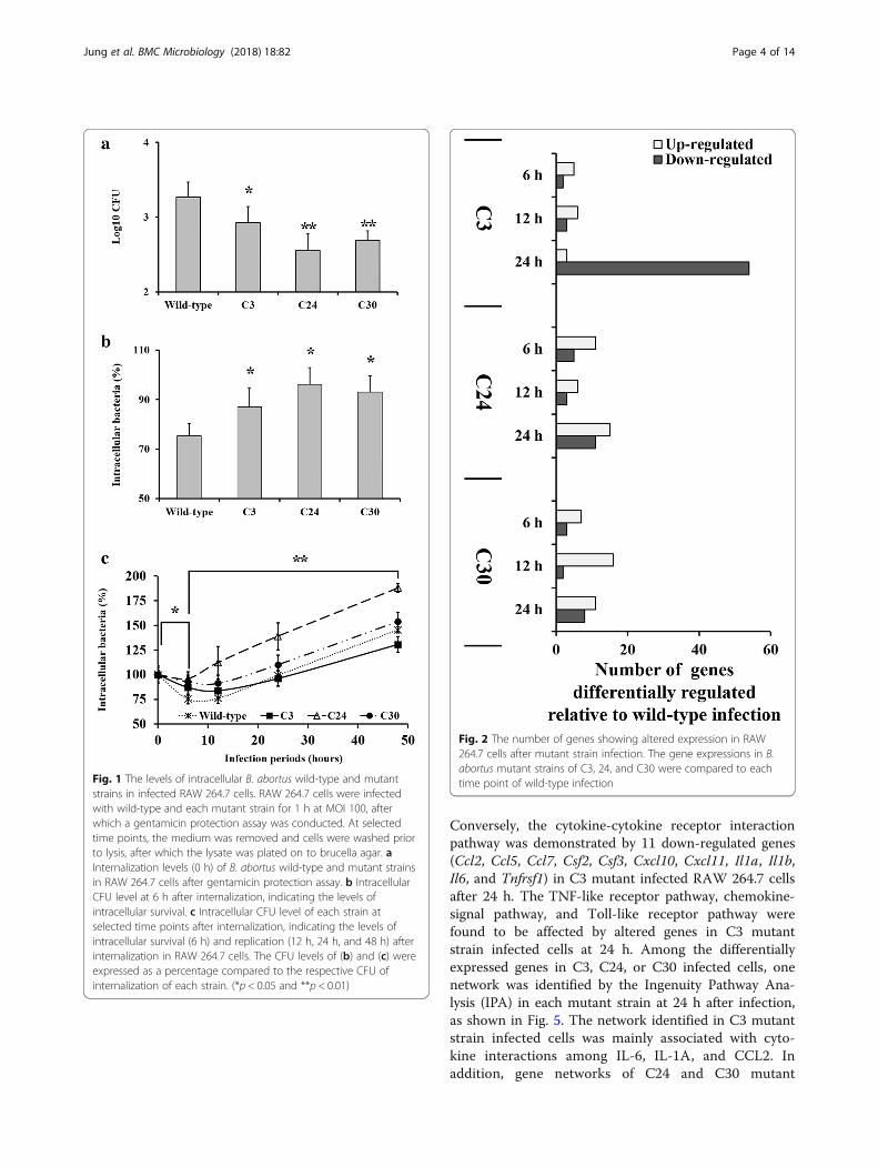
internalization, and intracellular replication of each Bru-cella strain in RAW 264.7 cells were investigated (Fig. 1and Additional file 3: Figure S2). Although the highestinternalization level was observed in B. abortuswild-type (Fig. 1a, p < 0.05 and p < 0.01), the decreasewas steeper than that observed in of the mutant strains,between 0 h and 6 h (Fig. 1b and c, p < 0.05). This steepdecrease demonstrated that the wild-type has a lowerlevel of intracellular survival as compared to the mutantstrains. According to the CFU slope level increasebetween 12 h and 48 h, lower levels of intracellular repli-cation were observed in the C3 and C30 mutant strainsas compared to the wild-type (Fig. 1c, p < 0.01).
Identification of the transposon insertion site in B.abortus mutant strainsThe insertion sequence of B. abortus mutant C3 waslocated at the 1,033,916 bp - 1,034,502 bp of chromo-some II, B. abortus mutant C24 was at the 113,672 bp -114,206 bp, and B. abortus mutant C30 was at the529,584 bp - 530,364 bp region. From sequencing re-sults, ABC transporter permease (BruAb2_1031), ABCtransporter substrate-binding protein (BruAb2_0113)and alkyl hydroperoxide reductase D (ahpD) were iden-tified as the genes disrupted by transposon insertion inmutant strains C3, C24, and C30, respectively.
Determination of differentially expressed genes ofinfected RAW 264.7 cellsFollowing infection of B. abortus wild-type for 6 h, 12 h,and 24 h, we observed more than 2-fold change in theexpression of 58, 202, and 1766 genes, respectively, as
Jung et al. BMC Microbiology (2018) 18:82 Page 2 of 14

compared to uninfected cells (Additional file 4: Table S2).The mutant strain infected RAW 264.7 cells also showeddifferent gene expression levels when compared to unin-fected cells (Additional file 5: Figure S3 and Additional file 6:Figure S4). The change in expression levels at 6 h, 12 h,and 24 h, respectively, were observed in: 48, 157, and 1239genes in C3 mutant strain infected cells; 25, 164, and 1422genes in C24 mutant strain infected cells; and 58, 202, and1766 genes in C30 mutant strain infected cells.In comparison to the wild-type infected RAW 264.7
cells, the C3 mutant strain infected cells showed 7, 9,and 57 altered genes at 6 h, 12 h, and 24 h, respectively(Fig. 2 and Additional file 7: Figure S5), whereas C24and C30 infections showed number of altered genes lessthan 20 at all-time points (Fig. 2 and Additional file 7:Figure S5). Additional file 7: Figure S5 shows the medianof normalized hybridization signals of genes with alteredtranscription comparing the wild-type and mutant straininfected RAW 264.7 cells at each time point. Whencompared to wild-type infected RAW 264.7 cells, allgenes showing altered expression level of > |2| in B.abortus mutant strains infected cells are presented inAdditional file 8 Table S3, Additional file 9 Table S4, andAdditional file 10 Table S5.
Gene set enrichment analysis of infected RAW 264.7 cellsThe genes showing altered expression were categorizedby gene set enrichment analysis using the Protein Ana-lysis Through Evolutionary Relationships (PANTHER)classification database, to demonstrate coordinatedchanges in pre-specified sets of related genes. Genesshowing different regulation in mutant-infected RAW264.7cells compared to wild-type infected cells werecategorized by molecular functions and biological pro-cesses, as shown in Figs. 3 and 4, respectively. Mostaltered genes in C3 mutant strain infected cells for 24 hwere down-regulated, compared to the wild-type infec-tions. These regulated genes were mostly involved in themolecular functions of binding (Fig. 3b) and biologicalprocesses of cellular process, response to stimulus, andbiological regulation (Fig. 4b). Also, most biological pro-cesses of cells were down-regulated in C3 mutant-infectedRAW 264.7cells, while only few genes were-upregulatedin the C24 mutant-infected cells (Fig. 4a). In case of C24
and C30 mutant strain infected cells, no significant resultsfor pre-specified sets in the PANTHER classification sys-tem were observed.The altered gene expressions in wild-type-infected
RAW 264.7 cells compared to uninfected cells were alsocategorized by the PANTHER classification database.Following B. abortus wild-type infection, most of thegenes showing different expression were up-regulated at6 h post infection. Gene enrichment analysis revealedthat these genes were associated with the immunesystem and cell defense processes (Ccl2, Gbp7, H2-T24,Ier3, Irg1, Irf7, Kdm6b, Ltb, Nfkbia, Nfkbiz, Oas2, Oasl2,Ptgs2, Slfn2, Tnf, Trim30a). Among these genes, Casp4,Ier3, Ifi204, Ltb, Nfkbia, Tnf, Xaf1, and Zc3h12a werealso categorized to be associated with apoptosis. At 12 hpost infection, RAW 264.7 cells showed down-regulationin 30 genes and up-regulation in 172 genes. The nucleo-some core and DNA binding were found to be impactedby the down-regulated genes (Hist1h2ag, Hist1h2bj,Hist1h3g, Hist1h3c2, Serpinb1a, and Serpinb9b). Of the172 genes up-regulated by Brucella infection, the 10 mostup-regulated genes were associated with immune re-sponse (Irg1, Ifi44l, Ifit1, Ifi204, Oasl2, Ifi202b, H2-T24,Irf7, Cmpk2, and Usp18). In addition, genes associatedwith apoptosis were also up-regulated (Bcl2a1d, Cd40,Hck, Ltb, Nfkbia, Pim1, Rassf4, Stat1, Stat2, Tnf, Tnfrsf1b,Tnfrsf9). Maximum altered gene expression was observedafter 24 h infection (Additional file 5: Figure S3 andAdditional file 6: Figure S4). Genes showing alteredexpression were associated with 10 molecular functionsand 14 biological processes. Most of the differentiallyexpressed genes were involved in two molecular func-tion categories (catalytic activity and binding) and twobiological process categories (metabolic process andcellular process).
Analyses of affected pathways and gene networksfollowing mutant strain infectionsPathway mapping using the Kyoto Encyclopedia ofGenes and Genomes (KEGG) database revealed that24 h B. abortus infection led to cell cycle arrest. Whencompared to the wild type infected cells, no KEGG path-way was represented by more than 10 genes in the C24and C30 mutant strain infected RAW 264.7 cells.
Table 1 Different characteristics of RAW 264.7 cells infected with mutant strains, as compared to wild-type infected cells
Strains Nitric oxide (μg/mL) IL-6 (pg/mL) TNF-α (pg/mL) Growth rates (%)
Wild-type 0.57 ± 0.10 3.61 ± 2.64 1219.58 ± 38.91
C3 0.24 ± 0.03** N.D.1) 1183.69 ± 81.12 87.7 ± 27.9
C24 22.62 ± 0.11** N.D. 1439.00 ± 71.73** 104.0 ± 1.1
C30 38.81 ± 0.22** 41.64 ± 8.72** 1334.62 ± 43.72* 111.4 ± 16.1
The growth rates are presented as the relative percentage compared to that of the wild-type, when growth rate of wild-type is considered as 100%. N.D.1),non-detected, the product levels of IL-6 in RAW 264.7 cells infected with C3 and C24 mutant strains were close to or below detectable levels of the ELISA system.(*p < 0.05 and **p < 0.01)
Jung et al. BMC Microbiology (2018) 18:82 Page 3 of 14
Conversely, the cytokine-cytokine receptor interactionpathway was demonstrated by 11 down-regulated genes(Ccl2, Ccl5, Ccl7, Csf2, Csf3, Cxcl10, Cxcl11, Il1a, Il1b,Il6, and Tnfrsf1) in C3 mutant infected RAW 264.7 cellsafter 24 h. The TNF-like receptor pathway, chemokine-signal pathway, and Toll-like receptor pathway werefound to be affected by altered genes in C3 mutantstrain infected cells at 24 h. Among the differentiallyexpressed genes in C3, C24, or C30 infected cells, onenetwork was identified by the Ingenuity Pathway Ana-lysis (IPA) in each mutant strain at 24 h after infection,as shown in Fig. 5. The network identified in C3 mutantstrain infected cells was mainly associated with cyto-kine interactions among IL-6, IL-1A, and CCL2. Inaddition, gene networks of C24 and C30 mutant
Fig. 1 The levels of intracellular B. abortus wild-type and mutantstrains in infected RAW 264.7 cells. RAW 264.7 cells were infectedwith wild-type and each mutant strain for 1 h at MOI 100, afterwhich a gentamicin protection assay was conducted. At selectedtime points, the medium was removed and cells were washed priorto lysis, after which the lysate was plated on to brucella agar. aInternalization levels (0 h) of B. abortus wild-type and mutant strainsin RAW 264.7 cells after gentamicin protection assay. b IntracellularCFU level at 6 h after internalization, indicating the levels ofintracellular survival. c Intracellular CFU level of each strain atselected time points after internalization, indicating the levels ofintracellular survival (6 h) and replication (12 h, 24 h, and 48 h) afterinternalization in RAW 264.7 cells. The CFU levels of (b) and (c) wereexpressed as a percentage compared to the respective CFU ofinternalization of each strain. (*p < 0.05 and **p < 0.01)
Fig. 2 The number of genes showing altered expression in RAW264.7 cells after mutant strain infection. The gene expressions in B.abortus mutant strains of C3, 24, and C30 were compared to eachtime point of wild-type infection
Jung et al. BMC Microbiology (2018) 18:82 Page 4 of 14

strains were on the prostaglandin-endoperoxide syn-thase 2 and tripartite-motif protein 30, respectively.
Validation of microarray dataTo verify the microarray results, gene expression levelsof IL-1β, IL-6, Csf2, and Gadd45b in the microarrayRNA samples were investigated by quantitative real timeRT-PCR (qRT-PCR). The C3 mutant strain infectedRAW 264.7 cells showed significant changes in geneexpression level compared to wild-type infected cells.Validation was therefore performed based on these genesshowing different expression levels. Furthermore, themicroarray results revealed that pathways of “Responsesof cytokine and immune defense”, and “Apoptosisprocess” were down-regulated in C3 mutant straininfected cells compared to wild-type infections. Hence,qRT-PCR for microarray validation was carried out usinggenes associated with these pathways (IL-1β, IL-6, Csf2,and Gadd45b). As shown Fig. 6, all genes evaluated byqRT-PCR showed similar expression levels as the micro-array results.
DiscussionBrucella spp. are facultative intracellular bacteria likeSalmonella enterica and Listeria monocytogenes. How-ever, they express uncanonical virulence factors such asvirulence regulator proteins and phosphatidylcholine,
rather than classic virulence factors such as protease,exotoxin, cytolysins, and capules [1–3]. Moreover, Bru-cella spp. utilize and modulate normal functions of hostcells, such as macrophages, to establish successful strat-egies for intracellular survival [5, 14, 15]. Therefore,characterization of the host macrophage-Brucella inter-action is very important to identify the pathogenicityand infection mechanism of Brucella [22]. Microarray isa powerful approach that enables understanding the hostresponses at the global gene transcription level, therebyproviding a plethora of information regarding the inter-actions associated with cell responses to antigens at themolecular level [15, 23]. In this study, microarray ana-lysis was used to compare the responses between macro-phages infected with B. abortus wild-type and B. abortusmutant strains, to demonstrate the Brucella genes thataffect the interactions between host immune cells andthe bacteria.Previous studies of genes associated with patho-
genicity of bacteria have been conducted using ran-dom insertion mutants generated by transposableelements [18, 19, 23, 24]. B. abortus mutant strainsused in the current study were generated usingtransposon mutagenesis and single insertions certi-fied in our previous study [18, 23, 25], in which Bru-cella genes associated with internalization and earlyhost immune responses were investigated using 2 h
Fig. 3 Categorization by molecular function of genes, showing different expression levels between B. abortus infections. The different expressionlevels in B. abortus mutant strain infected RAW 264.7 cells were compared to the wild-type infected cells at 24 h after infection. a Up-regulatedgenes. b Down regulated genes
Jung et al. BMC Microbiology (2018) 18:82 Page 5 of 14

infection experiments. In this study, we focused onthe genes affecting interactions between host immunecells and Brucella, which would affect the survival strategyof the bacterium through longer infection period. Infec-tion periods longer than 12 h are sufficient for Brucella toform replicative Brucella-containing vacuoles [26]. Amongthe mutant strains generated in our previous study [18],C3, C24, and C30 strains, which showed unique character-istics compared to the wild-type (Additional file 2: FigureS1 and Table 1), were selected for this study. The selectionwas carried out based on the growth rate and differencesin responses of RAW 264.7 cells to infection withwild-type and mutant strains. The product levels of IL-6,TNF-α, and NO in infected cells, which are represen-tative biomaterials associated with bactericidal re-sponses of macrophage to bacterial infection [27–29],were used as criteria for identifying differences in re-sponses of infected cells.Macrophages have been reported to show early im-
mune responses (such as inflammation) following B.abortus infection [6, 14, 15, 23, 30, 31]. Inflammation isa protective response to B. abortus infection observed inthe early stages of infection, and coordinated by cyto-kines and chemokines [15, 30]. In this study, wild-type
infected RAW 264.7 cells showed up-regulation in genesassociated with immune responses (Ccl2, Gbp7, H2-T24,Ier3, Irg1, Irf7, Kdm6b, Ltb, Nfkbia, Nfkbiz, Oas2, Oasl2,Ptgs2, Slfn2, Tnf, Trim30a). Of these, Casp4, Ier3, Ifi204,Ltb, Nfkbia, Tnf, Xaf1, and Zc3h12a were also catego-rized as being associated with apoptosis, which plays amajor role in responses to brucellosis by exposing thebacterium to the extracellular environments, where itencounters immune system components such as anti-bodies and the complement system, thereby reducingbacterial replication [6, 14, 15, 31]. In this study, mostresponses of RAW 264.7 cells at 6 h were focused ondefense activities of macrophages. These results corre-sponded to the intracellular survival and replication afterinternalization in RAW 264.7 cells, demonstrated by adecrease in intracellular B. abortus wild-type at 6 h afterinternalization (Fig. 1b).Following 12 h of infection, the nucleosome core and
DNA binding were found to be down-regulated(Hist1h2ag, Hist1h2bj, Hist1h3g, Hist1h3c2, Serpinb1a,and Serpinb9b). Considering nucleosomes are selectiveproducts during apoptosis [32, 33], it is assumed thatthe apoptotic process of macrophages was affected bydown-regulated genes due to Brucella infection.
Fig. 4 Categorization by biological process of genes, showing different expression levels between B. abortus infections. The different expressionlevels in B. abortus mutant strain infected RAW 264.7 cells were compared to wild-type infected cells at 24 h after infection. a Up-regulatedgenes. b Down regulated genes
Jung et al. BMC Microbiology (2018) 18:82 Page 6 of 14

Interestingly, the most up-regulated genes were involvedin the immune responses (Irg1, Ifi44l, Ifit1, Ifi204, Oasl2,Ifi202b, H2-T24, Irf7, Cmpk2, and Usp18), as were thegenes associated with apoptosis (Bcl2a1d, Cd40, Hck,Ltb, Nfkbia, Pim1, Rassf4, Stat1, Stat2, Tnf, Tnfrsf1b,Tnfrsf9). These results indicate that the professionalmacrophages, namely the RAW 264.7 cells, elicited
immune responses to clear Brucella infection consist-ently, concurrent with the activities of Brucella to eludethe clearance efforts of the host [6, 14, 15, 23, 31, 34].KEGG pathway mapping showed that B. abortus
wild-type infected cells were down-regulated in cell cyclethrough down-regulation of pathways such as cell cycle(42 genes), purine metabolism (26 genes), pyrimidine
Fig. 5 Identified network of the genes with altered expression in mutant strain infected RAW 264.7 cells. The different gene expression levels inB. abortus mutant strain infected RAW 264.7 cells at 24 h after infection were compared to the wild-type infected cells. Green color indicatesdown-regulation and arrows indicate directional relationships. a C3. b C24. c C30
Fig. 6 Validation of microarray data via quantitative RT-PCR. Relative expression level was determined by the 2-ΔΔCt method with normalization tothe housekeeping gene, β-actin
Jung et al. BMC Microbiology (2018) 18:82 Page 7 of 14
metabolism (24 genes), and DNA replication (23 genes)at 24 h [35]. The p53 pathway, which inhibits cell prolif-eration and then induces apoptosis [36, 37], was alsodown-regulated. Those down-regulations were relatedwith inhibition of cell death, which allows the survival ofbrucellae by avoiding exposure to the more hostile extra-cellular environment, thereby inducing more replicationof B. abortus [31]. Moreover, it was observed that genesinvolved in phagosome formation were down-regulated(Scarb1, Tuba1a, Tuba4a, Tuba1c, Tubb5, Tubb4b, andTubb6). Brucella is known to inhibit phagosome forma-tion and phagosome-lysosome fusion to avoid hostimmune responses [9, 14, 38, 39]. In addition to thesestrategies, modification of cell cycle arrest, which maypermit inhibition of apoptosis [15, 22], seemed to bemore dominant than other down-regulated factors suchas cytokine interaction and phagolysosome formation,considering the number of genes involved in cell arrest.Conversely, immune responses of RAW 264.7 cells to B.abortus infection for 24 h were focused on the cytokineinteractions, as described in previous studies [11, 15, 31].The up-regulation of the KEGG pathway of TNF-signalingand cytokine-cytokine receptor interaction was observedin this study. Moreover, these pathways involved genesthat were differentially expressed by more than 20-fold,such as Ccf1, Il6, and Ccl5.Although the overall transcriptional responses were
similar between mutant strains and wild-type of B. abor-tus, notable difference were observed at the differenttime points. The 54 down-regulated genes in C3 mu-tants at 24 h were mainly involved in cell immuneresponses associated with cytokine-cytokine receptorinteraction in the KEGG pathway database, which aremainly involved in immune responses elicited by RAW264.7 cells to clear Brucella [4, 14, 15, 23]. Moreover,these genes were involved in the apoptosis process,which plays an important role in intracellular survival ofBrucella [4, 15, 40]. The gene network analysis demon-strated that IL-1α, IL-6, tripartite-motif protein 30(Trim30), prostaglandin-endoperoxide synthase 2 (PTGS2;also known as cyclooxygenase-2, COX-2), and CCL2 weredown-regulated, showing interaction. These genes mediatethe macrophage polarization to M1, a common response ofmacrophages to bacterial infections, which includes genesencoding TNF, IL-1β, IL-6, and CCL2 [41]. IL-6 plays acentral role in the response of regulatory T cells to micro-bial infections, and is known to be a predominant mediatorof the acute phase response in inflammation triggered byinfection [42]. IL-6 synthesis is induced by COX-2 activa-tion [42]. This positive relationship was found in the net-work of RAW 264.7 cells infected with C3 mutant strain.Several studies have proved the important role of IL-6 inBrucella infection. Indeed, IL-6 is known to play a role inthe induction of acquired cellular resistance to intracellular
bacteria by macrophage activation [43–45]. Important rolesof IL-6 were also reported in the regulation of MHC II ex-pression and antigen processing [45, 46]. Previous studyfurther demonstrated that IL-6 inhibited intracellular repli-cation through its control effects on endocytosis andendosome-phagosome fusion [47]. IL-1 has been reportedto play an important role in protective responses againstBrucella infection [48]. Trim30 regulates the NF-κB path-way as a negative feedback via the degradation of TAB2and TAB3, resulting in inhibition of TRAF6 ubiquitylation,leading to the inhibition of NF-κB activity [49, 50].Activation of the NF-κB pathway results in the induc-tion of Type I IFN and other pro-inflammatorycytokines, thereby inducing protective effects againstinfection [51]. Down-regulation of Trim30d expres-sion was presumed to be caused by lower NF-κBactivity in C3 mutant strain infected cells. Thisdown-regulation of genes associated with the protect-ive immune responses against Brucella infection wasconfirmed by the quantification of NO, IL-6 andTNF-α in C3 mutant strain infected RAW 264.7 cells.In summary, the C3 mutant strain had the ability toinhibit the protective immune responses, via cytokineproduction.The load of intracellular bacteria has differing effects
on the gene expression in infected RAW 264.7 cells.However, there was no significant difference in the geneexpression in the 12 h microarray analysis, between cellsinfected with wild-type and C24 mutant strains,although the RAW 264.7 cells were infected with higherCFU number of C24 mutants than wild-type strain(Fig. 1). In case of 24 h microarray analysis, eventhough differences in intracellular bacteria CFU num-ber were observed among the strains of wild-type,C24, and C30 mutants, there was no significant dif-ference of gene expression level in the infected RAW264.7 cells compared to the wild-type infected cells.These results suggest that different gene expressionsobserved in the RAW 264.7 cells infected with C3mutant compared to the cells infected with thewild-type were not significantly affected by the intra-cellular bacterial CFU load.The ABC transporter permease (BruAb2_1031) has a
role in dipeptide import [20]. Insertion of transposoninto BruAb2_1031 was confirmed in the C3 mutantstrain by BLASTIN analysis. The disruptive effects ofBruAb2_1031 on the pathogenesis of B. abortus are notclearly understood. However, considering the importantroles of peptide uptake in bacterial nutrition associatedwith growth and replication, especially in intracellularbacteria, it is plausible that mutation at BruAb2_1031might affect the growth and virulence of B. abortus[52, 53]. These agreed with our results which showedslow growth and lower replication levels of C3
Jung et al. BMC Microbiology (2018) 18:82 Page 8 of 14

mutant in brucella broth and RAW 264.7 cells, re-spectively. Although the exact roles of the gene in B.abortus infection are not clear, this result implicatedthat mutation at BruAb2_1031 causes the inhibitionin uptake of nutrition, thereby inducing the decreaseof growth and intracellular replication.Along with the effect on bacterial nutrition, disruption
of ABC transporter affects the internalization and intra-cellular survival. Previous studies demonstrated that themutation at genes associated with the transporter system(such as VirB, cgt, and cydC) reduced the internalizationand intercellular survival in B. abortus [54–56]. Theseresults implicate that disruption of the ABC transportersystem generally exerts a negative effect on internaliza-tion, and the intracellular survival and replication ofBrucella. However, a high level of intracellular survivalwas observed in the C3 mutant strain at 6 h after intern-alization, in spite of low levels of internalization andintracellular replication (Fig. 1) [6, 14, 15, 23, 30, 31].This result was thought to be induced by reducedimmune responses of RAW 264.7 cells to C3 mutantstrain infection, which were agreeable with the results ofmicroarray.ABC permease utilizes the periplasmic binding protein
(PBPs) to capture substrate and present it at the intakevestibules of the membrane translocator unit [57]. It istherefore presumed that the mutation at the BruAb2_1031gene could induce changes in constituents of the bacterialenvelope, even though they were small. This perturbationin bacterial envelope was speculated to alter the antigensinteracting with macrophages, thereby reducing theimmune responses of macrophage to infection of C3 mu-tant strain in this study [14, 31, 58]. This phenomenonwas also supported by microarray analysis where down-regulation of the gene expression in C3 mutant infectedRAW 264.7 cells was observed, especially in genes associ-ated with M1 polarization of macrophages [58].In the C24 mutant strain infected RAW 264.7 cells,
most genes altered in expression were classified as pre-dicted genes. The IPA network revealed the low immuneresponse of RAW 264.7 cells to C24 mutant strain infec-tion through down-regulation of COX-2, which isnormally up-regulated in response to inflammatory andpro-inflammatory responses [59]. However, no other sig-nificant differences were observed in immune responsesor apoptosis between RAW 264.7 cells infected withC24 mutant strain and wild-type strain. The C30 mutantinfected RAW 264.7 cells also showed slight differencesin gene expression compared to the wild-type infectedcells. Similar to C24 mutant strain infected RAW 264.7cells, most of the altered genes in the C30 mutant straininfected cells were predicted genes. There were nonotable differences between cells infected with wild typeor C30 mutant strain, except for down-regulation of
Trim30. Therefore, further analyses on these predictedgenes are required to demonstrate the role ofBruAb2_0113 and ahpD in brucellosis.Altogether, the C3 mutant strain showed high intracel-
lular survival in RAW 264.7 cells compared to thewild-type. These cells also showed down-regulation ofgenes associated with protective immune responses toBrucella infection. Our results suggest that the C3mutant strain has more enhanced strategies for intracel-lular survival than the wild-type. This enhanced intracel-lular survival ability could be presumed to be elicited bythe mutation of BruAb2_1031, which is considered to bea cause of antigen alteration that could reduce the im-mune responses of macrophages in brucellosis.The BruAb2_1031 gene (ABC transporter permease) is
known to play a role in the transporter of peptides as acomponent of the ABC transporter [20]. However, ourstudy showed BruAb2_1031 mutation is effective againstBrucellosis. Our findings suggest that the ABC trans-porter permease could be a potential antigen in thedevelopment of Brucella vaccine in the future. Inaddition, to clarify a role of ABC transporter permease,changes in the function of ABC transporter by mutationof BruAb2_1031 gene need to be further investigated,and effects of BruAb2_1031 gene mutation on B. abortusshould be identified.
ConclusionsCharacteristics of mutant strains generated by trans-poson mutagenesis were investigated by infection experi-ments in RAW 264.7 cells. BLASTIN analysis revealedthat the ABC transporter permease (BruAb2_1031) wasmutated in the C3 mutant strain, which showed a higherintracellular survival rate in RAW 264.7 cells than thewild strain. This enhancement is presumed to be elicitedby the mutation of BruAb2_1031, which is considered asa cause of antigen alteration that could reduce theimmune responses of macrophages in brucellosis. Also,down-regulation of the protective immune response-re-lated functions were observed in C3 mutant strain in-fected RAW 264.7 cells. This study reported for the firsttime that mutation of the BruAb2_1031 gene in B. abor-tus reduces the defense responses of RAW 264.7 cells toBrucella infection, indicating that the molecules associ-ated with BruAb2_1031 gene have a role in immuneresponses of macrophages, and therefore its mutationenhanced the intracellular survival of B. abortus.
MethodsBacterial strains and cell lineIn our previous study, 132 mutant strains were gener-ated from B. abortus 1119–3 strain (wild-type) [18, 23]using EZ-Tn5™ Transposome complexes (Epicentre® Bio-technologies, USA) [18]. Brucellae were cultured in
Jung et al. BMC Microbiology (2018) 18:82 Page 9 of 14

brucella broth or agar (Difco, USA) and Brucella mutantstrains were cultured with 30 μg/mL of kanamycin. Themurine leukemic monocyte macrophage line, RAW264.7, was obtained from the Korea Cell Line Bank(KCLB No.40071, Korea) and grown at 37 °C in 5% CO2
atmosphere in Roswell Park Memorial Institute medium(RPMI) 1640 (Gibco, USA) containing 10% fetal bovineserum (FBS; Gibco), penicillin (100 U/mL), andstreptomycin (100 μg/mL). All procedures of thebacterial experiment were approved by the Seoul Na-tional University Institutional Biosafety Committee(SNUIBC-R160314–1).
Growth rates of B. abortus mutant strainsThe bacterial growth rates were measured in brucellabroth without antibiotics, according to the standardcurve of CFU versus optical density. The level of bacter-ial growth rates was presented as the relative percentagecompared to that of the wild-type, where the growth ratelevel of wild-type was regarded as 100%. Growth meas-urement experiments were independently conductedtwo times.
Selection of B. abortus mutant strainsAmong the generated mutants by same electroporationof transposome, 26 mutant stains showed defective in-ternalization (Additional file 2 Figure S1 and Additionalfile 1: Table S1). Because transposon mutagenesis couldcause defective growth [60], the mutant strains weregrouped based on the growth rate, as follows: group Aof mutant strains showed more than 10% reduction ingrowth rate; group B of mutant strains showed similargrowth rate compared to that of wild-type; group C ofmutant strains showed more than 10% increment ingrowth rate. Among the mutant strains in each group,strains showing distinctive characteristics were selectedfor further studies.The selection of mutant strains was carried out based
on the differences in production levels of NO, IL-6, andTNF-α in RAW 264.7 cells responding to infection witheach strain. Briefly, RAW 264.7 cells were cultured at aconcentration of 8.0 × 10⁴ cells/cm2 in 6-well cultureplates, in 2 mL of media containing 2% FBS. After8 h-culture, cells were infected with B. abortus wild-typeor mutant strains at a multiplicity of infection (MOI) of100 [18]. Following centrifugation at 150×g for 10 minat room temperature, the infected cells were incubatedat 37 °C and 5% CO2 for 24 h. The NO production insupernatants of infected cells was measured using Griessreaction, as described previously [61]. The amounts ofIL-6 and TNF-α in supernatants were measured using acytokine ELISA kit (eBioscience, USA) according to themanufacturer’s instructions. All experiments were inde-pendently carried out twice.
Internalization, intracellular survival, and intracellularreplication in RAW 264.7 cellsThe levels of internalization, intracellular survival, andintracellular replication in RAW 264.7 cells were alsoinvestigated. RAW 264.7 cells were infected with B.abortus wild-type and mutant strains at an MOI of 100[6, 18]. After 1 h, the medium was removed and replen-ished with medium containing 2% FBS and 30 μg/ml ofgentamicin (gentamicin protection assay). The cells werethen washed prior to lysis, and lysates were plated on tobrucella agar at 0 h, 6 h, 12 h, 24 h, and 48 h after genta-micin treatment. The levels of internalization (0 h),intracellular survival (6 h) and replication for each strain(12 h, 24 h, and 48 h) were expressed by CFU. The CFUchanges of intracellular bacteria were investigatedthrough three repeated experiments.
Identification of transposon insertion sitesIn our previous study, we had confirmed the singleinsertion of Tn5 transposome by Southern blot analysis[25]. The sequence of insertion site and disrupted genein mutant strains were identified using PCR andalignment of search tool, as described previously[18]. DNA sequences from the PCR product wereused to identify the insertion sites using analysis B.abortus chromosome II with circus (http://circos.ca/guide/genomic/).
Macrophage infection and RNA preparation formicroarrayMacrophage infection was conducted using B. abortuswild-type and mutant strains at an MOI 10 [62, 63]. Fol-lowing centrifugation at 150×g for 10 min at roomtemperature, the infected cells were incubated at 37 °Cunder 5% CO2 atmosphere for 6 h, 12 h, and 24 h. Afterthree washes with DPBS (Gibco, USA), the RNA wasextracted from the infected cells at each time point usingan RNeasy mini kit (Qiagen, Netherlands), as describedby the manufacturer. The purity and integrity of RNAwas determined by denaturing gel electrophoresis, theOD260/280 ratio, and analysis on an Agilent 2100 Bioa-nalyzer (Agilent Technologies, USA). All RNA sampleswere quantified, divided into aliquots, and stored at −80 °C until use.
Microarray hybridizationRNA amplification, labeling, array hybridization, andscanning were conducted by Macrogen Inc. (Seoul,Korea). The Affymetrix Whole Transcript Expressionarray process was conducted according to the manufac-turer’s protocols using a GeneChip Whole TranscriptPLUS reagent kit (Affymetrix, USA). Briefly, the Gene-Chip Whole Transcript Amplification kit (Affymetrix)was used for cDNA synthesis. The sense cDNA was
Jung et al. BMC Microbiology (2018) 18:82 Page 10 of 14

then fragmented and biotin-labeled with terminaldeoxynucleotidyl transferase, using a GeneChip WholeTranscript Terminal labeling kit (Affymetrix). Ap-proximately 5.5 μg of labeled DNA target was hybrid-ized to the Affymetrix GeneChip Mouse 2.0 ST Arrayfor 16 h at 45 °C, which covers more than 35,000 tran-scripts. After washing step, hybridized arrays were stainedon a GeneChip Fluidics Station 450 and scanned on aGCS3000 Scanner (Affymetrix). Signal values were com-puted using the Affymetrix® GenChip™ Command Consolesoftware (AGCC).
Raw data preparation and statistical analysisRaw data extracted by AGCC were summarized andnormalized by the robust multi-average (RMA) methodimplemented in Affymetrix® Expression Console™ Soft-ware. The results were exported with gene level RMAanalysis, and subjected to the differentially expressed gene(DEG) analysis. Statistical significance of the expressiondata was determined using LPE test and fold change, inwhich the null hypothesis was that no difference existsamong groups. False discovery rate was controlled byadjusting the p-value using the Benjamini-Hochberg algo-rithm. The genes that showed different expression levelsin RAW 264.7 cells infected with B. abortus mutantstrains compared to that of wild-type, were determinedbased on a p-value < 0.05 and a fold-change > |2|. Datasetof microarray results has been deposited in Gene Expres-sion Omnibus (GEO, http://www.ncbi.nlm.nih.gov/geo/query/acc.cgi?acc=GSE79264) and are accessible throughGEO series accession number GSE79264.
Microarray data analysisGenes showing significantly altered expressions weresubjected to gene set enrichment analysis using thePANTHER classification database (http://www.pantherd-b.org). Differently expressed genes were categorized bybiological processes and molecular functions using thePANTHER classification database based on means of
Fisher’s exact test, to identify coordinated changes inpre-defined sets of related genes.The changes in cell process were derived from interac-
tions among the differently expressed genes, which formthe functional pathways and networks [64]. Therefore,genes showing altered expressions were analyzed usingthe KEGG database to identify systemic informationrepresenting functional aspects of each gene. For apathway mapping term to be considered significant,pathways represented by fewer than 10 genes were fil-tered out for identification of the most affected path-ways [65]. In addition, the Qiagen’s IPA (IngenuitySystems Inc., USA) was performed for biological pro-cesses, canonical pathways, and networks analysis.
Verification of microarray resultsTo verify the microarray results, three genes associatedwith responses of cytokines and immune defense againstBrucella infection, and one gene connected to the apop-tosis process of the cell, were selected and subjected toqRT-PCR (Table 2). Total RNA was reverse transcribedusing a QuantiTect Reverse Transcription kit (Qiagen),according to the manufacturer’s protocols. TheqRT-PCR reaction was carried out with cDNA synthe-sized from 24 ng of RNA using the Rotor-Gene SYBRGreen PCR kit (Qiagen) and Rotor-Gene Q real timePCR cycler (Qiagen), under the following conditions:45 cycles at 95 °C for 15 s followed by 45 s at 60 °C [35].The gene expression levels were analyzed by the 2-ΔΔCt
method based on the house-keeping gene, β-actin, as areference [35, 66].
StatisticsData are presented as the mean ± standard deviation(SD). Statistical significance was analyzed by Student’st-test or repeated measures of ANOVA (Tukey’s HSDtest for post-test of multiple comparison) using SPSSversion 23.0 software (SPSS, USA). The statistical signifi-cance of differences was set at value of p < 0.05.
Table 2 Primers used for qRT-PCR
Genes Primers (5′-3′) Relevant function in brucellosis Gene Accession
IL-1β F CAACCAACAAGTGATATTCTCCATG Responses of cytokine and immune defense NM_008361
R GATCCACACTCTCCAGCTGC
IL-6 F CTCTGCAAGAGACTTCCATCCA Responses of cytokine and immune defense NM_031168
R GACAGGTCTGTTGGGAGTGG
Csf2 F GAGGATGTGGCTGCAGAATTTAC Responses of cytokine and immune defense NM_009969
R CTTCTACCTCTTCATTCAACGTGAC
Gadd45b F CTGATGAATGTGGACCCCGA Apoptosis process NM_008655
R CCTCTGCATGCCTGATACCC
Jung et al. BMC Microbiology (2018) 18:82 Page 11 of 14
Additional files
Additional file 1: Table S1. Growth of B. abortus wild-type and mutantstrains at each time point. CFU was calculated using the standard curveof CFU versus optical density. (PDF 19 kb)
Additional file 2: Figure S1. Characteristics of RAW 264.7 cells infectedwith B. abortus mutant strains. (a) Internalization was investigated usingRAW 264.7 cells infected with B. abortus wild-type and mutant strains atan MOI of 100. (b), (c), (d) Product levels of NO, IL-6, and TNF-α in RAW264.7 cells responding to infection with each strain (MOI 100) was mea-sured 24 h after infection. Based on the growth rate, mutant strains inthis study were divided into the three groups as follow: group A of mu-tant strains showed more than 10% reduction in growth rate; group B ofmutant strains showed similar growth rate compared to that of wild-type; group C of mutant strains showed more than 10% increment ingrowth rate. In this study, RNA samples from the RAW 264.7 cells infectedwith mutant strain (C3, C24, and C30) were subjected to microarray ana-lysis. The product level of IL-6 in RAW 264.7 cells infected with C3 andC24 mutant strains were close to or below detectable levels of the ELISAsystem. (TIF 939 kb)
Additional file 3: Figure S2. The CFU numbers of intracellular B.abortus wild-type and mutant strains in RAW 264.7 cells. RAW 264.7 cellswere infected with wild-type and each mutant strain for 1 h at MOI 100,after which a gentamicin protection assay was conducted. At the se-lected time points, the medium was removed and cells were washedprior to lysis; the lysate was then plated on to brucella agar. IntracellularCFU (Log10) numbers of each strain at selected time points after internal-ization was evaluated, which indicates the levels of intracellular survival(6 h) and replication (12 h, 24 h, and 48 h) at each time point after intern-alization in RAW 264.7 cells (*p < 0.05 and **p < 0.01). (TIF 503 kb)
Additional file 4: Table S2. The genes showing altered expression inRAW 264.7 cells after B. abortus infection. The different expression levelsin B. abortus infected RAW 264.7 cells were compared to uninfected cells.(PDF 1128 kb)
Additional file 5: Figure S3. Categorization by molecular function ofgenes showing different expression levels after infection. The differentexpression levels in B. abortus wild-type and mutant strain infected RAW264.7 cells were compared to uninfected cells. (a) Up-regulated genes. (b)Down regulated genes. (TIF 922 kb)
Additional file 6: Figure S4. Categorization by biological process ofgenes showing different expression levels after infection. The differentexpression levels in B. abortus wild-type and mutant strain infected RAW264.7 cells were compared to uninfected cells. (a) Up-regulated genes. (b)Down regulated genes. (TIF 936 kb)
Additional file 7: Figure S5. Scatter plots showing different geneexpressions. The different gene expression levels in B. abortus mutantstrain infected RAW 264.7 cells were compared to cells infected withwild-type at 6 h, 12 h, and 24 h after infection. Genes showing differentexpression levels are indicated by red dots. (TIF 2217 kb)
Additional file 8: Table S3. The genes showing altered expression inRAW 264.7 cells after C3 mutant strain infection. The different expressionlevels in B. abortus C3 mutant strain infected RAW 264.7 cells werecompared to wild-type infected cells. (PDF 52 kb)
Additional file 9: Table S4. The genes showing altered expression inRAW 264.7 cells after C24 mutant strain infection. The different expressionlevels in B. abortus C24 mutant strain infected RAW 264.7 cells werecompared to wild-type infected cells. (PDF 41 kb)
Additional file 10: Table S5. The genes showing altered expression inRAW 264.7 cells after C30 mutant strain infection. The different expressionlevels in B. abortus C30 mutant strain infected RAW 264.7 cells werecompared to wild-type infected cells. (PDF 37 kb)
AbbreviationsCCL2: Chemokine (C-C motif) ligand 2; CFU: Colony forming unit; COX-2: Cyclooxygenase-2; DEG: Differentially expressed gene; DPBS: Dulbecco’sPhosphate-Buffered Saline; FBS: Fetal bovine serum; GRO-α: Growthregulated oncogene alpha; IFN-γ: Interferon-gamma; IL-12: Interleukin-12; IL-
6: Interleukin-6; IL-8: Interleukin-8; IPA: Ingenuity Pathway Analysis;KCLB: Korea Cell Line Bank; KEGG: Kyoto Encyclopedia of Genes andGenomes; LPS: Lipopolysaccharide; MCP-1: Monocyte chemotactic protein 1;MIP1 α/β: Macrophage inflammatory protein alpha/beta; MOI: Multiplicity ofinfection; NO: Nitric oxide; PANTHER: Protein analysis through evolutionaryrelationships; PCR: Polymerase chain reaction; PTGS2: Prostaglandin-endoperoxide synthase 2; qRT-PCR: Quantitative real time polymerase chainreaction; RANTES: Regulated on activation, Normal T cell expressed andsecreted; RPMI: Roswell Park Memorial Institute medium; TNF-α: Tumornecrosis factor alpha; Trim30: Tripartite-motif protein 30
AcknowledgementsThe authors and this work was supported by NRF grant of MSIP (No.2014R1A2A2A01007291), Korea Health Industry Development Institute(HI16C2130), BK21 PLUS and Research Institute for Veterinary Science, SeoulNational University, Seoul, Republic of Korea.
FundingThis work was supported by NRF grant of MSIP (No. 2014R1A2A2A01007291),Korea Health Industry Development Institute (HI16C2130), BK21 PLUS andResearch Institute for Veterinary Science, Seoul National University, Seoul,Republic of Korea. The funders had no role in study design, data collectionand interpretation, writing the manuscript and the decision to submit thework for publication.
Availability of data and materialsThe data that support the findings of this study are available from thecorresponding author HSY upon reasonable request.
Authors’ contributionsThis study included the efforts of MJ involved in data analysis and drafting ofmanuscript. SS was involved in mutant strain characterization. YBI wasinvolved in mutant strain characterization. WBP was involved in transposoninsertion site identification. HSY was involved in experimental design andsupervision of the experiments and manuscript revision. All authors read andapproved the final manuscript and publication.
Ethics approval and consent to participateNot applicable.
Consent for publicationNot applicable.
Competing interestsThe authors declare that they have no competing interests.
Publisher’s NoteSpringer Nature remains neutral with regard to jurisdictional claims inpublished maps and institutional affiliations.
Author details1Department of Infectious Diseases, College of Veterinary Medicine, SeoulNational University, Seoul, Republic of Korea. 2Institute of Green Bio Scienceand Technology, Seoul National University, Pyeongchang, Republic of Korea.3Present address: Department of Microbiology, Research Institute of LifeSciences, Gyeongsang National University School of Medicine, Jinju 52727,Republic of Korea.
Received: 2 May 2017 Accepted: 19 July 2018
References1. Neta AVC, Mol JP, Xavier MN, Paixão TA, Lage AP, Santos RL. Pathogenesis
of bovine brucellosis. Vet J. 2010;184:146–55.2. Moreno E, Moriyón I. Brucella melitensis: a nasty bug with hidden credentials
for virulence. Proc Natl Acad Sci. 2002;99:1–3.3. Roop RM II, Gee JM, Robertson GT, Richardson JM, Ng WL, Winkler ME.
Brucella stationary-phase gene expression and virulence. Annu RevMicrobiol. 2003;57:57–76.
4. Ficht TA. Intracellular survival of Brucella: defining the link with persistence.Vet Microbiol. 2003;92:213–23.
Jung et al. BMC Microbiology (2018) 18:82 Page 12 of 14

5. Skendros P, Pappas G, Boura P. Cell-mediated immunity in humanbrucellosis. Microbes Infect. 2011;13:134–42.
6. Gross A, Terraza A, Ouahrani-Bettache S, Liautard JP, Dornand J. In vitroBrucella suis infection prevents the programmed cell death of humanmonocytic cells. Infect Immun. 2000;68:342–51.
7. Porte F, Liautard JP, Köhler S. Early acidification of phagosomes containingBrucella suis is essential for intracellular survival in murine macrophages.Infect Immun. 1999;67:4041–7.
8. Pizarro-Cerdá J, Méresse S, Parton RG, van der Goot G, Sola-Landa A, Lopez-Goñi I, et al. Brucella abortus transits through the autophagic pathway andreplicates in the endoplasmic reticulum of nonprofessional phagocytes.Infect Immun. 1998;66:5711–24.
9. Byndloss MX, Tsolis RM. Brucella ssp. virulence factors and immunity. AnnuRev Anim Biosci. 2016;4:111–27.
10. Eskra L, Canavessi A, Carey M, Splitter G. Brucella abortus genes identifiedfollowing constitutive growth and macrophage infection. Infect Immun.2001;69:7736–42.
11. Golding B, Scott DE, Scharf O, Huang LY, Zaitseva M, Lapham C, et al.Immunity and protection against Brucella abortus. Microbes Infect. 2001;3:43–8.
12. Zheng K, Chen DS, Wu YQ, Xu XJ, Zhang H, Chen CF, et al. MicroRNAexpression profile in RAW264. 7 cells in response to Brucella melitensisinfection. Int J Biol Sci. 2012;8:1013–22.
13. Jiang X, Leonard B, Benson R, Baldwin CL. Macrophage control of Brucellaabortus: role of reactive oxygen intermediates and nitric oxide. CellImmunol. 1993;151:309–19.
14. Pei J, Ficht TA. Brucella abortus rough mutants are cytopathic formacrophages in culture. Infect Immun. 2004;72:440–50.
15. Eskra L, Mathison A, Splitter G. Microarray analysis of mRNA levels fromRAW264. 7 macrophages infected with Brucella abortus. Infect Immun. 2003;71:1125–33.
16. Sung KY, Jung M, Shin MK, Park HE, Lee JJ, Kim S, et al. Induction ofimmune responses by two recombinant proteins of Brucella abortus, outermembrane proteins 2b porin and cu/Zn superoxide dismutase, in mousemodel. J Microbiol Biotechnol. 2014;24:854–61.
17. Ko J, Splitter GA. Molecular host-pathogen interaction in brucellosis: currentunderstanding and future approaches to vaccine development for miceand humans. Clin Microbiol Rev. 2003;16:65–78.
18. Cha SB, Rayamajhi N, Lee WJ, Shin MK, Jung MH, Shin SW, et al. Generationand envelope protein analysis of internalization defective Brucella abortusmutants in professional phagocytes, RAW 264.7. FEMS Immunol MedMicrobiol. 2012;64:244–54.
19. van Opijnen T, Camilli A. Transposon insertion sequencing: a new tool forsystems-level analysis of microorganisms. Nat Rev Microbiol. 2013;11:435–42.
20. Jenner DC, Dassa E, Whatmore AM, Atkins HS. ATP-binding cassette systemsof Brucella. Comp Funct Genomics. 2009;2009:354649.
21. Steele KH, Baumgartner JE, Valderas MW, Roop RM. Comparative study ofthe roles of AhpC and KatE as respiratory antioxidants in Brucella abortus2308. J Bacteriol. 2010;192:4912–22.
22. He Y, Reichow S, Ramamoorthy S, Ding X, Lathigra R, Craig JC, et al. Brucellamelitensis triggers time-dependent modulation of apoptosis and down-regulation of mitochondrion-associated gene expression in mousemacrophages. Infect Immun. 2006;74:5035–46.
23. Cha SB, Lee WJ, Shin MK, Jung MH, Shin SW, Yoo AN, et al. Earlytranscriptional responses of internalization defective Brucella abortus mutantsin professional phagocytes, RAW 264.7. BMC Genomics. 2013;14:426.
24. Vidal JE, Chen J, Li J, McClane BA. Use of an EZ-Tn5-based randommutagenesis system to identify a novel toxin regulatory locus in Clostridiumperfringens strain 13. PLoS One. 2009;4:e6232.
25. Park WB, Im YB, Jung M, Yoo HS. Molecular characteristics of Brucellaabortus mutants generated using EZ-Tn5Tm pMODTm-3 transposon system. JPrev Vet Med. 2015;39:144–52.
26. Gorvel JP, Moreno E. Brucella intracellular life: from invasion to intracellularreplication. Vet Microbiol. 2002;90:281–97.
27. Wong SS, Zhou HR, Marin-Martinez M, Brooks K, Pestka JJ. Modulation of IL-1β, IL-6 and TNF-α secretion and mRNA expression by the trichothecenevomitoxin in the RAW 264.7 murine macrophage cell line. Food ChemToxicol. 1998;36:409–19.
28. MacMicking J, Xie QW, Nathan C. Nitric oxide and macrophage function.Annu Rev Immunol. 1997;15:323–50.
29. Hamza T, Li B. Differential responses of osteoblasts and macrophages uponStaphylococcus aureus infection. BMC Microbiol. 2014;14:207.
30. Chen F, Ding X, Ding Y, Xiang Z, Li X, Ghosh D, et al. Proinflammatorycaspase-2-mediated macrophage cell death induced by a rough attenuatedBrucella suis strain. Infect Immun. 2011;79:2460–9.
31. Roop RM II, Gaines JM, Anderson ES, Caswell CC, Martin DW. Survival of thefittest: how Brucella strains adapt to their intracellular niche in the host. MedMicrobiol Immunol. 2009;198:221–38.
32. Amoura Z, Chabre H, Koutouzov S, Lotton C, Cabrespines A, Bach JF, et al.Nucleosome-restricted antibodies are detected before anti-dsDNA and/orantihistone antibodies in serum of MRL-Mp lpr/lpr and +/+ mice, and arepresent in kidney eluates of lupus mice with proteinuria. Arthritis Rheum.1994;37:1684–8.
33. Ren Y, Tang J, Mok M, Chan AW, Wu A, Lau C. Increased apoptoticneutrophils and macrophages and impaired macrophage phagocyticclearance of apoptotic neutrophils in systemic lupus erythematosus. ArthritisRheum. 2003;48:2888–97.
34. Rossetti C, Galindo C, Everts R, Lewin H, Garner H, Adams L. Comparativeanalysis of the early transcriptome of Brucella abortus–infected monocyte-derived macrophages from cattle naturally resistant or susceptible tobrucellosis. Res Vet Sci. 2011;91:40–51.
35. Jung M, Shin MK, Jung YK, Yoo HS. Modulation of macrophage activities inproliferation, lysosome, and phagosome by the nonspecificimmunostimulator, mica. PLoS One. 2015;10:e0117838.
36. Harris SL, Levine AJ. The p53 pathway: positive and negative feedbackloops. Oncogene. 2005;24:2899–908.
37. Hoe KK, Verma CS, Lane DP. Drugging the p53 pathway: understanding theroute to clinical efficacy. Nat Rev Drug Discov. 2014;13:217–36.
38. Harrison RE, Grinstein S. Phagocytosis and the microtubule cytoskeleton.Biochem Cell Biol. 2002;80:509–15.
39. Döhmer PH, Valguarnera E, Czibener C, Ugalde JE. Identification of a type IVsecretion substrate of Brucella abortus that participates in the early stages ofintracellular survival. Cell Microbiol. 2014;16:396–410.
40. Tian M, Qu J, Han X, Zhang M, Ding C, Ding J, et al. Microarray-basedidentification of differentially expressed genes in intracellular Brucellaabortus within RAW264. 7 cells. PLoS ONE. 2013;8:e67014.
41. Benoit M, Desnues B, Mege JL. Macrophage polarization in bacterialinfections. J Immunol. 2008;181:3733–9.
42. Chen BC, Liao CC, Hsu MJ, Liao YT, Lin CC, Sheu JR, et al. Peptidoglycan-induced IL-6 production in RAW 264.7 macrophages is mediated bycyclooxygenase-2, PGE2/PGE4 receptors, protein kinase a, IκB kinase, andNF-κB. J Immunol. 2006;177:681–93.
43. Zhan Y, Cheers C. Differential induction of macrophage-derived cytokinesby live and dead intracellular bacteria in vitro. Infect Immun. 1995;63:720–3.
44. Delpino MV, Barrionuevo P, Macedo GC, Oliveira SC, Di Genaro S, Scian R,et al. Macrophage-elicited osteoclastogenesis in response to Brucella abortusinfection requires TLR2/MyD88-dependent TNF-α production. J Leukoc Biol.2012;91:285–98.
45. Xavier MN, Winter MG, Spees AM, Nguyen K, Atluri VL, Silva TM, et al. CD4+T cell-derived IL-10 promotes Brucella abortus persistence via modulation ofmacrophage function. PLoS Pathog. 2013;9:e1003454.
46. Barrionuevo P, Cassataro J, Delpino MV, Zwerdling A, Pasquevich KA,Samartino CG, et al. Brucella abortus inhibits major histocompatibilitycomplex class II expression and antigen processing through interleukin-6secretion via toll-like receptor 2. Infect Immun. 2008;76:250–62.
47. Pizarro-Cerdá J, Desjardins M, Moreno E, Akira S, Gorvel JP. Modulation ofendocytosis in nuclear factor IL-6 (−/−) macrophages is responsible for a highsusceptibility to intracellular bacterial infection. J Immunol. 1999;162:3519–26.
48. Skyberg JA, Thornburg T, Kochetkova I, Layton W, Callis G, Rollins MF, et al.IFN-γ-deficient mice develop IL-1-dependent cutaneous andmusculoskeletal inflammation during experimental brucellosis. J Leukoc Biol.2012;92:375–87.
49. Bowie AG. TRIM-ing down tolls. Nat Immunol. 2008;9:348–50.50. Shi M, Deng W, Bi E, Mao K, Ji Y, Lin G, et al. TRIM30α negatively regulates
TLR-mediated NF-κB activation by targeting TAB2 and TAB3 for degradation.Nat Immunol. 2008;9:369–77.
51. McNab FW, Rajsbaum R, Stoye JP, O’Garra A. Tripartite-motif proteins andinnate immune regulation. Curr Opin Immunol. 2011;23:46–56.
52. Braibant M, Gilot P, Content J. The ATP binding cassette (ABC) transportsystems of Mycobacterium tuberculosis. FEMS Microbiol Rev. 2000;24:449–67.
53. Green RM, Seth A, Connell ND. A peptide permease mutant ofMycobacterium bovis BCG resistant to the toxic peptides glutathione and S-Nitrosoglutathione. Infect Immun. 2000;68:429–36.
Jung et al. BMC Microbiology (2018) 18:82 Page 13 of 14
54. Roset MS, Ciocchini AE, Ugalde RA, de Iannino NI. Molecular cloning andcharacterization of cgt, the Brucella abortus cyclic β-1, 2-glucan transportergene, and its role in virulence. Infect Immun. 2004;72:2263–71.
55. O'Callaghan D, Cazevieille C, Allardet-Servent A, Boschiroli ML, Bourg G,Foulongne V, et al. A homologue of the Agrobacterium tumefaciens VirB andBordetella pertussis Ptl type IV secretion systems is essential for intracellularsurvival of Brucella suis. Mol Microbiol. 1999;33:1210–20.
56. Truong QL, Cho Y, Barate AK, Kim S, Hahn TW. Characterization andprotective property of Brucella abortus cydC and looP mutants. Clin VacImmunol. 2014;21:1573–80.
57. Jones PM, George AM. The ABC transporter structure and mechanism:perspectives on recent research. Cell Mol Life Sci. 2004;61:682–99.
58. Mosser DM, Edwards JP. Exploring the full spectrum of macrophageactivation. Nat Rev Immunol. 2008;8:958–69.
59. Kalinski P. Regulation of immune responses by prostaglandin E2. J Immunol.2012;188:21–8.
60. Kim S, Watarai M, Kondo Y, Erdenebaatar J, Makino SI, Shirahata T. Isolationand characterization of mini-Tn5Km2 insertion mutants of Brucella abortusdeficient in internalization and intracellular growth in HeLa cells. InfectImmun. 2003;71:3020–7.
61. Sreejayan, MNA R. Nitric oxide scavenging by curcuminoids. J PharmPharmacol. 1997;49:105–7.
62. Lee JJ, Kim DH, Kim DG, Lee HJ, Min W, Rhee MH, et al. Toll-like receptor 4-linked Janus kinase 2 signaling contributes to internalization of Brucellaabortus by macrophages. Infect Immun. 2013;81:2448–58.
63. Pei J, Turse JE, Wu Q, Ficht TA. Brucella abortus rough mutants inducemacrophage oncosis that requires bacterial protein synthesis and directinteraction with the macrophage. Infect Immun. 2006;74:2667–75.
64. Kanehisa M, Goto S, Furumichi M, Tanabe M, Hirakawa M. KEGG forrepresentation and analysis of molecular networks involving diseases anddrugs. Nucleic Acids Res. 2010;38:D355–60.
65. Waters KM, Masiello LM, Zangar RC, Tarasevich BJ, Karin NJ, Quesenberry RD,et al. Macrophage responses to silica nanoparticles are highly conservedacross particle sizes. Toxicol Sci. 2009;107:553–69.
66. Livak KJ, Schmittgen TD. Analysis of relative gene expression data usingreal-time quantitative PCR and the 2−ΔΔCT method. Methods. 2001;25:402–8.
Jung et al. BMC Microbiology (2018) 18:82 Page 14 of 14