Embed Size (px)
Citation preview

DePaul Discoveries DePaul Discoveries
Volume 3 Issue 1 Article 10
2014
Generation of Chimeric Antibody Light Chain Plasmid Generation of Chimeric Antibody Light Chain Plasmid
Lily Arendt [email protected]
Follow this and additional works at: https://via.library.depaul.edu/depaul-disc
Part of the Chemistry Commons
Recommended Citation Recommended Citation Arendt, Lily (2014) "Generation of Chimeric Antibody Light Chain Plasmid," DePaul Discoveries: Vol. 3 : Iss. 1 , Article 10. Available at: https://via.library.depaul.edu/depaul-disc/vol3/iss1/10
This Article is brought to you for free and open access by the College of Science and Health at Via Sapientiae. It has been accepted for inclusion in DePaul Discoveries by an authorized editor of Via Sapientiae. For more information, please contact [email protected].

Generation of Chimeric Antibody Light Chain Plasmid
Lily Arendt*
Department of Chemistry, DePaul University
ABSTRACT In order to determine the distance between the antigen-binding site and the crystallizable
fragment (Fc) region of an antibody using single-molecule Förster Resonance Energy Transfer, dyes must
be attached to these locations. To accomplish this, the variable regions of the light and heavy chains of an
antibody can be modified to introduce dye-reactive amino acids. Initial attempts to alter the variable light
chain (VL) gene included trying to ligate a 350 bp variable gene into an 11,000 bp plasmid. When these
attempts were unsuccessful, the variable light chain gene was ligated into a commercially available 3500
bp plasmid.
INTRODUCTION
Antibodies are comprised of four polypeptide
chains including two identical light chains and
two identical heavy chains (Figure1).1 One end
of each of the two heavy chains interacts with
the other to form the crystallizable fragment (Fc)
region of the antibody, which is the lower lobe
and the other ends interact with the light chains
to form the upper lobes. The two upper lobes are
separated from the lower lobe by an unstructured
hinge region. The presence of this hinge region
provides segmental flexibility between the lobes
____________________________________
*Faculty Advisor: Dr. Cathrine A. Southern
Department of Chemistry
Research Completed in Autumn 2013
Author Contact: [email protected]
and grants antibodies a larger range to search for
antigens.2
Binding of effector molecules to the
Fc region can result in an immune response
within the body. The two upper lobes of the
antibody are where the fragment antigen-binding
(Fab) regions reside. If the two Fab regions
engage a single antigen, the possibility of
forming an immune complex, or a group of
antibodies bound to the antigen, is increased.
The formation of immune complexes is
necessary for the generation of an immune
response and it is affected by antibody
flexibility, or the ability of an antibody to take
on a variety of structures. Further researching
the possible antibody conformations will lead to
a better understanding of the variety of
structures antibodies exhibit.
1
Arendt: Generation of Chimeric Antibody Light Chain Plasmid
Published by Via Sapientiae, 2014
METHODS
Examining the distance between the Fc and Fab
regions can reveal details of antibody structures.
This distance can be seen in crystal structures,
which provide information about one particular
antibody conformation.1 It is of particular
interest to determine whether or not on
structure is preferred or if a multitude of Fab
distances are possible. Discovering how this
distribution of conformations is affected when
Fc receptor binding occurs is also of interest.
This information on the structure of the IgG
antibody could lead to an enhanced
understanding of the medicinal properties of
antibodies.
The technique of single-molecule Förster
Resonance Energy Transfer (FRET) can be used
to determine the distance between the Fc and
Fab regions. In order to use the technique, dyes
Figure 1: Crystal structure of an IgG antibody. The
Fc region is attached to the upper lobes by a hinge
region. Antibody drawn from the PDB file 1IGT.
Harris, L.J.; Larson, S.B.; Hasel, K.W.; McPherson,
A. (1997) Biochemistry 36, 1581.
Isolate Plasmid Open Plasmid via
Enzymes
ce between the Fc and Fab
regions can reveal details of antibody structures.
This distance can be seen in crystal structures,
which provide information about one particular
It is of particular
interest to determine whether or not one
structure is preferred or if a multitude of Fab-Fc
distances are possible. Discovering how this
distribution of conformations is affected when
Fc receptor binding occurs is also of interest.
This information on the structure of the IgG
d to an enhanced
understanding of the medicinal properties of
molecule Förster
Resonance Energy Transfer (FRET) can be used
to determine the distance between the Fc and
Fab regions. In order to use the technique, dyes
molecules must be attached to those regions.
The dyes cannot be attached unless both the
variable light (VL) and var
chains are modified and dye
acids are introduced in the appropriate locations.
Through past experiments the variable heavy
chain and the Fc region have been mutated and a
dye-reactive cysteine residue was introduced in
the Fc region. The goal of this experiment was
to add a dye-reactive lysine residue to the Fab
region by manipulating the variable light chain
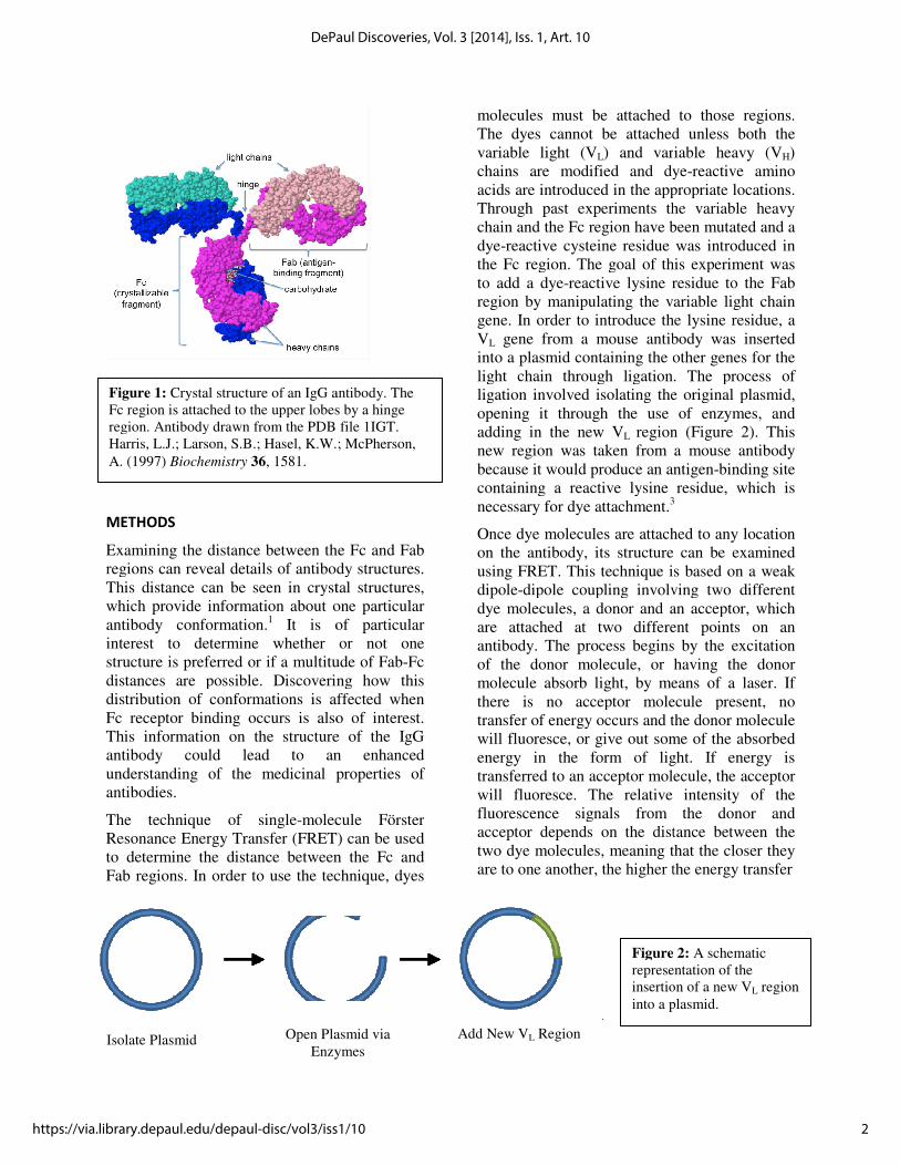
gene. In order to introduce the
VL gene from a mouse antibody was inserted
into a plasmid containing the other genes for the
light chain through ligation. The process of
ligation involved isolating the original plasmid,
opening it through the use of enzymes, and
adding in the new VL region (Figure 2).
new region was taken from a mouse antibody
because it would produce an antigen
containing a reactive lysine residue, which is
necessary for dye attachment.3
Once dye molecules are attached to any location
on the antibody, its structure can be examined
using FRET. This technique is based on a weak
dipole-dipole coupling involving two different
dye molecules, a donor and an acceptor, which
are attached at two different points on an
antibody. The process begins by the excitatio
of the donor molecule, or having the donor
molecule absorb light, by means of a laser. If
there is no acceptor molecule present, no
transfer of energy occurs and the donor molecule
will fluoresce, or give out some of the absorbed
energy in the form of light. If energy is
transferred to an acceptor molecule, the acceptor
will fluoresce. The relative intensity of the
fluorescence signals from the donor and
acceptor depends on the distance between the
two dye molecules, meaning that the closer they
are to one another, the higher the energy transfer
Crystal structure of an IgG antibody. The
Fc region is attached to the upper lobes by a hinge
region. Antibody drawn from the PDB file 1IGT.
Harris, L.J.; Larson, S.B.; Hasel, K.W.; McPherson,
Open Plasmid via
Enzymes
Add New VL Region
Figure 2:
representation of the
insertion of a new V
into a plasmid.
molecules must be attached to those regions.
The dyes cannot be attached unless both the
) and variable heavy (VH)
chains are modified and dye-reactive amino
acids are introduced in the appropriate locations.
Through past experiments the variable heavy
chain and the Fc region have been mutated and a
reactive cysteine residue was introduced in
c region. The goal of this experiment was
reactive lysine residue to the Fab
region by manipulating the variable light chain
gene. In order to introduce the lysine residue, a
gene from a mouse antibody was inserted
the other genes for the
light chain through ligation. The process of
ligation involved isolating the original plasmid,
opening it through the use of enzymes, and
region (Figure 2). This
new region was taken from a mouse antibody
se it would produce an antigen-binding site
containing a reactive lysine residue, which is 3
Once dye molecules are attached to any location
ts structure can be examined
using FRET. This technique is based on a weak
dipole coupling involving two different
dye molecules, a donor and an acceptor, which
are attached at two different points on an
antibody. The process begins by the excitation
of the donor molecule, or having the donor
molecule absorb light, by means of a laser. If
there is no acceptor molecule present, no
transfer of energy occurs and the donor molecule
will fluoresce, or give out some of the absorbed
ght. If energy is
transferred to an acceptor molecule, the acceptor
will fluoresce. The relative intensity of the
fluorescence signals from the donor and
acceptor depends on the distance between the
two dye molecules, meaning that the closer they
e another, the higher the energy transfer
Figure 2: A schematic
representation of the
insertion of a new VL region
into a plasmid.
2
DePaul Discoveries, Vol. 3 [2014], Iss. 1, Art. 10
https://via.library.depaul.edu/depaul-disc/vol3/iss1/10

efficiency. Single molecule FRET is beneficial
because it allows the distances between the dye
molecules on many individual molecules to be
examined. A histogram can then be constructed
with a distance value for each molecule instead
of simply the average distance i
experiment.
RESULTS
Initial attempts in the experiment included
amplifying the VL gene from a plasmid
containing a single chain variable fragment of a
mouse antibody called 84G3.3 The V
amplified using the polymerase chain reaction
(PCR). A ligation reaction between the V
and an 11,000 bp plasmid was carried out. To
determine if the ligation was successful,
restriction enzymes were used to cut the ligation
product to see if the expected 350 bp V
be observed. When analyzed on an agarose gel,
the results indicated that the ligation was
unsuccessful. The lack of success was attributed
to the difficulty involved in ligating a very small
fragment into a much larger one. To avoid this, a
commercially available pFuse 3500 bp plasmid
was then purchased, and was opened at the
EcoRI and BsiWI restriction enzyme cut sites in
order to allow for ligation of the VL
3).
Figure 3: Plasmid map for the pFuse light chain. The
pFuse map shows the locations of the EcoRI and
restriction enzyme cut sites where the VL gene was
inserted.
. Single molecule FRET is beneficial
because it allows the distances between the dye
molecules on many individual molecules to be
examined. A histogram can then be constructed
with a distance value for each molecule instead
of simply the average distance in the
nitial attempts in the experiment included
gene from a plasmid
containing a single chain variable fragment of a
The VL gene was
amplified using the polymerase chain reaction
(PCR). A ligation reaction between the VL gene
and an 11,000 bp plasmid was carried out. To
ermine if the ligation was successful,
restriction enzymes were used to cut the ligation
product to see if the expected 350 bp VL would
be observed. When analyzed on an agarose gel,
the results indicated that the ligation was
ess was attributed
to the difficulty involved in ligating a very small
fragment into a much larger one. To avoid this, a
commercially available pFuse 3500 bp plasmid
was then purchased, and was opened at the
WI restriction enzyme cut sites in
L gene (Figure
The newly ligated plasmid was then transformed
into E. coli cells and the DNA was replicated.
The DNA from colonies that grew was then
isolated. PCR was performed on the new ligated
pFuse plasmid, and the results were run on an
agarose gel. A band at the expected 350 bp mark
for the VL gene was observed, showing that the
ligation was successful (Figure 4).
CONCLUSION
After many attempts at ligating a V
into an 11,000 bp plasmid, the V
of the single chain variable region of a
mouse antibody was successfully ligated
into the smaller 3500 bp pFuse plasmid.
The 350 bp band that appeared on the
agarose gel demonstrated this success and
the DNA can now be sent out for
sequencing. Following this, the antibody
DNA will be shipped to a company that
will express and purify the mutated
antibody. Dyes will then be attached to
the antibody, and single
experiments will be performed on the dye
labeled antibodies to determine the F
distances present.
: Plasmid map for the pFuse light chain. The
RI and BsiWI
gene was
350 bp ����
1 2 3 4 5 6 7 8 9
Figure 4: Stained agarose gel of the variable
light chain gene from the uncut ligated pFuse
plasmid. Lanes 1-4 and 6-9 are ligated PCR
products with amplified VL
Lane 5 is the ladder for base pair reference.
The newly ligated plasmid was then transformed
cells and the DNA was replicated.
The DNA from colonies that grew was then
isolated. PCR was performed on the new ligated
the results were run on an
agarose gel. A band at the expected 350 bp mark
gene was observed, showing that the
ligation was successful (Figure 4).
After many attempts at ligating a VL gene
into an 11,000 bp plasmid, the VL region
of the single chain variable region of a
mouse antibody was successfully ligated
into the smaller 3500 bp pFuse plasmid.
The 350 bp band that appeared on the
agarose gel demonstrated this success and
the DNA can now be sent out for
owing this, the antibody
DNA will be shipped to a company that
will express and purify the mutated
antibody. Dyes will then be attached to
the antibody, and single-molecule FRET
experiments will be performed on the dye-
labeled antibodies to determine the Fc-Fab
1 2 3 4 5 6 7 8 9
agarose gel of the variable
light chain gene from the uncut ligated pFuse
9 are ligated PCR
L region at 350 bp.
Lane 5 is the ladder for base pair reference.
3
Arendt: Generation of Chimeric Antibody Light Chain Plasmid
Published by Via Sapientiae, 2014

REFERENCES
Harris, L.J.; Larson, S.B.; Hasel, K.W.;
McPherson, A. (1997) Biochemistry 36,
1581.
Huang, G. Steven, Yu-Shiun Chen, and Hsiao
Wei Yeh. "Measuring the Flexibility of
Immunoglobulin by Gold
Nanoparticles." Nano Letters 6.11
(2006): 2467-471. Print.
Wagner, J.; Lerner, R. A.; Barbas, C. F.
Science (1995) 270, 1797-1800.
4
DePaul Discoveries, Vol. 3 [2014], Iss. 1, Art. 10
https://via.library.depaul.edu/depaul-disc/vol3/iss1/10