Embed Size (px)
Citation preview

Page 1/21
A reduced level of the long non-coding RNA SNHG8activates the NF-kappaB pathway by releasingfunctional HIF-1alpha in a hypoxic in�ammatorymicroenvironmentChenxin Wang
Peking University School of StomatologyQiaolin Yang
Peking University School of StomatologyYineng Han
Peking University School of StomatologyHao Liu
Peking University School of StomatologyYue Wang
Peking University School of StomatologyYiping Huang
Peking University School of StomatologyYunfei Zheng
Peking University School of StomatologyWeiran Li ( [email protected] )
Peking University School of Stomatology https://orcid.org/0000-0001-9895-1143
Research
Keywords: hypoxia, in�ammation, long non-coding RNA, orthodontic tooth movement
Posted Date: August 30th, 2021
DOI: https://doi.org/10.21203/rs.3.rs-850529/v1
License: This work is licensed under a Creative Commons Attribution 4.0 International License. Read Full License

Page 2/21
Abstract
BackgroundA series of biochemical responses, including hypoxia and aseptic in�ammation, occur in periodontalligament cells (PDLCs) during periodontal tissue remodeling of orthodontic tooth movement (OTM).However, the role of long non-coding RNA (lncRNA) in these responses is still largely unknown. Weinvestigated the role of the lncRNA SNHG8 in hypoxic and in�ammatory responses during OTM, andexplored the underlying mechanisms.
MethodsThe expression pattern of SNHG8, and hypoxic and in�ammatory responses under compressive force,were analyzed by qRT-PCR, immunohistochemistry, and western blotting, in vivo and in vitro. The effect ofoverexpression or knockdown of SNHG8 on the nuclear factor-kappaB (NF-κB) pathway was evaluated.RNA sequencing was performed for mechanistic analysis. The interaction between SNHG8 and hypoxia-inducible factor (HIF)-1α was studied using catRAPID, RNA immunoprecipitation, and RNA pulldownassays. The effect of the SNHG8–HIF-1α interaction on the NF-κB pathway was determined by westernblotting.
ResultsThe NF-κB pathway was activated, and HIF-1α release was stabilized, in PDLCs under compressive forceas well as in OTM model rats. The SNHG8 level markedly decreased both in vivo and in vitro.Overexpression of SNHG8 decreased the expression levels of in�ammatory cytokines, thephosphorylation of p65, and the degradation of IκBα in PDLCs, whereas knockdown of SNHG8 reversedthese effects. Mechanically, RNA sequencing showed that differentially expressed genes were enriched incellular response to hypoxia after SNHG8 overexpression. SNHG8 binds to HIF-1α, thus preventing HIF-1from activating downstream genes, including those related to the NF-κB pathway.
ConclusionSNHG8 binds to HIF-1α. During OTM, the expression of SNHG8 dramatically decreased, releasing freefunctional HIF-1α and activating the downstream NF-κB pathway. These data suggest a novel lncRNA-regulated mechanism during periodontal tissue remodeling in OTM.
BackgroundOrthodontic treatment is the practice of moving the teeth through the dentoalveolar complex into moreadvantageous regions in the dental arch, thereby promoting esthetic tooth alignment and better

Page 3/21
occlusion(1). Orthodontic tooth movement (OTM) is characterized by alveolar bone remodeling inresponse to mechanical loads(2). Prior to alveolar bone remodeling, a series of complex and coordinatedbiochemical signals, including in�ammatory and hypoxic responses, occur in the localmicroenvironment(3, 4). When orthodontic force is applied, the periodontal ligament (PDL), a connectivetissue located between the alveolar bone and tooth roots(5), is compressed on the pressure side andstretched on the tension side(6). Periodontal ligament cells (PDLCs), which are speci�c to the PDL andessential for mechanotransduction during periodontal tissue remodeling(7), experience temporaryhypoxia due to reduced blood supply(8). Hypoxia-inducible factor-1α (HIF-1α), which responds tohypoxia(9), was elevated on both sides(8). HIF-1α promotes osteoclast activation in peripheral bloodmononuclear cells and accelerates osteoporotic bone loss in mice(10, 11). HIF-1α also modulates theexpression of genes involved in angiogenesis, and potentially also angiocrine factors in osteoblasts(12).Osteoclastogenesis, angiogenesis, and osteogenesis are important for alveolar bone remodeling(2).However, few studies have investigated the role of HIF-1α in PDLCs during OTM.
Periodontal tissue remodeling is accompanied by aseptic in�ammation(4). The NF-κB pathway isactivated by orthodontic stimuli, which triggers downstream biochemical events(13). Inhibition of NF-κBsigni�cantly blocked OTM and reduced osteoclast formation(14). Moreover, the regulation of OTM boneremodeling by osteoblast lineage cells and PDL �broblasts is dependent on NF-κB activation(15).Extensive crosstalk occurs between the HIF-1α and NF-κB pathways in immune cells, especially inpathological situations(16, 17). The crosstalk can be upstream or downstream, and may have activatingor inhibitory effects depending on the microenvironment and immune processes involved(16, 18).However, under the physiological condition of OTMs, whether activity in the NF-κB pathway in PDLCs isaffected by HIF-1α is unclear.
Long non-coding RNAs (lncRNAs), a class of RNAs of more than 200 nucleotides, regulate various cellularfunctions and gene expression via diverse mechanisms(19, 20). The lncRNA SNHG8 inhibits the sirtuin1-mediated NF-κB pathway by sponging miR-425‐5p in brain microvascular endothelial cells(21). Also,SNHG8 is upregulated in H9c2 rat cardiomyocytes under hypoxia and activates the NF-κB pathway(22).However, the expression pattern of SNHG8 and its effect on the NF-κB pathway during OTM are unclear.
We investigated whether SNHG8 responds to orthodontic stimuli, its effect on the NF-κB pathway inPDLCs during OTM, and the underlying mechanisms.
Methods
Animals and application of orthodontic forceAdult male Sprague–Dawley rats (160–180 g, 6–7 weeks old) were used for the OTM model. Theexperimental protocols were approved by the Animal Use and Care Committee of Peking University(LA2020033). Following anesthetization with pentobarbital sodium (5 mg/100 g body weight),mechanical force was applied. Nickel–titanium coil spring (wire size, 0.2 mm; diameter, 1 mm; Smart

Page 4/21
Technology, Beijing, China) was connected between the maxillary incisor and molar to provide a constantforce of approximately 50 g(23). After 0, 7, and 14 days, the rats were euthanized for further study.
Micro-computed tomography analysisOTM in specimens was observed by high-resolution micro-computed tomography (micro-CT; Siemens,Munich, Germany). Images were acquired by a Skyscan 1174 micro-CT system (Bruker, Belgium) with aneffective pixel size of 27 µm. All samples were placed in the same container and scanned with uniformparameters. The obtained CT images were imported into Mimics 19.0 software (Materialise, Belgium) for3D image reconstruction and segmentation.
ImmunohistochemistryFollowing �xation in 4% paraformaldehyde at 4°C for 24 h, the specimens were decalci�ed in 10%ethylenediaminetetraacetic acid for about 4 weeks. Next, the specimens were dehydrated in a series ofalcohols. The samples were embedded in para�n and 4-µm horizontal sections were depara�nized andrehydrated according to standard procedures. The slides were incubated with a primary antibody to HIF-1α (1:200; Proteintech Group, USA) at 4°C overnight, and then with biotinylated goat immunoglobulin Gfor 30 min. Images were obtained using an Olympus BX51 light microscope with an Olympus DP70camera (Olympus Optical, Tokyo, Japan). The integrated optical density was measured to quantitativelyanalyze HIF-1α staining using ImageJ software (NIH).
Cell isolation and cultureThis study was approved by the Ethics Committee of Peking University School of Stomatology(PKUSSIRB-201837096), and informed consent was obtained from all patients involved. Human PDLCswere isolated, cultured, and characterized. Brie�y, healthy premolars were obtained from three donors whounderwent orthodontic extraction. The teeth were immediately washed with sterile phosphate-bufferedsaline containing 10% penicillin/streptomycin (Gibco, Grand Island, NY, USA). Next, PDL tissues werescraped from the middle third of the root, cut into pieces, and digested in equal volumes of collagenaseand trypsin (Gibco) for 1 h at 37°C. The isolated cells were cultured in proliferation medium (α-modi�edEagle’s medium containing 10% fetal bovine serum and 1% penicillin/streptomycin) at 37℃ with 5% CO2.One week later, primary cells migrated outward from PDL tissues and were passaged using 0.25% trypsinat 80% con�uence. The cells were expanded and those at passages 4–8 were used for in vitro assays.
Application of compression stressPDLCs were seeded into six-well plates. After 80% con�uence was reached, a cover glass and metallicballs were placed on the cells. The compressive force was adjusted to 2 g/cm2 as previouslydescribed(24, 25) and maintained for 6, 12, and 24 h.
Cell transfectionThe small interfering RNAs (siRNAs) against SNHG8 (si-SNHG8), the siRNA control (si-NC) andrecombinant lentivirus containing the full-length SNHG8 (SNHG8, Gene Bank accession num

Page 5/21
ber: NR_003584.3), together with the control (NC), were designed by GenePharma Company (Suzhou,China). According to the manufacturer’s instructions, when they reached 70–80% con�uence, PDLCs wereseparately transfected with si-SNHG8 and si-NC, using Lipofectamine 3000 (Invitrogen, Carlsbad,California, USA) at 100 nM and Opti-MEM every 4 days. PDLCs were cultivated in medium containinglentivirus for 24 h and exposed to medium containing puromycin (10 ng/mL) to select stablyoverexpressing cells. The sequences used are listed in the supplementary table (Table S1).
RNA extraction and quantitative reverse-transcriptionpolymerase chain reactionTotal RNA was extracted from tissues or cells using TRIzol reagent (Invitrogen, Carlsbad, CA, USA), and 1µg of total RNA was reverse-transcribed into cDNA using a cDNA Reverse Transcription Kit (Takara,Tokyo, Japan) according to the manufacturer’s instructions. Quantitative reverse-transcriptionpolymerase chain reaction (qRT-PCR) was performed using SYBR Green PCR Master Mix (Roche, Meylan,France) on an ABI Prism 7500 Real-Time PCR System (Applied Biosystems, Foster City, CA, USA).Glyceraldehyde 3-phosphate dehydrogenase (GAPDH) was used as the endogenous normalizationcontrol. qRT-PCR was performed three times and the results were analyzed by the 2 − ΔΔCt relativeexpression method. The primers used are listed in the supplementary table (Table S2,S3).
Western blot analysisPDLCs were collected and lysed in radioimmunoprecipitation assay buffer. Protein content wasdetermined using a BCA kit (Thermo Fisher Scienti�c, Waltham, MA, USA). Proteins were separated by10% sodium dodecyl sulfate-polyacrylamide gel electrophoresis and electroblotted onto polyvinylidene�uoride membranes (Merck Millipore, Germany). After blocking, the membranes were incubated overnightat 4°C in the presence of primary antibodies against HIF-1α (Proteintech Group, USA), phos-p65 (CellSignaling Technology, USA), p65 (Abcam, Cambridge,UK), IκBα (Proteintech), and β-actin (ZSGB-Biotech,Beijing, China) at 1:1,000 dilution. After washing with TBST, the membranes were incubated with thecorresponding secondary antibodies (1:10000; ZSGB-Biotech) at room temperature for 1 h. The bandswere visualized by a Bio-Rad system (ChemiDocTM MP Imaging System, USA) using an enhancedchemiluminescence kit (Applygen, Beijing, China). ImageJ software was used to quantify bandintensities, the signals of which were normalized to that of β-actin or GAPDH.
RNA SequencingTotal RNA was extracted from the SNHG8 and NC groups using TRIzol reagent (Invitrogen). cDNAlibraries were constructed and samples were paired-end sequenced on the Illumina HiSeq 2000 platform.Whole transcriptome sequencing data were mapped to the human genome (hg38) using TopHat2. Weused HTSeq to count the genes and calculate the reads per kilobase transcriptome per million mappedreads (RPKM), to evaluate gene expression levels. Differentially expressed genes (DEGs) were de�nedbased on fold changes greater than or equal to 2.0 and a false discovery rate of less than 0.05. Thedatabase for Annotation, Visualization and Integrated Discovery was used for GO and KEGG pathway

Page 6/21
analyses. The high-throughput data were uploaded and the enriched biological GO terms and KEGGpathways were identi�ed.
Nuclear and cytoplasmic RNA extractionNuclear and cytoplasmic RNAs were separately extracted from PDLCs using a nuclear and cytoplasmicextraction reagent kit (Thermo).
Rapid prediction of the interaction between RNA and proteinThe CatRAPID online algorithm, which predicts interactions between RNA and proteins, was used toevaluate the interaction of SNHG8 and HIF-1α based on the secondary structure, hydrogen bonding, andmolecular interatomic forces. The interaction propensity is a measure of the interaction probabilitybetween one protein (or region) and one RNA (or region). This measure is based on the tendency of thecomponents of ribonucleoprotein complexes to exhibit speci�c physicochemical properties, which can beused to make predictions.
RNA immunoprecipitation assayRNA immunoprecipitation (RIP) assay was performed using the BersinBio RNA RIP Kit (BersinBio, China)according to the manufacturer’s instructions. Brie�y, the PDLCs were harvested, washed, and lysed. Ananti-HIF-1α antibody (Proteintech) was added to the supernatant and incubated overnight with gentlerotation. Magnetic beads were added to the samples and incubated for 1 h with gentle rotation. Unboundmaterials were washed off and RNAs bound to HIF-1α were puri�ed, reverse-transcribed, and analyzed byqRT-PCR.
RNA pulldown assayRNA pulldown assay was conducted using a magnetic RNA-protein pull-down kit (BersinBio). Brie�y,PDLCs were harvested, washed, and lysed. A biotin-labelled SNHG8 probe (Genepharma) was bound tothe magnetic beads and the RNA-bound beads were added to the supernatant. Proteins capable ofbinding to SNHG8 were obtained, puri�ed, and analyzed by western blotting.
Statistical analysisStatistical analyses were performed using SPSS software (version 20.0; IBM, Armonk, NY, USA). Resultsare expressed as means ± SD of three independent experiments. Student’s t-test and one-way analysis ofvariance (ANOVA) were performed. The threshold of statistical signi�cance was set at p < 0.05.
Results
Histological and molecular changes in periodontal tissueunder orthodontic tooth movement

Page 7/21
After application of orthodontic force using nickel-titanium coil spring (Fig. 1A) for 3 days, micro-CTshowed that the �rst molar had moved to the mesial, con�rming the OTM model (Fig. 1B).Immunohistochemical staining showed that the expression of HIF-1α was signi�cantly increased in thePDL tissue of the mesial compression side of the molars, and was higher at 7 than 14 days (Fig. 1C).After 7 days of force application, total RNA was extracted from the periodontal tissue of the �rst molars.qRT-PCR showed that the HIF-1α mRNA level was slightly increased, while those of IL-1β and TNF-α wereincreased signi�cantly. By contrast, the SNHG8 mRNA level decreased signi�cantly (Fig. 1D).
Mechanical force upregulates hypoxia and in�ammationand downregulates SNHG8 in PDLCsPDLCs were exposed to static compressive force (Fig. 2A). qRT-PCR showed that the mRNA levels of HIF-1α and the in�ammatory factors IL-1β, IL-6, IL-8 and TNF-α markedly increased over time. SNHG8 wasdownregulated in a time-dependent manner (Fig. 2B). Additionally, western blot showed that the proteinlevel of HIF-1α was initially upregulated and then downregulated, with a peak at 12 h. The upregulation ofphos-p65 and downregulation of IκBα indicated activation of the NF-κB pathway (Fig. 2C). The aboveresults were consistent with those of animal experiments.
SNHG8 inhibits the NF-κB signaling pathway in PDLCsThe in vivo and in vitro results showed that orthodontic force loading upregulated in�ammatory factorsand signi�cantly downregulated SNHG8. To determine whether SNHG8 was involved in the regulation ofthe NF-κB pathway in PDLCs, transfection was conducted to knock down or overexpress SNHG8.Fluorescence microscopy showed that after lentivirus transfection, about 80% of PDLCs expressed green�uorescent protein (Fig. 3A). qRT-PCR indicated that SNHG8 was downregulated by approximately 60% inthe knockdown group and increased by more than 100% in the overexpression group (Fig. 3B). The NF-κBsignaling pathway in PDLCs was activated by TNFα, and qRT-PCR showed that IL-1β, IL-6, and IL-8 weresigni�cantly upregulated in a concentration- and time-dependent manner (Fig. 3C). Therefore, 100 ng/mLfor 24 h was used in subsequent experiments. Knocking down SNHG8 caused upregulation of the IL-1β,IL-6, and IL-8 mRNA levels compared to the si-NC group, whereas overexpression of SNHG8downregulated those in�ammatory factors (Fig. 3D). Western blot analysis con�rmed that knockingdown SNHG8 increased the phos-p65 protein level and promoted the degradation of IκBα protein,whereas overexpressing SNHG8 decreased the phos-p65 protein level and reversed the degradation ofIκBα protein (Fig. 3E).
SNHG8 localizes to the nucleus and is closely associatedwith HIF-1αTo investigate the mechanism by which SNHG8 regulates the NF-κB pathway, RNA sequencing wasperformed after transfection with the SNHG8 vector. The results showed that 820 mRNAs, comprising

Page 8/21
282 upregulated and 538 downregulated mRNAs, were differentially expressed in the SNHG8overexpression group compared to the control group, as visualized in a volcano plot (Fig. 4A). GeneOntology (GO) enrichment analyses showed that the altered mRNAs were enriched in cellular response tohypoxia (Fig. 4B). Among the downregulated genes, 13 were related to the cellular response to hypoxia(Fig. 4C). qRT-PCR showed that SNHG8 was almost entirely localized to the nucleus in both groups,suggesting that SNHG8 functions in the nucleus (Fig. 4D). Literature reviews showed that AQP1, RORA,RGCC, VEGFA, PTGIS, and PPARGC1A are downstream target genes regulated by HIF-1. qRT-PCR veri�edthat the expression of these six genes was downregulated in the SNHG8 overexpression group.Additionally, the HIF-1α agonist dimethyloxalylglycine (DMOG)(26) reversed the inhibitory effect ofSNHG8 on HIF-1 downstream target genes (Fig. 4E), suggesting that SNHG8 prevents HIF-1-mediatedactivation of downstream target genes by interacting with HIF-1α.
SNHG8 directly binds to HIF-1αBased on the RNA sequencing data, we hypothesized that SNHG8 binds to HIF-1α. CatRAPID con�rmedan interaction between SNHG8 and HIF-1α. The interaction strength was 95%, indicating high speci�city.The discriminative power (DP) was 99%, suggesting high con�dence (Fig. 5A). Moreover, the interactionwas characterized by a binding peak (Fig. 5B). An RIP assay and qRT-PCR showed that, compared to IgG(control), HIF-1α had signi�cantly greater binding to SNHG8 (Fig. 5C). The interaction was veri�ed by RNApulldown assay using a biotinylated SNHG8 probe. Sense biotin-labeled DNA oligomers corresponding toSNHG8 bound most of the HIF-1α in the sample, whereas the antisense group showed little binding toHIF-1α, indicating that SNHG8 speci�cally binds to HIF-1α (Fig. 5D).
The effect of SNHG8 on NF-κB pathway is dependent onHIF-1αTo determine whether the regulatory effect of SNHG8 on the NF-κB pathway was HIF-1α-dependent, weknocked down SNHG8 after compressive force loading and used an inhibitor of HIF-1α, li�ciguat (YC-1)(27), to downregulate HIF-1α. Also, after overexpression of SNHG8, an HIF-1α agonist, DMOG, was used toupregulate HIF-1α. Western blotting showed that DMOG at 100 µM caused the greatest upregulation ofHIF-1α (Fig. 6A), and YC-1 at 100 µM inhibited HIF-1α accumulation after mechanical force was applied(Fig. 6B). The above concentrations were used in subsequent experiments. Western blot analysiscon�rmed that knocking down SNHG8 increased NF-κB pathway activation, whereas inhibition of HIF-1αreversed these effects (Fig. 6C). Overexpression of SNHG8 decreased activity in the NF-κB pathway,however, supplementation with HIF-1α reversed that effect (Fig. 6D). Collectively, these results indicatethat SNHG8 regulates the NF-κB pathway in an HIF-1α-dependent manner.
DiscussionIn this study, we con�rmed that stabilization of HIF-1α and activation of the NF-κB pathway occur duringperiodontal tissue remodeling in vivo and in vitro. OTM occurs via a variety of mechanisms, including

Page 9/21
mechanotransduction(7), local hypoxia(28), sterile in�ammation(4), angiogenesis(23),osteoclastogenesis(29), and osteogenesis(30). In bone metabolism, HIF-1α is related to angiogenesis andbone remodeling(11, 31), but the mechanism by which HIF-1α mediates periodontal tissue remodeling isunclear. In mice, NF-κB inhibition decreased OTM(14) and enhanced NF-κB activity decreasedosteogenesis in mesenchymal stem cells(32), indicating that the activation of NF-κB pathway plays anessential role in OTM. Therefore, investigation of hypoxic and in�ammatory responses and theirunderlying mechanisms in OTM is needed.
In this study, SNHG8 dramatically decreased during OTM, both in vivo and in vitro. SNHG8 is a tumor-associated lncRNA upregulated in various tumor types, increasing the proliferation, migration, invasion,and metastasis of cancer cells(33). In non-tumor diseases, upregulated SNHG8 serves as a competitiveendogenous RNA by sponging miR-425‐5p, and inhibits the SIRT1/NF‐κB signaling pathway to attenuatethe ischemia-induced microglial in�ammatory response(21). However, SNHG8 also affects myocardialinfarction by promoting activity in the NF‐κB pathway(22). This opposite phenomenon re�ects the hightissue or cell speci�city of lncRNAs and their diverse mechanisms of action, including transcriptionalregulation in cis or trans, organization of nuclear domains, and regulation of proteins or RNAs(19, 20). Inthis study, we �rst explored whether SNHG8 regulates the NF‐κB pathway in PDLCs. To rule out possibleinterference of other factors in the OTM complex microenvironment, we used the commonly recognizedactivator TNF-α to activate the NF‐κB pathway(34). The results con�rmed that, in PDLCs, SNHG8 inhibitsin�ammation and negatively regulates the NF‐κB pathway.
To further explore the mechanism by which SNHG8 regulates the NF-κB pathway, we performed RNAsequencing. The results con�rmed the anti-in�ammatory effect of SNHG8. However, SNHG8 also has afunction in the cellular response to hypoxia. Several downstream genes of HIF-1 related to hypoxia weresigni�cantly downregulated by SNHG8 overexpression, suggesting the importance of the interactionbetween SNHG8 and HIF-1α. Most lncRNAs localize to the nucleus and some exert their effects bybinding to transcription factors(20, 35). For instance, lincRNA-p21 binds von Hippel-Lindau (VHL) proteinand HIF-1α separately, disrupting their interaction and stabilizing HIF-1α to enhance glycolysis in cancercells(36). HIF-1 is an αβ-heterodimeric transcription factor, the HIF-1α subunit of which is stabilized byhypoxia, whereas the HIF-1β subunit is a constitutive nuclear protein. The two combine in the nucleus toform HIF-1, which binds to a variety of genes whose promoters contain the hypoxic response element andregulates their transcription(9, 37). In this study, SNHG8 was localized to the nucleus, indicating thatSNHG8 could binds to HIF-1α, thereby affecting the function of HIF-1 as a transcription factor.
The interaction strength between SNHG8 and HIF-1α predicted by catRAPID(38–40) was computed usinga reference set composed of 100 random protein and 100 random RNA sequences having the samelengths as the factors in this study. Strength values above 50% indicated high speci�city for theinteraction. The DP is a statistical measure of the interaction propensity, and represents the con�dence ofthe catRAPID prediction. DP values above 75% represent high-con�dence predictions. In this study, the DPand interaction strength between SNHG8 and HIF-1α were 99% and 95%, respectively, so the binding ishighly speci�c and reliable. Subsequent RIP and pulldown assays con�rmed the binding. However, we
Page 10/21
used the full-length sequence for prediction and validation. The speci�c binding site warrants furtherstudy.
The NF-κB pathway can be activated during OTM by a variety of mechanisms. Mechanical stimulationtriggers p65 phosphorylation directly(13). NF‐κB can also be activated by a series of cell-surfacereceptors, proin�ammatory cytokines, and κB kinase(41–43). In addition, NF‐κB is activated by thedeformation of blood vessels and recruitment of circulating monocytes and macrophages by theendothelium(2, 3). The effect of the HIF system on NF‐κB varies(18). HIF-1α activates NF‐κB to promotethe survival of neutrophils(44), and negatively regulates NF‐κB in T cells(45). However, the effect of HIF-1α on the NF‐κB pathway in PDLCs during OTM has not been demonstrated. In this study, we showedthat the activation of NF-κB in PDLCs under compressive force is HIF-1α-dependent, and that SNHG8interferes with this regulation by binding to HIF-1α directly; these �ndings improve our knowledge of OTM.However, lncRNAs can play multiple regulatory roles, so other factors may also be involved. Finally, theupstream regulation of SNHG8, and the in�uence of the mechanism on downstream osteoclastogenesis,require further exploration.
ConclusionOur results demonstrated that hypoxic and in�ammatory responses were induced by orthodontic force invivo and in vitro, causing HIF-1α stabilization and NF-κB activation. SNHG8 binds to HIF-1α and ismarkedly downregulated during OTM. Free functional HIF-1α is released to promote transcriptionalactivity, thereby activating the NF-κB pathway (Fig. 7). These �ndings suggest a regulatory mechanismfor tissue remodeling in OTM.
AbbreviationslncRNA, long non-coding RNA; OTM, orthodontic tooth movement; PDL, periodontal ligament; PDLCs,periodontal ligament cells; NF-κB, nuclear factor-kappaB; HIF-1α, hypoxia-inducible factor-1alpha; TNFα,tumor necrosis factor α; siRNA, small interfering RNA; qRT-PCR, quantitative reverse-transcriptionpolymerase chain reaction; ANOVA, one-way analysis of variance; DP, discriminative power; DMOG,dimethyloxalylglycine.
DeclarationsEthics approval and consent to participate
This study was approved by the Ethics Committee of Peking University School of Stomatology(PKUSSIRB-201837096). The experimental protocols were approved by the Animal Use and CareCommittee of Peking University (LA2020033).
Consent for publication
Page 11/21
Informed consent was obtained from all patients involved.
Availability of data and material
The datasets used and/or analysed during the current study are available from the corresponding authoron reasonable request.
Competing interests
The authors declare that they have no con�icts of interests with the contents of this
article.
Funding
This work was supported by the National Natural Science Foundation of China (No. 82071119,82071142).
Authors' contributions
Chenxin Wang was responsible for performing the experiments, collecting and analyzing the data, anddrafting and revising the manuscript. Qiaolin Yang and Yineng Han were responsible for collecting thedata. Hao Liu was responsible for analyzing the data. Yue Wang and Yiping Huang were responsible forthe study design, data analysis, and manuscript revision. Yunfei Zheng and Weiran Li were responsiblefor the study design, administrative support, and �nancial support.
Acknowledgements
All authors approved the �nal version of the manuscript.
References1. Andrews LF. The 6-elements orthodontic philosophy: Treatment goals, classi�cation, and rules for
treating. Am J Orthod Dentofac Orthop. 2015;148(6):883–7.
2. Krishnan V, Davidovitch Z. Cellular, molecular, and tissue-level reactions to orthodontic force. Am JOrthod Dentofacial Orthop. 2006;129(4):469.e1-32.
3. Krishnan V, Davidovitch Z. On a path to unfolding the biological mechanisms of orthodontic toothmovement. J Dent Res. 2009;88(7):597–608.
4. Li Y, Jacox LA, Little SH, Ko CC. Orthodontic tooth movement: The biology and clinical implications.Kaohsiung J Med Sci. 2018;34(4):207–14.
5. Beertsen W, McCulloch CA, Sodek J. The periodontal ligament: a unique, multifunctional connectivetissue. Periodontol 2000. 1997;13:20–40.

Page 12/21
�. Jeon HH, Teixeira H, Tsai A. Mechanistic Insight into Orthodontic Tooth Movement Based on AnimalStudies: A Critical Review. J Clin Med. 2021;10(8):1733.
7. Pavasant P, Yongchaitrakul T. Role of mechanical stress on the function of periodontal ligamentcells. Periodontol 2000. 2011;56(1):154–65.
�. Zhang X, Chen D, Zheng J, Deng L, Chen Z, Ling J, et al. Effect of microRNA–21 on hypoxia–inducible factor–1α in orthodontic tooth movement and human periodontal ligament cells underhypoxia. Experimental and Therapeutic Medicine. 2019;17.
9. Semenza GL. HIF-1 and mechanisms of hypoxia sensing. Curr Opin Cell Biol. 2001;13(2):167–71.
10. Hulley, Philippa A, Knowles, Helen J, et al. Hypoxia-inducible factor 1-alpha does not regulateosteoclastogenesis but enhances bone resorption activity via prolyl-4-hydroxylase 2. Journal ofPathology Journal of the Pathological Society of Great Britain & Ireland. 2017.
11. Lampert FM, Kütscher C, Stark GB, Finkenzeller G. Overexpression of Hif-1α in Mesenchymal StemCells Affects Cell-Autonomous Angiogenic and Osteogenic Parameters. J Cell Biochem.2016;117(3):760–8.
12. Pugh CW, Ratcliffe PJ. Regulation of angiogenesis by hypoxia: role of the HIF system. Nat Med.2003;9(6):677–84.
13. Zuo J, Archer LA, Cooper A, Johnson KL, Holliday LS, Dolce C. Nuclear Factor κB p65Phosphorylation in Orthodontic Tooth Movement. J Dent Res. 2007;86(6):556–9.
14. Xu L, Sun X, Zhu G, Mao J, Baban B, Qin X. Local delivery of simvastatin maintains tooth anchorageduring mechanical tooth moving via anti-in�ammation property and AMPK/MAPK/NF-kB inhibition.J Cell Mol Med. 2021;25(1):333–44.
15. Jeon HH, Yang C-Y, Shin MK, Wang J, Patel JH, Chung C-H, et al. Osteoblast lineage cells andperiodontal ligament �broblasts regulate orthodontic tooth movement that is dependent on NuclearFactor-kappa B (NF-kB) activation. The Angle Orthodontist; 2021.
1�. D'Ignazio L, Bandarra D, Rocha S. NF-κB and HIF crosstalk in immune responses. Febs j.2016;283(3):413–24.
17. Rius J, Guma M, Schachtrup C, Akassoglou K, Zinkernagel AS, Nizet V, et al. NF-kappaB links innateimmunity to the hypoxic response through transcriptional regulation of HIF-1alpha. Nature.2008;453(7196):807–11.
1�. Taylor CT, Colgan SP. Regulation of immunity and in�ammation by hypoxia in immunological niches.Nature Reviews Immunology. 2017.
19. Kopp F, Mendell JT. Functional Classi�cation and Experimental Dissection of Long Noncoding RNAs.Cell. 2018;172(3):393–407.
20. Sun Q, Hao Q, Prasanth KV. Nuclear Long Noncoding RNAs: Key Regulators of Gene Expression.Trends Genet. 2018;34(2):142–57.
21. Tian J, Liu Y, Wang Z, Zhang S, Yang Y, Zhu Y, et al. LncRNA Snhg8 attenuates microglialin�ammation response and blood–brain barrier damage in ischemic stroke through regulating miR-

Page 13/21
425-5p mediated SIRT1/NF-κB signaling. J Biochem Mol Toxicol. 2021;35(5):e22724.
22. Zhang Y, Bian Y. Long Non-Coding. RNA SNHG8 Plays a Key Role in Myocardial Infarction ThroughAffecting Hypoxia-Induced Cardiomyocyte Injury. Med Sci Monit. 2020;26:e924016-e.
23. Wang Y, Zheng Y, Li W. Compression loading of osteoclasts attenuated microRNA-146a-5pexpression, which promotes angiogenesis by targeting adiponectin. Sci China Life Sci. 2021.
24. Huang Y, Zhang Y, Li X, Liu H, Yang Q, Jia L, et al. The long non-coding RNA landscape of periodontalligament stem cells subjected to compressive force. Eur J Orthod. 2019;41(4):333–42.
25. de Araujo RMS, Oba Y, Kuroda S, Tanaka E, Moriyama K. RhoE regulates actin cytoskeletonorganization in human periodontal ligament cells under mechanical stress. Arch Oral Biol.2014;59(2):187–92.
2�. Zhang Y, Liu D, Hu H, Zhang P, Xie R, Cui W. HIF-1α/BNIP3 signaling pathway-induced-autophagyplays protective role during myocardial ischemia-reperfusion injury. Biomed Pharmacother.2019;120:109464.
27. Mao X, Nanzhang, Xiao J, Wu H, Ding K. Hypoxia-Induced Autophagy Enhances Cisplatin Resistancein Human Bladder Cancer Cells by Targeting Hypoxia-Inducible Factor-1α. J Immunol Res.2021;2021:8887437.
2�. Niklas A, Proff P, Gosau M, R?Mer P. The Role of Hypoxia in Orthodontic Tooth Movement. Int J Dent.2013;2013:841840.
29. Lu W, Li X, Yang Y, Yi J, Xie L, Zhao Z, et al. PTH/PTHrP in controlled release hydrogel enhancesorthodontic tooth movement by regulating periodontal bone remodaling. J Periodontal Res. 2021.
30. Jin A, Hong Y, Yang Y, Xu H, Huang X, Gao X, et al. FOXO3 Mediates Tooth Movement by RegulatingForce-Induced Osteogenesis. J Dent Res. 2021:220345211021534.
31. Kusumbe AP, Ramasamy SK, Adams RH. Coupling of angiogenesis and osteogenesis by a speci�cvessel subtype in bone. Nature. 2014;507(7492):323–8.
32. Lin T-h, Gibon E, Loi F, Pajarinen J, Córdova LA, Nabeshima A, et al. Decreased osteogenesis inmesenchymal stem cells derived from the aged mouse is associated with enhanced NF-κB activity. JOrthop Res. 2017;35(2):281–8.
33. Yuan X, Yan Y, Xue M. Small nucleolar RNA host gene 8: A rising star in the targets for cancertherapy. Biomed Pharmacother. 2021;139:111622.
34. Hayden MS, Ghosh S. Regulation of NF-κB by TNF family cytokines. Semin Immunol.2014;26(3):253–66.
35. Ernst C, Morton CC. Identi�cation and function of long non-coding RNA. Front Cell Neurosci.2013;7:168.
3�. Yang F, Zhang H, Mei Y, Wu M. Reciprocal regulation of HIF-1α and lincRNA-p21 modulates theWarburg effect. Mol Cell. 2014;53(1):88–100.
37. Semenza GL. Hypoxia-inducible factor 1 (HIF-1) pathway. Sci STKE. 2007;2007(407):cm8.
Page 14/21
3�. Li G, Jiang H, Zheng C, Zhu G, Xu Y, Sheng X, et al. Long noncoding RNA MRAK009713 is a novelregulator of neuropathic pain in rats. Pain. 2017;158(10):2042–52.
39. Liu S, Zou L, Xie J, Xie W, Wen S, Xie Q, et al. LncRNA NONRATT021972 siRNA regulates neuropathicpain behaviors in type 2 diabetic rats through the P2X7 receptor in dorsal root ganglia. Mol Brain.2016;9:44.
40. Blondeau JJ, Deng M, Syring I, Schrödter S, Schmidt D, Perner S, et al. Identi�cation of novel longnon-coding RNAs in clear cell renal cell carcinoma. Clin Epigenetics. 2015;7(1):10.
41. Mercurio F, Manning AM. NF-kappaB as a primary regulator of the stress response. Oncogene.1999;18(45):6163–71.
42. Liu Y, Ai Y, Sun X, Meng B, Chen X, Wu D, et al. Interleukin-20 Acts as a Promotor ofOsteoclastogenesis and Orthodontic Tooth Movement. Stem Cells Int. 2021;2021:5539962-.
43. Lisboa RA, Andrade MV, Cunhamelo JR. Toll-like receptor activation and mechanical forcestimulation promote the secretion of matrix metalloproteinases 1, 3 and 10 of human periodontal�broblasts via p38, JNK and NF-kB. Arch Oral Biol. 2013;58(6):731–9.
44. Walmsley SR, Print C, Farahi N, Peyssonnaux C, Johnson RS, Cramer T, et al. Hypoxia-inducedneutrophil survival is mediated by HIF-1alpha-dependent NF-kappaB activity. J Exp Med.2005;201(1):105–15.
45. Kurobe H, Urata M, Ueno M, Ueki M, Ono S, Izawa-Ishizawa Y, et al. Role of hypoxia-inducible factor1alpha in T cells as a negative regulator in development of vascular remodeling. Arterioscler ThrombVasc Biol. 2010;30(2):210–7.
Figures

Page 15/21
Figure 1
Histological and molecular changes in periodontal tissue under OTM. Adult male Sprague–Dawley ratswere used for the OTM model. (A) Application of orthodontic force using nickel-titanium coil spring. (B)Micro-CT show tooth movement. (C) Immunohistochemical staining of HIF-1α in the PDL of the mesialcompression side of the molars. The images in the second raw refer to the black frames in the �rst raw.The quantitative results are presented beside. (D) qRT-PCR results show the mRNA expression of SNHG8,

Page 16/21
HIF-1α,IL-1β and TNF-α in the periodontal tissue of the �rst molars. (Analysis of variance *p<0.05;***p<0.001; ****p<0.0001).
Figure 2
Mechanical force upregulates hypoxia and in�ammation and downregulates SNHG8 in PDLCs. Cells weresubjected to static compressive force for 0, 6,12, 24h. (A) Schematic diagram of compressive forceapplied. (B) qRT-PCR results show the mRNA expression of SNHG8, HIF-1α, IL-1β, IL-6, IL-8 and TNF-α inPDLCs after application of force. (C) Western blot analyses of HIF-1α, phos-p65 and IκBα afterapplication of force. Histograms show the quanti�cation of the band intensities. (Analysis of variance*p<0.05; **p<0.01; NS: non-signi�cance, P >0.05).
Page 17/21
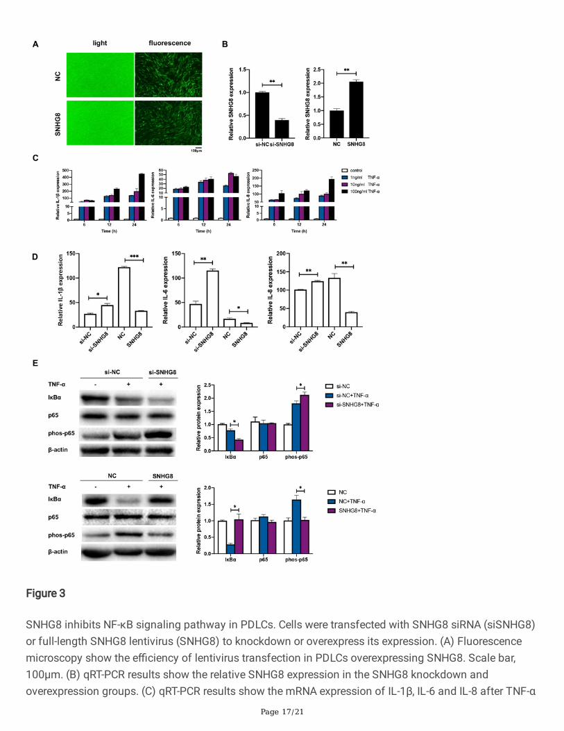
Figure 3
SNHG8 inhibits NF-κB signaling pathway in PDLCs. Cells were transfected with SNHG8 siRNA (siSNHG8)or full-length SNHG8 lentivirus (SNHG8) to knockdown or overexpress its expression. (A) Fluorescencemicroscopy show the e�ciency of lentivirus transfection in PDLCs overexpressing SNHG8. Scale bar,100μm. (B) qRT-PCR results show the relative SNHG8 expression in the SNHG8 knockdown andoverexpression groups. (C) qRT-PCR results show the mRNA expression of IL-1β, IL-6 and IL-8 after TNF-α

Page 18/21
stimulation. (D) qRT-PCR results show the relative RNA expression of IL-1β, IL-6 and IL-8 followingknockdown or overexpression of SNHG8 under TNF-α stimulation. (E) Western blot analyses show theeffects of SNHG8 knockdown or overexpression on the activation of NF-κB pathways induced by TNF-αin PDLCs. Histograms show the quanti�cation of band intensities. (Analysis of variance *p<0.05;**p<0.01; ***p<0.001).
Figure 4

Page 19/21
RNA sequencing following transfection with SNHG8. (A) Volcano plot of differentially expressed mRNAsin the control and SNHG8 groups. Red points: upregulated mRNAs; blue points: downregulated mRNAs.(B) The top 20 of gene ontology (GO) enrichment. Red frame: the altered mRNAs are enriched in cellularresponse to hypoxia. (C) Histograms show the genes related to the cellular response to hypoxia amongthe downregulated genes. (D) qRT-PCR results show that SNHG8 functions in the nucleus. (E) qRT-PCRresults show the mRNA expression of six HIF-1 mediated downstream genes, AQP1, RORA, RGCC, VEGFA,PTGIS, and PPARGC1A in the control group, SNHG8 group and SNHG8 supplemented with DMOG group.DMOG: dimethyloxalylglycine, an HIF-1α agonist. (Analysis of variance **p<0.01; ***p<0.001).
Figure 5
SNHG8 directly binds to HIF-1α. (A) The catRAPID heat-map shows the prediction of the interactionbetween SNHG8 and HIF-1α. The x- and the y-axes represent the indexes of the RNA and proteinsequences, respectively. The colors of the heat-map indicate the interaction score (ranging from -3 to +3)of the individual amino acid and nucleotide pairs. The interaction is identi�ed with con�dence (interactionpropensity=120 ; discriminative power=99%). (B) The interaction is characterized by a binding peak. Thex- and the y-axes represent the indexes of the RNA sequence and the interaction score, respectively. (C)qRT-PCR shows the RNA enrichment in the RIP assay using the anti-HIF-1α antibody in PDLCs. IgG wasused in the control group as the nonspeci�c protein. (D) Western blot analyses show the protein

Page 20/21
enrichment in the RNA pulldown assay. Histograms show the quanti�cation of band intensities. (Analysisof variance **p<0.01; ***p<0.001).
Figure 6
The effect of SNHG8 on NF-κB pathway is dependent on HIF-1α. (A) Western blot analyses of HIF-1α afterDMOG stimulation in PDLCs. (B) Western blot analyses of HIF-1α following application of force andstimulation of YC-1 (li�ciguat, an HIF-1α inhibitor ) in PDLCs. (C) Western blot analyses of phos-p65 and
Page 21/21
IκBα after the application of compressive force with or without SNHG8 knockdown, and with or withoutYC-1 stimulation, in PDLCs. (D) Western blot analyses of phos-p65 and IκBα after the application ofcompressive force with or without SNHG8 overexpression, and with or without DMOG stimulation, inPDLCs. All histograms show the quanti�cation of band intensities. (Analysis of variance *p<0.05;**p<0.01).
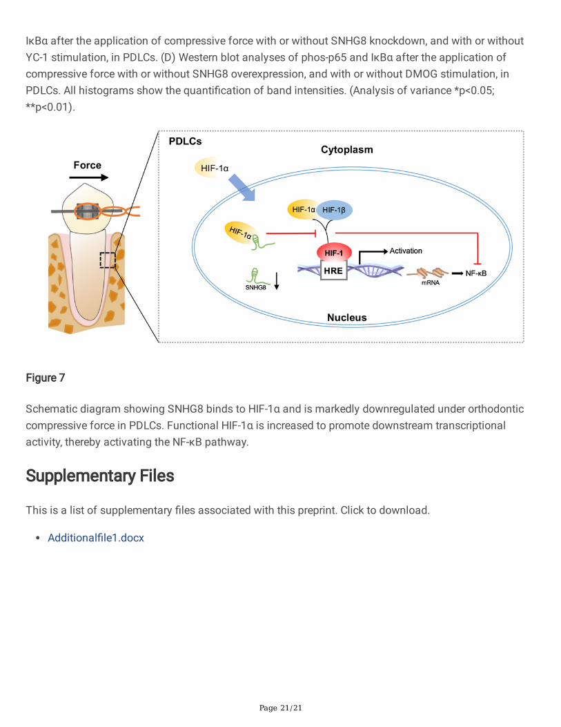
Figure 7
Schematic diagram showing SNHG8 binds to HIF-1α and is markedly downregulated under orthodonticcompressive force in PDLCs. Functional HIF-1α is increased to promote downstream transcriptionalactivity, thereby activating the NF-κB pathway.
Supplementary Files
This is a list of supplementary �les associated with this preprint. Click to download.
Additional�le1.docx