Embed Size (px)
Citation preview

Fractionation of Cells on a Discontinuous Ficoll Gradient
STUDYOF SUBPOPULATIONSOF HUMANT CELLS
USING ANTI-T-CELL ANTIBODIES FROMPATIENTS
WITH SYSTEMIC LUPUSERYTHEMATOSUS
WIESLAWGLiNsKI, M. ERc GERSHWIN,and Ai1mm D. STEINBERG
From the Arthritis and Rheumatism Branch, National Institute of Arthritis,Metabolism, and Digestive Diseases, National Institutes of Health,Bethesda, Maryland 20014
A B S T R A C T Patients with active systemic lupus ery-thematosus (SLE) had a decrease in a subpopulation ofcells (fraction D) when peripheral blood lymphocyteswere separated on a discontinuous Ficoll gradient. Pre-incubation of SLE cells at 370C for 30 min led to amarked decrease in this fraction, composed primarily ofthymus-derived (T) cells. Supernates of such preincuba-tions were found to cause a reduction in fraction D cellsfrom normal humans. The active factor in the supernatewas found to be an IgG antibody. Similarly, serum frompatients with active SLE produced a reduction in fractionD cells from normal donors. This activity was also foundin the IgG fraction, and could be absorbed with a pureT-cell population.
Depletion of macrophages and complement did not re-duce the SLE anti-T-cell antibody-mediated loss of cellsfrom fraction D; however, heat-aggregated humangammaglobulin led to impairment of the reaction. Thesefindings suggest that antibody-dependent direct lympho-cyte-mediated cytotoxicity may play a role in T-celllymphopenia of SLE.
It was further noted that the SLE anti-T-cell anti-bodies, in contrast to rabbit antihuman thymocyte serum,recognized fraction D cells but not fraction E cells fromnormals. Since both fractions are largely T cells, it ap-peared that the SLE serum was directed against cell-membrane antigenic determinants present on fractionD T cells, which were absent or reduced in quantity onfraction E T cells. Thus, evidence was presented indi-
Dr. Glinski's present address is the Department of Derma-tology, Academy of Medicine, Warsaw, Poland. He per-formed this work while a Visiting Fellow at the Derma-tology Branch, National Cancer Institute.
Received for publication 30 July 1975 and in revised form23 October 1975.
cating the presence of at least two subpopulations of Tcells in man. This was supported by differential absorp-tion of the anti-T-cell sera with fractions D and E.
INTRODUCTION
In an earlier study, peripheral blood cells from normalhumans and patients with systemic lupus were frac-tionated on discontinuous Ficoll gradients.' Two fractionscontained the great majority of thymus-derived (T)cells: fractions D and E. Patients with active systemiclupus erythematosus (SLE)' had a selective reduction incells from fraction D. Previous observations of anti-lymphocyte antibodies in patients with active SLE (1-5)suggested to us that cells from fraction D might be re-duced by such antibodies. This was a particularly appeal-ing hypothesis in view of the T-cell specificity of someof these antibodies (6, 7) as well as their ability to al-ter T-cell functions (8, 9).
In addition, the relatively greater loss of T cells fromfraction D as compared to T cells from fraction E sug-gested that subpopulations of human T cells might berecognized by SLE anti-T-cell antibodies. The presentstudy was, therefore, undertaken to further investigatethese possibilities. The relative loss of cells from fraction
' Glinski, W., M. E. Gershwin, and A. D. Steinberg. 1975.Fractionation of cells on a discontinuous Ficoll gradient.Study of peripheral blood lymphocyte subpopulations in nor-mal humans and patients with systemic lupus erythematosus.Submitted for publication.
aAbbreviations used in this paper: ADDLC, antibody-dependent direct lymphocyte-mediated complement-independ-ent cytotoxicity; ATS, antihuman thymocyte serum; HBSS,Hank's balanced salt solution; PBL, peripheral blood lym-phocytes; SLE, systemic lupus erythematosus; SRBC, sheepred blood cells; VBS, Veronal-buffered saline.
The Journal of Clinical Investigation Volume 57 March 1976*604-614604

D was found to be produced by an IgG antibody found inSLE sera. This antibody activity could be completely ab-sorbed by T cells. In addition, it recognized antigenicdeterminants present on normal T cells in fraction D butnot in fraction E, indicating that at least two T-cell sub-populations exist in humans.
METHODSPatients. 56 patients with SLE followed by the Ar-
thritis and Rheumatism Branch, National Institute of Ar-thritis and Metabolic Diseases, and 20 healthy volunteersof both sexes were studied. All patients met the diagnosticcriteria of American Rheumatism Association for SLE.Activity of the SLE was assessed by two observers basedupon clinical disease and graded as active or inactive. Onlypatients with clearly active or clearly inactive disease werestudied for this report. Patients receiving cytotoxic drugsor high doses of corticosteroids (> 15 mg/day prednisone)were excluded.
Separation of lymphocytes. Peripheral blood was drawnbetween 8:30 and 10:30 a.m. into preservative-free heparin(25 USP U/ml, Fellows Medical Mfg. Co., Oak Park,Mich.). The heparinized blood was diluted 1: 1 with cal-cium- and magnesium-free isotonic Hank's balanced saltsolution (HBSS) (Grand Island Biological Co., GrandIsland, N. Y.) and placed on a Ficoll-Hypaque gradient aspreviously described (10). Lymphocytes, thus separated,were washed three times with HBSS, and an aliquot ofthese unfractionated cells was removed for use in furtherexperiments. The remaining lymphocytes (the great ma-jority) were resuspended in a 5% Ficoll solution in HBSSand layered onto the discontinuous Ficoll gradient generally30 X 106 (25-40 X 10) cells per tube.
Discontinuous Ficoll gradient. Ficoll, (Sigma ChemicalCo., St. Louis, Mo., lot 14C-2350), a polysucrose polymerof mol wt 400,000, was dissolved in HBSS (Ca++- andMg"'-free) at pH 7.4, to obtain a 32% Ficoll solution. Thisstock solution was sterilized by passage through a 0.45-iumMillipore filter (Millipore Corp., Bedford, Mass.) and thefollowing concentrations (wt/vol) were prepared: 5, 9, 12,15, 17, 19, 21, 23, 25, and 30%. The Ficoll concentrationwas controlled in each solution by measurement of refrac-tive indices, which were linearly related to the density(11, 12). 3 ml of the Ficoll solutions was carefully layered
in sequence, in cellulose nitrate tubes (model 302237, 1 X 3iinches, Beckman Instruments, Inc., Spinco Div., Palo Alto,Calif.) beginning with the highest density preparation(30%o) at the bottom and ending with 9%o at the top. The
cells, suspended in 5% Ficoll, were added as the uppermostlayer. The tubes were centrifuged at 10'C for 30 min at16,000 g (force at bottom of tube) using an SW27 swinging-bucket rotor in a model L2-65B ultracentrifuge (BeckmanInstruments, Inc., Palo Alto, Calif.). After centrifugationthe lymphocytes were distributed at the interfaces. Theywere removed with Pasteur pipettes and placed into sepa-rate 16 X 125-mm plastic test tubes (Falcon Plastics, Ox-nard, Calif.) containing 5 ml of HBSS. The fractions werenamed sequentially from A to G for each interface startingat 9-12%o Ficoll and ending at 23-30% Ficoll. Cell viabilitywas more than 95%o in each fraction, as demonstrated bytrypan blue exclusion. The cells in fractions C, D, and Elooked relatively homogeneous under the microscope.
Lymphocyte counts. After fractionation, the cells in eachof eight fractions were washed twice in HBSS and counted. iCells were counted in a Coulter Counter (model ZBI,
Coulter Electronics, Inc., Hialeah, Fla.) . These countswere confirmed periodically using a hemocytometer.
Identification of T cells, bone marrow-derived (B) cells,and macrophages. The E-rosette test was used as a markerfor T lymphocytes. Cells from either the Ficoll-Hypaqueseparation or the Ficoll gradient were placed in HBSS ata concentration of 4 X 101/ml in HBSS. 100 Il of this cellpreparation was added to 4 X 10⁷ sheep red blood cells(SRBC), (SRBC: lymphocyte ratio of 10: 1) in 2,200 IAIof 50%o heat-inactivated, SRBC-absorbed fetal calf serum.The mixture was incubated at 370C for 30 min, followedby centrifugation at 200 g and incubation at 40C overnight.A rosette was defined as a lymphocyte surrounded by threeor more adherent SRBC. Viability of lymphocytes wasascertained by trypan blue exclusion and was always greaterthan 95%o. A single sheep was bled weekly for these ex-periments and the SRBCcollected in Alsever's solution. Allrosette assays were performed on SRBC less than 3 daysold.
The number and percentage of lymphocytes bearing com-plement receptors were determined by measuring the num-ber of lymphocytes binding three or more SRBC whichhad been sensitized with a IgM antibody to SRBC pluscomplement. A purified human IgM myeloma with antibodyactivity against SRBCwas used in a subagglutinating dilu-tion (kindly provided by Dr. Richard Wistar).
An equal volume of antibody and 2% SRBC were incu-bated at 370C for 45 min, washed three times in Veronal-buffered saline (VBS) containing calcium, magnesium, and1% bovine serum albumin, and resuspended to the originalvolume in VBS. Complement, 0.2 ml fresh-frozen mouseserum, was added to each 2 ml of antibody-coated SRBC,incubated at 370C for 45 min, washed three times in VBS,and diluted to a SRBC concentration of 0.5%o. To 100 1Aof this suspension, 4 X 0I lymphocytes in 100 ,ud VBS wereadded, incubated at 37°C for 45 min, and resuspended byrotation of the test tube. The cells were drawn up in a Pas-teur pipette by capillary action and enumerated.
Monocytes were identified by their ability to phagocytose1-/Am latex particles (1.099+0.0059 /Am diameter, Dow Chem-ical Co., Indianapolis, Ind.). Cell suspensions were incubatedwith latex particles at 370C for 1 h; centrifuged at 600 gfor 10 min, 800 g for 10 min, and finally 1,000 g for 10 minin medium, saving the precipitate each time. Finally, thecells were suspended in 100 bAl of medium, placed in ahemocytometer, and enumerated.
Preincubation of peripheral blood lymphocytes (PBL) ofSLE patients. 60-100-million lymphocytes from each of10 patients with SLE were placed in two to four separatetubes in the same absolute number. The cells were incu-bated at a concentration of 2 X 10⁷/ml at either 370C or atroom temperature (220C) for 30 min, and then separated onthe discontinuous Ficoll gradient.
The supernates from the 220 and 370C preincubation ex-periments were saved for further investigations and arecalled 220 and 370C supernates. Normal lymphocytes pre-incubated under the same conditions were similarly studied.
Preincubation of normal lymphocytes with SLE super-nates, SLE sera, or antihuman thymocyte serum (A TS).The effect of SLE supernates (220 and 370C), SLE sera,and ATS on the distribution of normal lymphocytes onthe discontinuous Ficoll gradient was next studied. The220 and 370C supernates (each 2.0-2.5 ml) were usedundiluted. SLE sera were inactivated at 560C for 30 minand used in various dilutions. ATS, prepared by weeklyinjections of human thymocytes (obtained during cardiacsurgery), was the gift of Dr. A. Ahmed. The ATS had
Fractionation of Cells on a Discontinuous Ficoll Gradient 605

been depleted of complement activity by heat inactivation andexhaustively absorbed with human tissues including eryth-rocytes and B cells from a patient with chronic lymphocyticleukemia. It was cytotoxic only for human T cells. It wasused in dilutions of 1: 50-1: 600. Typically, 4 X 10⁷ normalPBL (from the Ficoll-Hypaque separation) were suspendedin 1 ml of HBSS. The test serum was added and thevolume brought to 2 ml with additional HBSS. Thus, addi-tion of 0.25 ml SLE serum would represent a dilution of 1: 8(025 ml serum in a total volume of 2 ml). Controls con-sisted of (a) medium only and (b) normal human serum,type AB, which was previously heat inactivated.
The normal lymphocytes at a concentration of 2 X 10⁷/mlwere incubated at 370C for 1 h with one of these additionalsubstances and then separated on the discontinuous Ficollgradient. The total number of cells recovered as well asthe number of cells in each fraction was determined foreach treatment.
Depletion of macrophages. Macrophages were removedby iron particle phagocytosis. Suspensions of lymphocytes,isolated initially with Ficoll-Hypaque, were mixed 2: 1 (vol:vol) with lymphocyte-separator reagent (Technicon Instru-ments Corp., Tarrytown, N. Y.) which contains iron par-ticles. After 30 min at 370C, the cells were suspended andthe tube placed over a magnet. Iron particles and particle-containing cells were pulled to the bottom of the tube. Theremainder of the cells were aspirated and placed on aFicoll-Hypaque gradient for reisolation of the lymphocytes,free of remaining iron particles. As a control for this ex-periment, an aliquot of cells was carried through the sameprocedures, but without the addition of the lymphocyte-separator reagent.
Complement. To restore complement activity to the me-dium containing SLE serum, 5% fresh human AB serumwas added.
Blocking of cytotoxicity with aggregated human gammaglobulin. Human gamma globulin (human Cohn fractionII, E. R. Squibb & Sons, Princeton, N. J.) was dissolved inphosphate-buffered saline (pH 7.2) at a concentration of10 mg/ml and heated to 63°C for 20 min. The resultantaggregated gamma globulin was diluted to 1 mg/ml, and0.25 ml was added to 2.0 ml of a suspension of normal lym-phocytes (2 X 10⁷/ml). The mixture was incubated at 370Cfor 15 min after which 0.25 ml of SLE serum was added.The cells were incubated at 370C for an additional hour andthen separated on the discontinuous Ficoll gradient. Controllymphocytes were incubated in the medium (without aggre-gated gamma globulin) after which 0.25 ml of SLE serumwas added and the same procedures carried out. The numberof lymphocytes in each fraction and the total number oflymphocytes was determined for both control and aggre-gated gamma globulin preincubated cells. These were com-pared to a third aliquot of cells treated with medium onlyinstead of the SLE serum.
Study of IgG and IgM fractions of SLE sera. Serumproteins were precipitated with 50%o saturated ammoniumsulfate. The precipitate was centrifuged at 1,000 g for 30min and dissolved in distilled water. The solution wasdialyzed against Tris-buffered saline (pH 7.4) for 48 h andconcentrated to 0.5 ml with a collodion bag apparatus(Schleicher and Schuell, Inc., Keene, N. H.-bag 100, lot79432). The sample was subjected to gel filtration (Bio-Gel A-5m, operating range 10,000-5,000,000 mol wt, Bio-Rad Laboratories, Richmond, Calif.). The serum proteinconcentration in each tube was determined in a spectropho-tometer. The purity of the IgG and IgM fractions wasdetermined by gel diffusion with goat antihuman IgG and
goat antihuman IgM antisera (Hyland Div., Travenol Lab-oratories, Inc., Costa Mesa, Calif.). The uncontaminatedindividual fractions were concentrated to approximately theoriginal concentration in serum: IgG, 10 mg/ml, and IgM,1 mg/ml. In addition, individual SLE sera (one part) weremixed with four parts of rabbit antiserum to human IgG(purchased from Microbiological Associates, Bethesda, Md.,and found to precipitate only the IgG fraction of humanserum) and incubated at 4VC for 48 h. The precipitate wasremoved by centrifugation at 2,000 g for 1 h and the super-nate ultracentrifuged at 105,000 g for 30 min; then thesupernate of this procedure was studied. Similarly, thesupernate of 370C incubation of SLE cells was mixed withrabbit antihuman IgG serum (one part antiserum to fiveparts supernate), incubated at 4VC for 48 h, centrifuged at2,000 g for 1 h, ultracentrifuged as above, and the supernateused. The removal of IgG was confirmed by radial diffusion.
Absorption of SLE and ATS sera. Sera from three SLEpatients were diluted 1: 5 and absorbed with 109 lympho-cytes/ml. The lymphocytes were derived from a patientwith Sezary syndrome, kindly provided by Dr. R. Edelson.The leukocyte count of this patient was 48,000/mm' and99%o of these cells were lymphocytes with T-cell markers.The cells were incubated for 1 h at room temperature andfor 24 h at 4VC.
For study of the specificity of anti-T-cell antibodies in,SLE sera and in ATS, a 1: 5 dilution of SLE serum anda 1: 50 dilution of ATS were absorbed at 220C for 1 hand at 4VC for 24 h with cells of fraction D or fraction E(5 X 10⁸/ml) from a normal individual.
RESULTS
Distribution of E and EAC rosette-forming cells innormals. The cell type in each of these fractions wasidentified by determining the percentage of cells formingE or EAC rosettes as well as the percentage of macro-phages. The majority of cells in fractions D and Eformed E rosettes (Fig. 1). The relative percentage ofcells forming EAC rosettes rose as the density of Ficollincreased (Fig. 1). In contrast, fractions B and C werelargely (> 80%) composed of cells which did not formE or EAC rosettes (herein called null cells).
There were less than 5% macrophages (determined bylatex-particle phagocytosis) in any of the fractions, al-though almost 15% of the initial isolated Ficoll-Hypaquecells were macrophages. This was attributed to the ex-tensive number of washings in plastic tubes and the useof cellulose nitrate ultracentrifuge tubes; these cells ap-peared to stick to the latter.
Distribution of E and EAC rosette-forming cells inSLE. Patients with inactive SLE resembled normalcontrols in the relative distribution of E and EAC ro-sette-forming cells (Fig. 1). In contrast, patients withactive SLE had a reduction in E rosette-forming cells infractions D, E, and F + G with no change in EAC ro-settes (Fig. 1), leaving a relative increase in null cells inthose fractions.
Preincubation of PBL of SLE patients. Aliquots ofPBL from 10 patients with SLE were incubated at either220 or 37°C for 30 min and then separated on a dis-
606 W. Glinski, M. E. Gershwin, and A. D. Steinberg

°-° SLE active.-*-*-* 4SLE inactive
Wf Normalow 80_* \
z /
2I ____=1 1\\'\ E ROSETTESenz~I / h'Lu W 60-
C.)
D < ES0. U
20 EA
.II~~~~~~~~.
UNFRACTIONATED A + B C D E F + GCELLS FRACTION
FIGURE 1 Average values for three groups of patients arepresented. The percentage of lymphocytes in a given frac-tion forming E or EACrosettes is shown in the main figure,while the unfractionated results are presented to the left.The majority of cells in fraction A+B and C formedneither E nor EAC rosettes. Most cells in fractions D andE formed E rosettes. Normals (20 patients) and patientswith inactive SLE (12 patients) had similar values. Incontrast those with active SLE (16 patients) had reducedE rosettes in the heavier fractions as well as in unfrac-tionated cells.
continuous Ficoll gradient. Cell counts were performedbefore and after. No significant changes were observedafter incubation at 220C; however, a reduction in totallymphocyte count was observed after the 370C incubation(left-hand part of Fig. 2). It was found that the cells werebeing lost almost entirely from fraction D (center ofFig. 2). In 9 of the 10 patients, the reduction in PBLfrom fraction D was significant and is illustrated in theright-hand part of Fig. 2.
Preincubation of normal PBL. Normal lymphocytesincubated at 220 or 370C did not change in cell numberor distribution on the discontinuous Ficoll gradient in 12separate experiments. However, incubation of normallymphocytes with serum from a patient with active SLEled to a marked reduction in the number of cells in frac-tion D (illustrated in Fig. 3) without a concomitant in-crease in other fractions. This observation suggested thatthe SLE serum was eliminating cells from fraction D inthe same manner as cells were eliminated from fraction Dof SLE patients by 370C incubation. Furthermore, thesupernate of the 370C incubation of PBL from patientB. A. also reduced the number of cells in fraction D (Fig.
U)uj
0xa
0
zI u)
TOTAL A B C D E F+GFRACTION
FIGURE 2 Washed lymphocytes from 10 patients with activeSLE obtained by Ficoll-Hypaque separation of peripheralblood were incubated at either 220 or 370C for 30 minand then fractionated on a discontinuous Ficoll gradient.The total number of cells and the distribution of cells wasnot altered by the 220C incubation. In contrast, incubationat 370C led to a reduction in total number of cells (left-hand side of figure) and an alteration in the distribution(center of figure). The pronounced loss of cells from frac-tion D seen in the center of the figure is magnified on theright-hand side where individual values for the 10 patientsare presented.
xen-J-JUJU.0co
wz
Normal lymphocytesincubated at 370 C 1 h* medium
6 .--*-* SLE serum (BA)6 _ _ supernate 220 C
..-------supernate 370C5-
4 -,a\.
3 -
2
A B C D E F+GFRACTION
FIGURE 3 Washed lymphocytes from a patient (B. A.)with active SLE obtained by Ficoll-Hypaque separation ofperipheral blood were incubated at either 220 or 370C. Eachsupernate was subsequently incubated for 1 h at 370C withwashed lymphocytes from a normal volunteer (obtained byFicoll-Hypaque separation of peripheral blood). For com-parison, serum from patient B. A. was also incubated withan aliquot of the normal washed cells. Each aliquot oftreated cells (as well as one incubated with medium only)was then fractionated on a discontinuous Ficoll gradient.The number of cells in each fraction is presented in thefigure. The 220C supernate had no effect on the cell dis-tribution (compared to the medium control). In contrast,both the 370C supernate and the serum markedly reducedthe number of cells in fraction D.
Fractionation of Cells on a Discontinuous Ficoll Gradient 607

I ABLE I
Effect of SLE Serum, 37WCSupernate and IgG-depleted 37WCSupernate on the Distributionof Normal PBL from a Single Donor
Cell distribution on discontinuous Ficoll gradient*
Normal lymphocytes incubated Fractions Reduction in cellsat 37°C for I h with A + B + C D E F + G Total in fraction D
Medium 495,000 8,250,000 5,360,000 2,120,000 16,225,000(3.1 %)t (50.8%) (33.0%) (13.1%) (100.0%) -
Donor D. S.SLE serum 624,000 3,875,000 5,808,000 2,512,000 12,819,000
(3.9%)t (23.9%) (35.8%) (16.2%) (79.8%)t 52.9
370C supernate 660,000 4,988,000 5,538,000 2,791,000 13,977,000(4.1%) (30.7%) (34.1%) (17.2%) (86.1%) 39.6
370C supernate 827,000 7,880,000 6,180,000 1,670,000 16,557,000precipitated with rabbit (5.1%) (48.6%) (38.0%) (10.3%) (102.0%) 4.3antihuman IgG
* Absolute number of cells in each fraction.Percentage of cells given in parentheses; the total number of cells in medium control taken as 100%.
3). It was noted that the supernate of the 220C incuba-tion of B. A. cells did not alter the normal cell distribu-tion (Fig. 3). The same results were obtained in 40 ex-periments using 16 different sera from patients with
Normal lymphocytesincubated at 370C 1 h
-. *medium AL .---.. SLE serum (NM)
--------- e SLE serum 560 C /\b
L Z 2*SLE serum 560 C' 10 _ + complementXC,)-J-JLu
IL0
u
z5
5
0x 4
C,-u-3
LUC)u-30LU
2z
NORMALLYMPHOCYTESINCUBATEDAT 370C 1 h
* *-. MEDIUM
_---_ SLE SERUMI
A+B C
NORMALLYMPHOCYTESDEPLETEDOF MACROPHAGESINCUBATEDAT 370C 1 h
o-f-o MEDIUM
o---a SLE SERUM(AB)
I I I I
DFRACTION
E F + G
A BFRACTION
FIGURE 4 Washed PBL from -a normal volunteer wereincubated at 370C for 1 h with (a) serum from a patientN. M. with active SLE, (b) serum N. M. previouslyheated to 560C for 1 h to remove complement activity, (c)serum N. M. previously heated to 560C plus a fresh sourceof complement, or (d) medium only, and the cells fraction-ated on a discontinuous Ficoll gradient. All treatment groupscontaining serum N. M. had a reduction in cells in fractionD in comparison with the medium only treatment. Normalserum and serum from a patient with inactive SLE led to adistribution very similar to that of medium only (data notshown).
FIGuRE 5 Washed cells from a normal volunteer obtainedby Ficoll-Hypaque separation of peripheral blood weredivided into two aliquots. The first was depleted of macro-phages by iron-particle phagocytosis and reseparated free ofiron particles by a second Ficoll-Hypaque treatment. Thesecond aliquot was carried through the same procedureswithout the addition of iron particles. Each aliquot wasthen divided in half. One-half was incubated with serumfrom patient A. B., with active SLE, at 370C for 1 h. Theother half was incubated with medium only. All four ali-quots were then fractionated on the discontinuous Ficoll gra-dient. The number of cells in each fraction following eachtreatment is presented in the figure. The aliquot depletedof macrophages showed a reduction in fraction D followingtreatment with serum A. B. in comparison with mediumonly. This reduction was almost identical to that observedin the aliquot not depleted of macrophages.
608 W. Glinski, M. E. Gershwin, and A. D. Steinberg
eV

TABLE IIEffect of Serum from Patients with Active SLE on the Distribution of Normal PBL
Cell distribution on discontinuous Ficoll gradient*
Normal lymphocytes incubated Fractionsat37°Cfor 1 hwith A +B +C D E F +G Total
Medium 7.4 60.6 21.2 10.7 100.0Serum A. B. (1:10) 7.7 31.1 25.2 12.5 76.5Reductionj, % -0.3 29.5 -4.0 -1.8 23.5
Medium 11.1 53.1 20.2 15.6 100.0Serum M. N. (1:10) 12.8 14.7 15.9 13.4 56.8Reduction, % -1.7 38.4 4.3 2.2 43.2
Medium 17.0 47.9 26.2 8.8 100.0Serum D. S. (1:10) 14.4 22.2 31.7 12.1 80.4Reduction, % 2.6 25.7 -5.5 -3.3 19.6
Medium 4.2 61.1 20.2 14.5 100.0Serum A. S. (1:10) 3.4 47.0 19.4 10.9 80.7Reduction, % 0.8 14.1 0.8 3.6 19.3
Medium 6.1 52.7 27.9 13.3 100.0Serum J. U. (1:5) 5.5 15.9 28.6 13.7 63.7Reduction, % 0.6 36.8 -0.7 -0.4 36.3
Medium 7.4 58.0 22.8 11.8 100.0Serum B. K. (1:10) 6.8 30.4 21.6 10.6 69.4Reduction, % 0.6 27.6 1.2 1.2 30.6
Medium 6.8 58.5 22.4 12.3 100.0Serum B. C. (1:10) 5.8 41.2 23.0 12.7 82.7Reduction, % 1.0 17.3 -0.6 -0.4 17.3
Medium 14.3 44.8 20.2 20.7 100.0Serum B. H. (1:10) 12.5 21.1 23.6 18.8 76.0Reduction, % 1.8 23.7 -3.4 1.9 24.0
* Percentage of total cells of medium control in each fraction.$ Reduction = Percentage of all the medium-treated cells (total) that are reduced ina given fraction (a minus sign indicates an increase).
active SLE. An individual serum gave reproducible re-sults when run on separate occasions.
When the 370C supernate was depleted of IgG (byprecipitation with a rabbit antibody to human IgG), itno longer produced a reduction in cells in fraction D(Table I). This suggested that the antibody shed fromthe surface of T cells, rather than a nonspecific toxicsubstance in the supernate, was responsible for the re-duction in fraction D cells. The IgG class of the anti-body correlates with the serum anti-T-cell antibody asdiscussed below.
In several experiments, it was noted that serum frompatients with active SLE reduced fraction D, whereasserum from patients with inactive SLE did not. It wasrepeatedly observed that although both fractions D and Eare enriched in T cells, the SLE sera and supernatesmarkedly reduced the cells in fraction D, but had littleeffect on cells from fraction E.
The distribution of normal PBL on the discontinuousFicoll gradient was altered by SLE serum so as to re-semble the distribution of PBL from the serum donor.However, this was almost completely accounted for bythe changes in fraction D, with rather little change in theother fractions (Table II).
Studies of the mechanism of reduction in lymphocytesin fraction D complement. In the first study, the re-
quirement for the complement was investigated. SLEserum N. M. reduced the number of cells in fraction Dof normal PBL. Heating the serum to 560C to inactivatethe complement, or adding the complement, did not alterthe result (Fig. 4). These observations, which were re-
peated three times, suggested that the mechanism of lossof cells from fraction D was not complement mediated.
Macrophages. Normal peripheral blood was depletedof macrophages by the technique of iron-particle phago-cytosis and sedimentation in a magnetic field. Depletion
Fractionation of CeUs on a Discontinuous Ficoll Gradient 609
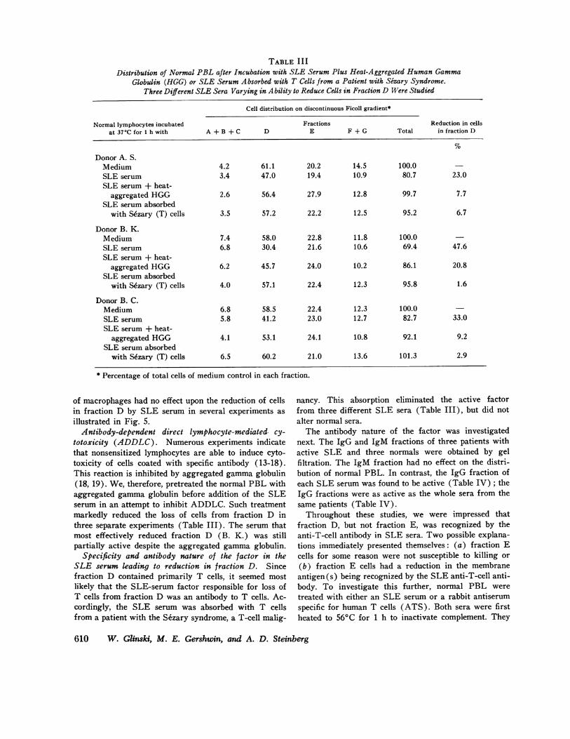
TABLE I I IDistribution of Normal PBL after Incubation with SLE Serum Plus Heat-Aggregated Human Gamma
Globulin (HGG) or SLE Serum Absorbed with T Cells from a Patient with Sezary Syndrome.Three Different SLE Sera Varying in Ability to Reduce Cells in Fraction D Were Studied
Cell distribution on discontinuous Ficoll gradient*
Normal lymphocytes incubated Fractions Reduction in cellsat 37'C for 1 h with A + B + C D E F + G Total in fraction D
Donor A. S.Medium 4.2 61.1 20.2 14.5 100.0SLE serum 3.4 47.0 19.4 10.9 80.7 23.0SLE serum + heat-
aggregated HGG 2.6 56.4 27.9 12.8 99.7 7.7SLE serum absorbed
with S6zary (T) cells 3.5 57.2 22.2 12.5 95.2 6.7
Donor B. K.Medium 7.4 58.0 22.8 11.8 100.0SLE serum 6.8 30.4 21.6 10.6 69.4 47.6SLE serum + heat-
aggregated HGG 6.2 45.7 24.0 10.2 86.1 20.8SLE serum absorbed
with Skzary (T) cells 4.0 57.1 22.4 12.3 95.8 1.6
Donor B. C.Medium 6.8 58.5 22.4 12.3 100.0SLE serum 5.8 41.2 23.0 12.7 82.7 33.0SLE serum + heat-
aggregated HGG 4.1 53.1 24.1 10.8 92.1 9.2SLE serum absorbed
with S~zary (T) cells 6.5 60.2 21.0 13.6 101.3 2.9
* Percentage of total cells of medium control in each fraction.
of macrophages had no effect upon the reduction of cellsin fraction D by SLE serum in several experiments asillustrated in Fig. 5.
Antibody-dependent direct lymphocyte-mediated cy-totoxicity (ADDLC). Numerous experiments indicatethat nonsensitized lymphocytes are able to induce cyto-toxicity of cells coated with specific antibody (13-18).This reaction is inhibited by aggregated gammaglobulin(18, 19). We, therefore, pretreated the normal PBL withaggregated gammaglobulin before addition of the SLEserum in an attempt to inhibit ADDLC. Such treatmentmarkedly reduced the loss of cells from fraction D inthree separate experiments (Table III). The serum thatmost effectively reduced fraction D (B. K.) was stillpartially active despite the aggregated gamma globulin.
Specificity and antibody nature of the factor in theSLE serum leading to reduction in fraction D. Sincefraction D contained primarily T cells, it seemed mostlikely that the SLE-serum factor responsible for loss ofT cells from fraction D was an antibody to T cells. Ac-cordingly, the SLE serum was absorbed with T cellsfrom a patient with the Sezary syndrome, a T-cell malig-
nancy. This absorption eliminated the active factorfrom three different SLE sera (Table III), but did notalter normal sera.
The antibody nature of the factor was investigatednext. The IgG and IgM fractions of three patients withactive SLE and three normals were obtained by gelfiltration. The IgM fraction had no effect on the distri-bution of normal PBL. In contrast, the IgG fraction ofeach SLE serum was found to be active (Table IV); theIgG fractions were as active as the whole sera from thesame patients (Table IV).
Throughout these studies, we were impressed thatfraction D, but not fraction E, was recognized by theanti-T-cell antibody in SLE sera. Two possible explana-tions immediately presented themselves: (a) fraction Ecells for some reason were not susceptible to killing or(b) fraction E cells had a reduction in the membraneantigen (s) being recognized by the SLE anti-T-cell anti-body. To investigate this further, normal PBL weretreated with either an SLE serum or a rabbit antiserumspecific for human T cells (ATS). Both sera were firstheated to 560C for 1 h to inactivate complement. They
610 W. Glinski, M. E. Gershwin, and A. D. Steinberg

TABLE IVEffect of the IgG Fraction of SLE Serum on the Distribution of Normal PBL
Cell distribution on discontinuous Ficoll gradient*
Normal lymphocytes incubated Fractions Reduction in cellsat 37°C for 1 h with A + B + C D E F + G Total in fraction D
Donor B. C.Medium 6.9 57.5 21.5 14.1 100.0SLE serum 8.8 35.0 23.0 13.4 80.2 39.1SLE serum IgG 9.9 21.2 26.7 15.0 72.8 59.7SLE serum IgM 10.6 53.8 24.1 12.5 101.0 6.4Normal serum lgG 14.6 51.2 23.5 13.2 102.5 11.0
Donor A. B.Medium 11.6 52.6 20.0 15.8 100.0SLE serum 8.7 30.9 21.2 12.5 73.3 41.3SLE serum IgG 10.8 31.9 22.1 17.9 82.7 39.4SLE serum IgM 9.4 48.1 20.5 16.2 95.2 8.5Normal serum IgG 7.7 50.2 20.9 17.4 96.1 4.8
Donor B. H.Medium 14.3 44.8 20.2 20.7 100.0SLE serum 12.5 21.1 23.6 18.8 76.0 52.9SLE serum IgG 11.3 15.0 26.1 19.6 72.0 66.5Normal serum IgG 11.1 46.7 21.3 21.8 100.9 -4.2
$ Percentage of total cells of medium control in each fraction.
were then incubated with normal lymphocytes at 370Cfor 1 h and the cells were fractionated on the discontinu-ous Ficoll gradient. The ATS led to a significant re-duction in cells in both fractions D and E. This was re-peatedly true in a number of experiments and over awide range of dilutions of this antiserum (1: 50-1: 600).In contrast, the SLE serum significantly reduced thecell number only in fraction D, again consistently andover a wide range of serum dilutions (1: 4-1: 128).
To further study differences between fractions D andE, the ATS and SLE sera were first absorbed with frac-tion D or fraction E from a normal donor. These ab-sorbed sera were then incubated with normal PBL at370C and the cells fractionated on the discontinuousFicoll gradient (Table V). Control unabsorbed SLE se-rum reduced the cell number in fraction D, but not infraction E. ATS reduced the number of cells in fractionD and E by approximately the same amount. Absorptionof the ATS with either fractions D or E markedly re-duced its activity (Table V). This suggested that thisantiserum was able to recognize cell surface-membranedeterminants common to both fractions. In contrast, ab-sorption of the SLE serum with the different fractionsgave different results. Absorption of the SLE serumwith fraction D reduced its activity towards fraction D,whereas absorption with fraction E had no significanteffect upon its activity toward fraction D (Table V).This suggested that the SLE serum recognized surface-
membrane antigeneic determinants of cells in fraction Dthat were diminished on or absent from fraction E cells.
DISCUSSION
It has been known that patients with active SLE havereduced numbers of total leukocytes (20-22) and lympho-cytes (22-24). Furthermore, they have been found tohave a reduction in T cells (24, 25) and T-cell functions(26-30). Finally, anti-lymphocyte and anti-T cell-anti-bodies have been found in patients with SLE (1-9). Thepresent study links these various observations.
Wehave observed that patients with active SLE havea reduction in the percentage of cells in fraction D, afraction obtained by separating PBL on a discontinuousFicoll gradient. Furthermore, this fraction is composedprimarily of T cells. In the present study, the mechanismof this reduction was investigated.
Preincubation at 370C of PBL from patients withactive SLE resulted in a loss of cells from fraction D.Supernates from this incubation were found to reducethe number of cells in fraction D of PBL from normaldonors. Similarly, SLE serum reduced the number ofcells in fraction D from normals. An IgG antibody withanti-T-cell specificity present in the SLE serum was
required for this effect. The reaction was independent ofboth complement and macrophage. Heat aggregatedgammaglobulin markedly inhibited the anti-T-cell cyto-
Fractionation of Celis on a Discontinuous Ficoll Gradient 611

TABLE VDifferential Absorption of SLE Serum and Rabbit A TS with Two T-Cell Subpopulations (Fractions D and E)
of Normal PBL. The Effect of These Absorbed Sera on the Distributions ofPBL from a Single Normal Donor Was Studied
Cell distribution on discontinuous Ficoll gradient*Reduction in cells in
Normal lymphocytes incubated Fractionsat 37°C for 1 h with A + B + C D E F + G Total Fraction D Fraction E
Medium 6.1 52.7 27.9 13.3 100.0
SLE serum J. U. (1:5)Unabsorbed 5.5 15.9 28.6 13.7 63.7 69.8 -2.5Absorbed with fraction D 6.1 42.1 28.7 15.5 92.4 20.1 -2.8Absorbed with fraction E 5.0 19.2 27.0 15.9 67.1 63.6 3.2
ATS (1:50)Unabsorbedt 5.7 22.6 14.8 12.1 55.2 57.2 47.0Absorbed with fraction D 6.7 51.3 30.6 14.9 103.5 2.7 -9.7Absorbed with fraction E 7.9 43.6 24.4 13.9 89.8 17.3 12.6
* Percentage of totol cells of medium control in each fraction.t Unabsorbed with fraction D or E. This serum had previously been absorbed as described in the methods.
toxicity, implicating Fc receptor-bearing cells. These ob-servations suggested that ADDLC (10-16) was an im-portant mechanism for T-cell loss in SLE patients, par-ticularly since the reactions were carried out at bodytemperature, 370C. The possibility of additional mecha-nisms for T-cell loss was suggested by only partial re-duction in activity of an active serum (B. K.) with theaggregated gammaglobulin.
It was further observed that the SLE anti-T-cell anti-bodies recognized antigeneic determinants on cells offraction D that were absent from or much reduced indensity on cells of fraction E. Since both fractions con-tained primarily T cells, it appeared that the SLE anti-bodies could distinguish among cells in the two fractionsleading to loss of cells only from fraction D. This isevidence for two subpopulations of T cells in normalhuman peripheral blood. The antihuman T-cell antibodiesin SLE sera were distinguishable from rabbit antihumanT-cell antibodies. The activity of the former was ab-sorbed to a much greater extent with fraction D thanfraction E, providing further evidence for membrane dif-ferences between cells in fractions D and E. In contrast,the rabbit antibody activity was equally absorbed withfractions D and E. Thus the rabbit anti-T-cell antibodiesrecognized all human T cells, whereas the SLE anti-T-cell antibodies recognized preferentially a subpopulationof human T cells.
Since only some of the T cells in fraction D wereeliminated by the SLE serum, fraction D itself may con-tain more than one subpopulation of T cells. Alterna-tively, soluble immune complexes in SLE sera mighthave inhibited the ADDLCreaction against antibody-
coated fraction D T cells, especially since immune com-plexes have been shown to inhibit ADDLC (31) andADDLCis reduced in SLE (32).
The IgG anti-T-cell antibodies active at 370C whichwere studied herein appear to be different from the IgManti-T-cell antibodies which are often more active at lowtemperatures (2, 4). The anti-T-cell specificity is analo-gous to that previously described (6, 7) and may be re-lated to the antibodies which inhibit the mixed-lympho-cyte culture (8, 9). The present studies are also in agree-ment with a previous observation, suggesting that IgGanti-T-cell antibodies in SLE are only minimally cyto-toxic (4). Previous reports of complement-dependentIgG antilymphocyte antibodies in SLE (3, 5) may in-volve sera with specificities different from those studiedin the present report. In fact, the existence of anti-lymphocyte antibodies with different specificities in SLEsera has recently been emphasized (4).
The effect of supernates from the present study maybe related to the "shedding" phenomenon previously de-scribed (33-35). The IgG nature of the supernatant anti-body is in agreement with the elution of IgG antibodyfrom SLE lymphocytes (36).
Multiple antibodies may be responsible for lymphocytedepletion in SLE. Different mechanisms may be responsi-ble for lymphocyte depletion in SLE. Different mecha-nisms may be responsible for the loss of different typesof cells. Patients with SLE may lose cells from fractionE by a mechanism different from that responsible for theloss of a subpopulation of cells from fraction D. Never-theless, the observations reported herein suggest thatADDLC is a likely mechanism for loss of at least one
612 W. Glinski, M. E. Gershwin, and A. D. Steinberg

T-cell subpopulation in SLE. The abnormalities in SLE-PBL distribution observed in a previous study using thistechnique' appear to be caused at least partially by ef-fects of antilymphocyte antibodies. The relatively greaterreduction in cells of fraction D is explainable on thebasis of specificity of SLE anti-T-cell antibodies forcells in fraction D.
New Zealand mice represent animal models of humanSLE (37). These mice spontaneously produce antibodiesto T cells (38), as well as antierythrocyte and antinu-clear antibodies. Before the appearance of overt auto-immune disease manifestations, New Zealand mice ap-pear to lose a subpopulation of T cells, which serves aregulatory or suppressor function (39-43). It is possiblethat a similar loss of regulatory T cells occurs in pa-tients with SLE. Investigations of this question are cur-rently in progress.
REFERENCES
1. Butler, W. T., J. T. Sharp, R. D. Rossen, M. D. Lidsky,K. K. Mittal, and D. A. Gard. 1972. Relationship of theclinical course -of systemic lupus erythematosus to thepresence of circulating lymphocytotoxic antibodies. Ar-thritis Rheum. 15: 231-238.
2. Ooi, B. S., A. R. Orlina, A. J. Pesce, N. Mendoza, L.Masaitis, and V. E. Pollak. 1974. Lymphotocytoxic anti-bodies in patients with systemic lupus erythematosus.Clin. Exp. Immunol. 17: 237-243.
3. Stastny, P., and M. Ziff. 1971. Antibodies against cellmembrane constituents in systemic lupus erythematosusand related diseases. I. Cytotoxic effect of serum frompatients with systemic lupus erythematosus (SLE) forallogeneic and for autologous lymphocytes. Clin. Exp.Immunol. 8: 543-550.
4. Winfield, J. B., R. J. Winchester, P. Wernet, S. M. Fu,Masaitis, and V. E. Pollak. 1974. Lymphocytotoxic anti-bodies to lymphocyte surface determinants in systemiclupus erythematosus. Arthritis Rheum. 18: 1-8.
5. Mittal, K. K., R. D. Rossen, J. T. Sharp, M. D. Lidsky,and W. T. Butler. 1970. Lymphocyte cytotoxic anti-bodies in systemic lupus erythematosus. Nature (Lond.).225: 1255-1256.
6. Lies, R. B., R. P. Messner, and R. C. Williams, Jr.1973. Relative T-cell specificity of lymphocytotoxinsfrom patients with systemic lupus erythematosus. Ar-thritis Rheum. 16: 369-375.
7. Thomas, D. B. 1973. Antibodies specific for human Tlymphocytes in cold agglutinin and lymphocytotoxic sera.Eur. J. Immunol. 3: 824-828.
8. Williams, R. C., Jr., R. B. Lies, and R. P. Messner.1973. Inhibition of mixed leucocyte culture responsesby serum and globulin fractions from certain patientswith connective tissue disorders. Arthritis Rheum. 16:597-605.
9. Wernet, P., and H. G. Kunkel. 1973. Antibodies to aspecific surface antigen of T cells in human sera in-hibiting mixed leucocyte culture reactions. J. Exp. Med.138: 1021-1026.
10. Boyum, A. 1968. Separation of leucocytes from bloodand bone marrow. Scand. J. Clin. Lab. Invest. 21 (Suppi.97): 109 pp.
11. Bach, M. K., and J. R. Brashler. 1970. Isolation of sub-populations of lymphocytic cells by the use of isotoni-cally balanced solutions of Ficoll. I. Development ofmethods and demonstration of the existence of a largebut finite number of subpopulations. Exp. Cell Res. 61:387-396.
12. Yu, D. T. Y., J. B. Peter, H. E. Paulus, and H. I.Machleder. 1973. Lymphocyte populations: separation bydiscontinuous density gradient centrifugation. J. Im-munol. 110: 1615-1622.
13. M6ller, E. 1965. Contact-induced cytotoxicity by lym-phoid cells containing foreign isoantigens. Science(Wash. D. C.). 147: 873-879.
14. Perlman, P., and G. Holm. 1969. Cytotoxic effects oflymphoid cells in vitro. Adv. Immunol. 11: 117-193.
15. Van Boxel, J. A., J. D. Stobo, W. E. Paul, and I.Green. 1972. Antibody-dependent lymphoid cell-mediatedcytotoxicity: no requirement for thymus-derived lym-phocytes. Science (Wash. D. C.). 175: 194-196.
16. Van Boxel, J. A., W. E. Paul, M. M. Frank, and I.Green. 1973. Antibody-dependent lymphoid cell-mediatedcytotoxicity: role of lymphocytes bearing a receptorfor complement. J. Immunol. 110: 1027-1036.
17. Wisloff, F., and S. S. Froland. 1973. Antibody-dependentlymphocyte-mediated cytotoxicity in man: no require-ment for lymphocytes with membrane-bound immuno-globulin. Scand. J. Immunol. 2: 151-157.
18. Scornick, J. C., H. Cosenza, W. Lee, H. Kohler, andD. A. Rowley. 1974. Antibody-dependent cell-mediatedcytotoxicity. I. Differentiation from antibody-independentcytotoxicity by "normal" IgG. J. Immunol. 113: 1510-1518.
19. Hallberg, T. 1974. Inhibition of cytotoxicity of nonim-mune human lymphocytes for sensitized chicken erythro-cytes by aggregated human IgG. Scand. J. Immunol.3: 117-125.
20. Goeckerman, W. H. 1923. Lupus erythematosus as asystemic disease. JAMA (J. Am. Med. Assoc.). 80:542-547.
21. Michael, S. R., I. L. Vural, F. A. Bassen, and L.Schaefer. 1951. The hematologic aspects of disseminated(systemic) lupus erythenmatosus. Blood. 6: 1059-1072.
22. Harvey, A. M., L. E. Schulman, P. A. Tumulty, C.L. Conley, and E. H. Schoenrich. 1954. Systemic lupuserythematosus: review of the literature and analysis of138 cases. Medicine (Baltimore). 33: 291-437.
23. Delbarre, F., A. Pompidou, A. Kahan, H. Brouilhet,A. Le G6, and B. Amor. 1971. Study of blood lympho-cytes during systemic lupus erythematosus. Pathol. Biol.19: 379-387.
24. Scheinberg, M. A., and E. S. Cathcart. 1974. B celland T cell lymphopenia in systemic lupus erythematosis.Cell. Immunol. 12: 309-314.
25. Messner, R. P., F. D. Lindstr6m, and R. C. Williams,Jr. 1973. Peripheral blood lymphocyte cell surfacemarkers during the course of systemic lupus erythemato-sus. J. Clin. Invest. 52: 3046-3056.
26. Suciu-Foca, N., J. A. Buda, T. Thiem, and K. Reem-tsma. 1974. Impaired responsiveness of lymphocytes inpatients with systemic lupus erythematosus. Clin. Exp.Immunol. 18: 295-301.
27. Horwitz, D. A. 1972. Impaired delayed hypersitivity insystemic lupus erythematosus. Arthritis Rheum. 15:353-359.
28. Hahn, B. H., M. K. Bagby, and C. K. Osterland. 1973.Abnormalities of delayed hypersensitivity in systemiclupus erythematosus. Am. J. Med. 55: 25-31.
Fractionation of Cells on a Discontinuous Ficoll Gradient 613

29. Malave, I., Z. Layrisse, and M. Layrisse. 1975. Dose-dependent hyporrieactivity to phytohemagglutinin in sys-temic lupus erythematosus. Cell. Immunol. 15: 231-236.
30. Rosenthal, C. J., and E. C. Franklin. 1975. Depressionof cellular-mediated immunity in systemic lupus erythe-matosus. Relation to disease activity. Arthritis Rheum.18: 207-217.
31. MacLannan, I. C. M. 1972. Competition for receptorsfor immunoglobulin on cytotoxic lymphocytes. Clin. Exp.Immunol. 10: 275-283.
32. Schneider, J., W. Chin, G. J. Friou, S. M. Cooper, B.Harding, R. L. Hill, and F. P. Quismorio. 1975. Re-duced antibody-dependent cell-mediated cytotoxicity insystemic lupus erythematosus. Clin. Exp. Immunol.20: 187-192.
33. Wernet, P., M. Fotino, R. Thoburn, A. Moore, and H.G. Kunkel. 1973. Blockage of lymphocyte surface anti-gens and the shedding phenomenon in systemic lupuserythematosus (SLE). Arthritis Rheum. 16: 137.(Abstr.)
34. Winchester, R. J., J. B. Winfield, F. Siegal, P. Wernet,Z. Bentwich, and H. G. Kunkel. 1974. Analysis of lym-phocytes from patients with rheumatoid arthritis andsystemic lupus erythematosus. Occurrence of interferingcold-reactive antilymphocyte antibodies. J. Clin. Invest.54: 1082-1092.
35. Messner, R. P., M. S. Kennedy, and J. G. Jelinek. 1975.Antilymphocyte antibodies in systemic lupus erythema-
tosus. Effect on lymphocyte surface characteristics.Arthritis Rheum. 18: 201-206.
36. Stastny, P., and M. Ziff. 1971. Lymphocyte and plateletautoantibodies in S.L.E. Lancet. 1: 1239-1240.
37. Talal, N., and A. D. Steinberg. 1974. The pathogenesisof autoimmunity in New Zealand Black mice. Curr.Top. Microbiol. Immunol. 64: 79-103.
38. Shirai, T., and R. C. Mellors. 1971. Natural thymocyto-toxic autoantibody and reactive antigen in New ZealandBlack and other mice. Proc. Nati. Acad. Sci. U. S. A.68: 1412-1415.
39. Steinberg, A. D. 1974. Pathogenesis of autoimmunity inNew Zealand mice. V. Loss of thymic suppressorfunction. Arthritis Rheum. 17: 11-14.
40. Barthold, D. R., S. Kysela, and A. D. Steinberg. 1974.Decline in suppressor T cell function with age in femaleNZB mice. J. Immunol. 12: 9-16.
41. Gerber, N. L., J. A. Hardin, T. M. Chused, and A. D.Steinberg. 1974. Loss with age in NZB/W mice ofthymic suppressor cells in the graft-vs-host reaction.J. Immunol. 113: 1618-1625.
42. Dauphinee, M. J., and N. Talal. 1975. Reversible resto-ration by thymosin of antigen-induced depression ofspleen DNA synthesis in NZB mice. J. Immunol. 114:1713-1716.
43. Gershwin, M. E., and A. D. Steinberg. 1975. Suppres-sion of autoimmune hemolytic anemia in New Zealand(NZB) mice by syngenic young thymocytes. Clin. Im-munol. Immunopathol. 4: 38-45.
614 W. Glinski, M. E. Gershwin, and A. D. Steinberg