Embed Size (px)
Citation preview
The Radiation Biology of Dose Fractionation:Determinants of Effect
E. Day Werts, Ph.D.Department of Radiation Oncology
West Penn Allegheny Radiation Oncology Network
Allegheny General Hospital
The Radiation Biology of Dose Fractionation:The Radiation Biology of Dose Fractionation:Determinants of EffectDeterminants of Effect
E. Day Werts, Ph.D.E. Day Werts, Ph.D.Department of Radiation OncologyDepartment of Radiation Oncology
West Penn Allegheny Radiation Oncology West Penn Allegheny Radiation Oncology NetworkNetwork
Allegheny General HospitalAllegheny General Hospital
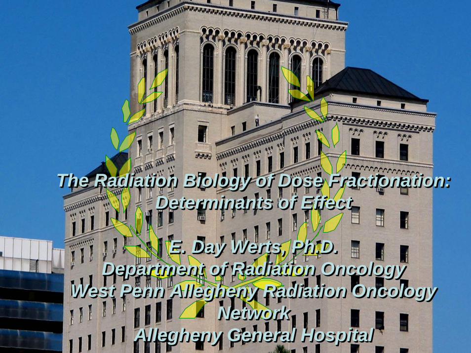
Historical Perspectives
1. Strandquist Plot2. NSD (ret and neuret)3. Time, Dose & Fractionation (TDF)4. Linear Quadratic
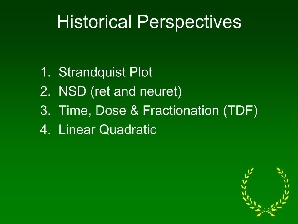
Strandquist Plot - 1944• Standard fractionation – 2Gy/dy x 5/wk• 250kvP x-ray machine• Skin morbidty vs control of skin cancer
EJ Hall, Radiobiology for the Radiologist 2nd ed p277,1978

Ellis NSD Equation - 1967
Ellis used the iso-effect data for skin from Strandquist and proposed that the tolerance dose for normal tissues (D rads) was related to overal treatment time (T days) and the number (N) of fractions.
D = (NSD)(T0.11)(N0.24)NSD = nominal single dose (rets)
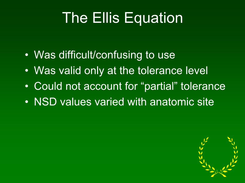
The Ellis Equation
• Was difficult/confusing to use• Was valid only at the tolerance level• Could not account for “partial” tolerance• NSD values varied with anatomic site

TDF Tables - 1972
• Based upon the NSD equation which was derived from the Strandquist data
• Tables generated for 1,2,3,4 or 5 fx/day
• Independent of NSD and therefore generally applicable to any tissue

Orton & Ellis, BJR 46:529-537, 1973
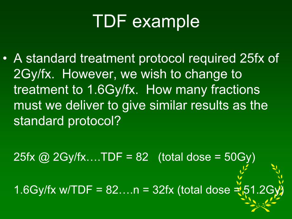
TDF example
• A standard treatment protocol required 25fx of 2Gy/fx. However, we wish to change to treatment to 1.6Gy/fx. How many fractions must we deliver to give similar results as the standard protocol?
25fx @ 2Gy/fx….TDF = 82 (total dose = 50Gy)
1.6Gy/fx w/TDF = 82….n = 32fx (total dose = 51.2Gy)

Critique of Power Law Formulas(NSD/TDF)
• Iso-effects in Strandquist plots are curved, not linear as req’d by the NSD formula
• Assume all normal tissues behave like skin and tumors like squamous cell CA
• Tissues differ in fractionation sensitivity therefore there can be no single exponent for “N”

Critique cont’d
• NSD underestimates late tissue reactions after large dose fractions
• There is NO appreciable time factor for late tissue reactions
• The formula dose not adequately account for the proliferation response in handling treatment time

A New Way of Looking at Iso-effect Data - 1982
Defines the shape of isoeffect curves
Describes sensitivity to changes in fraction size
α/β = intercept/slope
HR Withers, Cancer 55:2086, 1985
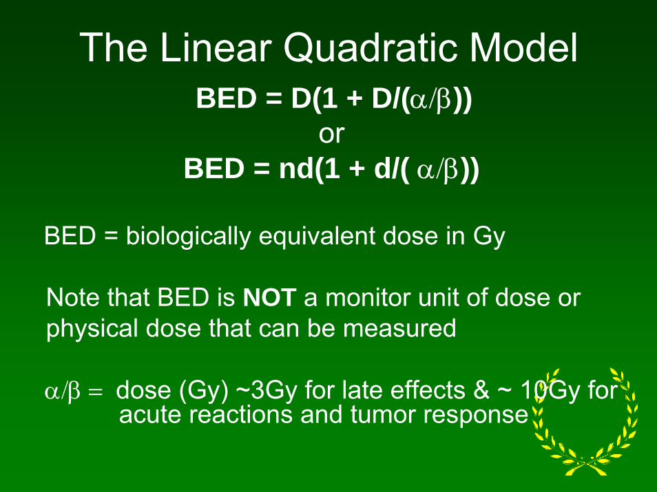
The Linear Quadratic ModelBED = D(1 + D/(α/β))
orBED = nd(1 + d/( α/β))
BED = biologically equivalent dose in Gy
Note that BED is NOT a monitor unit of dose or physical dose that can be measured
α/β = dose (Gy) ~3Gy for late effects & ~ 10Gy for acute reactions and tumor response
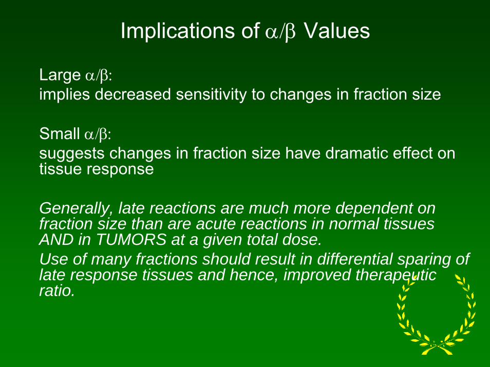
Implications of α/β Values
Large α/β:implies decreased sensitivity to changes in fraction size
Small α/β:suggests changes in fraction size have dramatic effect on tissue response
Generally, late reactions are much more dependent on fraction size than are acute reactions in normal tissues AND in TUMORS at a given total dose. Use of many fractions should result in differential sparing of late response tissues and hence, improved therapeutic ratio.
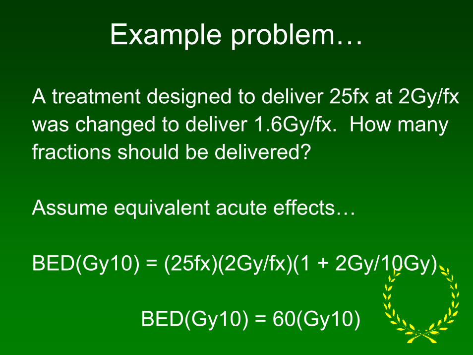
Example problem…
A treatment designed to deliver 25fx at 2Gy/fxwas changed to deliver 1.6Gy/fx. How many fractions should be delivered?
Assume equivalent acute effects…
BED(Gy10) = (25fx)(2Gy/fx)(1 + 2Gy/10Gy)
BED(Gy10) = 60(Gy10)
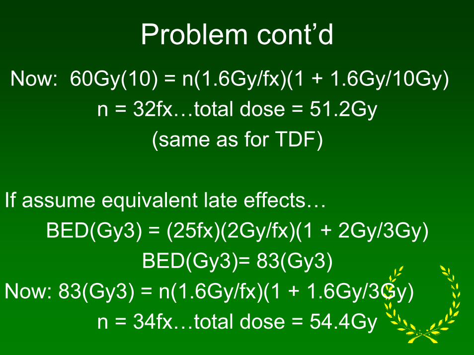
Problem cont’dNow: 60Gy(10) = n(1.6Gy/fx)(1 + 1.6Gy/10Gy)
n = 32fx…total dose = 51.2Gy(same as for TDF)
If assume equivalent late effects…BED(Gy3) = (25fx)(2Gy/fx)(1 + 2Gy/3Gy)
BED(Gy3)= 83(Gy3)Now: 83(Gy3) = n(1.6Gy/fx)(1 + 1.6Gy/3Gy)
n = 34fx…total dose = 54.4Gy
Biologic Factors that Influence Fractionation Response
Repair
Volume
Oxygenation/Reoxygenation

Elkind (split dose) Repair - 1965
Shibamato, et al IJROBP 59:1484-1490, 2004
Changes in the surviving fraction of EMT6 (○) and SCCVII (●)cells after a total dose of 8 Gy as a function of the interval (h)between two doses of 4 Gy.
Fx. S
urvi
ving
Dose
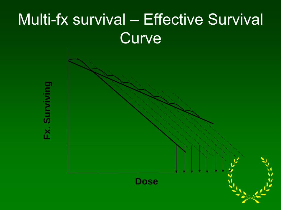
Multi-fx survival – Effective Survival Curve
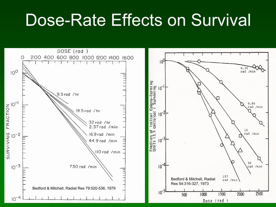
Dose-Rate Effects on Survival
Bedford & Mitchell, RadiatRes 54:316-327, 1973
Bedford & Mitchell, Radiat Res 79:520-536, 1979
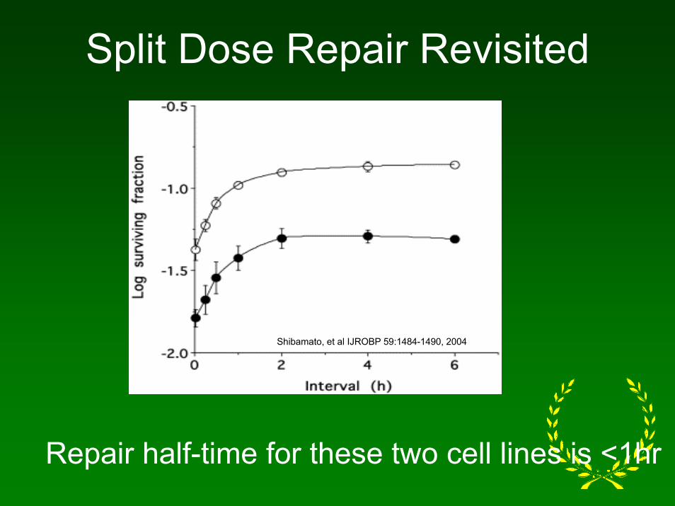
Split Dose Repair Revisited
Shibamato, et al IJROBP 59:1484-1490, 2004
Repair half-time for these two cell lines is <1hr
Repair Half-Time
For most tissues is…minutes to few hours
Implications of Short Repair Half-Time:
IMRT…A cautionary word…or two…
1. Typical step and shoot mode IMRT can require 5-9 treatment fields with 20-40 segments resulting in over 100 segments for the IMRT treatment plan.
2. Total treatment delivery time may be prolonged to 15 – 45 minutes
3. With short repair half-times, repair of radiation damage during the treatment will potentially decrease the cell killing that would have been expected
Cautionary Word…cont’d
4. By comparison, typical EBRT beam-on time and fraction-delivery times are on the order of 1-2 and 3-5 minutes.
5. Therefore, calculations of IMRT plans based upon conventional EMRT plans may underestimate the total dose necessary to maintain similar tumor control…if the repair half-times of the tumors are very short.
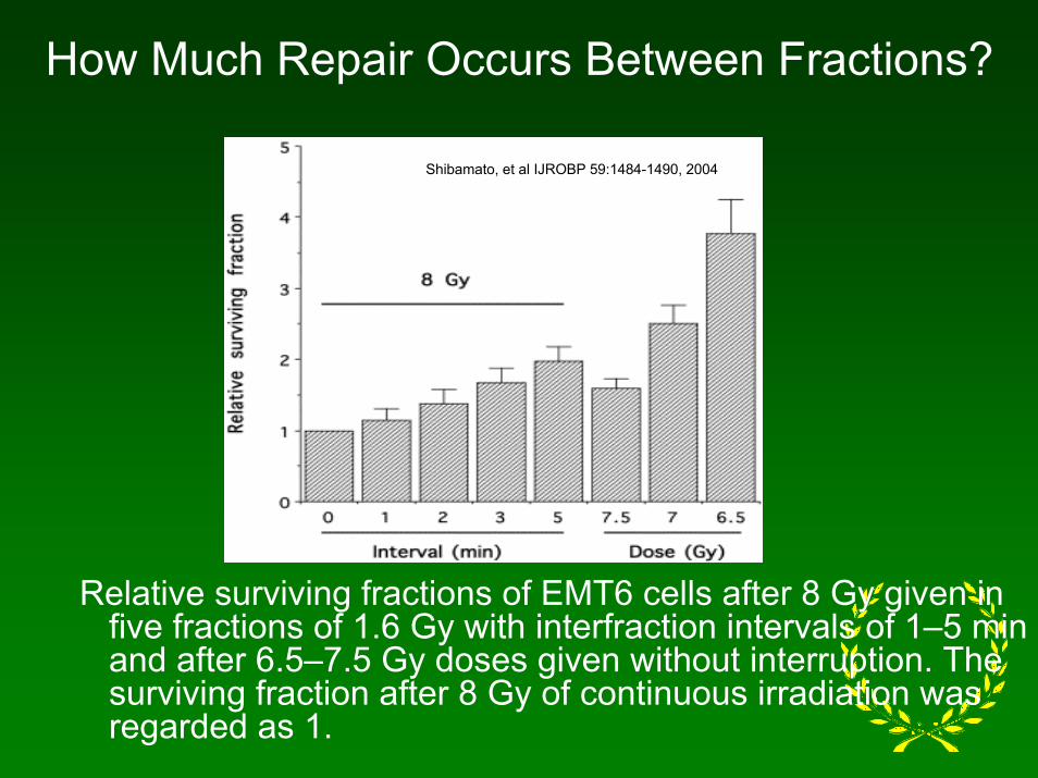
How Much Repair Occurs Between Fractions?
Shibamato, et al IJROBP 59:1484-1490, 2004
Relative surviving fractions of EMT6 cells after 8 Gy given in five fractions of 1.6 Gy with interfraction intervals of 1–5 min and after 6.5–7.5 Gy doses given without interruption. The surviving fraction after 8 Gy of continuous irradiation was regarded as 1.
Oxygen Effect & Oxygen Enhancement Ratio
EJ Hall, Radiobiology for the Radiologist, 2nd ed, p82, 1978
EJ Hall, Radiobiology for the Radiologist 5th ed. p95, 2000
The Oxygen Effect andEffect of Hypoxia on Repair
1. Contradictory evidence suggests that repair of sublethal damage may be inhibited by hypoxia.
Therefore, hypoxic tumor cells may be less able to repair radiation damage than the surrounding normal tissue thereby permitting therapeutic gain.
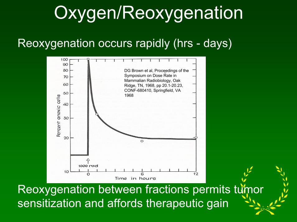
Oxygen/ReoxygenationReoxygenation occurs rapidly (hrs - days)
Reoxygenation between fractions permits tumor sensitization and affords therapeutic gain
DG Brown et al, Proceedings of the Symposium on Dose Rate in Mammalian Radiobiology, Oak Ridge, TN, 1968, pp 20.1-20.23, CONF-680410, Springfield, VA 1968
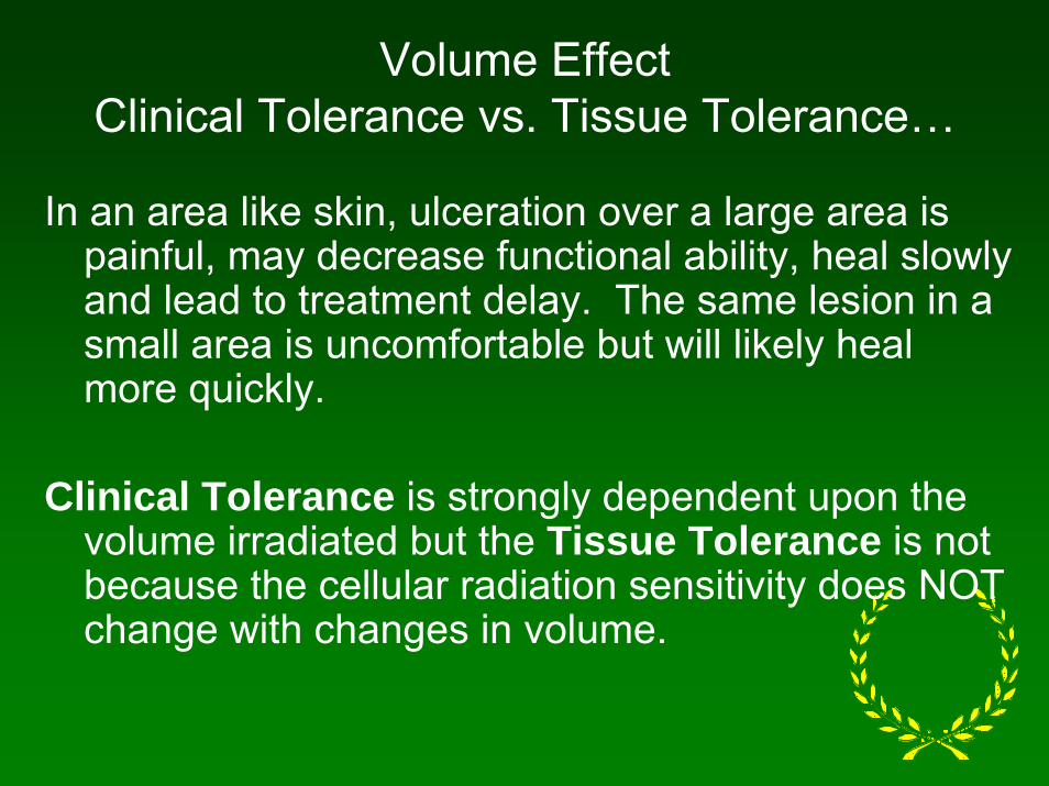
Volume EffectClinical Tolerance vs. Tissue Tolerance…
In an area like skin, ulceration over a large area is painful, may decrease functional ability, heal slowly and lead to treatment delay. The same lesion in a small area is uncomfortable but will likely heal more quickly.
Clinical Tolerance is strongly dependent upon the volume irradiated but the Tissue Tolerance is not because the cellular radiation sensitivity does NOT change with changes in volume.
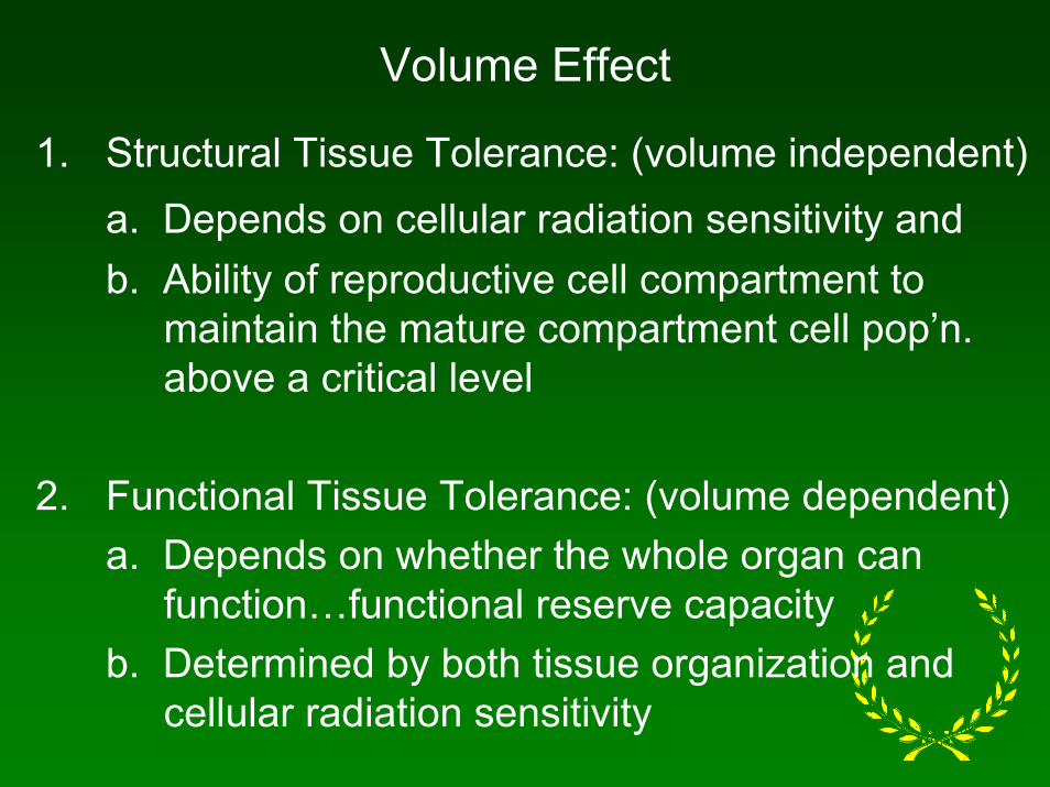
Volume Effect
1. Structural Tissue Tolerance: (volume independent)a. Depends on cellular radiation sensitivity andb. Ability of reproductive cell compartment to
maintain the mature compartment cell pop’n. above a critical level
2. Functional Tissue Tolerance: (volume dependent)a. Depends on whether the whole organ can
function…functional reserve capacityb. Determined by both tissue organization and
cellular radiation sensitivity
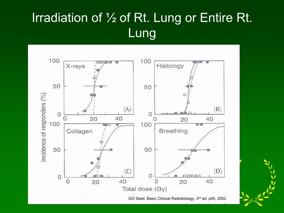
Irradiation of ½ of Rt. Lung or Entire Rt. Lung
GG Steel, Basic Clinical Radiobiology, 3rd ed, p45, 2002
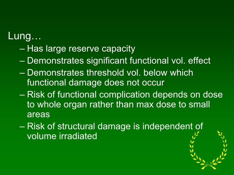
Lung…– Has large reserve capacity– Demonstrates significant functional vol. effect – Demonstrates threshold vol. below which
functional damage does not occur– Risk of functional complication depends on dose
to whole organ rather than max dose to small areas
– Risk of structural damage is independent of volume irradiated
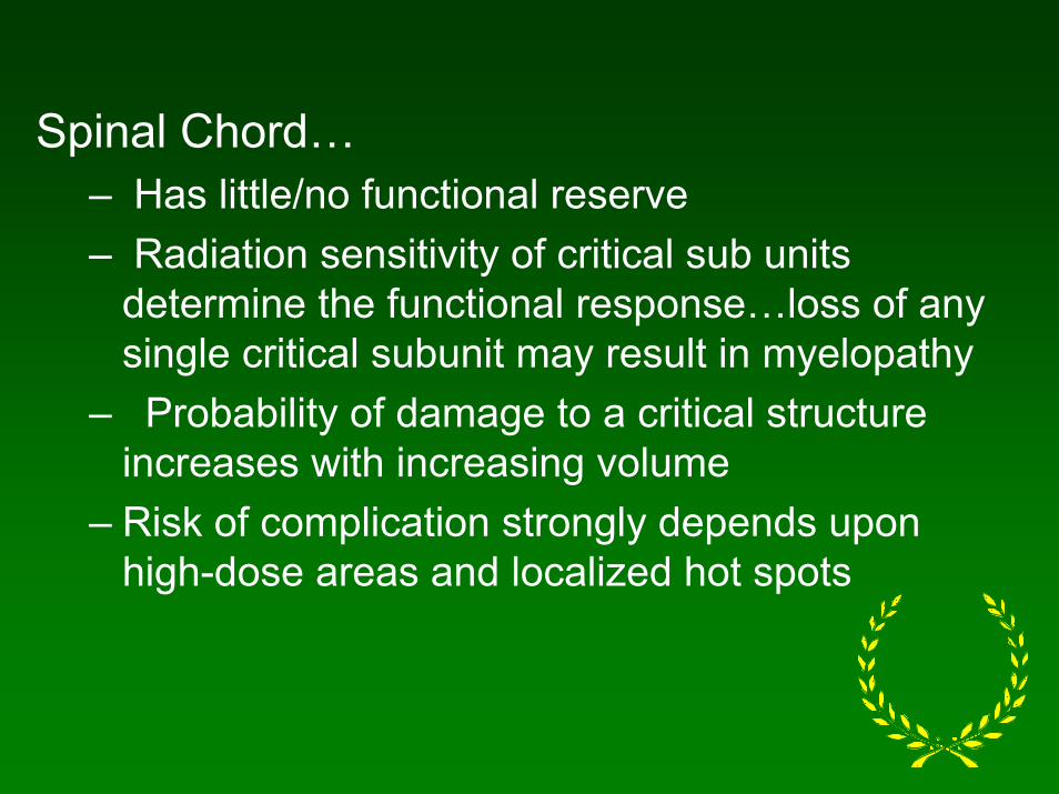
Spinal Chord…– Has little/no functional reserve – Radiation sensitivity of critical sub units
determine the functional response…loss of any single critical subunit may result in myelopathy
– Probability of damage to a critical structure increases with increasing volume
– Risk of complication strongly depends upon high-dose areas and localized hot spots
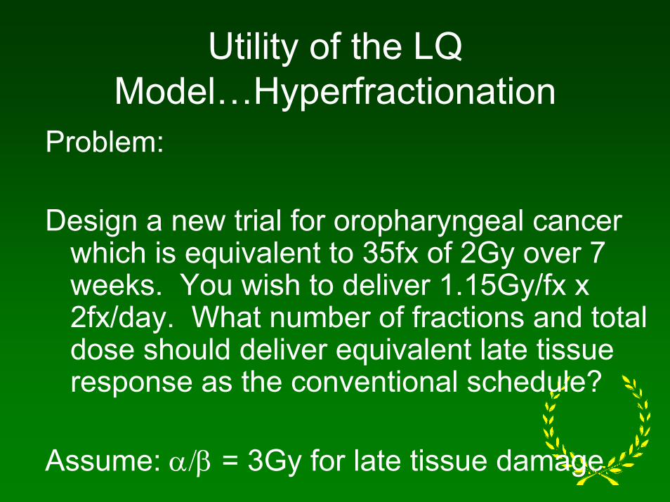
Utility of the LQ Model…Hyperfractionation
Problem:
Design a new trial for oropharyngeal cancer which is equivalent to 35fx of 2Gy over 7 weeks. You wish to deliver 1.15Gy/fx x 2fx/day. What number of fractions and total dose should deliver equivalent late tissue response as the conventional schedule?
Assume: α/β = 3Gy for late tissue damage
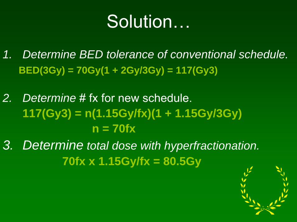
Solution…
1. Determine BED tolerance of conventional schedule.BED(3Gy) = 70Gy(1 + 2Gy/3Gy) = 117(Gy3)
2. Determine # fx for new schedule.117(Gy3) = n(1.15Gy/fx)(1 + 1.15Gy/3Gy)
n = 70fx3. Determine total dose with hyperfractionation.
70fx x 1.15Gy/fx = 80.5Gy
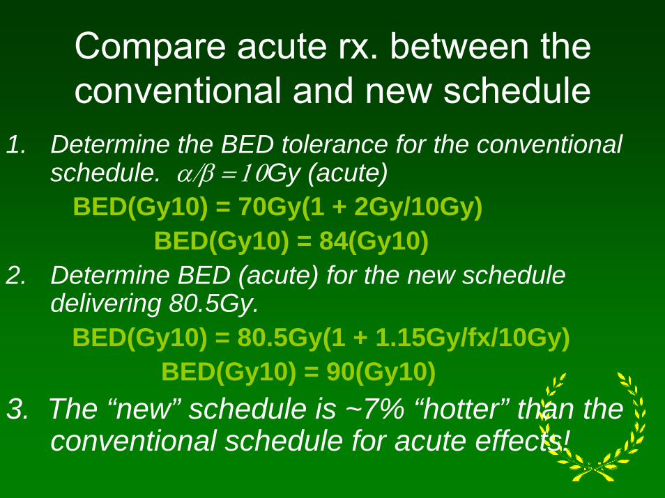
Compare acute rx. between the conventional and new schedule
1. Determine the BED tolerance for the conventional schedule. α/β = 10Gy (acute)
BED(Gy10) = 70Gy(1 + 2Gy/10Gy)BED(Gy10) = 84(Gy10)
2. Determine BED (acute) for the new schedule delivering 80.5Gy.
BED(Gy10) = 80.5Gy(1 + 1.15Gy/fx/10Gy)BED(Gy10) = 90(Gy10)
3. The “new” schedule is ~7% “hotter” than the conventional schedule for acute effects!
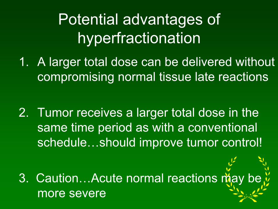
Potential advantages of hyperfractionation
1. A larger total dose can be delivered without compromising normal tissue late reactions
2. Tumor receives a larger total dose in the same time period as with a conventional schedule…should improve tumor control!
3. Caution…Acute normal reactions may be more severe
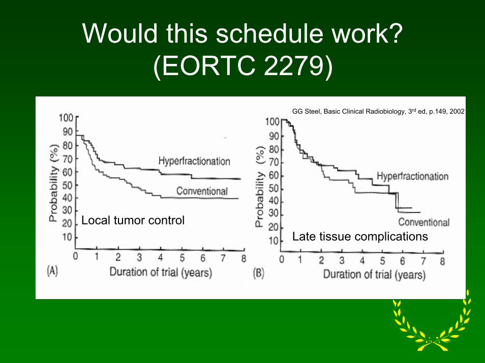
Would this schedule work?(EORTC 2279)
Local tumor controlLate tissue complications
GG Steel, Basic Clinical Radiobiology, 3rd ed, p.149, 2002
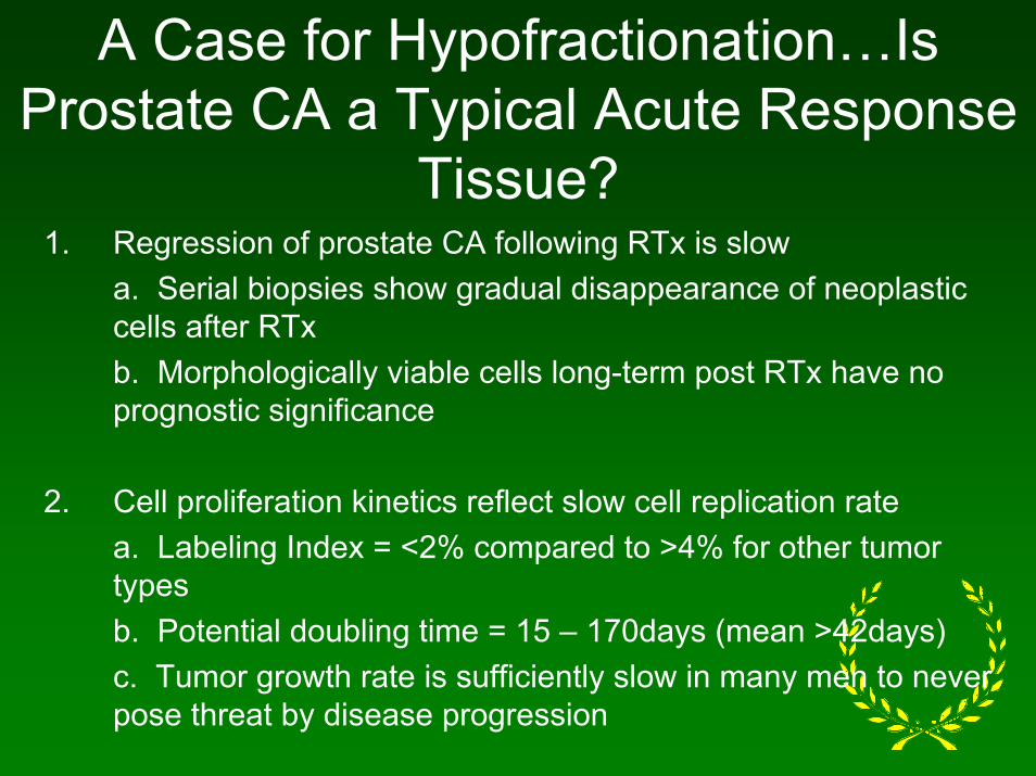
A Case for Hypofractionation…Is Prostate CA a Typical Acute Response
Tissue?1. Regression of prostate CA following RTx is slow
a. Serial biopsies show gradual disappearance of neoplastic cells after RTxb. Morphologically viable cells long-term post RTx have no prognostic significance
2. Cell proliferation kinetics reflect slow cell replication ratea. Labeling Index = <2% compared to >4% for other tumor typesb. Potential doubling time = 15 – 170days (mean >42days)c. Tumor growth rate is sufficiently slow in many men to never pose threat by disease progression
Is Prostate CA a Typical Acute Response Tissue? – cont’d
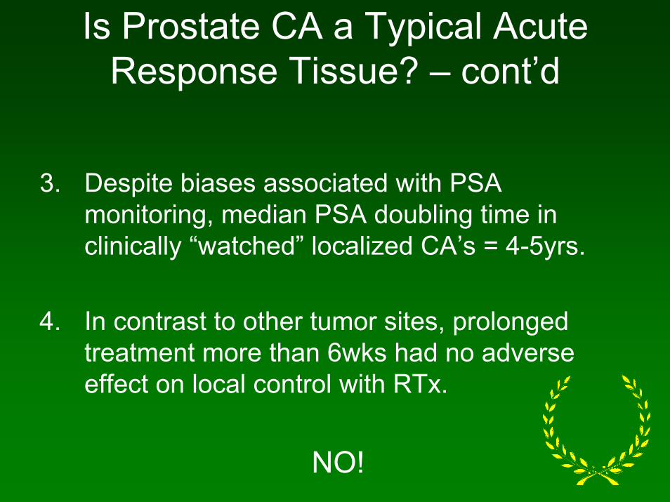
3. Despite biases associated with PSA monitoring, median PSA doubling time in clinically “watched” localized CA’s = 4-5yrs.
4. In contrast to other tumor sites, prolonged treatment more than 6wks had no adverse effect on local control with RTx.
NO!
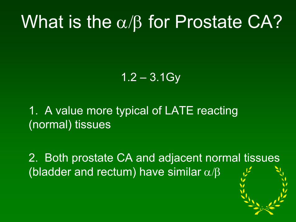
What is the α/β for Prostate CA?
1.2 – 3.1Gy
1. A value more typical of LATE reacting (normal) tissues
2. Both prostate CA and adjacent normal tissues (bladder and rectum) have similar α/β
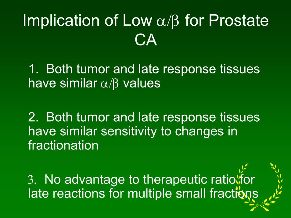
Implication of Low α/β for Prostate CA
1. Both tumor and late response tissues have similar α/β values
2. Both tumor and late response tissues have similar sensitivity to changes in fractionation
3. No advantage to therapeutic ratio for late reactions for multiple small fractions
Implication of Low α/β for Prostate CA – cont’d
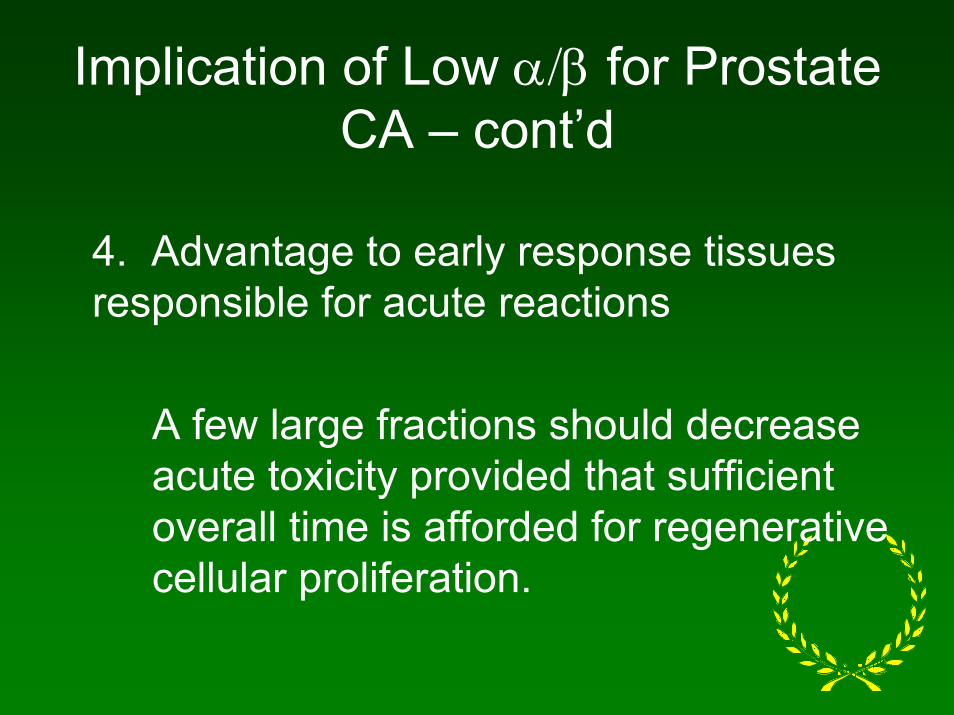
4. Advantage to early response tissues responsible for acute reactions
A few large fractions should decrease acute toxicity provided that sufficient overall time is afforded for regenerative cellular proliferation.
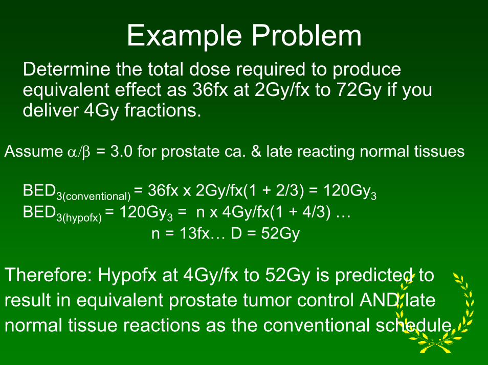
Example ProblemDetermine the total dose required to produce equivalent effect as 36fx at 2Gy/fx to 72Gy if you deliver 4Gy fractions.
Assume α/β = 3.0 for prostate ca. & late reacting normal tissues
BED3(conventional) = 36fx x 2Gy/fx(1 + 2/3) = 120Gy3BED3(hypofx) = 120Gy3 = n x 4Gy/fx(1 + 4/3) …
n = 13fx… D = 52Gy
Therefore: Hypofx at 4Gy/fx to 52Gy is predicted toresult in equivalent prostate tumor control AND latenormal tissue reactions as the conventional schedule
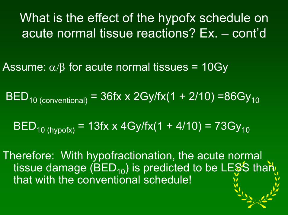
What is the effect of the hypofx schedule on acute normal tissue reactions? Ex. – cont’d
Assume: α/β for acute normal tissues = 10Gy
BED10 (conventional) = 36fx x 2Gy/fx(1 + 2/10) =86Gy10
BED10 (hypofx) = 13fx x 4Gy/fx(1 + 4/10) = 73Gy10
Therefore: With hypofractionation, the acute normal tissue damage (BED10) is predicted to be LESS than that with the conventional schedule!
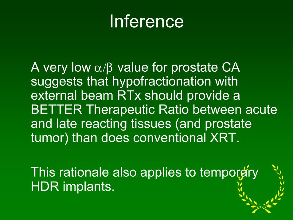
Inference
A very low α/β value for prostate CA suggests that hypofractionation with external beam RTx should provide a BETTER Therapeutic Ratio between acute and late reacting tissues (and prostate tumor) than does conventional XRT.
This rationale also applies to temporary HDR implants.
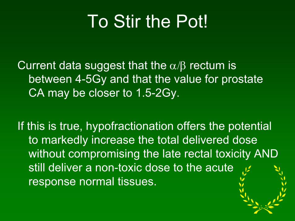
To Stir the Pot!
Current data suggest that the α/β rectum is between 4-5Gy and that the value for prostate CA may be closer to 1.5-2Gy.
If this is true, hypofractionation offers the potential to markedly increase the total delivered dose without compromising the late rectal toxicity AND still deliver a non-toxic dose to the acute response normal tissues.
Consequences of Low α/β• Should produce tumor control and late
responses as good as or better than conventional RTx for:
a. EBRT with intensity modulationb. EBRT and HDR boostc. HDR monotherapy
2. Early sequelae may be reduced because the acute response tissues have a larger α/β than the prostatic CA
3. Fewer EBRT fractions
CAUTIONS
1. Must establish appropriate treatment doses using a low value for α/β if equivalent tumor control to conventional RTx is desired.
2. Extreme fractionation (1-2fx) is unwise as it limits reoxygenation.
3. With EBRT, setup errors further mitigate against 1 - 2 fx.