Embed Size (px)
Citation preview

For Review O
nly
FIRST DETECTION OF WHITE SPOT SYNDROME VIRUS
(WSSV) IN WILD MUDCRAB Scylla spp. (de Haan, 1883)
FROM SETIU WETLANDS, MALAYSIA.
Journal: Songklanakarin Journal of Science and Technology
Manuscript ID SJST-2017-0056.R1
Manuscript Type: Original Article
Date Submitted by the Author: 17-Jul-2017
Complete List of Authors: Norizan, Nurshafiqah; University Malaysia Terengganu, School of Fisheries
and Aquaculture Sciences Sharoum, Faizah; University Malaysia Terengganu, School of Fisheries and Aquaculture Sciences Hassan, Marina; institute of Tropical Aquaculture, Musa, Najiah; Universiti Malaysia Terengganu, Musa, Nadirah; Universiti Malaysia Terengganu, Dept Fisheries Science Abdul Wahid, Mohd Effendy; University Malaysia Terengganu, School of Fisheries and Aquaculture Sciences Zainathan, Sandra Catherine; University Malaysia Terengganu, School of Fisheries and Aquaculture Sciences; Universiti Malaysia Terengganu, Institute of Marine Biotechnology
Keyword: mudcrab, Scylla spp., White Spot Syndrome Virus, PCR, Setiu Wetlands
For Proof Read only
Songklanakarin Journal of Science and Technology SJST-2017-0056.R1 Zainathan

For Review O
nly
1
FIRST DETECTION OF WHITE SPOT SYNDROME VIRUS (WSSV) IN WILD
MUDCRAB Scylla spp. (de Haan, 1883) FROM SETIU WETLANDS, MALAYSIA.
NURSHAFIQAH NORIZAN
1,2,FAIZAH SHAHAROM-HARRISON
1, MARINA HASSAN
3, NAJIAH
MUSA1, NADIRAH MUSA
1, MUHD EFFENDY ABD WAHID
1,2, SANDRA CATHERINE
ZAINATHAN1,2*
1School of Fisheries and Aquaculture Sciences, 21030 Universiti Malaysia Terengganu, Malaysia
2Institute of Marine Biotechnology, 21030 Universiti Malaysia Terengganu, Malaysia 3Institute of Tropical Aquaculture, 21030 Universiti Malaysia Terengganu, Malaysia
*Corresponding author: [email protected] / Office: +6096685028/ Mobile: +60179261392/ Fax:
+6096685002
Abstract
In this study, tissue samples from 90 wild mudcrabs (Scylla spp. including S. olivacea, S.
tranquebarica and S. paramamosain) collected during pre-monsoon, monsoon and post-
monsoon in Setiu Wetlands, Terengganu, Malaysia were screened for the presence of white spot
syndrome virus (WSSV) by PCR. This study was conducted to detect the presence or absence of
WSSV in wild Scylla spp. from Setiu Wetlands at different time of sampling. WSSV DNA was
detected in 36% of the mudcrabs. The DNA sequence of a 941 bp genome region amplified from
a crab by PCR was identified to be most similar (99% nucleotide sequence, 98% amino acid
sequence) to a WSSV strain detected in Mexico (KU216744.1) and Taiwan WSSV 419 strain
(AY850066.1). The data indicate that mudcrabs in the Setiu Wetland might act as a WSSV
reservoir of risk to shrimp aquaculture. Our findings are the first detection of WSSV form wild
mudcrabs, Scylla spp. from Setiu Wetlands, Terengganu, Malaysia.
Keywords: Mudcrab, Scylla spp., White Spot Syndrome Virus, PCR, Setiu Wetlands
Page 2 of 25
For Proof Read only
Songklanakarin Journal of Science and Technology SJST-2017-0056.R1 Zainathan
123456789101112131415161718192021222324252627282930313233343536373839404142434445464748495051525354555657585960

For Review O
nly
2
Introduction
The 23,000ha Setiu Wetland resides between the Setiu River Basin and larger Setiu-Chalok-
Bari-Merang Basin Wetland in Terengganu on the east coast of the Malaysian Peninsular. The
wetland possess high biodiversity and unique ecosystems including mangrove swamps and
supports small-scale but economically-important oyster farming and brackish water cage, pond
and pen culture of Scylla spp. mudcrabs (Azwad, 2013). Scylla spp. present in intertidal zones
and estuaries in the Setiu Wetland are dominated by Scylla olivacea (S. olivacea)(Herbst, 1796),
followed by Scylla tranquebarica (S. tranquebarica) (Fabricius, 1798) and Scylla paramamosain
(S. paramamosain) (Estampador, 1949) (Keenan, Davie, & Mann, 1998; Zaidi, Hilmi,
Ikhwanuddin, & Bachok, 2011).
Mudcrab culture is widely extensive with extremely low input and it has been accompanied with
the development of many infectious disease, especially due to viruses, which cause mass
mortalities during disease outbreak resulting in vast economic loss. Many new diseases have
been reported with significant pathogenicity and high impact to the production of mudcrab
across the world including White Spot Baculovirus (WSBV), White Spot Syndrome Virus
(WSSV), Mud Crab Reovirus (MCRV) and Muscle Necrosis Virus (MNV) (Anderson & Prior,
1992; Chen, Lo, Chiu, Chang, & Kou, 2000; Song et al., 2007; Weng, Guo, Sun, Chan, & He,
2007; Jithendran, Poornima, Balasubramanian, & Kulasekarapandian, 2010; Somboonna et al.,
2010; Owen, Liessmann, La Fauce, Nyugen, & Zeng, 2010; Bonami, & Zhang, 2011; Liu, Qian,
& Yan, 2011; Deng et al., 2012).
One of the most severe viral infections in Scylla sp. is the WSSV infection. WSSV, which was
discovered in 1992 in Southeast Asia, has become one of the most serious viral pathogen (Flegel,
2006). To date, no decapod crustacean from marine and brackish or freshwater sources has been
Page 3 of 25
For Proof Read only
Songklanakarin Journal of Science and Technology SJST-2017-0056.R1 Zainathan
123456789101112131415161718192021222324252627282930313233343536373839404142434445464748495051525354555657585960

For Review O
nly
3
reported to be resistant (Flegel, 1997; Lightner, 1996; Lo & Kou, 1998; Maeda et al., 2000;
Stentiford, Bonami, & Alday-Sanz, 2009). WSSV has a wide host range and it has been observed
not only in shrimp but also in crabs and other arthropods (Lo et al., 1996a; Lo et al., 1996b).
Among these carriers, mudcrabs have been considered to be a threat to the shrimp farms because
they are generally believed [based on a report for Scylla serrata (Forskål, 1775)] to be highly
tolerant to WSSV and remain infected for a long period of time without signs of disease (Flegel,
2006; Somboonna et al., 2010). WSSV can be reisolated from previously infected mudcrabs
(Villareal, 2005; Flegel, 2006; Somboonna et al., 2010). S. olivacea and S. tranquebarica have
been reported to be susceptible to WSSV infection (Somboonna et al., 2010). S. olivacea and S.
paramamosain showed wide variation in response to challenge with WSSV as S. olivacea was
shown to be more susceptible than S. paramamosain (Somboonna et al., 2010).
The East Coast fisherman in Setiu Wetlands depend on wild species such as Portunus pelagicus,
Scylla spp., Squilla mantis, Macrobrachium lancesteri and cultured species, Litopenaeus
vannamei for their living stipend. An outbreak of WSSV infection has affected one of the shrimp
farm in Setiu Wetlands in March 2015 (Unpublished). This incident has raised questions
whether: the WSSV infections could have been transmitted horizontally via water to the
surrounding rivers and wetlands area or WSSV infected wild crustaceans had transmitted the
disease to the cultured farm.
Wild organisms in natural reservoirs might represent a potential source of viral disease, thus it is
necessary to understand the dynamics of viruses in the environment, in terms of the mode of
infection, transmission and natural reservoir, which is dependent on its prevalence (Macias-
Rodriguez et al., 2014). Thus, this paper describes the first detection of WSSV in wild Scylla
spp. from Setiu Wetlands, Terengganu, Malaysia.
Page 4 of 25
For Proof Read only
Songklanakarin Journal of Science and Technology SJST-2017-0056.R1 Zainathan
123456789101112131415161718192021222324252627282930313233343536373839404142434445464748495051525354555657585960

For Review O
nly
4
Materials and Method
Sample collection
A total of 90 samples of wild Scylla spp. including Scylla olivacea (S. olivacea) (n = 69), Scylla
tranquebarica (S. tranquebarica) (n = 8) and Scylla paramamosain (S. paramamosain) (n = 13)
were collected from Setiu Wetlands, Terengganu (N 05° 40.709, E 102° 42.774). The sampling
trips were conducted during the post-monsoon (March 2015), pre-monsoon (August 2015) and
monsoon seasons (December 2015). The water quality parameters (pH, temperature, salinity and
dissolved oxygen) were measured using YSI Multiparameter Water Quality Meter and API®
Freshwater Aquarium Master Test Kit (Mars Fishcare North America, Inc). The samples were
kept in polystyrene box and brought back alive to laboratory of Aquatic Organisms Health in
Universiti Malaysia Terengganu. The length and weight of the samples were measured and any
external and internal clinical signs were observed. The gills and internal organs (hepatopancreas,
stomach and heart) were collected for sample processing and further analysis by PCR was
conducted based on Lo et al. (1996a, 1996b) and Nunan and Lightner (2011).
DNA extraction
The DNA extraction of the samples was conducted using NucleoSpin Tissue Extraction Kit
(Macherey-Nagel) according to the protocol provided by the manufacturers. About 25 mg of the
pooled organs (hepatopancreas, heart and stomach) and gills were cut up to small pieces and
placed in a 1.5 mL microcentrifuge tube. The extracted DNA was stored at -20°C prior to PCR
analysis.
Page 5 of 25
For Proof Read only
Songklanakarin Journal of Science and Technology SJST-2017-0056.R1 Zainathan
123456789101112131415161718192021222324252627282930313233343536373839404142434445464748495051525354555657585960

For Review O
nly
5
PCR amplification
The conventional nested PCR was carried out according to the method by Lo et al. (1996) a and
b as described in OIE (2015). The primers were based on the sequence published by Lo et al.
(1996a, 1996b): Forward primer 146F1- (5’-ACT-ACT-AAC-TTC-AGC-CTA-TCTAG-3’) and
reverse primer 146R1- (5’-TAA-TGC-GGG-TGT-AAT-GTT-CTT-ACG-A-3’) for primary
reaction with the expected size of 1447 bp. Followed by the second primer set 146F2 - (5’-GTA-
ACT-GCC-CCT-TCC-ATC-TCC-A-3’) and 146R2 - (5’-TAC-GGC-AGC-TGC-TGC-ACC-
TTG-T-3’) in the nested reaction which were expected to yield an amplicon of 941 bp. A total of
24 µL PCR mixture containing: 12.5 µL 2X MyTaqMix (Bioline), 10.5 µL Rnase-free water, 0.5
µL 146F1 (10 µM) and 0.5 µL 146R1 (10 µM) were added to 1 µL extracted DNA. For the
primary reaction, the PCR thermal profile was programmed as followed: a cycle of 94°C for 4
minutes, followed by 39 cycles of 94°C for 1 minute, 55°C for 1 minute, and 72°C for 2 minutes
and a final 5-minutes extension at 72°C.
The nested reaction was carried out with a total of 24 µL PCR mixture containing: 12.5 µL 2X
MyTaqMix (Bioline), 10.5 µL Rnase-free water, 0.5 µL 146F2 (100 µM) and 0.5 µL 146R2
(100 µM) were added to 1 µL PCR product. The amplification was conducted with the following
programme: a cycle of 94°C for 4 minutes, followed by 39 cycles of 94°C for 1 minute, 55°C for
1 minute, and 72°C for 2 minutes and a final 5-minutes extension at 72°C. The amplified PCR
products from both reactions were visualized by gel electrophoresis through 2% (w/v) agarose
gel in TAE buffer and stained with SYBR®
Safe DNA gel stain (Invitrogen) for 45 minutes at 70
volt.
Page 6 of 25
For Proof Read only
Songklanakarin Journal of Science and Technology SJST-2017-0056.R1 Zainathan
123456789101112131415161718192021222324252627282930313233343536373839404142434445464748495051525354555657585960

For Review O
nly
6
Gel extraction and DNA sequencing analysis
Each sample representative from PCR positive samples from each host species was chosen for
sequencing analysis to examine the sequence difference of WSSV between the different host
species.The expected bands from sample 5 from site 5 (S5-5, host species: S. paramamosain),
sample 6 from site 5 (S5-6, host species: S. olivacea) and sample 1 from site 3 (S3-1, host
species: S. tranquebarica) were excised and purified using NucleoSpin® Gel and PCR Clean-up
(Macherey-Nagel) based on the standard protocols. The purified DNA was diluted into 10 ngµL-
1 as required by the protocol before it was sent for sequencing to First Base Laboratory
(Selangor, Malaysia).
Phylogenetic analysis
The DNA sequencing results were used for the phylogenetic analysis. The sequences were used
to interrogate the NCBI BLAST database to confirm its likely identity. Then the multiple
alignment were aligned using Clustal X2.0.12 with other WSSV- related sequences. Finally, the
phylogenetic tree was inferred among the sequences of WSSV obtained in this study with known
sequences contained in GenBank using Molecular Evolutionary Genetics Analysis (MEGA).
Statistical analysis
The effects of time of sampling on the prevalence of virus was determined using one-way
ANOVA analysis. ANOVA analyses were performed using SPSS®
statistic software version
19.0. Values were identified as significantly different if p < 0.05. The Tukey HSD post hoc test
Page 7 of 25
For Proof Read only
Songklanakarin Journal of Science and Technology SJST-2017-0056.R1 Zainathan
123456789101112131415161718192021222324252627282930313233343536373839404142434445464748495051525354555657585960

For Review O
nly
7
was applied at a significance level of α ≤ 0.05, to determine differences between the explanatory
variable.
Results
No clinical signs were observed on all of the samples; externally and internally. A total of 11
samples were detected with the presence of Octolasmis sp. on their gills. The average weight
and carapace width of the samples were tabulated according to the different seasons (Table 1).
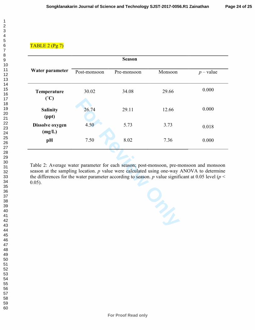
Water parameter at sampling location
A significant difference was observed between the water parameter and the three seasons (Table
2). These results indicated the occurrence of fluctuations in water parameters during the three
seasons.
Conventional nested PCR for the detection of WSSV inScylla spp.
A total of 33 samples (36%) of Scylla olivacea (S. olivacea), Scylla tranquebarica (S.
tranquebarica) and Scylla paramamosain (S. paramamosain) sampled during post-monsoon,
pre-monsoon and monsoon season were found to be positive for the presence of WSSV. Higher
number of positives were detected during the pre-monsoon and monsoon season in Setiu
Wetlands; 26 samples than post-monsoon season, 7 samples. Based on the results, samples of S.
olivacea showed the highest presence (75.7%) of WSSV compared to S. paramamosain (12.1%)
and S. tranquebarica (12.1%) (Table 3).The primary reaction of conventional nested PCR
analysis of pooled gills and tissues (hepatopancreas, heart and stomach) demonstrated negative
results for the presence of WSSV (Figure 1). The secondary reaction of conventional nested PCR
Page 8 of 25
For Proof Read only
Songklanakarin Journal of Science and Technology SJST-2017-0056.R1 Zainathan
123456789101112131415161718192021222324252627282930313233343536373839404142434445464748495051525354555657585960

For Review O
nly
8
analysis of pooled samples of gills and tissues (hepatopancreas, heart and stomach) demonstrated
positive band at expected size of 941bp for the presence of WSSV (Figure 1).
A significant difference was observed between the season and the prevalence of WSSV as
determined by one-way ANOVA (p< 0.05; p = 0.01). Tukey post-hoc test revealed that the
prevalence of WSSV infection demonstrated a significant difference between the pre-monsoon
and monsoon season (α < 0.05; α = 0.008). The total prevalence of WSSV infection was not
influenced by the different number of mud crabs sampled during post-monsoon season (n=25)
pre-monsoon season (n=45) and monsoon season (n=20). Comparison of total positives for
WSSV infection based on the season showed highest prevalence during monsoon season (Figure
2).
Sequencing and phylogenetic analysis.
A multiple alignment of nucleotide sequences of the amplified PCR products confirmed that
sample S5-5 ( S. paramamosain), S5-6 (S. olivacea) and S3-1(S. tranquebarica) were from the
same member of WSSV. These results confirmed that all the positive samples was from the same
member of WSSV. S5-6 (S. olivacea) and S3-1(S. tranquebarica) showed 99% while S5-5 (S.
paramamosain) showed 98% sequence similarity to the whole genome of WSSV Mexican strain
WSSV-MX08 (GenBank accession no. KU216744.1).
Multiple alignment of nucleotide sequences of the amplified PCR product confirmed that
samples S5-5 (S. paramamosain) were from the same lineage with WSSV Mexican strain
(WSSV-MX08) (Rodriguez-Anaya et al., 2016), samples S5-6 (S. olivacea) with Taiwan
WSSV419 strain (Reville et al., 2005) and samples S3-1 (S. tranquebarica) with WSSV
complete genome strain (van Hulten et al., 2001) with minor variations (Figure 3).
Page 9 of 25
For Proof Read only
Songklanakarin Journal of Science and Technology SJST-2017-0056.R1 Zainathan
123456789101112131415161718192021222324252627282930313233343536373839404142434445464748495051525354555657585960

For Review O
nly
9
Discussion
The water quality parameters which was taken at each of the sampling location include
temperature, salinity, dissolve oxygen (DO) and pH. The temperature range (28°C - 32°C) was
normal for tropical coastal waters (Alongi et al., 2009). The lowest salinity was recorded at site
3, in the downstream of Setiu Wetlands (combination of freshwater and seawater) during the
monsoon season. The highest salinity was recorded at sampling site 6, which was located in the
inshore area, during the pre-monsoon. Scylla spp. are not known to be affected by elevated
salinity due to their high tolerancy to wide range of salinities (FAO, 2011). The dissolve oxygen
(DO) was in range of 3.58 mgL-1
to 5.74 mgL-1
with the lowest DO recorded due to aquaculture
activities at the site. Aquaculture discharges contain high levels of organic matter such as feed
and faeces. The degradation of organic matter by microorganism will consume more oxygen,
hence, lowering the DO content in water (Hargrave, Duplisea, Pfeiffer, & Wildish, 1993) and
Scylla spp. can tolerate low oxygen level (FAO, 2011). The results for the water parameters were
consistent with previous study regarding water quality at Setiu Wetlands where aquaculture
activities can affect water quality parameter (Suratman, Hussein, Latif, & Weston, 2014).
A total of 33 samples of Scylla spp. (36%) including S. olivacea, S. tranquebarica and S.
paramamosain sampled from March to December 2015 demonstrated positive result for the
presence of WSSV. The result from this study is consistent with several papers which showed
positive result for WSSV infection in Scylla spp. using nested PCR (Lo et al., 1997; Otta et al.,
1999; Chen et al., 2000; Vaseeharan, Jayakumar, & Ramasamy, 2003; Somboonna et al., 2010;
Page 10 of 25
For Proof Read only
Songklanakarin Journal of Science and Technology SJST-2017-0056.R1 Zainathan
123456789101112131415161718192021222324252627282930313233343536373839404142434445464748495051525354555657585960

For Review O
nly
10
Liu et al., 2011; Joseph, Roswin, Anbu Rajan, Surendran, & Lalitha, 2015). Natural WSSV
infections have been found in wild-caught and farmed mud crabs in various stages in many
countries of Asiatic region (Lo et al., 1996a; Flegel, 1997; Flegel, 2006; Kou, Peng, Chiu, & Lo,
1998; Otta et al., 1999; Lavilla-Pitogo, Marcial, Pedrajas, Quinitio, & Millamena, 2001). Natural
benthic larvae of wild Scylla serrata were found to be positive for WSSV infection at 60%
prevalence (Lo et al., 1997). WSSV infection (60% prevalence) was detected in benthic larvae of
wild mud crab, Scylla serrata captured from Taiwan’s coastal water (Chen et al., 2000). WSSV
was detected on diseased cultured mud crabs collected from Zhejiang Province of China during
2006–2008 which showed a prevalence of 34.82% (Liu et al., 2011).
The number of WSSV positive Scylla spp. were detected throughout the three seasons; monsoon
season in December (14%), pre-monsoon season in August (14%) and monsoon season in March
(8%). Consistent prevalence for WSSV infections in mud crab population captured in August
and September were shown by Chen et al. (2000). The results of one-way ANOVA showed that
there was a significant difference between the three seasons and the prevalence of WSSV
infection in wild mud crab, Scylla spp. This result is consistent with Chen et al. (2000) that stated
the infection rate of WSSV might be persistent throughout the year (Chen et al., 2000). The high
prevalence of WSSV infection during monsoon season demonstrated that WSSV outbreak is
affected by the change of environmental factors; which will depress the immune system of
crustacean (Gunalan, Soundarapandian, & Dinakaran, 2010). Temperature is the most important
environmental factor as it has a profound effect on disease expression. The average water
temperatures between 18°C to 30°C can play an important role in inducing outbreaks of WSSV
(Vidal, Granja, Aranguren, Brock, & Salazar, 2001). This study showed that the water
temperature during monsoon season is lower compared to post-monsoon and pre-monsoon
Page 11 of 25
For Proof Read only
Songklanakarin Journal of Science and Technology SJST-2017-0056.R1 Zainathan
123456789101112131415161718192021222324252627282930313233343536373839404142434445464748495051525354555657585960

For Review O
nly
11
season due to the incoming freshwater during the rainy season (Sankar, Ramamoorthy,
Sakkaravarthi, & Vanitha, 2011). Low water temperature has been known to influence the
dispersal of WSSV (Jiravanichpaisal, Soderhall, & Soderhall, 2004; Gunalan et al., 2010).
A BLAST search of the WSSV_S5-5 (S. paramamosain) segments showed 98% sequence
identity with MX08 (KU216744.1), CN01 (KT995472.1), CN02 (KT995470.1), CN03
(KT995471.1), EG3 (KR083866.1), K-LV1 (JX515788.1), Taiwan WSSV 419 (AY850066.1),
CM-VN1 (JX564899.1), White Spot Syndrome Virus with partial sequence (AF361753.1) and
WSSV complete genome (AF369029.1). The published sequences compared to S5-5 (S.
paramamosain) were mostly sequences from cultured white shrimp, Litopenaeus vannamei (L.
vannamei). WSSV_S5-6 (S. olivacea) and WSSV_S3-1 (S. tranquebarica) were found to be
99% similar with the above published sequences. S5-5 (S. paramamosain) showed 98%
similarity with the whole genome of a WSSV Mexican strain (WSSV-MX08), which was the
first WSSV isolated from cultured L. vannamei from Mexican farms (Rodriguez-Anaya et al.,
2016). White spot syndrome virus (WSSV) was first discovered in 1992 from Taiwanese
cultured Penaeus japonicus (Flegel, 2006). Since then, WSSV has spread across Asian countries
including Vietnam, Thailand, Korea, India, China, and Malaysia (Nakano et al., 1994; Flegel,
1997; Mohan, Shankar, Kulkarni, & Sudha, 1998; Zhan et al., 1998; Wang, Nunan, & Lightner,
2000). The sequence similarity of S5-5 (S. paramamosain) to L. vannamei indicated that the
WSSV infections could have been transmitted to the wild environment from farm or vice versa.
WSSV can be transmitted horizontally and vertically (Lo & Kou, 1998; Chou, Huang, Wang, Lo,
& Kou, 1998; Otta et al., 1999). The horizontal transmission of WSSV was shown to be via
ingestion and waterborne routes (Chou et al., 1998). The water disposal from shrimp farm was
shown to transmit WSSV infectious agent into the natural enzootic areas (Lo & Kou, 1998;
Page 12 of 25
For Proof Read only
Songklanakarin Journal of Science and Technology SJST-2017-0056.R1 Zainathan
123456789101112131415161718192021222324252627282930313233343536373839404142434445464748495051525354555657585960
For Review O
nly
12
Esparza-Leal et al., 2009). A cannibalistic behavior of mud crab also contributes to the
transmission of WSSV (Lo et al., 1997). Consequently, the virus can be transmitted to whole
population of the cultured or wild crab and shrimp (OIE, 2015).
However, the phylogenetic tree analysis showed that sample WSSV_S5-6 (S. olivacea) shared a
common ancestry relationships with the Taiwan WSSV 419 strain (GenBank accession no.
AY850066.1) while sample WSSV_S3-1 (S. tranquebarica) with the WSSV complete genome
strain (GenBank accession no. AF369029.2). Based on the published sequences; WSSV Mexican
strain (WSSV-MX08) (Rodriguez-Anaya et al., 2016), Taiwan WSSV 419 strain (Reville et al.,
2005) and WSSV complete genome (van Hulten et al., 2001), it is inheritable that WSSV was
originally found in L.vannamei and Penaeus monodon. Both published sequences; Taiwan
WSSV 419 strain (Reville et al., 2005) and WSSV complete genome (van Hulten et al., 2001)
originated from Thailand. Since Thailand is one of Malaysia’s neighboring country, WSSV
could be transmitted to the crustacean population via the horizontal route of infection through
release of ship ballast water and illegal importation of live post-larvae (Lo et al., 1997; Sanchez-
Martinez, Aguirre-Guzman, & Mejia-Ruiz, 2007; Muller, Andrade, Tang-Nelson, Marques, &
Lightner, 2010).
Genetic variation have been observed among the WSSV detected from the 3 different host
species of Scylla spp.. The host species play a role in the selection of a mutant within a viral
population which led to a genetic variation of WSSV (Wang et al., 2000). The genetic variation
of WSSV in different host species could be explained where the passage of the virus through
different hosts can induce a genomic alteration and alter the pathogenicity of the virus
(Waikhom, John, George, & Jeyaseelan, 2006). The non-static properties of WSSV genome
enable itself to adapt to any environmental condition (Sablok et al., 2010). This could further be
Page 13 of 25
For Proof Read only
Songklanakarin Journal of Science and Technology SJST-2017-0056.R1 Zainathan
123456789101112131415161718192021222324252627282930313233343536373839404142434445464748495051525354555657585960

For Review O
nly
13
strengthen by previous study that the occurrence of genomic mutations resulted from the
adaptation of the virus to different environmental conditions (Waikhom et al., 2006) and due to
the mutations in viral genome of WSSV after its introduction into the different countries (Muller
et al., 2010).
Conclusions
A total of 90 samples of wild Scylla spp. including Scylla olivacea (S. olivacea), Scylla
tranquebarica (S. tranquebarica) and Scylla paramamosain (S. paramamosain) were sampled
from Setiu Wetlands, Terengganu during post-monsoon, pre-monsoon and monsoon season. The
water parameter taken from the sampling sites showed a normal range for the Scylla spp. habitat.
A total of 33 samples sampled during post-monsoon, pre-monsoon and monsoon season were
found to be positive for the presence of WSSV. The sequenced sample of S5-6 (S. olivacea) and
S3-1 (S. tranquebarica) showed high similarity (99%) while S5-5 (S. paramamosain) showed
98% to the WSSV Mexican Strain (WSSV-MX08). In conclusion, our findings are the first
detection of WSSV form wild mudcrabs, Scylla spp. from Setiu Wetlands, Terengganu, Malaysia
(a new location).
Acknowledgements
This work was funded by the projects under the Niche-area Research Grant Scheme (NRGS Vot.
No. 53131).
Page 14 of 25
For Proof Read only
Songklanakarin Journal of Science and Technology SJST-2017-0056.R1 Zainathan
123456789101112131415161718192021222324252627282930313233343536373839404142434445464748495051525354555657585960

For Review O
nly
14
REFERENCES
Alongi, D. M., McKinnon, A. D., Brinkman R, Trott, L. A., Muhammad, C. U., Muawanah &
Rachmansyah (2009). The fate of organic matter derived from small-scale fish cage
aquaculture in coastal waters of Sulawesi and Sumatra, Indonesia. Aquaculture, 295, 60-
75.
Anderson, I. G., & Prior, H. G. (1992). Baculovirus infections in the mud crab, Scylla serrata,
and a freshwater crayfish, Cherax quadricarinatus, from Australia. Journal of
Invertebrate Pathology, 60, 265-273.
Azwad, M. N. (2013). WWF World Wildlife Fund for Nature Organization Malaysia. Project
Sustainable Management of Setiu Wetlands, Setiu – A wetland wonder. Retrieved from
http://www.wwf.org.my/about_wwf/what_we_do/freshwater_main/freshwater_conservin
g_freshwater_habitats/projects_sustainable_management_of_setiu_wetlands/
Bonami, J. R., & Zhang, S. (2011). Viral Disease in commercially exploited crabs: A review.
Journal of Invertebrate Pathology, 106, 6-17.
Chen, L. L., Lo, C. F., Chiu, Y. L., Chang, C. F., & Kou, G. H. (2000). Natural and experimental
infection of white spot syndrome virus (WSSV) in benthic larvae of mud crab Scylla
serrata. Diseases of Aquatic Organisms, 40, 157-161.
Chou, H. Y., Huang, C. Y., Wang, C. H., Lo, C. F., & Kou, G. H. (1998). Studies on
transmission of white spot syndrome associated baculovirus (WSBV) in Penaeus
monodon and P. japonicus via waterborne contact and oral ingestion. Aquaculture, 164,
263–276.
Deng, X. X., Lu, L., Qu, Y. J., Su, H. J., Li, G., Guo, Z. X.,...Weng, S. (2012). Sequence analysis
of 12 genome segments of mud crab reovirus (MCRV). Virology, 422, 185-194.
Esparza-Leal, H. M., Escobedo-Bonilla, C. M., Casillas-Hernández, R., Alvarez-Ruiz, P.,
Portillo-Clark, G., Valerio-García, R. C.,...Magallón-Barajas, F. J. (2009). Detection of
white spot syndrome virus in filtered shrimp–farm water fractions and experimental
evaluation of its infectivity in Penaeus (Litopenaeus) vannamei. Aquaculture, 292, 16–
22.
Food and Agriculture Organization of the United Nation. FAO Fisheries and Aquaculture
Department. (2011). Mud crab aquaculture – A practical manual. FAO Fisheries
Technical Paper, 508, Pp. 78. Retrieved from
http://www.fao.org/docrep/015/ba0110e/ba0110e00.htm
Flegel, T. W. (1997). Major viral diseases of the black tiger prawn (Penaeus monodon) in
Thailand. Journal of Microbiology and Biotechnology, 13, 433–442.
Page 15 of 25
For Proof Read only
Songklanakarin Journal of Science and Technology SJST-2017-0056.R1 Zainathan
123456789101112131415161718192021222324252627282930313233343536373839404142434445464748495051525354555657585960

For Review O
nly
15
Flegel, T. W. (2006). Detection of major penaeid shrimp viruses in Asia, a historical
perspective with emphasis on Thailand. Aquaculture, 158, 1-33.
Gunalan, B., Soundarapandian, P., & Dinakaran, G. K. (2010). The effect of temperature and pH
on WSSV infection in cultured marine shrimp, Penaeus monodon (Fabricius). Scientific
research, 5 (1), 28-33.
Hargrave, B. T., Duplisea, D. E., Pfeiffer, E., & Wildish, D. J. (1993). Seasonal changes in
benthic fluxes of dissolved oxygen and ammonia associated with marine culture Atlantic
Salmon. Marine Ecology Progress Series, 96, 249-257.
Jiravanichpaisal, P., Soderhall, K., & Soderhall, I. (2004). Effect of water temperature on the
immune response and infectiviti pattern of white spot syndrome virus (WSSV) in
freshwater crayfish. Fish and Shellfish Immunology, 17, 265-275.
Jithendran, K. P., Poornima, M., Balsubramanian, C. P., & Kulasekarapandian, S. (2010)
Diseases of mud crabs (Scylla spp.) Indian Journal of Fisheries, 57(3), 55-63.
Joseph, T. C., Roswin, J., Anbu Rajan, L., Surendran, P. K., & Lalithaa, K. V. (2015). White
spot syndrome virus infection: Threat to crustacean biodiversity in Vembanad Lake,
India. Biotechnology Reports, 7, 51–54.
Keenan, C. P., Davie, P. J. F., & Mann, D. L. (1998). A Revision of Genus Scylla de Haan, 1833
(Crustacea: Decapoda: Brachyura: Portunidae). Raffles Bulletin of Zoology, 46, 217-
245.
Kou, G. H., Peng, S. E., Chiu, Y. L., & Lo, C. F. (1998). Tissue distribution of white spot
syndrome virus (WSSV) in shrimp and crabs. In T.W. Flegel (Eds.), Advances in Shrimp
Biotechnology (pp. 267–271). National Center for Genetic Engineering and
Biotechnology, Bangkok.
Lavilla-Pitogo, C. R., Marcial, H. S., Pedrajas, S. A. G., Quinitio, E. T., & Millamena, O. M.
(2001). Problems associated with tank-held mud crab (Scylla spp.) broodstock. Asian
Fisheries Science, 14, 217-224.
Lightner, D. V. (1996) Handbook of Shrimp Pathology and Diagnostic Procedures for Diseases
of Cultured Penaeid Shrimp.World Aquaculture Society, Baton Rouge, Louisiana.
Liu, W., Qian, D., & Yan, X. J. (2011). Studies on pathogenicity and prevalence of white spot
syndrome virus in mud crab, Scylla serrata (Forskal), in Zhejiang Province, China.
Journal of Fish Diseases, 34, 131-138.
Lo, C. F., Leu, J. H., Chen, C. H., Peng, S. E., Chen,Y. T., Chou, C. M., Yeh, P.Y., Huang, C.J.,
Chou, H.Y., Wang, C.H. & Kou, G. H. (1996a). Detection of baculovirus associated with
Page 16 of 25
For Proof Read only
Songklanakarin Journal of Science and Technology SJST-2017-0056.R1 Zainathan
123456789101112131415161718192021222324252627282930313233343536373839404142434445464748495051525354555657585960
For Review O
nly
16
White Spot Baculovirus Virus (WSBV) in penaeid shrimps using polymerase chain
reaction. Diseases of Aquatic Organisms, 25, 133-141.
Lo, C. F., Ho, C. H., Peng, S. E., Chen, C. H., Hsu, H. C., Chiu, Y.L., Chang, C.F., Liu, K.F., Su,
M.S., Wang, C.H., & Kou, G. H. (1996b.) White spot syndrome baculovirus (WSBV)
detected in cultured and captured shrimp, crab and other arthropods. Diseases of Aquatic
Organisms, 27, 215-225.
Lo, C. F., Ho, C. H., Chen, C. H., Liu, K. F., Chiu, Y. L., Yeh, P. Y., Peng, S.E., Hsu, H.C., Liu,
H.C., Chang, C.F., Su, M.S., Wang, C.H., & Kou, G. H. (1997). Detection and tissue
tropism of white spot syndrome baculovirus (WSBV) in captured brooders of Penaeus
monodon with a special emphasis on reproductive organs. Diseases of Aquatic
Organisms, 30, 53-72.
Lo, C. F., & Kou, G. H. (1998). Virus-associated White Spot Syndrome of Shrimp in Taiwan: a
review. Fish Pathology, 33, 365–371.
Macias-Rodriguez, N. A., Manon-Rios, N., Romero-Romero, J. L., Camacho-Beltran, E.,
Magallanes Tapia, M. A., Leyva-Lopez, N. E., Hernandez-Lopez, J., Magallón-Barajas,
F.J., Perez-Enriquez, R., Sanchez-Gonzalez, S., & Mendez-Lozano, J. (2014). Prevalence
of viral pathogens WSSV and IHHNV in wild organisms at the Pacific Coast of
Mexico. Journal of Invertebrate Pathology, 116, 8-12.
Maeda, M., Itami, T., Mizuki, E., Tanaka, R., Yoshizu, Y., Doi, K., Chisa, Y.A., Takahashi, Y.,
& Kawarabata, T. (2000). Red Swamp Crawfish (Procambarus clarkii): An Alternative
Experimental Host in the Study of White Spot Syndrome Virus. Acta Virologica, 44, 371–
374.
Mohan, C. V., Shankar, K. M., Kulkarni, S., & Sudha, P. M. (1998). Histopathology of cultured
shrimp showing gross signs of yellow head syndrome and white spot syndrome during
1994 Indian epizootics. Diseases of Aquatic Organisms, 34, 9–12.
Muller, I. C., Andrade, T. P. D., Tang-Nelson, K. F. J., Marques, M. R. F., & Lightner. (2010).
Genotyping of white spot syndrome virus (WSSV) geographical isolates from Brazil and
comparison to other isolates from the Americas. Diseases of Aquatic Organisms, 88, 91-
98.
Nakano, H., Koube, H., Umezawa, S., Momoyama, K., Hiraoka, M., Inouye, K., & Oseko, N.
(1994). Mass mortalities of cultured kuruma shrimp, Penaeus japonicus, in Japan in
1993: epizootiological survey and infection trails. Fish Pathology, 29, 135–139.
Nunan, L. M., & Lightner, D. V. (2011). Optimized PCR assay for detection of White Spot
Syndrome Virus (WSSV). Virology Methods, 171, 318-321.
Page 17 of 25
For Proof Read only
Songklanakarin Journal of Science and Technology SJST-2017-0056.R1 Zainathan
123456789101112131415161718192021222324252627282930313233343536373839404142434445464748495051525354555657585960

For Review O
nly
17
OIE (Office International des Epizooties/World Animal Health Organization). (2015). White
Spot Diseases. Manual of Diagnostic Test for Aquatic Animals. Retrieved from
http://www.oie.int/fileadmin/Home/eng/Health_standards/aahm/2009/2.2.05_WSD.pdf.
Otta, S. K., Shubha, G., Joseph, B., Chakraborty, A., Indrani, K., & Iddya, K. (1999).
Polymerase chain reaction (PCR) detection of White Spot Syndrome Virus (WSSV) in
cultured and wild crustaceans in India. Diseases of Aquatic Organisms, 38, 67-70.
Owen, L., Liessmann, L., La Fauce, K., Nyugen, T., & Zeng, C. (2010). Intranucleur baciliform
virus and hepatopancreatic parvovirus (PmergDNV) in the mud crab Scylla serrata
(Forskal) of Australia. Aquaculture, 310, 47-50.
Reville, C., AL-Beik, J., Meehan-Meola, D., Xu, Z. K., Goldsmith, M. L., Rand, W., & Alcivar-
Warren, A. (2005). White Spot Syndrome Virus in frozen shrimp sold at Massachusetts
supermarket. Journal of Shellfish Research, 24, 285-290.
Rodriguez-Anaya, L. Z., Gonzalez-Galaviz, J. R., Casillas-Hernandez, R., Lares-Villa, F.,
Estrada, K., Ibarra-Gamez, J. C., & Sanchez-Flores, A. (2016). Draft genome sequence of
white spotsyndrome virus isolated from cultured Litopenaeus vannamei in Mexico.
Genome Announcement, 4(2), e01674-15.
Sablok, G., Sanchez-Paz, A., Wu, X. M., Ranjan, J., Kuo, J., & Bulla, I. (2012). Genome
dynamics in three different geographical isolates of white spot syndrome virus (WSSV).
Virology, 157, 1-6.
Sanchez-Martinez, J. G., Aguirre-Guzman, G., & Mejia-Ruiz, H. (2007). White Spot Syndrome
Virus in cultured shrimp: A review. Aquaculture research, 38, 1339-1354.
Sankar, G., Ramamoorthy, K., Sakkaravarthi, K., & Vanitha, S. (2011). Prevalence of shrimp
viral pathogen (WSSV) in marine ecosystem. Aquaculture, Aquarium, Conservation &
Legislation - International Journal of the Bioflux Society, 4(1), 40-45.
Somboonna, N., Mangkalanan, S., Udompetcharaporn, A., Krittanai, C., Sritunyalucksana, K.
& Flegel, T.W. (2010). Mud crab susceptibility to disease from white spot syndrome
virus in species-dependent. Bio Med Central Research Notes, 3, 315.
Song, X. H., Cheng, J. X., Zhu, M. X., Cai, C. F., Yang, C. G., & Shen, Z. H. (2007). Pathogenic
factors of albinism in hepatopancreas of Chinese mitten crab Eriocheir sinensis
(Decapoda:Grapsidae). Journal of Fishery Sciences of China, 14, 762-769.
Stentiford, G. D., Bonami, J. R., & Alday-Sanz, V. (2009). A critical review of susceptibility of
crustaceans to Taura Syndrome, yellow head disease and white spot disease and
Page 18 of 25
For Proof Read only
Songklanakarin Journal of Science and Technology SJST-2017-0056.R1 Zainathan
123456789101112131415161718192021222324252627282930313233343536373839404142434445464748495051525354555657585960

For Review O
nly
18
implications of inclusion of these diseases in European legislation. Aquaculture, 291, 1-
17.
Suratman, S., Hussein, A. N. A. R., Latif, M. T., & Weston, K. (2014). Reassessment of Physico-
Chemical Water Quality in Setiu Wetlands, Malaysia. Sains Malaysiana, 43, 1127-1131.
Van Hulten, M. C. W., Witteveldt, J., Peters, S., Kloosterboer, N., Tarchini, R., Fiers, M.,
Sandbrink, H., Lankhorst, R.K., & Vlak, J. M. (2001). The white spot syndrome virus
DNA genome sequence. Virology, 286, 7–22.
Vaseeharan, B., Jayakumar, R., & Ramasamy, P. (2003). PCR-based Detection of White Spot
Syndrome Virus (WSSV) in Cultured and Captured Crustaceans in India. Letters in
Applied Microbiology, 37, 443-447.
Vidal, O. M., Granja, C. B., Aranguren, F., Brock, J. A., & Salazar, M. (2001). A Profound
Effect of Hyperthermia on Survival of Litopenaeus vannamei Juveniles Infected with
White Spot Syndrome Virus. World Aquaculture Society, 32, 364-372.
Villarreal, L. P. (2005). Viruses and the evolution of life. Pp. 198-200. Washington (DC): ASM
Press.
Waikhom, G., John, K. R., George, M. R., & Jeyaseelan, M. J. P. (2006). Differential host
passaging alters pathogenicity and induces genomic variation in white spot syndrome
virus. Aquaculture, 261, 54–63.
Wang, Q., Nunan, L. M., & Lightner, D. V. (2000). Identification of genomic variation among
geographic isolates of white spot syndrome virus using restriction analysis and Southern
blot hybridization. Diseases of Aquatic Organisms, 43, 175-181.
Weng, S. P., Guo, Z. X., Sun, J. J., Chan, S. M., & He, J. G. (2007). A reovirus disease in
cultured mud crab, Scylla serrata in southern China. Journal of Fish Diseases, 30, 133-
139.
Zaidi, Z. M., Hilmi, M. G., Ikhwanuddin, M., & Bachok, Z. (2011).Population Dynamics of Mud
Crab, Scylla olivacea from Setiu Wetland Areas, Terengganu, Malaysia. Proceedings of
Universiti Malaysia Terengganu Annual Symposium 2011.
Zhan, W. B., Wang, Y. H., Fryer, J. L., Yu, K. K., Fukuda, H., & Meng, Q. X. (1998). White
spot syndrome virus infection of cultured shrimp in China. Journal of Aquatic Animal
Health, 10, 405–410.
Page 19 of 25
For Proof Read only
Songklanakarin Journal of Science and Technology SJST-2017-0056.R1 Zainathan
123456789101112131415161718192021222324252627282930313233343536373839404142434445464748495051525354555657585960

For Review O
nly
FIGURES
FIGURE 1 (Pg7)
Figure 1: A) Primary PCR amplification of tissues from Scylla spp. from Setiu Wetlands. 1)
Lane 1 (from left) 100 bp DNA ladder, 2) Lane 2-9: samples, 3) Lane 10: Negative control, 4)
Lane 11 and 12: Synthetic positive control of WSSV, 1447 bp. B) Nested PCR amplification of
tissues from Scylla spp. from Setiu Wetland 1) Lane 1 (from left): 100bp DNA ladder, 2) Lane 2-
8: Tissue samples, 3) Lane 10: Negative control, 4) Lane 11: Synthetic positive control of
WSSV, 941 bp. 5) Lane 7 and 8 - Positive results for the presence of WSSV 941bp, 6) Lane 2
until 6 - Negative results for the presence of WSSV.
Page 20 of 25
For Proof Read only
Songklanakarin Journal of Science and Technology SJST-2017-0056.R1 Zainathan
123456789101112131415161718192021222324252627282930313233343536373839404142434445464748495051525354555657585960

For Review O
nly
FIGURE 2 (Pg 8)
Figure 2: Mean score of the prevalence of WSSV infection in wild mud crab depend on the
season. p < 0.05 (p = 0.01). A different lower case letters denote a significant differences for a
Tukey post-hoc test, α < 0.05. Letter a and aa : no significant differences, α > 0.05; a = 0.890, aa
= 0.052 and Letter b: significant differences, α < 0.05; ab = 0.008.
Page 21 of 25
For Proof Read only
Songklanakarin Journal of Science and Technology SJST-2017-0056.R1 Zainathan
123456789101112131415161718192021222324252627282930313233343536373839404142434445464748495051525354555657585960

For Review O
nly
FIGURE 3 (Pg 8)
Figure 3: Phylogenetic neighbour-joining tree deduced from analysis nucleotide sequences of
WSSV strain. A BLAST search of the WSSV_S5-5, WSSV_S5-6 and WSSV_S3-1 segments
showed similarities sequence identity with MX08 (KU216744.1), CN01 (KT995472.1), CN02
(KT995470.1), CN03 (KT995471.1), EG3 (KR083866.1), K-LV1 (JX515788.1), Taiwan WSSV
419 (AY850066.1), CM-VN1 (JX564899.1), White Spot Syndrome Virus with partial sequence
(AF361753.1) and WSSV complete genome (AF369029.1).
Page 22 of 25
For Proof Read only
Songklanakarin Journal of Science and Technology SJST-2017-0056.R1 Zainathan
123456789101112131415161718192021222324252627282930313233343536373839404142434445464748495051525354555657585960

For Review O
nly
TABLE
TABLE 1 (Pg 7)
Season
Post-monsoon Pre-monsoon Monsoon
Average weight
(g)
63.35 101.16 136.75
Average carapace
length (cm)
7.22 7.44 8.59
Table 1: Average weight and carapace length of mud crab samples during post-monsoon, pre-
monsoon and monsoon season.
Page 23 of 25
For Proof Read only
Songklanakarin Journal of Science and Technology SJST-2017-0056.R1 Zainathan
123456789101112131415161718192021222324252627282930313233343536373839404142434445464748495051525354555657585960
For Review O
nly
TABLE 2 (Pg 7)
Water parameter
Season
Post-monsoon Pre-monsoon Monsoon p – value
Temperature
(˚C)
30.02 34.08 29.66 0.000
Salinity
(ppt)
26.74 29.11 12.66 0.000
Dissolve oxygen
(mg/L)
4.50 5.73 3.73 0.018
pH 7.50 8.02 7.36 0.000
Table 2: Average water parameter for each season; post-monsoon, pre-monsoon and monsoon
season at the sampling location. p value were calculated using one-way ANOVA to determine
the differences for the water parameter according to season. p value significant at 0.05 level (p <
0.05).
Page 24 of 25
For Proof Read only
Songklanakarin Journal of Science and Technology SJST-2017-0056.R1 Zainathan
123456789101112131415161718192021222324252627282930313233343536373839404142434445464748495051525354555657585960

For Review O
nly
TABLE 3 (Pg 7)
Time/
Season
Species Number
tested
Positive for
WSSV
infection
Total positives
March 2015
(Post-
monsoon)
Scylla olivacea
Scylla tranquebarica
Scylla Paramamosain
12
4
9
2
3
2
7
August 2015
(Pre-
monsoon)
Scylla olivacea
Scylla tranquebarica
Scylla paramamosain
40
2
3
11
0
2
13
December
2015
(Monsoon)
Scylla olivacea
Scylla tranquebarica
Scylla Paramamosain
17
2
1
12
1
0
13
Total 90 33
Table 2: Summary of PCR analyses for the detection of WSSV in Scylla spp. from Setiu
Wetlands during post-monsoon, pre-monsoon and monsoon season.
Page 25 of 25
For Proof Read only
Songklanakarin Journal of Science and Technology SJST-2017-0056.R1 Zainathan
123456789101112131415161718192021222324252627282930313233343536373839404142434445464748495051525354555657585960