Embed Size (px)
DESCRIPTION
lecture
Citation preview

PLEURAL EFFUSION
Prepared By:
Honey May R. Vicente

Objectives:
• To review the etiology and basic pathophysiology concepts related to pleural effusion.
• To understand indications for thoracentesis.• To outline a systematic approach to diagnosing a cause
of effusion.• To be able to differentiate exudative from transudative
effusions.• To understand the basic principles of initial management
of pleural effusions.

Normal lung pleural effusion
Picture used with permission (Allibone, 2006, p.56)

Physiology of the normal lung
• The lungs are soft, spongy, cone-shaped organs located in the chest cavity.
• They are separated by the mediastinum and the heart.
• There are 3 lobes on the right lung and 2 lobes on the left lung.

Pleura
-serous fluid that allows for the parietal pleura (outer lining) and visceral pleura (inner lining) to glide over each other without separation (Porth, 2005, p. 639)
-contains about 5-15ml of fluid at one time-Pleural fluid is produced by the parietal
pleura and absorbed by the visceral pleura as a continuous process. (Drummond Hayes, 2001, p. 32)
-about 100-200ml of fluid circulates though the pleural space within a 24-hour period (Brubacher & Holmes Gobel, 2003)
-has an alkaline pH of about 7.64 (Drummond Hayes, 2001, p. 33)

Layers of the lung
Pleural Space
Picture used with permission Allibone, 2006
Rib Cage
Lung
• thin, transparent, serous membrane which lines the thoracic cavity• a potential space between the parietal pleura and visceral pleura

Layers of the lung
Parietal Pleura• Lines the thoracic
cavity, including the thoracic cage, mediastinum, and diaphragm
• Contains sensory nerve endings that can detect pain
Picture used with permission Allibone, 2006
Lung
Rib Cage

Layers of the lung
Visceral Pleura• Lines the entire
surface of the lung• Contains NO sensory
nerve endings that detect pain
Picture used with permission Allibone, 2006
Lung
Rib Cage

The normal lung
The lungs are supplied with blood via the pulmonary and bronchial circulations.
• Pulmonary circulation: supplied from the pulmonary artery and provides for gas exchange function of the lungs.
• Bronchial circulation: distributes blood to the conducting airways and supporting structures of the lung.

The normal lung• Intrapulmonary
pressure-the pressure within the alveoli -as the chest expands on inspiration the intrapulmonary pressure becomes more negative, which causes air to be sucked into the lungs.
(Allibone, 2006, p. 56)
• Intrapleural pressure-Negative pressure is created in the pleural space as the thoracic cage enlarges and the lungs recoil during normal inspiration -negative pressure may be lost if fluid collects in the pleural space, making the lung unable to expand fully.
(Allibone, 2006, p. 56)

The normal lung
• cells within the pleura are primarily mesothelial cells that line the surfaces of the pleural membranes and some white blood cells (WBC).
• The visceral pleura absorbs fluid, which then drains into the lymphatic system and returns to the blood
• Protein in the circulation and balanced pressures keep excessive amounts of fluid from seeping out of the blood vessels into the pleural space
(Pumonary Channel, 2007)

Pleural effusion
• Created by an abnormal collection of fluid in the pleural space
• Seen in chest X-ray with presence of about 200ml pleural fluid
• Fluid in X-ray seen as a dense, white shadow with a concave upper edge (fluid level)
(Allibone, 2006)
Used with permission (Allibone, 2006, p. 59)
Click on the pleural effusion in the picture!

Pleural Effusion
Fluid accumulates in the pleural space by three mechanisms:
-increased drainage of fluid into the space
-increased production of fluid by cells in the space
-decreased drainage of fluid from the space
(pulmonary channel, 2007)

Pleural Effusion
• The build-up of fluid presses on the lung, making it difficult for the lung to expand fully.
• Part or all of the lung may then collapse(National Cancer Institute, 2007)
Pleural Effusion
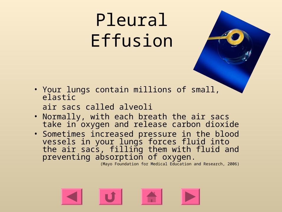
• Your lungs contain millions of small, elastic air sacs called alveoli
• Normally, with each breath the air sacs take in oxygen and release carbon dioxide
• Sometimes increased pressure in the blood vessels in your lungs forces fluid into the air sacs, filling them with fluid and preventing absorption of oxygen.
(Mayo Foundation for Medical Education and Research, 2006)

Pleural Effusions
Malignancy accounts for about 40% of symptomatic pleural effusions, with congestive heart failure and infection being the other leading causes
(National Cancer Institute, 2006)
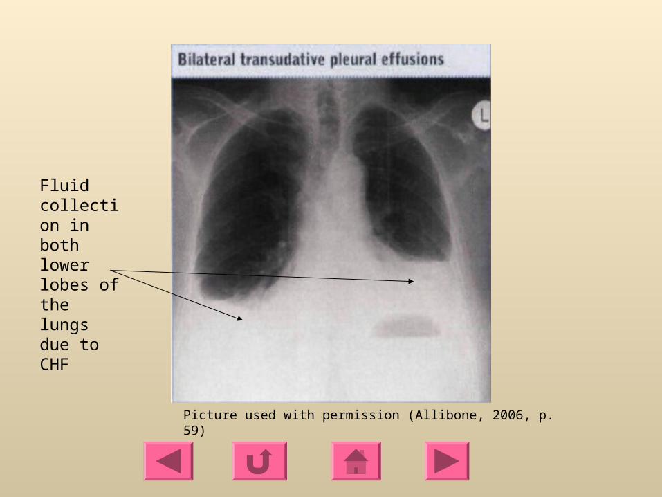
Fluid collection in both lower lobes of the lungs due to CHF
Picture used with permission (Allibone, 2006, p. 59)

Main causes of a Pleural Effusion
• Congestive Heart Failure (CHF)
• Liver failure
• Infection
• Atelectasis
• Cancer
• TraumaClick on home
icon when finished viewing
these topics

Congestive Heart Failure
CHF• As the heart fails, pressure in the vein going through the lungs starts
to rise.• Due to the heart’s inability to move blood from the pulmonary
circulation into the arterial side of systemic circulation, there is a decrease in cardiac output, an increase in left atrial and ventricular end-diastolic pressures, and congestion in the pulmonary circulation.
• As the pressure increases, fluid is pushed into the air spaces (alveoli)
• This fluid then leaks from the alveoli into the pleural space• This fluid creates a pleural effusion and interrupts normal oxygen
movement through the lungs, resulting in shortness of breath

CHF
• CHF is the most common cause of pleural effusion. • Frequently the effusions are bilateral (approximately 75% of the
time) but may occur alone on either side with the right side being more common.
• Fluid is usually straw colored, with low white blood cell counts (<500 cells/mm3) and a mononuclear cell predominance.
• With severe congestive heart failure, fluid may persist in spite of vigorous diuresis.
(National Lung Health Education Program, 2000)
Back

Liver Failure
• Negative intrapleural pressure may lead to a transudative effusion due to peritoneal fluid from ascites moving across the diaphragm into the chest
(Current Therapy, 2001, p. 208)

Infection
• Pneumonia
-inflammation of the lung structures, specifically the alveoli and bronchioles
• WBCs accumulate in response to infection and inflammation leading to empyema

Atelectasis
• Atelectasis is an incomplete expansion of the lung which leads to collapse of the alveoli
• Increased negative intrapleural pressure can lead to the collection of fluid in the portion of the lung which is not expanding
• This can cause an effusion by fluid leaking out of the lung and into the chest cavity
• Atelectasis typically leads to small pleural effusions not requiring surgical intervention

Cancer
• Impaired lymphatic drainage of the pleural space due to obstruction by a tumor
• Typically due to the interference with the visceral pleura (which absorbs pleural fluid)
• A tumor can obstruct pulmonary veins, preventing fluid from being reabsorbed into the bloodstream
• A tumor can perforate the thoracic duct • Shedding of malignant cells into the pleural space,
decreasing reabsorption of pleural fluid back into the lymphatic system (Brubacher & Holmes Gobel, 2003, p. 1)

Trauma
• Increased capillary permeability as a result of inflammation
• Fluid (most often, blood) may collect in the lung cavity as a result of trauma to the lung

Pleural fluid types
• Transudate
• Exudate
• Empyema
• Chyle
• HemothoraxClick on home
icon when finished viewing
these topics

Transudate
• Clear, pale yellow, watery substance• Influenced by systemic factors that alter the
formation or absorption of fluid• Increase in hydrostatic pressure • Decrease in plasma oncotic pressure • Contains few protein cells• Common causes: CHF and liver or kidney
disease

Exudate• Pale yellow and cloudy substance• Influenced by local factors where fluid absorption is
altered (inflammation, infection, cancer) • Rich in protein (serum protein greater than 0.5)• Ratio of pleural fluid LDH and serum LDH is >0.6• Pleural fluid LDH is more the two-thirds normal upper
limit for serum• Rich in white blood cells and immune cells• Always has a low pH• Common causes: pneumonia, cancer, and trauma

Empyema
• Pus• Yellow, cloudy, and foul odor• Most likely due to pneumonia, lung
abscess, infected chest wounds• Has a pH > 7.2
(Drummond Hayes, 2001, p. 33)

Chyle
• Milky fluid• Consists of lymph and fat• Chyle leaks from the thoracic duct
-due to lymphatic obstruction (tumor) or trauma
• High triglyceride levels found in fluid analysis

Hemothorax
• Blood • Usually results from chest injury• A blood vessel ruptures into the pleural space or
a bulging area into the aorta (aortic aneurysm) leaks blood into the pleural space
• Can occur as a result of bleeding from the ribs, chest wall, pleura, and the lung

Signs and symptoms
• Dyspnea• Cough, usually non-productive• Pleuritic chest pain• Chest pressure• Hypoxemia• Decreased breath sounds on the affected side• Some people may exhibit no symptoms!

Diagnosis
• Chest radiograph (x-ray)-able to distinguish >200ml of fluid
• Chest ultrasound-locates small amounts or isolated loculated pockets of fluid-able to give precise position of accumulation
• Computed Tomography (CT) scan-Differentiates between fluid collection, lung abcess, or tumor

DiagnosisFluid analysis confirms a pleural effusion
Normal pleural fluid has the following characteristics: • clear ultrafiltrate of plasma• pH 7.60-7.64• protein content less than 2% (1-2 g/dL)• fewer than 1000 WBCs per cubic millimeter• glucose content similar to that of plasma• lactate dehydrogenase (LDH) level less than 50% of plasma and
sodium• potassium and calcium concentration similar to that of the interstitial
fluid (Abrahamian, 2005, p. 2 of 28)

Non-surgical Treatment Options
• Thoracentesis
• tPA
• Chemical Pleurodesis
• Pleurx catheter

Thoracentesis• A needle is inserted into
the chest wall to remove the collection of fluid
• 50-100ml of fluid is sent for analysis
• Determines the type of fluid (transudate or exudate)
Picture used with permission (Allibone, 2006, p. 60)

Thoracentesis
• Not a permanent solution, fluid may reaccumulate after a few days
• Will temporarily relieve symptoms• Potential complications include bleeding,
infection, and pneumothorax

tPA (alteplase)
• Thrombolytic enzyme• Converts plasminogen to the enzyme plasmin,
which degrades fibrin clots• Lyses thrombi and emboli• May be administered into the chest tube
catheter to restore patency and improve drainage
• The patient is instructed to move positions frequently to distribute the medication throughout the lung

Chemical Pleurodesis
• Sclerosing agents used: Talc, bleomycin, or doxycyline
• Administered through a chest tube to create inflammation and subsequent fusion of the parietal and visceral pleura
• Fluid is then unable to accumulate in this potential space

Chemical Pleurodesis
• The goal of chemical pleurodesis is to cause an irritation between the two layers covering the lung.
• The sclerosant irritates the pleurae which results in inflammation and causes the pleurae to stick together.
• The procedure can be done at the bedside or in the operating room.
• Do not administer with any anti-inflammatory agents

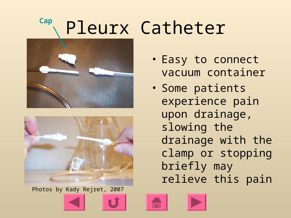
Pleurx Catheter
• Small, flexible tube inserted into the chest to drain fluid from around the lungs
• Contains a one-way valve that prevents air from entering and fluid from leaking out when capped
• Allows for intermittent home drainage using a vacuum bottle
Picture used with permission from Denver Biomedical

Pleurx Catheter
Picture used with permission from Denver Biomedical
In chest wall where fluid is accumulating

Pleurx Catheters
• Catheters are typically drained every one to two days• Keeping the lung fairly free of fluid, will most likely
permanently stop the fluid from building up, so that the catheter can be removed.
• The catheter may remain until fluid quits draining from the lung
• The length of time a catheter will remain varies from patient to patient, ranging from a few weeks to several months.

Pleurx Catheter
• Beneficial for patients who are independent and able to perform self drainage
• Minimizes the time spent in the hospital• Patients are instructed to drain up to 1,000ml of
fluid at one time• Patients are instructed to call MD if drainage is
<50ml on three consecutive sessions• Patients are able to wear usual clothing and
continue usual activities
Pleurx Catheter
• Easy to connect vacuum container
• Some patients experience pain upon drainage, slowing the drainage with the clamp or stopping briefly may relieve this pain
Cap
Photos by Kady Rejret, 2007

Pleurx
Photo by Kady Rejret, 2007
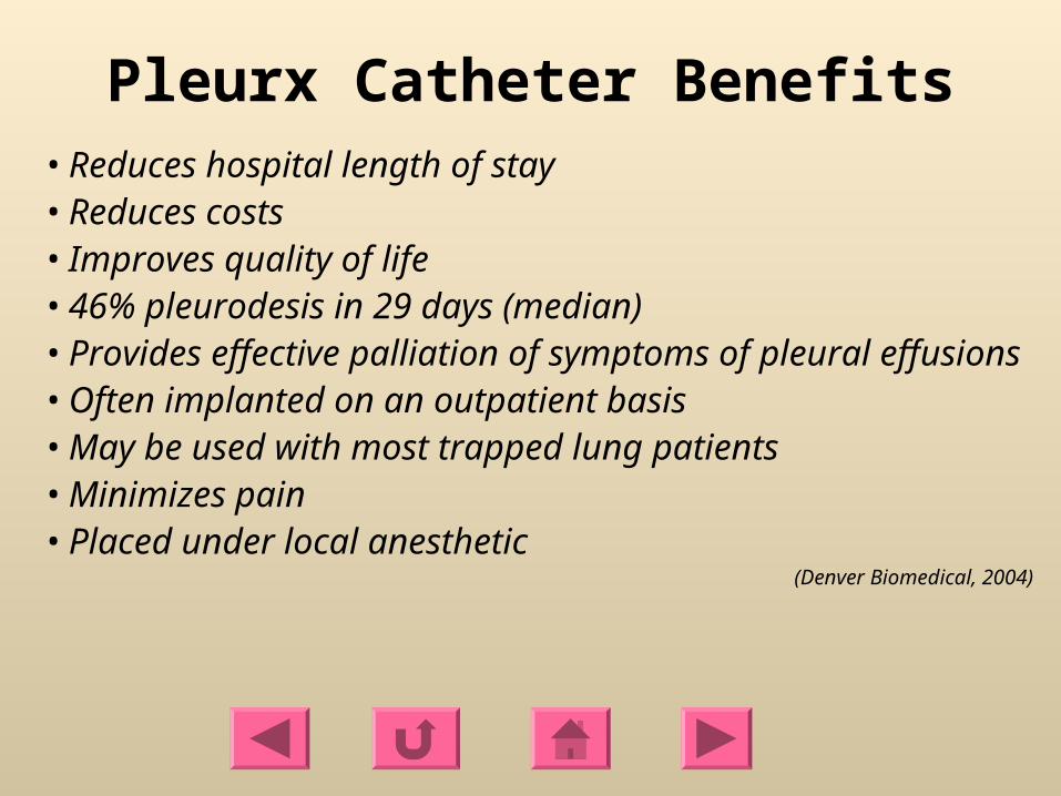
Pleurx Catheter Benefits• Reduces hospital length of stay• Reduces costs• Improves quality of life• 46% pleurodesis in 29 days (median)• Provides effective palliation of symptoms of pleural effusions• Often implanted on an outpatient basis• May be used with most trapped lung patients• Minimizes pain• Placed under local anesthetic
(Denver Biomedical, 2004)