Embed Size (px)
DESCRIPTION
Faculty of allied medical sciences. Histopathology and cytology (MLHC-201). Liver Pathology Supervision : Prof. Dr. Noha Ragab. Outcomes. By the end of this lecture, the student will be able to identify: 1-Steatosis 2-Benign tumours of the liver 3-Malignant tumours of the liver. - PowerPoint PPT Presentation
Citation preview

Histopathology and cytologyHistopathology and cytology
((MLHC-201MLHC-201))
Faculty of allied medical Faculty of allied medical sciencessciences
Liver PathologyLiver Pathology
Supervision : Supervision : Prof. Dr. Noha RagabProf. Dr. Noha Ragab

OutcomesOutcomes
By the end of this lecture, the student By the end of this lecture, the student will be able to identifywill be able to identify::
11--SteatosisSteatosis
22--Benign tumours of the liverBenign tumours of the liver
33--Malignant tumours of the liverMalignant tumours of the liver
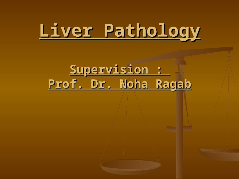
11.. Fatty changes (steatosis)Fatty changes (steatosis)
It is a reversible conditionIt is a reversible condition
Grossly Grossly :: Liver enlargement, yellow and greasyLiver enlargement, yellow and greasy
MicroscopicallyMicroscopically:: Centrilobular micro-vesicular steatosis Centrilobular micro-vesicular steatosis
(reversible)(reversible) Eventual fibrosis around the central vein Eventual fibrosis around the central vein
(irreversible)(irreversible)
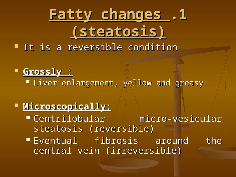
This liver is slightly enlarged and has a pale yellow appearance, seen both on the capsule and cut surface.
Fatty changes (steatosis)Fatty changes (steatosis)

The lipid accumulates in the hepatocytes as vacuoles. These vacuoles have a clear appearance with H&E staining.
Fatty changes Fatty changes (steatosis)(steatosis)
LIVER TUMORSLIVER TUMORS
BENIGN LIVER BENIGN LIVER TUMORSTUMORS
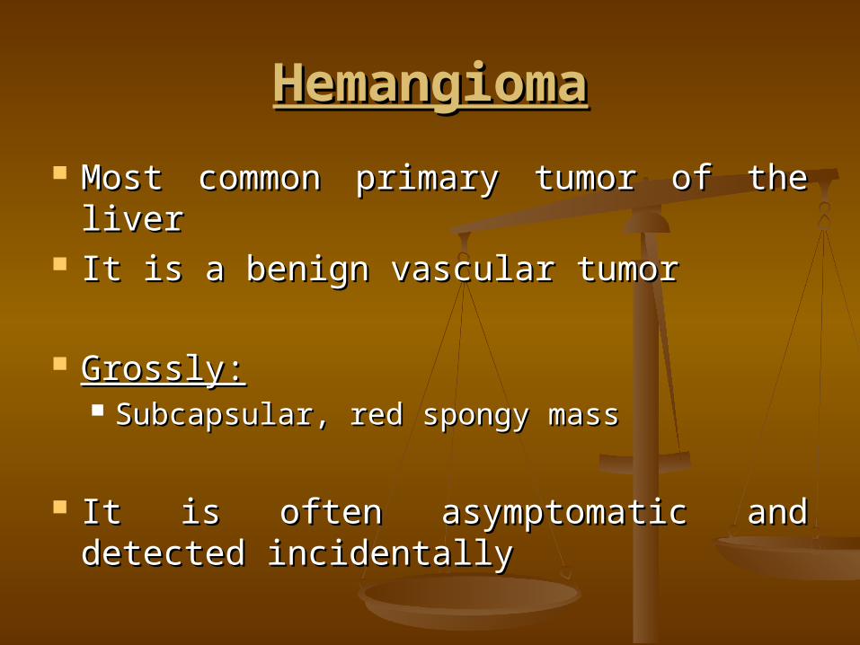
HemangiomaHemangioma
Most common primary tumor of the liverMost common primary tumor of the liver It is a benign vascular tumorIt is a benign vascular tumor
Grossly:Grossly: Subcapsular, red spongy massSubcapsular, red spongy mass
It is often asymptomatic and detected It is often asymptomatic and detected incidentallyincidentally

This is a benign hemangioma of the liver just beneath the capsule.
HemangiomaHemangioma
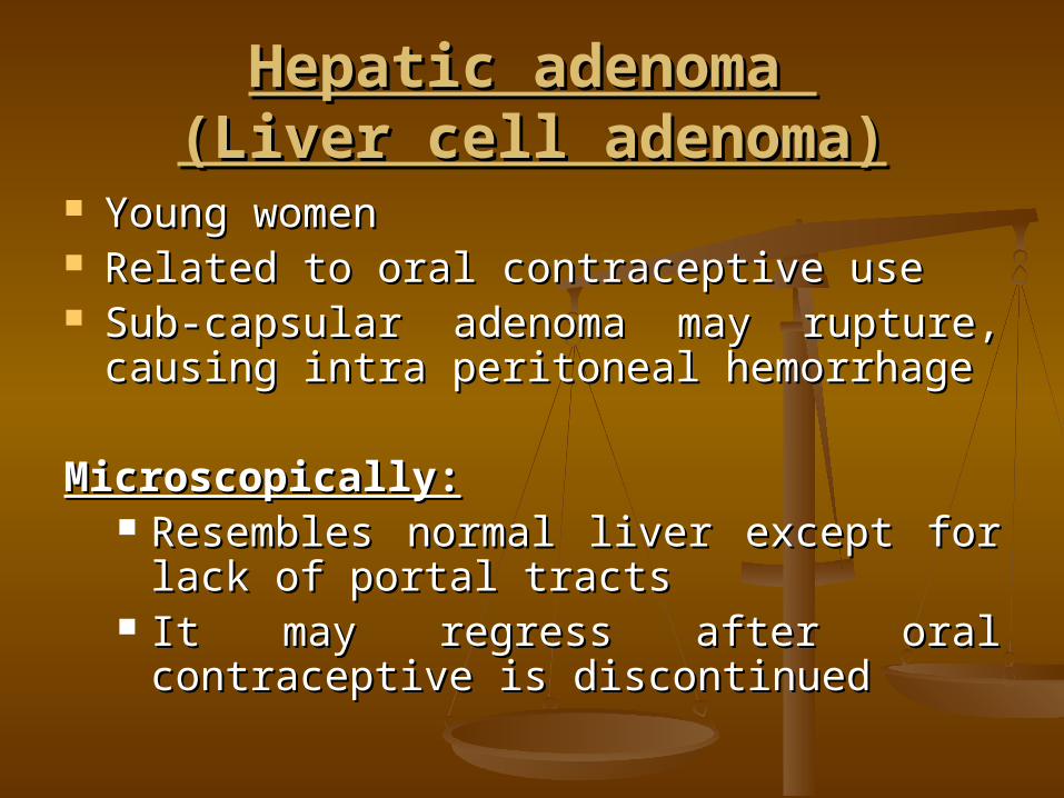
Hepatic adenoma Hepatic adenoma (Liver cell adenoma)(Liver cell adenoma)
Young womenYoung women Related to oral contraceptive useRelated to oral contraceptive use Sub-capsular adenoma may rupture, Sub-capsular adenoma may rupture,
causing intra peritoneal hemorrhagecausing intra peritoneal hemorrhage
Microscopically:Microscopically: Resembles normal liver except for lack Resembles normal liver except for lack
of portal tractsof portal tracts It may regress after oral contraceptive is It may regress after oral contraceptive is
discontinueddiscontinued
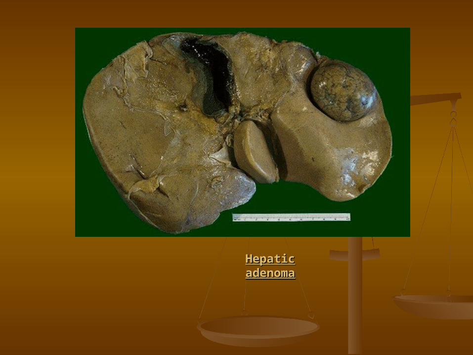
Hepatic Hepatic adenomaadenoma

Hepatic Hepatic adenomaadenoma
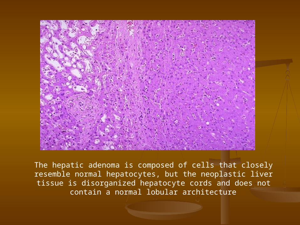
The hepatic adenoma is composed of cells that closely resemble normal hepatocytes, but the neoplastic liver tissue is
disorganized hepatocyte cords and does not contain a normal lobular architecture

MALIGNANT LIVER MALIGNANT LIVER TUMORSTUMORS

Hepatocellular carcinoma (HCC)Hepatocellular carcinoma (HCC)
Definition:Definition:
Hepatocellular Carcinoma (HCC) is a Hepatocellular Carcinoma (HCC) is a Malignant Tumor that Derives from Malignant Tumor that Derives from Hepatocytes or Their PrecursorsHepatocytes or Their Precursors
Risk factors include the followingRisk factors include the following::
1-HEPATITIS B: 1-HEPATITIS B: There is a strong association between HBV There is a strong association between HBV
infection and HCC. infection and HCC. Most (>80%) cases of HCC associated with Most (>80%) cases of HCC associated with
HBV infection occur in patients with cirrhosis.HBV infection occur in patients with cirrhosis. The repeated cycles of liver cell regeneration The repeated cycles of liver cell regeneration
in chronic hepatitis initiate the emergence of a in chronic hepatitis initiate the emergence of a neoplastic clone. neoplastic clone.
The worldwide use of a vaccine for HBV should The worldwide use of a vaccine for HBV should significantly decrease the prevalence of HCC in significantly decrease the prevalence of HCC in the future.the future.
2-HEPATITIS C:2-HEPATITIS C: HCV infection is present in about 50% of HCV infection is present in about 50% of
cases of HCC. cases of HCC. Most patients infected with HCV who Most patients infected with HCV who
develop HCC have underlying cirrhosis. develop HCC have underlying cirrhosis.
3-Haemochromatosis 3-Haemochromatosis
4-Alcoholic cirrhosis4-Alcoholic cirrhosis
Pathological picturePathological picture::
Grossly:Grossly: HCCs appear grossly as soft, HCCs appear grossly as soft,
hemorrhagic tan masses in the liver. hemorrhagic tan masses in the liver.
In some cases, a large solitary tumor In some cases, a large solitary tumor occupies a portion of the liver; in occupies a portion of the liver; in other instances, many smaller other instances, many smaller tumors are found. tumors are found.
The tumor has a tendency to grow The tumor has a tendency to grow into portal veins and may extend into into portal veins and may extend into the vena cava and even the right the vena cava and even the right atrium through the hepatic veins. atrium through the hepatic veins.
Metastases occur widely, although Metastases occur widely, although the most common sites are the lungs the most common sites are the lungs and portal lymph nodes.and portal lymph nodes.
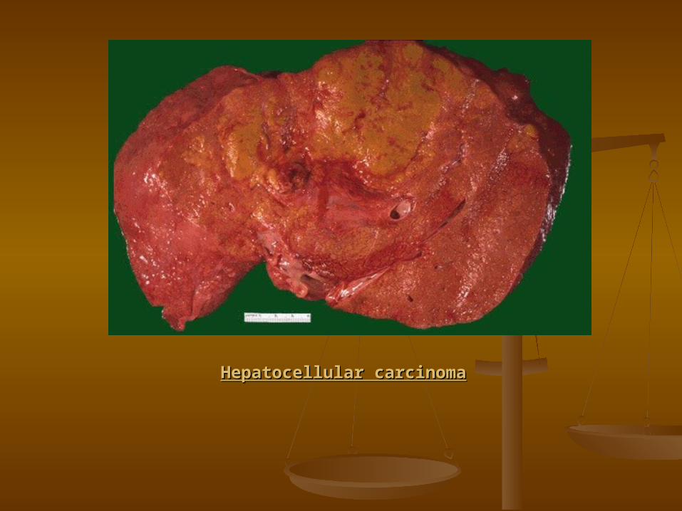
Hepatocellular carcinomaHepatocellular carcinoma
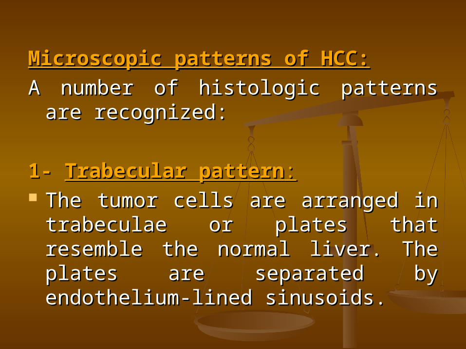
Microscopic patterns of HCC:Microscopic patterns of HCC:
A number of histologic patterns are A number of histologic patterns are recognized: recognized:
1- 1- Trabecular patternTrabecular pattern:: The tumor cells are arranged in The tumor cells are arranged in
trabeculae or plates that resemble trabeculae or plates that resemble the normal liver. The plates are the normal liver. The plates are separated by endothelium-lined separated by endothelium-lined sinusoids. sinusoids.
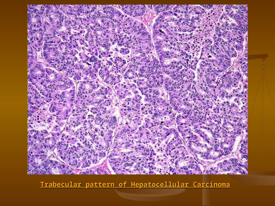
Trabecular pattern of Hepatocellular CarcinomaTrabecular pattern of Hepatocellular Carcinoma
2- 2- Pseudoglandular patternPseudoglandular pattern(adenoid, acinar)(adenoid, acinar)
In this variety, malignant In this variety, malignant hepatocytes are arranged around a hepatocytes are arranged around a lumen and thus resemble glands. lumen and thus resemble glands. The lumina may contain bile. The lumina may contain bile.
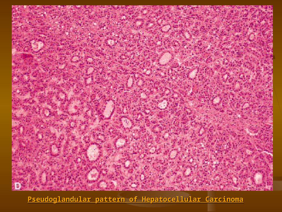
Pseudoglandular pattern of Hepatocellular CarcinomaPseudoglandular pattern of Hepatocellular Carcinoma
3- 3- Fibrolamellar HCCFibrolamellar HCC The tumor is composed of large, The tumor is composed of large,
eosinophilic, neoplastic hepatocytes eosinophilic, neoplastic hepatocytes arranged in clusters and surrounded arranged in clusters and surrounded by delicate collagen fibers. by delicate collagen fibers.
The prognosis is considered more The prognosis is considered more favorable than in most cases of HCC.favorable than in most cases of HCC.
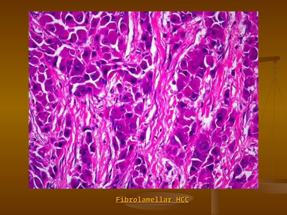
Fibrolamellar HCCFibrolamellar HCC

Clinical Features:Clinical Features:
1.1. HCC usually presents as a painful and HCC usually presents as a painful and enlarging mass in the liver.enlarging mass in the liver.
2.2. HCC may be associated with a variety of HCC may be associated with a variety of paraneoplastic manifestations (e.g., paraneoplastic manifestations (e.g., polycythemia, hypoglycemia, polycythemia, hypoglycemia, hypercalcemia) as a result of ectopic hypercalcemia) as a result of ectopic hormone production by the tumor.hormone production by the tumor.
3.3. Alpha-Fetoprotein levels are often Alpha-Fetoprotein levels are often elevated in HCC elevated in HCC

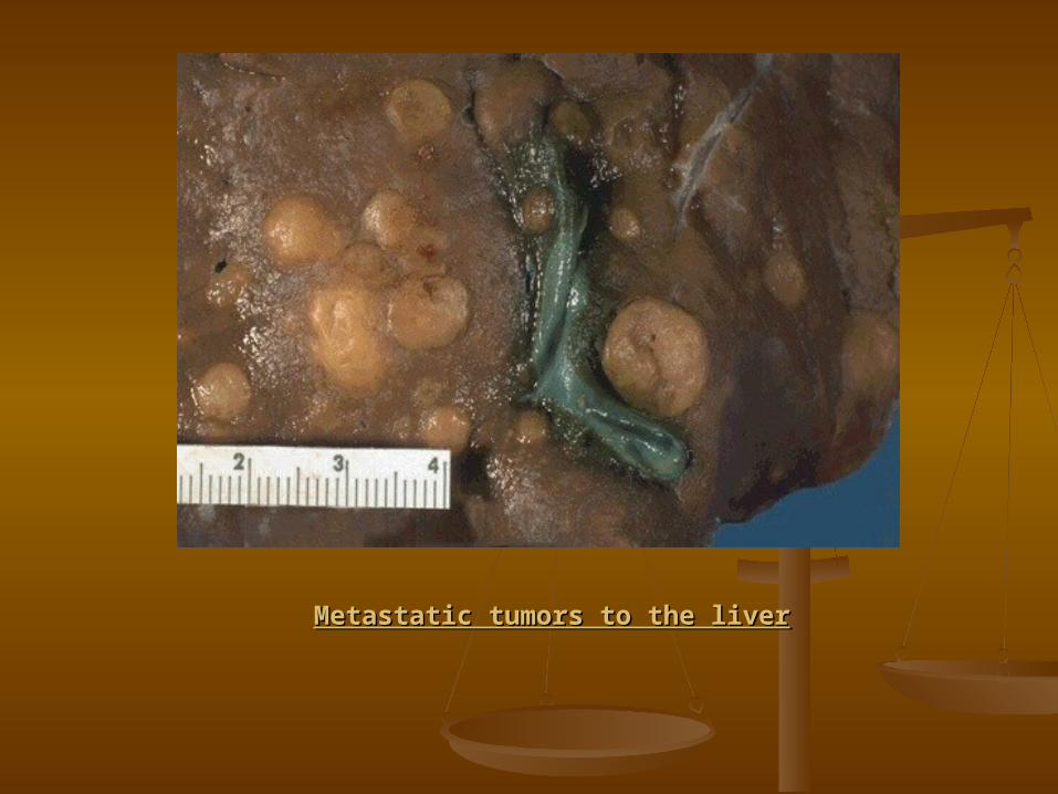
Metastatic tumors to the liverMetastatic tumors to the liver:: Metastatic Cancer is the Most Common Malignant Metastatic Cancer is the Most Common Malignant
Tumor of the LiverTumor of the Liver
Metastatic cancers, including the gastrointestinal Metastatic cancers, including the gastrointestinal tract, breast, and lung. Also pancreatic carcinoma tract, breast, and lung. Also pancreatic carcinoma and malignant melanoma.and malignant melanoma.
The first indication of a metastatic tumor is The first indication of a metastatic tumor is frequently an unexplained increase in the serum frequently an unexplained increase in the serum alkaline phosphatase level. alkaline phosphatase level.
Most patients die within a year of the diagnosis of Most patients die within a year of the diagnosis of

Metastatic tumors to the liverMetastatic tumors to the liver
Metastatic tumors to the liverMetastatic tumors to the liver

QuestionsQuestions
11 - -What are the microscopic characters What are the microscopic characters of steatosisof steatosis? ?
22 - -What are the microscopic characters What are the microscopic characters of hepatic adenomaof hepatic adenoma? ?
33 - -Define Hepatocellular carcinomaDefine Hepatocellular carcinoma..
44 - -What is the first indication of a What is the first indication of a metastatic tumourmetastatic tumour ? ?

Practical ExamPractical Exam
يوم العملى امتحان إجراء يوم سيتم العملى امتحان إجراء سيتمالموافق الموافق االحد فى فى 29/12/201329/12/2013االحد
. . G516G516مدرج مدرج المجموعة حضور يتم ان المجموعة على حضور يتم ان على
من من االولى المجموعة 3030: : 1010 االولى المجموعة و ومن من الثانية 3030: : 1212 . .الثانية

THANK YOUTHANK YOUANDAND
GOOD LUCKGOOD LUCK