Embed Size (px)
Citation preview

EXPERIMENTAL CARCINOGENESIS Bronchial Intramural Adenocarcinomas in Rats
From X-ray Irradiation of the Chest PAUL GROSS, MD, EMIL A. PFITZER, ScD,* JOSEPH WATSON, PHD, ROBERT T. P. DETREVILLE, MD, ScD, MARIANNE KASCHAK, BS,
ETHEL B. TOLKER, BS, AND MARY ANN BABYAK, BS
One hundred eighty-six rats and 202 hamsters, that survived the respective time periods when the first lung cancer was diagnosed in each species, had been given an exposure of 3000 R or 4000 R of x-irradiation applied externally to the chest. About 43% of the rats, but only 2% of the hamsters, developed lung cancer. To enhance the prevalence of lung cancer, some of the irradiated animals were injected intratracheally with 9, 10-dimethyl-1, 2-benzanthracene (DMBA). Lung cancer prevalence in irradiated animals injected with DMBA was no higher than that in irradiated animals not so injected. Intratracheal injections of DMBA in nonirradiated rats did not result in tumor production but, in nonirradiated hamsters, DMBA caused a 6% prevalence of lung cancer. The mortality rate of both animal species showed a more than additive effect in animals given the higher x-ray dosage plus intratracheal injections of either DMBA or iron oxide alone. The lung cancers in the irradiated rats predomi- nantly affected bronchi (86%) and were mostly adenocarcinomas (88%). The cancers were generally intramural and were covered by an intact and nor- mal surface epithelium. Their origin appeared to be from aberrant intramural glands. These glands are not found in the bronchi of germ-free rats but do occur in bronchi thickened by severe endemic chronic bronchitis.
HE CL‘SI‘OhIARY PRACTICE OF ESTlhlATING T the carcinogenic potential of a material by repetitive applications 3n the skin of mice does not necessarily provide answers regard- ing the ability of this material to produce lung cancers when inhaled. There is a need for a reliable and sensitive method for esti- mating the potential of a material to cause lung cancer when introduced into the lungs of animals.
In a previous study, it was found that ex- posure of hamsters to roentgen rays ap- plied externally to the chest resulted in a lzyo lung cancer prevalence, all squamous cell carcinomas.1 In these animals there were, in addition, diffuse cellular changes with
From the Industrial Hygiene Foundation, 5231 Cen- tre Avenue, Pittsburg, Pa. 15232, and the Depart- ment of Occupational Health, Graduate School of Public Health, University of Pittsburgh, Pittsburgh.
Supported by Grant No. R01 CA 08033-03 PTHB. + Kettering Laboratory, University of Cincinnati,
Eden & Bethesda Avenues, Cincinnati, Ohio 45219. Received for publication December 12, 1968.
much atypia, pointing to a widespread, near- neoplastic effect of the irradiation. With these findings in mind, it was thought that irradi- ated tissues with such atypical changes might be more sensitive than normal tissues to the subsequent exposure to carcinogens. Whereas the lungs of normal animals can be expected to remain unresponsive to a weak carcinogen, lung tissue “sensi tized” by x-ray irradiation, as found previously in hamsters,’ might con- ceivably respond to such a weak carcinogen with features of a malignant neoplasm.
The purpose of this study was, therefore, to test the above hypothesis in hamsters and rats by comparing the prevalence of lung can- cer in irradiated animals given intratracheal doses of 9, 10-dimethyl-I, 2-benzanthracene (DMBA) with the prevalence of lung cancer in irradiated controls and with such prev- alence in nonirradiated DMBA controls.
Although the results of this study did not support the above hypothesis, they are of in- terest to students of carcinogehesis and, there- fore, are reported here.
1046

No. 5 EXPERIMENTAL CARCINOGENESIS * Gross et al. 1047
METHOD AND MATERIALS
Two hundred and three rats (average wt, 250 g) and 208 hamsters* (average wt, 115 g), which had been pastured for over 2 weeks to allow recovery from the effects of shipment, were given radiation exposures from the same therapy x-ray unit used previously1 under the following slightly modified operating condi- tions: 110 kv, 6.4 to 6.8 ma (for a total ex- posure of 3000 R) and 8.6 to 9.4 ma (for a total exposure of 4000 R); 0.6 mm A1 inher- ent filtration plus 1.83 mm A1 added filtra- tion; target to center line of animal holder, 10.85 cm. A 2 cm diameter lead-shielded col- limator was used to localize the x-ray beam to the chest region. The rats were 9 weeks old and the hamsters, 14 weeks old, when the radiation exposures were initiated.
Unanesthetized animals were held in po- sition during irradiation in open-ended cyl- inders made of 1/2” (12.5 mm) wire mesh. The diameter was such as to accommodate the animal without discomfort, that is, 3.7 cm x 15 cm for hamsters, and 6 cm x 25 cm for rats, yet the snugness prevented the ani- mal from turning around. Snugly-fitting metal plungers attached to Y2’’ (12.5 mm) aluminum rods were inserted into both open ends of the wire cylinder to immobilize the animals. The plungers were clamped in posi- tion by means of adjustable hose clamps. The protuding aluminum rods of the plungers were fastened to sockets of a barbecue-spit motor which rotated the immobilized ani- mals at 7 rpm during the x-ray exposure. Exposure was limited to the chest region and lasted 8 min on each of 5 consecutive days. The exposure rate for animals that received a total of 4000 R and 3000 R was 100 R/min and 75 R/min, respectively. Exposure values were measured every second or third exposure
+ Purchased From Hilltop Lab Animals, Inc., Scott- dale, Pa. 15683.
+ + 7, 12 Dimethylbenz (a) anthracene obtained from Distillation Products Industries, Eastman Organic Chemicals Department, Rochester, N.Y. 10003. Its purity was listed as “Eastman Grade.”
t Ferric oxide, red, anhydrous (jeweler’s rouge), purity not listed, obtained from Fisher Scientific Com- pany, Pittsburgh, Pa. 15219. Its arithmetic mean par- ticle size was 1 . 1 3 ~ and its surface mean diameter,
SMr. Bruce Phillips of the National Institutes of Health and Dr. Helmut A. Gordon, Associate Research Professor of Lobund Institute, University of Notre Dame, provided these lungs.
2.20k.
with an NBS calibrated, 250 R Victoreen R- meter, that was placed in a wooden phantom inside the rotating wire mesh cylinder.
Film densitometer measurements at the target to midline distance of 10.85 cm indi- cated a uniform exposure over a circular area with a diameter of 2.4 cm. This area was ex- tended to a total diameter of 5.5 cm due to scatter. These highly collimated x-rays do not result in a uniform exposure of the thoracic content of the rat.
Eight weeks (average) after the x-ray ex- posure, some of the irradiated animals were injected with DMBA suspended in water, using Fe,O, (jeweler’s rouge) as a carrier, or with aqueous suspensions of Fe203 alone. As controls, some of the irradiated animals were pastured without having been injected intra- tracheally. Other controls consisted of non- irradiated animals injected intratracheally with DMBA or with the carrier alone and untreated laboratory controls. The intratra- cheal injections were given either in 4 or 16 doses. When given in 16 doses they were spaced 1 week apart; when given in 4 doses, 4 weeks apart. The experimental setup is outlined in Tables 1 and 2.
An acetone solution of a weighed amount of DMBA** was added to an amount of jeweler’s rouge (Fe203)t to result in a ratio of lOOyg DMBA to 3 mg Fe203. The acetone was removed by evaporation and the iron- oxide DMBA mixture was suspended in water with the aid of a drop of nonionic de- tergent (TweentR)) so that 3 mg Fe203 was contained in 0.5 ml suspension.
The animals were fed Purina Laboratory Chow, ad lib., and their drinking water con- tained 0.025% chlortetracycline hydrochlo- ride. They were housed in wire mesh cages; rats, 2 and 3 in a cage, and hamsters, 5 in a cage. Most of the irradiated animals died spontaneously. The survivors were killed with ether 20 to 22 months after the x-ray exposure.
Autopsies were performed on all animals. The lungs of all but 4 animals were dis- tended with 4% formaldehyde solution at a pressure of 10 to 12 cm of water. Because 4 of the animals had been cannibalized, their lungs were not available. Portions of the vari- ous extrapulmonary tumors were removed and placed in the same fixative for later microscopic examination. Sections from the lungs of 4 germ-free ratst were studied to ob- tain data on the anatomy of “normal” rat bronchi.

I048 CANCER May 1969 VOl. 29
TABLE 1. Tabular Protocol of Rats Irradiated and Injected Intratracheally
Intratracheal injections No.
Radiation No. DhlBA FelOI No. 9-mpnth R Group rats Pg mR doses survivors
4000
A B C D E
Total .4 B
3000 C D
Total control-1 control-2
None control4 control -4 control-5 Total Grand total
20 20 20 20 43
123 20 20 20 20 80 20 20 10 10 10 70
2 73
100 400 - - -
100 400 -
-
100 400 -
3 12 3
12 - - 3
12 3
12 -
3 12 3
12
16 4
16 4 - - 16 4
16 4
16 4
16 4
-
- - -
~
18 19 15 14 42
108 20 19 20 19 78 18 20 6
10 10 64
250
RESULTS
Rats: The groups of rats irradiated with 4000 R and injected intratracheally with DMBA or with the carrier (Fe,O,) alone had mortality rates varying from 75 to 90% in 21 to 22 months. Yet, the group of rats given irradiation alone had only a 35% mortality at this time; other nonirradiated control groups injected intratracheally with DMBA or only with the carrier had mortality rates of less than 25% (Fig. 1). A baseline mortality of
10% is indicated by the untreated laboratory control group. Whether the rats irradiated with 4000 R received intratracheal injections of DMBA plus carrier or of the carrier alone, produced no consistent effect on the mor- tality rate.
It is apparent from the mortality curves. (Fig. 1) that the mortality rates of the groups irradiated with 4000 R and injected intra- tracheally are greater than the sums of the mortality rates caused by the irradiation alone and by the intratracheal injections.
TABLE 2. Tabular Protocol of Hamsters Irradiated or Injected Intratracheally
Intratracheal injections NO.
Radiation No. DMB.4 Fe?OP NO. 2 %-month. R Group hamsters M m?z doses survivors
3000
None
A 15 100 3 3 15 400 12 1 22
3 4 9 R 25
10 12 - 27
4000 C 28
46 D
119 46 E
Total 124 A 10 100 3 3 10
-100 12 2 28 3 4 10
R 28 10
12 2 27 C
8 D 28
83 E 8
Total 84 control- 1 14 100 3 3 13 control-2 26 400 12 2 22
3 4 4 12 1 15
control-3 5 control-4 15
10 64
control-5 10 Total 70
- 266 Grand total 2 78
- 3 -
- - - - - -
- - - - - - - -
- - - - - - - - - -

so. 5
loor EXPERIMENTAL CARCINOGENESIS - Gross et al. 1049
90
80 Groups given 4000r Groups given 30001- -----
)r 70 c .- - 0
0 + 60
50
6 40 30
20
10
I
c c
L
n
boratory Control
I I I I I -0 2 4 6 8 10 12 14 16 18 20 22
Months FIG. 1. Graphical representation of mortality rates of the different groups of rats: A = 1.6 mg
DMBA intratrachcally in 16 doses with Fe,O, as carrier; B = 1.6 mg DMBA intratracheally in 4 doses with Fe,O, as carrier; C = no DMBA. Fe,O, intratracheally in 16 doses; D = no DMBA. Fe,O, intratracheally in 4 doses, and E = radiation alone.
alone. This more than additive effect is much less marked at the 3000 R level of irradiation.
As seen in Table 3, there was an overall incidence of 80 lung cancers among the 186 irradiated rats that had survived 9 months or more after the x-ray exposure, giving a prev- alence of 43%. (The 9-month survival period was chosen because the first lung cancer among these animals made its appearance at 9% months.) Although the average prev- alence of lung cancer in the groups irradiated with 4000 R and injected with DMBA was 49y0, compared with the 280/, average prev- alence in the groups irradiated with the same dose but not injected with DMBA (groups C and D), this different rate of tumor pro- duction was not observed in the groups ir- radiated with 3000 R. Here the average prev- alence of lung cancer was 41% with DMBA and 40y0 without DMBA. Furthermore, the 49y0 prevalence of lung cancer in rats irradi- ated with 4000 R and injected with DMBA is practically equal to the 50y0 prevalence in the group given the same dose of x-ray ir- radiation but no DMBA. No significant dif- ference was noted when the average prev- alence of lung cancer in the groups irradiated
with 4000 R was compared with that of the groups irradiated with 3000 R (43% vs. 41y0). In addition, the difference in the average prevalence of lung cancer in the groups ir- radiated with 4000 R and injected with DMBA (490/,) and that in the groups given 3000 R and no DMBA injections (40oj,) is not significant. All of these data point to the fact that the intratracheal injections of DMBA in these x-ray irradiated rats did not contribute to a greater prevalence of lung cancer than was caused by the x-ray irradiation alone.
In Slyo of the irradiated rats, the cancers were central, located in the wall of a major bronchus, In 24y0 of the rats, the cancers were peripheral. However, 9% of the rats had both, a central cancer and also a peri- pheral one. Four of the peripheral cancers (5%) could be identified as originating in a smaller bronchus. Since the percent distribu- tion of central and peripheral cancers in the different groups did not vary significantly, the treatment with DMBA had no effect on the location of the tumor.
T h e centrally located adenocarcinoma was generally recognized by the presence of nu- merous irregularly distributed tubular glands

1050 CANCER May 1969 Vol. 23
situated within the wall of a major bronchus that was greatly thickened and profusely in- filtrated by lymphocytes (Fig. 2). These glands were irregular in shape, lined by epithelium that varied from flat to columnar, and the lumen often contained desquamated cells. The tumor glands infiltrated not only the bronchial wall, but also the loose tissues lying between the bronchus and the accompanying branch of the pulmonary artery or vein (Fig. 3). Although the bronchial mucosal epithe- lium overlying the infiltrating tumor was generally intact and normal (Fig. 3), in some instances ulceration was present (Fig. 2). In no rat could an origin of a carcinoma be traced to the bronchial surface epithelium.
Inasmuch as a study of bronchial walls in the lungs of germ-free rats failed to reveal the presence of glandular structures beneath
the mucosa, the origin of the bronchial ade- nocarcinoma was difficult to explain until tu- bular glands similar to those in the tumors were observed in untreated laboratory con- trol, as well as in other nonirradiated con- trol animals. Here these glands were found in the submucosa and deeper tissues of bron- chial walls greatly thickened by endemic chronic bronchitis. As in the case of the bronchial adenocarcinoma, the surface epi- thelium overlying the glandular structures in the control of animals was ciliated and ap- parently normal. The tubular glands were relatively few in number when found in the control animals (Fig. 4). They were small (50 to 70p in diameter), lined by cuboidal cells and their lumens contained clusters of desquamated cells. The presence of the in- traluminal cells often made recognition of
FIG. 2. (Left) Portion of a main bronhcus a short distance below the carina shows glandular tumor tissue between the bronchial wall and a branch of the pulmonary artery. The bronchial wall is greatly thickened and infiltrated with lymphocytes. The surface epithelium is denuded. A few tumor glands are seen in the left lower edge of the bronchial wall. From a rat irradiated with 4000 R and killed 22 months later (H and E, X150).
FIG. 3. (Right) A large bronchus between a pulmonary vein (above) and artery (below) show- ing glandular tumor tissue within a thickened bronchial wall and also between bronchus and vein. The bronchial surface epithelium is intact and essentially normal. From a rat irradiated with 4000 R and killed 211h months later (H and E, ~40).

No. 5 EXPERIMENTAL CARCINOCENESIS - Gross et ai. 1051
the glands difficult because of the heavy in- flammatory infiltrate of the surrounding tis- sues (Fig. 4).
In an attempt to explain the presence of these aberrant glands in the chronically in- flamed bronchial wall, a search was made for invaginations of the bronchial mucosa. Al- though a few such invaginations were found, the demonstrations were not altogether con- vincing. Another possible explanation under consideration, particularly of the deeper ly- ing glands, was that the glands represented alveolar remnants which became isolated as the peribronchial inflammatory tissue moved down to involve the adjoining parenchyma. Although fibrous and cellular thickening of alveolar walls abutting on thickened bron- chial walls could readily be demonstrated,
i t was difficult to apply this explanation to the more superficially situated glands.
Some difficulty was encountered in a num- ber of irradiated animals in which the gland- ular structures were not only moderately in- creased in number over that seen in control animals, but also showed bizarre shapes (Fig. 5). Rather than categorizing these as cancers (which some of them might have been), they were classed as atypical hyperplasia of aber- rant epithelium. The character of the epithe- Iium in most of these tumors was usually of no help in determining their neoplastic char- acter. The nucleocytoplasmic ratio was usu- ally within normal limits. There was no hy- perchromatism or other nuclear abnormal- ity and mitoses were rare. The diagnosis was, however, facilitated in many of the bronchial
FIG. 4. (Left) Aberrant glands in the wall of an inflamed bronchus a short distance below the carina. T h e glands extend deeply into the cellular inflammatory tissue and are lined by Battened to cuboidal epithelium. These glands would stand out more prominently i f their lumens did not contain desquamated cells. R’ote the apparently normal ciliated surface epithe- lium. From an untreated laboratory control rat killed a t a time corresponding to 223h months after the irradiation exposures (H and E, ~ 3 0 0 ) .
FIG. 5 . (Riwht) A thickened bronchial wall with adjacent wall of pulmonary artery. The bronchial wall is permeated throughout its thickness by glandular structures of various sizes, shapes, and orientation. All contain desquamated cells. This was considered a questionable case and was not included in the tabulation of lung cancer in spite of the proximity of the glands to the arterial wall. From a rat exposed to 3000 R and injected with DMBA (Group B). Killed 22 months after the x-ray exposure (H and E, ~ 1 5 0 ) .

1052 CANCER May 1969 Vol. 23
cancers where the tubular glands were found intermingled with tumor cell aggregations having only miniature lumens and tumor cell clusters without lumens (Fig. 3).
In one animal, the adenocarcinomatous tis- sue, still covered by nonneoplastic columnar epithelium, extended as a polyp into the bronchial lumen (Fig. 6). Most of the periph- eral tumors were also adenocarcinomas (Fig. 7) and appeared to be independent of bron- chi. There were 5 peripheral adenocarcinomas in which an ulcerated bronchus could be identified in their center (Fig. 8). Some of the peripheral cancers exhibited portions in which the glandular tissue was well differ- entiated, often papillary, and resembled that of an adenoma (Fig. 8).
Nearly all of the squamous cell cancers were peripheral tumors and were only par- tially differentiated (Fig. 9). The one squam-
ous cell carcinoma that was associated with much keratin production had originated in the lining of a bronchiectatic cavity.
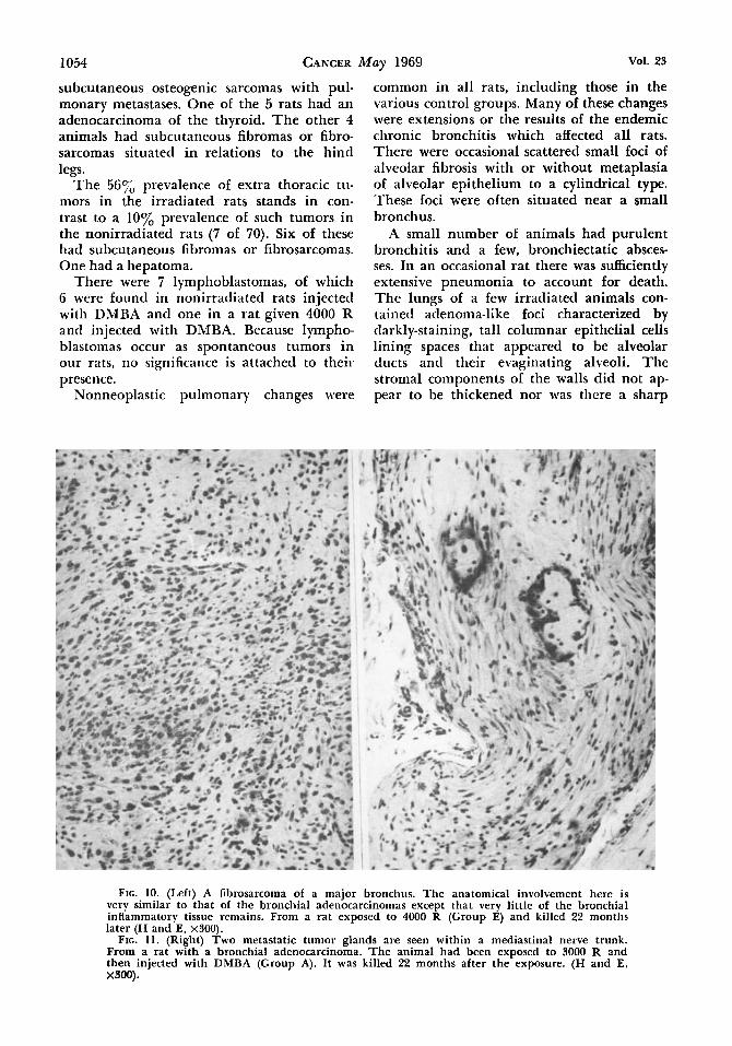
Three well-differentiated fibrosarcomas were found (Fig. 10). They involved the wall of major bronchi and their location greatly resembled that of the bronchial adenocarci- nomas, being situated partly in the bronchial wall and partly in the tissue between the bronchial wall and the accompanying large artery or vein.
Metastases from the pulmonary cancers were observed in 3 rats. One of these con- sisted of glandular structures within a medi- astinal nerve trunk (Fig. 11) another con- sisted of irregular tumor glands occupying the cortex of a satellite lymph node (Fig. 12), and the third was a satellite lymph node completely replaced by partially differenti- ated squamous epithelium.
FIG. 6. (Left) T h e wall of B large bronchus which is diffusely infiltrated by glandular tumor tissue that also cxtcnds as a polyp into the bronchial lumen. Note also the proximity of the tumor to the wall of a branch of the pulmonary artery (lower portion of the field). Except for short stretches, the surface epithelium is normal. From a rat exposed to 3000 R and injected with DMBA (Group A). Killed 22 months after the exposure (H and E, X40).
FIG. 7. (Right) A peripheral adenocarcinoma. T h e pleural surface is a t the top of the field. Although the tumor is large. the adjoining lung tissue shows no compression. From a rat exposed to 4000 R and injected with DMBA (Group ,A). I t died 17 months after the irradiation (H and E, ~ 4 0 ) .

so. 5 1053
FIG. 8. (Left) A “peripheral” adenocarcinoma which has in its center an ulcerated bronchus (middle of right half of field). The bronchial wall and adjacent structures are infiltrated by partially differentiated adenocarcinomatous tissue, whereas well-differentiated papillary tumor tissue resembling that of an adenoma is seen at the bottom of the field. From a rat exposed to 4000 R and injected with DMBA (Group B). It died 93/4 months after the irradiation (H and E, x40).
FIG. 9. (Right) A peripheral squamous cell carcinoma. The pleural surface is at the top of the field. This animal, dead 16~/, months after irradiation with a dose of 4000 R (Group E), was 1 of only 3 with a demonstrable metastasis (H and E, Xl50).
About 88% of all rats with lung cancer had adenocarcinomas (range, 75 to 100% in the various groups), and 11% of the rats had squamous cell carcinomas (range, 7 to 25%). Four of the 9 squamous cell carcinomas were associated with adenocarcinomas and one of the former originated in a large bronchiecta- tic cavity. Several of the adenocarcinomas were multicentric; some showing different discrete foci of bronchial involvement, others showing multiple peripheral tumors.
Of the 3 fibrosarcomas originating in the wall of major bronchi, one was associated with an adenocarcinoma of the same bron- chus. I n 3 rats, the lung cancers were undif- ferentiated. One was a carcinoma, one a sar- coma, and the third could not be classified. All 3 were central tumors involving a major bronchus. The distribution of the various types of lung cancers among the different
animal groups (Table 3) suggests that treat- ment with DMBA had no effect on the prev- alence of any specific type of lung cancer. Also of importance is the fact that in non- irradiated rats the injections of Fe203. with or without DMBA, did not cause the production of a single lung tumor.
Two rats, irradiated with 4000 R developed mediastinal, extrapulmonary sarcomas. One of these rats injected with DMBA had a rhab- domyosarcoma and the other, a radiation control (group E), had a fibrosarcoma.
Slightly over one-half of all x-ray irradi- ated rats (103 of 184 9-month survivors) had extrathoracic tumors. In all but 5 of these animals, the tumors were situated in the re- gion of irradiation, subcutaneously. Most of the subcutaneous tumors were fibromas or fibrosarcomas, 2 of the latter with pulmonary metastases, but there were also 2 rats with
1054 CANCER May 1969 Vol. 23
subcutaneous osteogenic sarcomas with pul- monary metastases. One of the 5 rats had an adenocarcinoma of the thyroid. T h e other 4 animals had subcutaneous fibromas or fibro- sarcomas situated in relations to the hind legs.
The 56% prevalence of extra thoracic tu- mors in the irradiated rats stands in con- trast to a 10% prevalence of such tumors in the nonirradiated rats (7 of 70). Six of these had subcutaneous fibromas or fibrosarcomas. One had a hepatoma.
There were 7 lymphoblastomas, of which 6 were found in nonirradiated rats injected with DMBA and one in a rat given 4000 R and injected with DMBA. Because lympho- blastomas occur as spontaneous tumors in our rats, no significance is attached to their presence.
Nonneoplastic pulmonary changes were
common in all rats, including those in the various control groups. Many of these changes were extensions or the results of the endemic chronic bronchitis which affected all rats. There were occasional scattered small foci of alveolar fibrosis with or without metaplasia of alveolar epithelium to a cylindrical type. These foci were often situated near a small bronchus.
A small number of animals had purulent bronchitis and a few, bronchiectatic absces- ses. In an occasional rat there was sufficiently extensive pneumonia to account for death. The lungs of a few irradiated animals con- tained adenoma-like foci characterized by darkly-staining, tall columnar epithelial cells lining spaces that appeared to be alveolar ducts and their evaginating alveoli. The stromal components of the walls did not ap- pear to be thickened nor was there a sharp
FIG. 10. (Left) A fibrosarcoma of a major bronchus. The anatomical involvement here is very similar to that of the bronchial adenocarcinomas except that very little of the bronchial inflammatory tissue remains. From a rat exposed to 4000 R (Group E) and killed 22 months later (H and E, x300).
FIG. 11. (Right) Two metastatic tumor glands are seen within a mediastinal nerve trunk. From a rat with a bronchial adenocarcinoma. The animal had been exposed to 3000 R and then injected with DMBA (Group A). It was killed 22 months after the exposure. (H and E, XSOO).

No. 5 EXPERIhlENTAL CARCINOGENESIS Gross et a[. 1055
line of separation between these lesions and the adjoining normal tissues. The fact that the spatial relationship of the gland-like struc- tures resembled that of normal lung tissue suggested an adenomatoid change rather than a true adenoma.
In the sections, it was not possible to dis- tinguish between the iron oxide injected alone and that injected admixed with DMBA. Neither the microscopic appearance of the dust nor the tissue reaction allowed a dis- tinction of one from the other.
The iron oxide was found loose in alveoli in the form of small oval, jet black blobs that could have been macrophages. These were often loosely aggregated but never in num- bers large enough to obstruct an alveolus. There was little or no reaction of the alveo- lar walls to the presence of the dust.
Atypical cellular changes such as have been described in irradiated hamster lungs1 were not present in the irradiated rat lungs.
Hamsters: The groups of animals irradi- ated with 4000 R generally had the highest mortality rate (Fig. 13). In group A of this series, 80% of the animals had died within 3 months after the x-ray exposure, whereas it required 14 months for the radiation control group (E) to reach this mortality rate. The nonirradiated, DMBA control group had an 80% mortality rate by 17 months, in contrast to the untreated laboratory control group which did not attain this mortality until about 21 months. However, some of the mor- tality rates are incongruous and lack an ex- planation; for example, the excessively high mortality of group C irradiated with 3000 R and the excessively low mortality of group D that was also irradiated with 3000 R.
Based on the number of hamsters surviv- ing 2% months after the x-ray exposure, the total number of lung cancers found in all groups was 6, giving a prevalence of 3% (Ta- ble 4). Of these 6 cancers, 3 were lympho- blastomas, 2, squamous cell carcinoma, and one, an unclassified, undifferentiated malig- nant tumor. Two cancers each were found among the irradiated animals injected with DMBA (3%), among animals irradiated but not injected with DMBA (273, and among nonirradiated animals injected with DMBA (6%). The undifferentiated tumor was found in a hamster that had died 2y2 months after the x-ray exposure; the squamous cell cancers, in animals dead 7% and 9% months, respectively, after the x-ray exposure or the
FIG. 12. A satellite lymph node with partial re lace inent of cortical lymphoid tissue by tumor gf)ands: From a rat with a peripheral adenocarcinoma. The animal belonged in the same group as that of Fig. 11 and was killed at the same time. (H and E, ~300).
DMBA injections, and the lymphoblastomas, 11, 14, and 18% months, respectively, after the x-ray exposure or the DMBA injections. It may be well to point out that the lym- phoblastomas appeared to be confined to the lungs and, contrary to our experience with rats, lymphoblastomas have not developed spontaneously in our hamsters.
Less important changes in the irradiated hamsters included the encirclement of the chest region by a zone of white, depigmented fur. The lungs of the irradiated hamsters showed focal recent hemorrhage, acute and chronic pneumonitis, focal emphysema (sub- pleural), foci of alveolar proteinosis, and atypical proliferation of alveolar cells with enlargement and hyperchromatism of nuclei, often associated with squamous metaplasis (Fig. 14).
There were also regions of metaplasia of alveolar epithelium to a columnar type so that the involved alveoli resembled glandu-

1056 CANCER May 1969 Vol. 23
The Production of Lung TABLE 3.
All Total No lung no. survi- cancers
dose Group* group months No. %+ A 20 18 10 56 B 20 19 8 42
.A + B 40 37 18 49 4000 R C 20 1s 2 13
D 20 14 6 43 40 29 8 28
E 43 42 22 52 C + D
Irradiation in ving 9 -
Central Central Periph- Small* (large and pe-
era1 bronchus bronchus) ripheral cancers cancers cancers cancers11
No. % No. % No. % No. % -___- -
2 20 1 10 8 80 1 10 2 2 5 1 1 3 6 7 5 - - 4 22 2 11 14 78 1 6 1 5 0 - - 1 5 0 - - 1 1 7 - - 5 8 3 - - 2 2 s - - 6 7 5 - - 2 9 I 5 2 0 9 1 - -
A + B /C + D + E 123 108 48 44 8 17 3 6 41 85 1 2 A 20 20 8 40 2 25 1 13 6 75 1 13 B 20 19 8 4 2 3 3 8 - - 7 88 2 25
40 39 16 41 5 31 1 6 13 81 3 19 3000 R C 20 20 9 4 5 3 3 3 - - 7 78 I 11
D 20 19 7 3 s 3 4 3 - - 4 5 7 - - 40 39 16 40 6 38 - - 11 70 1 6 80 78 32 41 11 34 1 3 24 75 4 13
Grand Total 203 186 80 43 19 24 4 5 65 81 5 6
h + B
C + D A + B + C + D
~ ~ ~
* A = 1.6 mg DMBA intratracheally in 16 doses with Fez03 as carrier. B = 1.6 mg DMBA intratracheally in 4 doses with Fez03 as carrier. C = No DMBA. Fez03 intratracheally in 16 doses. D = No DMBA. Fez03 intratracheally in 4 doses. E = Radiation alone.
lar structures. All of these changes have been described in a previous publication' and need not be enlarged upon here.
oxide with or without DMBA. as well as the paucity of tissue reaction to it, were similar to those seen in rat lungs.
The character and distribution of the iron Focal alveolar hemorrhage and cellular
Months FIG. 13. Graphical representation of mortality rates of the different groups of hamsters:
A = irradiation plus DMBA in 3 doses; B = irradiation plus DMBA in 1 or 2 doses; C = irra- diation plus FgO, in 3 doses; D = irradiation plus Fe,O, in 2 doses, and E = irradiation only.

No. 5 EXPERIMENTAL CARCINOCENESIS - Gross et al. 1057
Cancer in X-rav Irradiated Rats
Adenocar- No rats Total no. cinoma and Adenocar- Undif- with sub- No rats rats with
Squamous squamous cinoma feren- cutaneous with extrapul- Adenmar- cell car- cell Fibrosar- and fibro- tiated chest tumors monary
cinoma cinoma carcinoma coma sarcoma cancer tumors elsewhere tumors
NO. % No. % No. % No. % No. % No. ’% No. % No. % No. % 9 90 2 2 0 1 1 0 1 1 0 1 1 0 - - 10 56 2 10 12 67 6 75 1 1 3 - - - - - - 1 1 3 6 3 2 1 5 7 3 7
15 83 3 1 7 1 6 1 6 1 6 1 6 1 6 4 2 3 8 1 9 5 1 4 2 7 - - 4 27 2 100 - - - - - - - - - -
5 8 3 - - - - _ - - - 1 1 7 9 6 4 - - 9 64 7 8 8 - - - - - - - - 1 13 13 45 - - 13 45
1 8 8 2 4 1 8 2 9 1 5 - - 1 5 31 78 1 3 32 80 40 83 7 1 5 3 6 2 4 1 2 3 6 6 0 5 7 1 4 6 4 6 0
7 88 2 2 5 1 1 3 - - - - - - 8 4 0 - - 8 40 12 63 7 8 8 - - - - 1 1 3 - - - - 20 51 14 88 2 1 3 1 7 1 7 - - - -
9 100 - - - - - - - - - - 1 1 55 1 5 12 60 7 3 7 - - 7 37 7 100 - - - - - - - - - -
18 49 - - 18 49 16 100 - - - - - - - - - - 3 0 9 3 2 7 1 3 1 3 - - - - - - 38 49 1 1 39 50 70 88 9 1 1 4 5 3 4 1 1 3 4 9 8 5 3 5 3 1 0 3 5 6
12 63 - - 20 51 - -
~~ ~ ~ ~~
+ Percentage based on nine-month survivors. * Peripheral cancer in which a smaller bronchus could be identified as its origin. 1 1 Both, Central and Peripheral cancers present.
atypia were also found in the lungs of non- irradiated hamsters which had been injected intratracheally with DMBA. However, the prevalence of alveolar hemorrhage in the nonirradiated, DMBA-treated animals was only one-half of that in the irradiated series (14% versus 28’%), whereas the prevalence of cellular atypia was the same in both series of animals (1 1 yo).
COMMENTS
The results of this study suggest that most of the bronchogenic carcinomas in x-ray ir- radiated rats had developed only because of the presence of aberrant glands in the bron- chial wall. The production of these glands in their abnormal location was, in turn, as- sociated with a severe chronic bronchitis. Although i t seems more likely that the aberrant glands in thickened bronchial walls are derived from alveolar remnants rather than from invaginations of bronchial surface epithelium, the cancers developing from these aberrant glands are unlike those situated in peripheral lung tissue and originating from alveolar epithelium. Whereas the peripheral alveolar carcinomas in rat lungs are com- monly sharply defined, rounded nodules, composed of closely apposed glands or squa-
mous cell masses with a paucity of intervening stroma,2 the present bronchial tumors char- acteristically are not circumscribed and in- filtrate the soft tissues situated between the major bronchus and its accompanying large vessels. The stroma of the bronchial carci- nomas is appreciably more abundant and dense than it is in alveolar cancers, a feature which is associated with smaller glandular structures, often with diminutive lumens. The stromal preponderance may be such that, instead of acini, tumor cell clusters without lumens may be found. These observations indicate that the cancers derived from aber- rant glands in the inflammatory peribron- chial tissues are different from alveolar periph- eral cancers and deserve to be classified as bronchial cancers, particularly since the in- flammatory peribronchial tissue has become an integrated component of the bronchial wall. Although this pathogenetic concept of a bronchogenic carcinoma may apply onIy to rats afflicted with endemic chronic bronchitis, it has, nevertheless, interesting implications. For example, the failure of a cancer to de- velop from the bronchial surface epithelium of these rats and the apparently normal status of the surface epithelium covering extensive intramural cancers suggests that the normal bronchial surface epithelium in the rat is re-

1058 CANCER May 1969 Vol. 23
markably resistant to the oncogenic effect of ionizing radiations, but that epithelium al- tered by chronic inflammation (as exempli- fied by the aberrant glands) is highly suscep tible. An analogous origin of the bronchial fibrosarcoma from the bronchial inflamma- tory stroma is also suggested.
Twenty-five years ago, Burrows and Clark- son3 described the development of sarcomas in 14 of 18 rabbits irradiated with x-rays of 600 or 2000 R in the inguinal region in which an inflammation had previously been pro- duced. These tumors, arising at the site of irradiation, suggested that tissues altered by inflammation had an increased susceptibility to develop cancer when irradiated since con- trol rabbits with normal inguinal regions failed to develop tumors when similarly ir- radiated.
If the above pathogenetic concept is soundly based, the susceptibility of rats to de- velop bronchogenic cancer from x-ray irradi- ation applied to the intact chest should be greatly reduced when the prevalence and severity of endemic chronic bronchitis is min- imized. This may actually have occurred in a previous study‘ in which rats had been given approximately the same dose af irradiation from the same equipment. In that study, only 2 of 35 irradiated rats developed lung cancer (6%). However, in contrast to the present series of animals in which endemic chronic bronchitis was universally present and often severe, the rats of the previous study had relatively “clean” lungs.
The failure of DMBA to contribute to a greater prevalence of lung cancer or to alter the prevalence of the types or locations of the lung cancers suggests that the dosage of this carcinogen may have been inadequate or that the target tissue at which DMBA was directed was other than the aberrant glands of the bronchial wall. Although the first possibility is quite likely and is supported by the fact that DMBA without irradiation produced no tumor, the second should not be dismissed too lightly. The fact that the bronchial can- cers were often at some depth in the thickened bronchial wall and generally also isolated from the surface by an intact epithelial cover with its mucous blanket suggests that the DMBA had little opportunity to exert a car- cinogenic effect on the epithelium of the ab- errant bronchial glands.
From the point of view of mortality, there was an apparent more-than-additive effect be-
tween the higher dose of x-ray irradiation and the intratracheal injections. The nature of this effect is not clear. It is apparent that it is not the nature of the injected material which contributed to the excessive mortality, and also that 3000 R is close to the cut-off value below which this effect will not manifest it- self in rats. Previous experience with applica- tions of carcinogens4 and dilute NaOH2 to the tracheal mucosa has indicated that dam- age to these tissues is associated with a con- siderable mortality, usually caused by medi- astinitis and empyema. Presumably the x-ray irradiation also damaged the tracheal wall and this, in association with the mechanical trauma connected with the tracheal intuba- tion, could account for the excessive mortality in this study. However, mediastinitis or pleu- ral empyema was rarely encountered in these animals, nor could many of the deaths be ascribed to pneumonia.
I t is interesting to note that although there was a significant difference in the mortality rate of the rats given 4000 R plus intratra- cheal injections compared with that of the rats given 3000 R, this difference in x-ray ir- radiation dosage had no effect on the prev- alence of lung cancer in these animals. As previously indicated, the overall 43y0
prevalence of rats with lung cancer is con- sidered to be a conservative estimate because lesions not frankly malignant (Fig. 5) have not been included in the tabulation. A pos- sibility, therefore, exists that the percentage of rats with central (bronchial) cancers may even be higher than the 88% listed.
The demonstration of a bronchial origin in the center of a peripheral lung cancer in 4 of 19 rats with such tumors raises the ques- tion of a bronchial origin in some of the other lung tumors in which the section may have been cut tangential to the bronchus. This, of course, suggests that the prevalence of rats with truly peripheral cancers, i.e., arising from alveolar epithelium, may be considerably less than the tabulated 24y0.
Comparing the lung cancers observed in the present study with those produced in rats with chrysotile asbestos dust,2 similarities as well as significant differences are noted (Ta- ble 5). There is not too much difference in the cancer prevalence, 31% vs. 43%, asbestos and irradiation series, respectively; or in the prevalence of adenocarcinoma, 7 1 % (asbestos) vs. 88% (irradiation); and squamous cell car- cinoma, 14% (asbestos) vs. 11% (irradiation).

No. 5 EXPERIMENTAL CARCINOGENESIS * Gross et al. 1059
TABLE 4. The Production of Lung Cancers in Hamsters
Irradiation
Cancers No.
Group* animalst No. % Type of cancer
A B
4000 R C D E
A + B + C + D + E A B
3000 R C D E
A + B + C + D + E A B
C D
None .4 + B
Laboratory controls Total irradiated + DMBA Irradiated without DMBA DMBA without irradiation total
15 1 22 1 9 0 27 0 46 1
119 3 10 0 28 0 10 0 27 1 8 0 83 1 13 0 ~~
22 2 35 2 15 0 10 0 9 0 75 2 121 2 35 2 237 6
7 Undifferentiated cancer 5 squamous cell carcinoma 0 0 2 Lymphoblastoma 3 0 0 0 3 Lymphoblastoma 0 1 0 Squamous cell carcinoma 9 lymphoblastoma 6 0 0
* A = DMBA with Fez08 as carrier injected intratracheally in 16 doses. B = DMBA with Fez08 as carrier injected intratracheally in 4 doses. C = Fez03 injected intratracheally without DMBA, 4 times. D = Fez08 injected intratracheally without DMBA, twice. E = No intratracheal injection.
+ Animals surviving 2% months or more after x-ray exposure or intratracheal injections.
The location of the cancers was, however, entirely different. No bronchial (central) tu- mor was found among the asbestotic rats as compared with the 81% (or better) prevalence of bronchial tumors in the present irradiation series. Here, clearly a difference in target tis- sue would explain the difference in the loca- tion of the lung cancers. Insofar as the target tissue of asbestos dust in rats is concerned, there is little question but that the respira- tory portion of the lung rather than the tubu- lar air-conducting apparatus is under attack, whereas the bronchi are primarily involved in rats irradiated with x-rays.
It has been noted in previous investigations that experimental lung cancers in rats2 and hamsters*# 4 do not metastasize readily. The
b FIG. 14. An alveolar duct with evaginating alveoli
that are narrowed or completely occluded by cells. The cells seem to be metaplastic and hyperplastic alveolar cells. They possess vesicular nuclei that often overlap one another and show a loss of polarity. Considerable variation in the size and shape of the nuclei is present. True squamous cells are found only at the top of the field. This is a fairly common lesion in these animals and is considered to be atypia rather than neoplasia. From a hamster irradiated with 4000 R and injected with DMBA (Group B). It died 71/4 months after the irradiation. (H and E, ~300).

1060 CANCER May 1969 VoI. 23
TABLE 5. Comparison of Lung Cancers in Asbestotic Rats3 with Those in X-ray Irradiated Rats
Location No % rats % Squamous %
Carcinogen survivors* cancer Cancert carcinoma' cancer' sarcoma* Bronchial11 Peripheral7 No with % Adeno- cell Fibro-
Asbestos Dust 91 28 31 71 14 25 0 100
X-ray Irradiation 186 80 43 88 11 4 81 24
* Sixteen-month survivors in the asbestos study: so chosen because the first cancer was diagnosed at that time. Nine-month survivors in the present study because the first cancer was diagnosed 9 months after the irradiation. + Based on the number of survivors. * Based on total number of rats with lung cancer. 1 1 Based on number of rats with cancer of bronchus (large and small). 7 Based on number of rats with peripheral tumor minus those showing origin from a smaller bronchus.
lung cancers in the present study are no ex- ceptions. In only 3 rats were metastases found (Fig. 11 and 12).
Over one-half (537') of the irradiated rats had developed extrathoracic tumors involv- ing subcutaneous tissues. This is in contrast to a 10% prevalence of such tumors in non- irradiated rats and points to the high sus- ceptibility of the rat subcutaneous tissues to develop tumors not only in response to for- eign materials and chemicals, but also to x-ray irradiation.
As with the irradiated rats, there appears to be a greater than additive effect on the mortality rate of hamsters when intratracheal injections are superimposed on irradiation. This effect is more pronounced with the higher irradiation dose (Fig. 13).
There are a number of unexplained in- congruities in the mortality rates of the ir- radiated hamsters not only within the present series (as seen in Fig. 13), but also when compared with the mortality of the irradi- ated hamsters of a previous study' that had been exposed to approximately the same dose of x-ray irradiation from the same apparatus.
In the previous study, the mortality rate of irradiated hamsters was 74% in less than 5 months (42 of 57 animals), whereas the mor- tality rate of irradiated hamsters (not injected intratracheally) in the present series was only 25y0 in 5 months.
Perhaps the most important incongruity (which also remains unexplained) is the very low prevalence of lung cancer in irradiated hamsters of the present study. There were only 4 animals with lung cancer inclusive of 2 lymphoblastomas out of a total of 202 ir- radiated hamsters (prevalence of 2%) in con- trast to a prevalence of 12% (squamous cell carcinomas) reported in the previous study.'
I n contrast to the lack of response of rat pulmonary tissue to the administration of DMBA, 2 of 35 nonirradiated hamsters given this carcinogen intratracheally developed lung cancer; one, a squamous cell carci- noma, the other, a lymphoblastoma. However, the combination of DMBA administration and x-ray irradiation did not increase the prevalence of lung cancer in hamsters over that caused by the irradiation alone (Table 4).
REFERENCES
1. DeVilIiers, A. J., and Gross, P.: Morphologic 3. Burrows, H., and Clarkson, J. R.: The role of in- flammation in the induction of cancer by x-rays. Brit. J . Radiol. 16:381-382, 1943.
changes induced in the lungs of hamsters and by external radiation (x-rays): a study in experimental carcinogenesis. Cancer 19: 1399-1410, 1966.
2. Gross, p.9 deTreville, R. T. p.9 Talker. E. B-9 Kaschak, M., and Babyak, M. A.: Experimental as- bestosis, the development of lung cancer in rats with pulmonary deposits of chrysotile asbestos dust. Arch. Enuiron. Health 15543-355, 1967.
4. Gross, P., Tolker, E., Babyak, M. A., and Kaschak, M.: Experimental lung cancer in hamsters: repetitive intratracheal applications of two carcinogenic carbons. Arch. Enuiron. Health 11:59-65, 1965.