Embed Size (px)
Citation preview

Experimental and clinical aspects of otosclerosis and
stapes surgery
DOCTORAL (Ph.D.) THESIS
Péter Révész M.D.
Department of Otorhinolaryngology and Head and Neck Surgery
Supervisors: Imre Gerlinger M.D., full professor
Tamás Karosi M.D., associate professor
Leader of Program: István Kiss, full professor
Leader of Doctoral School: Gábor L. Kovács M.D., full professor
University of Pécs
Medical Faculty
2016

2
1 Introduction
1.1 The history of discovering stapes fixation
The stapes was first described by Giovanni Filippo Ingrassia (1510-1580), who characterized
the inner ear during cadaver studies (1546). It was Antonio Valsalva (1666-1723), who
recognized the so called otosclerosis accompanied by stapes fixation. In the book, titled De
aure humana tractatus he associated deafness with ossified and fixated stapes. The otosclerotic
focus affecting the foootplate of the stapes was first described by Adam Politzer (1835-1920)
in 1862.
1.2 The early conservative and surgical attempts to treat otosclerosis
The classical conservative treatments tended to mobilize the ossicular chain. To achieve a
mobile stapes, air inflation was applied with the Politzer balloon. Ernst Mach (1838-1916) was
the first, who attempted to mobilize the stapes in 1875. Miot reported 126 cases with stapes
mobilisation in 1896. Among these cases he diagnosed otosclerosis in 24 cases, and
demonstrated good results in 18 out of 24 cases. However, the surgical methods were
unsuccessful due to recurrent infections and the risk of developing intracranial abscess. At the
beginning of the 20th century stapes surgeries were abandoned for almost 2 decades, and
pharmaceutical approaches emerged due to poor surgical results and the statements of experts.
1.3 Fenestration methods
Róbert Bárány (1876-1936) obtained good results with the fenestration of the posterior
semicircular canal in 1910. Sourdille (1885-1961) developed a novel technique called
„tympanolabyrinthopexy” performed in 2 or 3 stages during aseptic conditions. Lempert
introduced one-stage approach for the fenestration of the lateral semicircular canal in 1938.
Shaumbaugh reported the 10 year postoperative results of 2100 cases of fenestration in 1949.
The majority (70%) of these cases maintained the hearing level measured 1 year
postoperatively.
1.4 Attempts to mobilize the stapes and stapediolysis
Samuel Rosen (1897-1981) was a succesful specialist applying the Lempert fenestration
method. Rosen realized, that mobilizing the fixed stapes prior to fenestration may yield better
hearing result as compared with fenestration. Holmgren, later on Fowler applied a method in

3
which the stapes footplate was intentionally fractured in the middle. The posterior part of the
footplate was mobilized, the anterior part was removed, and the posterior crus was pushed
cranially to achieve a mobile one-crus stapes attaining „stapediolysis”.
1.5 Stapedectomy
In the 1950s Howard House (1908-2003) was one of the first otologists to incorporate the stapes
mobilization technique popularized by Rosen in their practice besides the fenestration surgery.
John Shea (1924-2015) participated in one of the courses organized by Rosen in 1953. During
a discussion about the surgeries performed the day Shea suggested House to remove the stapes
and replace it with a prosthesis. Shea was impressed by Frederick L. Jack who reported good
results in a case of a double stapedectomy with a 10 year follow up in 1902. Shea performed
the first stapedectomy in 1955 with the interposition of a rod-like homograft cortical bone,
placed between the incus and the oval window covered with a thin connective tissue. The
hearing gain was remarkable, however the graft was rejected later on. Stapedectomy with teflon
prosthesis was first performed on may 1, 1956. During the operation performed under local
anaesthesia, the oval window was covered with a piece of vein and the prosthesis was placed
between the incus and the vein. This novel surgical method revolutionized the surgery of
otosclerosis and other stapes fixations.
1.6 Stapedotomy
Shea reported good results after creating a small hole in the footplate and placing a teflon
prosthesis without a vein graft. This technique was modified by Jean Marquet (1928-1991) from
Antwerp in 1963, and named it as platinotomy which was later renamed stapedotomy. He
achieved excellent results with his technique, while the rate of postoperative sensorineural
hearing loss remained low.
1.7 Laser stapedotomy
A new chapter began in middle ear surgery with the introduction of lasers. Owing to the basic
features of the laser beam (coherence, collimation, high energy), cutting, coagulating and
vaporising may also be possible with laser. According to different authors excellent hearing
results are achievable with laser, while the postoperative vertigo is milder and the period of
hospitalization is shorter as compared with the conventional stapedotomy technique.
4
1.8 Stapes prostheses
Several other pistons from different materials were introduced following the development of
the teflon prosthesis by Shea. Knox introduced the SMart piston made from nitinol in 2005.
The hook made of nitinol is available in an open condition, and becomes closed with the
application of heat. A few years later NiTiBOND piston was introduced which has heat memory
as well as the SMart nitinol piston but differs in shape.
2 Objectives
1. Demonstrating the features and the short-term hearing results achieved with the
NiTiBOND piston, the latest heat-memory prosthesis
2. Comparison of hearing results following the use of NiTiBOND versus Nitinol
prostheses in stapes surgery
3. Surgical management of particular cases as persistent stapedial artery, obliterative
otosclerosis, bilateral stapes fixation, otosclerosis leading to mixed hearing loss,
ptotic facial nerve, loose wire syndrome are demonstrated, and the required
conditions are discussed
4. The diagnostic role of temporal bone CBCT is discussed in otosclerosis
5. The diagnostic role of the HRCT and it’s ability in determining the extent of disease
regarding different types of stapes fixations are discussed
3 Comparison of hearing results following the use of NiTiBOND
versus Nitinol prostheses in stapes surgery: a retrospective
controlled study reporting short-term postoperative results
3.1 Introduction
With the introduction of the heat-memory nitinol (nickel-titanium alloy) pistons the manual
crimping has become avoidable. Owing to the heat-memory of the piston it adopts the
predefined shape when heat is applied. The applied SMart Nitinol and NiTiBOND pistons differ
from each other as regards the shape of their attachment loop. While the loop of the shape-
memory Nitinol piston has the shape of a shepherd’s crook, the loop of the NiTiBOND piston
forms a daisy shape when heated, which results in different extents of coverage of the surface
of the long process of the incus. According to our semi-quantitative calculations, NiTiBOND

5
covers only 25-30 % of the total perimeter of the long process of the incus, while the SMart
Nitinol covers two-third when closed.
3.2 Patienst and methods
Thirty-one patients underwent stapedotomy with use of the NiTiBOND prosthesis between
September 2012 and September 2014, and 39 patients received the Nitinol piston between
March 2006 and December 2012. The mean age was 43.8 years (range 22-61) and 46.9 years
(range 28-83) in the NiTiBOND and the Nitinol group, respectively. The inclusion criteria were
a normal-appearing tympanic membrane, a type A tympanogram, the absence of a stapedial
reflex and a substantial air-bone gap (ABG), resulting in a louder bone conduction (BC) than
air conduction (AC) with the 1024 Hz tuning fork and the absence of a vestibular evoked
myogenic potential on the affected side.
3.3 Results
Postoperative ABG closure within 10 dB was achieved in 77% of the cases in NiTiBOND and
in 59% in the Nitinol group (p = 0.10) at the 3-months follow up, the difference was not
statistically significant. The ABG closure was < 20 dB in all patients in both groups. The mean
preoperative ABG before NiTiBOND was 24.6 dB (SD 7.2), which declined to 7.6 dB (SD 4.7)
postoperatively (p < 0.001, Table 1, fig. 1). The mean ABG before Nitinol was 28.2 dB (SD:
10.1) preoperatively, which improved to 9.3 (SD 4.1, p < 0.001) following surgery (Table 1,
Fig. 1). The mean postoperative AC threshold at 4 kHz was 43.8 dB as compared with 51.6 dB
preoperatively for NiTiBOND (p = 0.007), and 36.4 dB as compared with 49.1 dB for Nitinol
(p < 0.001, Table 1). Neither the preoperative (p = 0.60), nor the postoperative (p= 0.13) 4-
frequency AC thresholds of the two groups were significantly different. The difference between
the mean preoperative (p = 0.33) and postoperative (p = 0.18) AC threshold at 4 kHz of the two
groups was not significant (Table 2). The measure overclosure was 1.4 dB for NiTiBOND (p =
0.12) and 6 dB for the Nitinol group (p < 0.001). The mean 3-frequency (1, 2 and 4 kHz) BC
threshold postoperatively was 26.3 dB (SD 13.2), as compared with 27.1 dB (SD 8.8)
preoperatively for NiTiBOND (p = 0.20), and 18.5 dB (SD 7.4) as compared with 22.9 dB (SD
8.5) for Nitinol (p < 0.001, Fig. 2).

6
Table 1: Comparison of the preoperative and 3-month postoperative hearing results within the
two surgical groups
Fig. 1: Preoperative and 3-month postoperative mean air-bone gap (ABG) in the NiTiBOND
and Nitinol groups. Bars indicate 1 SD
Neither intraoperative complications nor significant hearing deterioration occured in the groups
(Table 2).
Variables NiTiBOND
p Nitinol
p Preop. Postop. Preop. Postop.
Mean ABG,
dB (SD) 24.6 (7.2) 7.6 (4.7) p <0.001 28.2 (10.1) 9.3 (4.1)
p <0.001
Mean BC, dB
(SD) 26.1 (7.2) 24.7 (12.7) p=0.12 22.6 (7) 16.6 (5.8) p <0.001
Mean AC, dB
(SD) 50.7 (8.9) 32.3 (15.6) p <0.001 50.9 (14.4) 25.9 (6.2) p <0.001
Mean BC
(1,2 and 4
kHz), dB
(SD)
27.1 (8.8)
26.3 (13.2)
p=0.20 22.9 (8.5) 18.5 (7.4) p <0.001
AC 4 kHz,
dB (SD) 51.6 (18.5) 43.8 (22.1) p=0.007 49.1 (21) 36.4 (15) p <0.001

7
Table 2: Comparison of the preoperative and 3-month postoperative hearing results between
the two surgical groups
Fig. 2: Preoperative and 3-month postoperative means of the 3-frequency (1, 2 and 4 kHz) bone
conduction (BC) threshold in the NiTiBOND and Nitinol groups. Bars indicate 1 SD
3.4 Discussion
We compared the 3-month postoperative hearing results of patients who underwent
stapedotomy for stapes fixation with the application of either a NiTiBOND or a Nitinol stapes
piston prosthesis. It has been hypothesized that lower incus coverage by the NiTiBOND piston
as compared with Nitinol is therefore advantageous, leading to a decrease in the chance of incus
necrosis, which can be the underlying pathology in up to 49% of revision surgeries. The
Variables Preop.
p Postop.
p NiTiBOND Nitinol NiTiBOND Nitinol
Mean ABG,
dB (SD) 24.6 (7.2) 28.2 (10.1) p=0.17 7.6 (4.7) 9.3 (4.1)
p=0.059
Mean BC, dB
(SD) 26.1 (7.2) 22.6 (7) p=0.008 24.7 (12.7) 16.6 (5.8) p=0.002
Mean AC, dB
(SD) 50.7 (8.9) 50.9 (14.4) p=0.60 32.3 (15.6) 25.9 (6.2) p=0.13
Mean BC (1,2
and 4 kHz),
dB (SD)
27.1 (8.8) 22.9 (8.5) p=0.009 26.3 (13.2) 18.5 (7.4) p=0.006
AC 4 kHz, dB
(SD) 51.6 (18.5) 49.1 (21) p=0.33 43.8 (22.1) 36.4 (15) p=0.18

8
application of the NiTiBOND piston results in easier manipulation as compared with the SMart
Nitinol piston, with similar air-bone gap closure ratio. The daisy-shaped attachement loop of
the NiTiBOND may decrease the chance of incus necrosis, as it covers less mucosal surface
and the laser beam is aimed more distal to the core of the long process of the incus, as compared
with Nitinol.
4 Special surgical interventions in stapes surgery
4.1 Introduction
With the introduction of laser stapedotomy the special cases of stapes surgery, such as
obliterative otosclerosis, persistent stapedial artery causing mixed hearing loss and pulsatile
tinnitus and the ptotic facial nerve associated with stapes fixation seemed manageable. In
selected cases the bilateral simultaneous stapedotomy have also become manageable with good
results. The advanced otosclerosis affecting the cochlea may result in mixed, moderately severe
hearing loss. This condition can be managed recently by the combination of the Vibrant
Soundbridge (VSB) placed on the incus and stapedotomy in one stage. Rarely the fixation of
the piston may be unsuccessful leading to the loose wire syndrome, characterized by the
fluctuation of auditory acuity and perception of distorted sounds.
4.2 Patients and methods
4.2.1 Case report 1: management of persistent stapedial artery causing vertigo and tinnitus
A 35-year-old female presented at our department in 2012 with left-sided progressive, severe
mixed hearing loss, left-sided pulsatile tinnitus, headache and vertigo. Explorative
tympanotomy was performed in June 2012 following examinations. During the surgical
intervention a persistent stapedial artery was revealed running on the surface of the stapes
footplate. Following repeated embolizations of the vessel and the KTP laser assisted partial
stapedectomy, the pulsatile tinnitus and headache ceased, while the hearing improved.
According to the angiography, the artery was proved to be the middle meningeal artery arising
from the internal carotid artery.
9
4.2.2 Case report 2: obliterative otosclerosis
A 45 year-old-male presented at our department with a bilateral, progressive hearing loss since
2011. Explorative tympanotomy was performed in September 2013. During the surgical
intervention an excessively thick and rigid stapes footplate was found. KTP laser vaporisation
was carried out on the posterior surface of the footplate, then a 0.8 mm microdrill was applied
alternatively. The stapedotomy was performed with the microdrill. The ossicular chain was
reconstructed with a NiTiBOND piston measuring 4.75 x 0.6 mm. The right ear was managed
surgically in the same manner in March 2014 due to the similar findings as compared with the
contralateral ear. The 2-week postoperative audiogram revealed a 5 dB air-bone gap on the right
side, as compared with 6.75 dB ont he left side. No significant bone-conduction threshold shift
was recorded related to cochlear trauma.
4.2.3 Case report 3: bilateral stapedotomy
A 60-year-old female presented at our department with a progressive, bilateral conductive
hearing developed in 2010. Bilateral explorative tympanotomy was performed simultaneously
in May 2015. During the surgical intervention fixed stapes was found both sides and
simultaneous bilateral KTP laser stapedotomy was performed. The ossicular chain was
reconstructed with NiTiBOND piston both sides. Following uneventful surgery 5 dB air-bone
gap was measured on the 1-month postoperative audiogram. On the left side 5 dB overclosure
was recorded. No signs of cochlear damage occured.
4.2.4 Case report 4: „power stapes”
A 35-year-old female presented at our department with a progressive left-sided hearing loss in
2014. Following retroauricular incision, mastoidectomy and posterior tympanotomy was
performed and fixed stapes was found. The floating mass transducer of the VSB was placed on
the long process of the incus. Laser stapedotomy was performed and SMart Nitinol piston
measuring 5.4 x 0.6 mm was implanted. Fifty-two dB functional gain was achieved with the
VSB and 11.25 dB improvement was measured due to the stapedotomy. With the VSB turned
on, the word reception score reached 85 % as compared with 0 % with the device turned off.
4.2.5 Case report 5: ptotic facial nerve
A 27-year-old male presented at our department with a right-sided hearing loss and tinnitus
existing for 6 months. Explorative tympanotomy was performed in July 2015, while fixed

10
stapes was found. The facial nerve revealed running over the posterior surface of the stapes
footplate. The edge of the promontorium sorrounding the stapes footplate was drilled due to the
narrow condition. Following the removal of the stapes suprastructure KTP laser vaporisation
was performed on the posterior third of the stapes footplate, the stapedotomy was carried out
with a microdrill and the ossicular chain was reconstructed with the latest Nitiflex piston. The
hearing improved, and the facial nerve function remained intact.
4.2.6 Case report 6: „loose wire” syndrome
A 54-year-old female patient presented at our department with a progressive bilateral
conductive hearing loss. KTP laser stapedotomy was carried out, whereby a self-crimping heat
memory NitiBOND piston was implanted to reconstruct the ossicular chain. Following surgery
however, the hearing did not improve and the patient complained about distorted sound
perception and fluctuation of auditory acuity. Revision surgery was carried out due to settled
complaints, whereby a loose connection was revealed between the head of the NiTBOND piston
and the long process of the incus. The rod of the piston was found to be placed accurately in
the stapedotomy whole. The connection between the head of the piston and the incus was
reinforced with KTP laser and glass ionomer cement. Following the revision surgery the
distorted sound perception disappeared and the hearing slightly improved.
4.3 Discussion
During the surgical managements of stapes fixations rare anatomic and pathologic conditions
may be revealed such as persistenst stapedial artery, obliterative otosclerosis or ptotic facial
nerve. Hearing rehabilitation in case of advanced otosclerosis leading to mixed moderate to
severe hearing loss is a challenge. Besides the modern stapes prostheses available to close air-
bone gap, the active middle ear implants facilitate the achievement of remarkable functional
gain. In case of unchanged postoperative hearing, distorted sound perception and fluctution in
audiotory acuity the loose wire syndrome should be taken into consideration. These unique and
challenging situations in stapes surgery seem manageable recently with the support of modern
instrumentation and adequate surgical experience. As long as the the majority of cases in stapes
surgery can be managaed without laser and microdrill, we state that these situations require
advanced surgical experience and the support of modern instrumentation including a laser with
a hand-piece, a microdrill and a stapes prosthesis made of nitinol.

11
5 Diagnostic value of cone-beam CT in histologically confirmed
otosclerosis
5.1 Introduction
Otosclerosis is a unique bone dyscrasia of the human otic capsule that is characterized by
pathologically increased new bone formation. Modern imaging techniques introduced new
insights into the preoperative evaluation of various osseous disorders of the human temporal
bone, which have been confirmed by several studies in stapes fixation. Preoperative detection
of otosclerosis-like hypodense foci has great clinical significance, since it might correspond to
the severity and progression of hearing loss. Cone-beam computed tomography (CBCT) is a
relatively new imaging method that is widely used in dentistry. The emitted X-ray dose by the
CBCT is significantly less as compared with the high-resolution computed tomography
(HRCT) and the scanning procedure (20–40 s) and the reconstruction time (2 min) are also
significantly shorter than those in HRCT. Present study investigates the correlations between
CBCT scans and postoperative histopathologic findings in patients with stapes ankylosis to
assess the role of CBCT in the preoperative diagnosis of otosclerosis.
5.2 Patienst and methods
5.2.1 Patients
In this study 102 temporal bone CBCT scans were performed. Thirty-two patients (64 ears)
with stapes ankylosis were included in the study, who underwent stapedectomy with
postoperative histopathologic analysis of the removed stapes footplates. The study group
consisted of 24 females and 8 males (female/male ratio 3:1). The mean age of patients was
32.57 years (range 26–53 years). The diagnosis of stapes fixation was based on clinical,
audiometric, tympanometric and CBCT findings.
5.3 Cone-beam computed tomography (CBCT) scans and image review
Cone-beam computed tomography scans of the temporal bone were performed by a multi-slice
CBCT scanner with 0.4 mm section thickness by axial, sagittal and coronal imaging. All
examinations were performed without contrast material and imaging included the entire
temporal bone. CBCT scans were evaluated by the grading system of Marshall et al.

12
5.4 Histopathologic analysis
A total of 32 ankylotic stapes footplates were fixed in 10 % (w/v) buffered formaldehyde and
decalcified in 0.5 M Na-EDTA (sodium ethylene-diamino-tetraacetate, 72 h, 4 °C) containing
0.02 % (w/v) sodium azide. Specimens were embedded in 15 % (w/v) purified gelatin (24 h, 56
°C) and refixed in 4 % (w/v) paraformaldehyde (24 h, 20 °C). Gelatin blocks were
cryoprotected in 20 % (w/v) sucrose solution (2 h, 4 °C) and sectioned into 10 µm slides at -25
°C (MNT-200, Slee, Mainz, Germany). Slides were stored in 0.1 M PBS (phosphate-buffered
saline) containing 0.03 % (w/v) sodium azide at 4 °C. Sections were processed to conventional
hematoxylin and eosin (H.E.) staining.
5.5 Results
Ankylotic stapes footplates (n = 32) removed by stapedectomy were analyzed through
conventional H.E. staining, respectively. Histopathologic results were correlated to the
preoperative CBCT scans and the audiometric findings. Histologic diagnosis of otosclerosis
was established in all ankylotic stapes footplates. Otosclerotic stapes footplates were affected
by single otosclerotic foci. Among these specimens, foci of otosclerosis were considered to be
active in 21 stapes footplates and inactive in 11 cases (Table 3).
Table 3: Preoperative cone-beam computed tomography findings in otosclerosis correlated to
audiologic data
aCone-beam computed tomography
b Sensitivity of CBCT correlated to histopathologic confirmation of otosclerosis
c Specificity of CBCT correlated to histopathologic confirmation of otosclerosis
d Air-bone gap average at 0.5–1–2 kHz frequencies
e Bone conduction average at 0.5–1–2 kHz frequencies
f Hypodensity detected in the otic capsule
g No hypodensity detected in the otic capsule
Histology of
the ankylotic
stapes (n=32)
Temporal bone CBCT1(n=32)
Sensiti
vity2
(%)
Speci-
ficity3
(%)
ABG4
(0.5-1-
2 kHz,
dB)
BC5
(0.5-1-
2 kHz,
dB)
Positive findings6
Negative
findings7 Oval
window
niche
Round
window Pericochlear
Otosclerosis
(n=32, 100%) 21 0 0 11 65.62 100 22.4 7.3
Active (n=21,
65.6%) 21 0 0 0 100 100 17.9 6.8
Inactive (n=11,
34.4%) 0 0 0 11 0 0 36.1 8.5

13
During the further analysis, ears were divided into three groups: (1) active otosclerosis (n = 21);
(2) inactive otosclerosis (n = 11); and (3) contralateral ears (n = 32) without histopathologic
findings. According to our results, both histologically active and inactive cases of otosclerosis
displayed pure conductive hearing loss. According to the audiometric findings, otosclerosis was
bilateral in 15 patients (46.87 %). There was a statistically significant association between the
histopathologic activity of otosclerosis and ABG averages at 0.5–1–2 kHz frequencies (p <0.05,
Mann–Whitney’s U probe). In contrast, no statistically significant association was found with
BC averages in the two histopathologic groups of otosclerosis. Among the ears with otosclerosis
(n = 32), CBCT revealed 21 positive findings indicating a sensitivity for otosclerosis as 65.62
%. In the active otosclerosis group (n = 21), the sensitivity of CBCT for otosclerosis increased
to 100 %, while in case of inactive otosclerosis (n = 11), sensitivity levels decreased to 0 %.
According to CBCT findings, otosclerosis was bilateral in 18 patients (56.25 %), which was
higher than those that revealed by preoperative audiometric estimation findings (46.87 %). We
have found a statistically significant and inverse association between the CBCT grades and
ABG averages in ears with active or inactive otosclerosis (p <0.001, Mann–Whitney’s U probe).
However, it could not be confirmed in the contralateral ear group. On the contrary, CBCT
grades did not present statistically significant association with BC averages in the group of ears
with different histopathologic activities of otosclerosis.
6 Discussion
In the present study, we demonstrated associations between CBCT scans and audiometric
findings in patients with histologically confirmed otosclerosis. In our series, however, CBCT
seemed to be highly sensitive for non-symptomatic otosclerotic foci overestimate revealing
bilateral otosclerosis as 56.25 %, in contrast to 46.87 % prevalence revealed by pure-tone
audiometry. We have found statistically significant associations between the ABG averages and
CBCT grades including the location of hypodense lesions in patients with histologically
confirmed otosclerosis. This association followed an inverse function: histologically active
otosclerosis with less ABG averages was characterized by positive CBCT findings; however,
histologically inactive cases with larger ABG averages displayed negative CBCT scans.
Temporal bone CBCT is a useful imaging method in the preoperative evaluation of
histologically active fenestral otosclerosis. In the lack of histologic findings, inactive
otosclerosis and non-otosclerotic stapes fixations may occur as differential diagnostic
difficulties during imaging. Its overall sensitivity falls away from that of histologic analysis;

14
however, it is continuously evolving due to the introduction of higher resolution CT techniques
and more powerful analyzer softwares. In critical interpretation, CBCT seems to be reliable tool
in the preoperative diagnosis of otosclerosis; however, further studies are necessary to assess
the precise diagnostic values of this imaging technique.
7 Comparative analysis of preoperative diagnostic values of HRCT
and CBCT in patients with histologically diagnosed otosclerotic
stapes footplates
7.1 Introduction
Nowadays, the most exact diagnosis of otosclerosis is still based on the postoperative
histopathologic analysis of the removed ankylotic stapes footplates. However, there are several
limitations: most ear surgeons prefer stapedotomy or partial stapedectomy by piston technique,
which are not suitable methods to obtain whole stapes footplate specimens. Furthermore, in
some cases of early otosclerosis, the stapes footplate is not affected by spongiotic lesions, which
can lead to misdiagnosis. In the latest years, range of interest for preoperative imaging in
different types of conductive hearing losses with normal tympanic membrane significantly
increased. There are two main causes: (1) preoperative imaging helps to avoid ‘blinded surgery’
a.k.a. explorative tympanotomy; (2) most of the patients would like to know detailed
informations regarding the problem in their middle ear cavities before they give their consent
for surgery. Positivity of X-ray-based imaging has been reported as an important prognostic
factor of surgical success rates according to the extension and location of radiologically
hypodense bone lesions of the otic capsule. According to the previous studies, high-resolution
computed tomography (HRCT) is the first imaging method of choice in the evaluation of
structural disorders in the human temporal bone in cases of condutive hearing loss (CHL) and
mixed hearing loss (MHL) with normal tympanic membranes. Present study investigates the
correlations between CBCT and HRCT scans and postoperative histopathologic findings in
patients with histologically confirmed otosclerosis to assess and compare the usefulness of
CBCT and HRCT in the preoperative diagnosis of otosclerosis.

15
7.2 Patients and methods
7.2.1 Patients
A total of 131 temporal bone CBCT and HRCT scans were performed. Finally, 43 patients (86
ears) with stapes ankylosis were included in the study, who underwent stapedectomy with
postoperative histopathologic analysis of the removed stapes footplates. The study group
consisted of 29 females and 14 males (female/male ratio 2.07). The mean age of patients was
36.11 years (range 16–59 years). The diagnosis of stapes fixation was based on clinical,
audiometric, tympanometric, CBCT and HRCT findings. This study was strictly focused on
histologically confirmed otosclerosis to avoid statistical bias. Specificity data could be
calculated, since we used age- and gender-matched patients without otological disorders, who
underwent CBCT examinations due to various dental problems. In case of HRCT, we used
another group of patients, who underwent HRCT examinations due to idiopathic sudden
sensorineural hearing loss (ISSNHL).
7.3 CBCT and HRCT scans and image review
Cone-beam computed tomography scans of the temporal bone were performed by a multi-slice
CBCT scanner with 0.4 mm section thickness by axial, sagittal and coronal imaging. All
examinations were performed without contrast material, and imaging included the entire
temporal bone. HRCT scans of the temporal bone were performed by a 16-section CT scanner
with 0.625 mm section thickness and both axial and coronal imaging. The deviceoptimized and
controller softwares and the devices themselves allow the above-mentioned resolutions. All
examinations were performed without contrast material, and imaging included the entire
temporal bone. CBCT and HRCT scans were evaluated by the modified grading system of
Marshall et al. Our modification was the following: (1) grade 0 is a negative finding; (2) grade
1 is fenestral lesion and (3) grades 2a, b, c–3 are retrofenestral or fenestral–retrofenestral
lesions.
7.3.1 Histopathologic analysis
A total of 32 ankylotic stapes footplates were fixed in 10 % (w/v) buffered formaldehyde and
decalcified in 0.5 M Na-EDTA (sodium ethylene-diamino-tetraacetate, 72 h, 4 °C) containing
0.02 % (w/v) sodium azide. Specimens were embedded in 15 % (w/v) purified gelatin (24 h, 56
°C) and refixed in 4 % (w/v) paraformaldehyde (24 h, 20 °C). Gelatin blocks were
16
cryoprotected in 20 % (w/v) sucrose solution (2 h, 4 °C) and sectioned into 10 µm slides at -25
°C (MNT-200, Slee, Mainz, Germany). Slides were stored in 0.1 M PBS (phosphate-buffered
saline) containing 0.03 % (w/v) sodium azide at 4 °C. Sections were processed to conventional
hematoxylin and eosin (H.E.) staining.
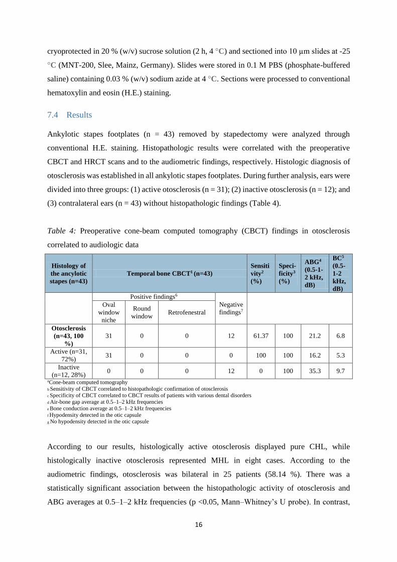
7.4 Results
Ankylotic stapes footplates (n = 43) removed by stapedectomy were analyzed through
conventional H.E. staining. Histopathologic results were correlated with the preoperative
CBCT and HRCT scans and to the audiometric findings, respectively. Histologic diagnosis of
otosclerosis was established in all ankylotic stapes footplates. During further analysis, ears were
divided into three groups: (1) active otosclerosis (n = 31); (2) inactive otosclerosis (n = 12); and
(3) contralateral ears (n = 43) without histopathologic findings (Table 4).
Table 4: Preoperative cone-beam computed tomography (CBCT) findings in otosclerosis
correlated to audiologic data
Histology of
the ancylotic
stapes (n=43)
Temporal bone CBCT1 (n=43)
Sensiti
vity2
(%)
Speci-
ficity3
(%)
ABG4
(0.5-1-
2 kHz,
dB)
BC5
(0.5-
1-2
kHz,
dB)
Positive findings6
Negative
findings7 Oval
window
niche
Round
window Retrofenestral
Otosclerosis
(n=43, 100
%)
31 0 0 12 61.37 100 21.2 6.8
Active (n=31,
72%) 31 0 0 0 100 100 16.2 5.3
Inactive
(n=12, 28%) 0 0 0 12 0 100 35.3 9.7
aCone-beam computed tomography
b Sensitivity of CBCT correlated to histopathologic confirmation of otosclerosis
c Specificity of CBCT correlated to CBCT results of patients with various dental disorders
d Air-bone gap average at 0.5–1–2 kHz frequencies
e Bone conduction average at 0.5–1–2 kHz frequencies
f Hypodensity detected in the otic capsule
g No hypodensity detected in the otic capsule
According to our results, histologically active otosclerosis displayed pure CHL, while
histologically inactive otosclerosis represented MHL in eight cases. According to the
audiometric findings, otosclerosis was bilateral in 25 patients (58.14 %). There was a
statistically significant association between the histopathologic activity of otosclerosis and
ABG averages at 0.5–1–2 kHz frequencies (p <0.05, Mann–Whitney’s U probe). In contrast,

17
no statistically significant association was found with BC averages in the two histopathologic
group of otosclerosis. Among the ears with otosclerosis (n = 43), CBCT revealed 31 positive
findings indicating a sensitivity for otosclerosis as 61.37 %. In the active otosclerosis group (n
= 31), the sensitivity of CBCT for otosclerosis increased to 100 %; while in case of inactive
otosclerosis (n = 12), sensitivity levels decreased to 0 %. According to CBCT findings,
otosclerosis was bilateral in 21 patients (48.83 %), which was smaller than the preoperative
audiometric estimation (58.14 %). However, this finding was not statistically significant. As
replication of previously reported data, a statistically significant and inverse association was
found between CBCT grades and ABG averages in ears with active or inactive otosclerosis (p
<0.001, Mann–Whitney’s U probe). Among all otosclerosis cases (n = 43), HRCT revealed 40
positive findings indicating a sensitivity for otosclerosis as 76.29 %. In the active otosclerosis
group (n = 31), the sensitivity of HRCT for otosclerosis increased to 100 %, while in case of
inactive otosclerosis (n = 12), sensitivity levels decreased to 59.3 % (Table 5).
Table 5: Preoperative high-resolution computed tomography (HRCT) findings in otosclerosis
correlated to audiologic data
Histology of
the ancylotic
staes (n=43)
Temporal bone HRCT1 (n=43)
Sensiti
vity2
(%)
Speci-
ficity3
(%)
ABG4
(0.5-1-
2 kHz,
dB)
BC5
(0.5-1-
2 kHz,
dB)
Positive findings6
Negatíve
findings7 Oval
window
niche
Round
window Retrofenestral
Otosclerosis
(n=43, 100
%)
31 2 13 3 76.29 100 21.2 6.8
Active (n=31,
72%) 31 1 9 0 100 100 16.2 5.3
Inactive
(n=12, 28%) 7 1 4 3 59.3 100 35.3 9.7
a High-resolution computed tomography
b Sensitivity of HRCT correlated to histopathologic confirmation of otosclerosis
c Specificity of HRCT correlated to HRCT results of patients with idiopathic sudden sensorineural hearing loss
d Air-bone gap average at 0.5–1–2 kHz frequencies
e Bone conduction average at 0.5–1–2 kHz frequencies
f Hypodensity detected in the otic capsule
g No hypodensity detected in the otic capsule
We have found statistically significant association between HRCT grades (fenestral or
retrofenestral) and ABG (p <0.05) and BC averages (p <0.001) in the contralateral ears. HRCT
grades also showed statistically significant association with BC (p <0.001) and ABG averages
(p <0.001) comparing the groups of active and inactive otosclerosis.

18
7.5 Discussion
In this study, various statistical associations were demonstrated between CBCT and HRCT
scans and audiometric findings in patients with histologically confirmed otosclerosis. The
weakness of our study is the relatively low number of subjects, however; is a comprehensive
imaging and histologic study that is able to assess the sensitivity and specificity values of CBCT
and HRCT scans in histologically confirmed otosclerosis. Sensitivity levels of HRCT scans
have been reported as 70.5–84.5 % in patients with stapes fixation. In histologically confirmed
cases, specificity levels were estimated as 100 %. According to our previous and present
observations, in the group of histologically confirmed otosclerosis, CBCT showed 61.37–65.62
% overall sensitivity, which was lower than that of previous reports. According to our
observations, in case of otosclerosis, HRCT showed 76.29 % overall sensitivity, which is more
robust, compared to the sensitivity levels of CBCT. In our series, however; CBCT seemed to
be less sensitive for non-symptomatic otosclerotic foci revealing bilateral otosclerosis as 48.83
%, in contrast to the 58.14 % prevalence revealed by pure tone audiometry. In contrast, HRCT
showed a good correspondence (55.81 %) with audiological findings in the clinical assessment
of bilateral cases. Histologically active otosclerosis with less ABG averages was characterized
by positive CBCT findings; however, histologically inactive cases with larger ABG averages
displayed negative CBCT scans. On the contrary, HRCT grades (fenestral or retrofenestral)
showed a statistically significant association with ABG and BC averages in the contralateral
ears and also in the histologically confirmed group of stapes footplates depending on the
histologic activity of otosclerosis. Regarding to HRCT findings, in case of inactive otosclerosis,
the sensorineural component of hearing impairment shows a strong correlation with the severity
and extension of otosclerosis. However, it cannot be confirmed by CBCT. Preoperative
evaluation of the extension of otosclerosis by combined application of HRCT and audiometry
has a great clinical significance, since patients with far advanced cochlear otosclerosis may
have benefit from cochlear, DACS or BAHA implantation depending on the bone conduction
threshold rather than stapedectomy or stapedtotomy. In conclusion, temporal bone HRCT is a
useful imaging method in the preoperative evaluation of different types of stapes fixations and
may serve as a reliable tool in the assessment of disease extension. Its sensitivity is much higher
for retrofenestral lesions and for inactive otosclerosis than that of CBCT. Therefore, it can be
stated that HRCT must be the first choice of temporal bone imaging. As we have previously
concluded, preoperative HRCT scan may serve as a valuable imaging method in the planning
19
of stapes surgery. It helps to avoid serious complications and unnecessary stapes surgeries by
the detection of several abnormalities in the middle or inner ear, such as large vestibular
aqueduct, dehiscent facial canal, superior semicircular canal dehiscence, round window
obliteration, persisting stapedial artery and malleus head fixation. Temporal bone CBCT is a
reliable imaging method in the preoperative evaluation of histologically active fenestral
otosclerosis. Other authors have reported that selected anatomic structures of the temporal bone
(n = 16) were clearly reconstructed by CBCT and no discrepancies were found compared to
HRCT findings. These results indicate that CBCT may also serve as a choice of temporal bone
imaging in case of CHL. Nevertheless, CBCT is a cheap, easy and a rapid imaging method that
is characterized by considerably lower radiation dose than HRCT with normal tympanic
membranes. Its overall sensitivity falls away from that of HRCT and histologic analysis;
however; it is continuously evolving due to the introduction of more powerful analyzer
softwares.
8 Summary
1. Applying laser technique and the NiTiBOND piston together we newly developed a
reliable method in stapes surgery which promise excellent short-term hearing results
2. We experienced easier manipulation with similar air-bone gap closure achieved with
NiTiBOND piston as compared with the SMart Nitinol piston, while the risk of incus
necrosis may continue to decrease
3. We introduced the surgical management of particular situations in stapes surgery,
such as stapes fixation associated with persistent stapedial artery, obliterative
otosclerosis, bilateral stapes fixation, otosclerosis leading to mixed hearing loss,
ptotic facial nerve, loose wire syndrome initially in the hungarian literature
4. The temporal bone CBCT was demonstrated to be a reliable and useful imaging
method in the preoperative evaluation of histologically active fenestral otosclerosis
5. We introduced HRCT as the imaging method of choice in temporal bone imaging to
differentiate between the types of stapes fixations preoperatively and to serve as a
reliable tool in the assessment of disease extension
20
9 Publications related to the thesis
1. Révész P, Harmat K, Háromi I, Ráth G, Karosi T, Molnár K, Gerlinger I. Különleges
stapes sebészeti megoldások – esetismertetések és irodalmi áttekintés. Fül-Orr-
Gégegyógyászat 2016; 62: 9-16.
2. Révész P, Szanyi I, Ráth G, Bocskai T, Lujber L, Piski Z, Karosi T, Gerlinger I.
Comparison of hearing results following the use of NiTiBOND versus Nitinol
prostheses in stapes surgery: a retrospective controlled study reporting short-term
postoperative results. Eur Arch Otorhinolaryngol 2016; 273:1131–6 IF:1.545
3. Liktor B, Révész P, Csomor P, Gerlinger I, Sziklai I, Karosi T. Diagnostic value of
cone-beam CT in histologically confirmed otosclerosis. Eur Arch Otorhinolaryngol
2014; 271 (8): 2131-8. IF: 1.545
4. Révész P, Liktor B, Liktor B, Sziklai I, Gerlinger I, Karosi T. Comparative analysis of
preoperative diagnostic values of HRCT and CBCT in patients with histologically
diagnosed otosclerotic stapes footplates. Eur Arch Otorhinolaryngol 2016; 273: 63-72.
IF:1.545
10 Further publications
1. Szabadi É, Török L, Révész P, Burián A, Gerlinger I, Lujber L. Dobhártyapótlás új
lehetőségének bemutatása állatkísérletes modellen. Fül-Orr-Gégegyógyászat 2010; 56
(3): 182.
2. Révész P, Burián A, Szabadi É, Bakó P, Gerlinger I, Pytel J, Lujber L. Subglotticus
lokalizációjú rhabdomyosarcoma fiatal férfibetegünknél. Fül-Orr-Gégegyógyászat
2010; 56 (3): 178.
3. Révész P, Gerlinger I. A fiatalság és az mp3 lejátszók – fokozott kockázat, kevés
óvintézkedés. Fül-Orr-Gégegyógyászat 2011; 57 (3): 35–38.
4. Lujber L, Révész P. Childhood Laryngeal Rhabdomyosarcoma Causing Acute Airway
Obstruction. Otolaryngol Head Neck Surg August 2011; 145 (2): 354-5.
5. Gerlinger, I., Révész, P., Piski, Z., Burián, A., Móricz, P.: Eseménytelen laser
stapedotomiát követő késői nervus facialis paresis esetismertetés és irodalmi áttekintés
Fül-Orr-Gégegyógyászat 2011; 57 (3): 133-7.
6. Piski, Z., Mózes, R., Burián, A., Révész, P., Gerlinger, I., Pytel, J.: Korral járó
hallászavarok korai kimutatása. Fül-Orr-Gégegyógyászat 2011; 57 (3): 138-44.
7. Juhász K, Gombos K, Szirmai M, Révész P, Magda I, Gocze K, Ember I. DMBA
induces deregulation of miRNA expression of let-7, miR-21 and miR-146a in CBA/CA
mice. In Vivo 2012; 26 (1):113-7.

21
8. Juhász K, Gombos K, Gocze K, Wolher V, Szirmai M, Révész P, Magda I, Sebestyén
A, Ember I. Effect of N-methyl-N-nitrosourea on microRNA expression in CBA/Ca
mice. Journal of Enviromental and Occupational Science 2012; 1(2): 77-82.
9. Szanyi I, Ráth G, Móricz P, Somogyvári K, Révész P, Gerlinger I, Orsós Zs, Ember I,
Kiss I. Effects of cytochrome P450 1A1 (CYP 1A1) and UGT-glucuronyltransferase
1A1 (UGT 1A1) allelic polymorphisms on the risk of development and the prognosis
of head and neck tumors. Eur J Cancer Prev 2012; 21(6):560-8.
10. Móricz P, Somogyvári K, Burián A, Piski Z, Révész P, Gerlinger I. Hypertonicitás
okozta aphonia megoldása myotomiával hangprotézis beültetés után Fül-Orr-
Gégegyógyászat 2013; 59(2): 45-6.
11. Juhász K, Gombos K, Szirmai M, Gőcze K, Wolher V, Révész P, Magda I, Sebestyén
A, Németh A, Ember I. Very early effect of DMBA and MNU on microRNA
expression. In Vivo 2013; 27 (1):113-7.
12. Gobel G, Szanyi I, Révész P, Bauer M, Gerlinger I, Németh Á, Ember I, Gocze K,
Gombos K. Expression of NFkB1, GADD45A and JNK1 in salivary gland carcinomas
of different histotypes. Cancer Genomics Proteomics 2013; 10 (2):81-7.
13. Révész P, Szabadi É, Járai T, Tornóczky T, Gerlinger I, Lujber L. Chorda tympani
neurinoma. Fül-Orr-Gégegyógyászat 2013; 57 (4).
14. Ráth G, Katona G, Bakó P, Török L, Révész P, Tóth E, Gerlinger I. Application of
ionomer cement onto the stapedial footplate: Impact on the perilymphatic aluminum
level. Laryngoscope 2014; 124(2): 541-4.
15. Gerlinger I, Bakó P, Piski Z, Révész P, Ráth G, Karosi T, Lujber L. KTP laser
stapedotomy with a self-crimping, thermal shape memory Nitinol piston: follow-up
study reporting intermediate-term hearing. Eur Arch Otorhinolaryngol 2014; 271(12):
3171-7.
16. Orosz E, Gombos K, Révész P, Kiss I, Pytel J, Gerlinger I, Szanyi I. MicroRNA
expression profiles in squamous cell carcinomas of the meso- and hypopharynx. Orv
Hetil 2014; 155(27):1063-70.
17. Révész P, Piski Z, Burián A, Harmat K, Gerlinger I. Delayed Facial Paralysis following
Uneventful KTP Laser Stapedotomy: Two Case Reports and a Review of the Literature.
Case Rep Med 2014; 2014:971362. doi: 10.1155/2014/971362.
18. Szirmai M, Juhász K, Bertha A, Gombos K, Gőcze K, Magda I, Révész P, I Ember.
Potential chemopreventive effect of “Procont” on miRNA expression in CBA/Ca mice.
European Medical Health and Pharmaceutical Journal 2012; 3: 24-28.
19. Szanyi I, Gerlinger I, Lujber L, Szabadi É, Burián A, Révész P, Ember I, Kiss I.
Onkogén és tumorszuppresszor génexpresszió változások biológiai markerként való
alkalmazása malignus fej-nyaki daganatokban. Fül-Orr-Gégegyógyászat 2011; 57 (2):
66-72.
20. Glavanits M, Millei L, Révész P. „Keep smiling” – fogpótlás, rekonstrukciós-,
szépészeti sebészet, arcátültetés. In: Révész Gy (szerk.) Az emberi arc: Tanulmányok a
pszichológia, orvostudomány, mesterséges intelligencia és a képzőművészet
területeiről. Pécs: Pro Pannonia Kiadó, 2010. P. 135-149.

22
11 Ackowledgements
I am grateful to my tutors, Professor Imre Gerlinger and Dr. Tamás Karosi for their help and
support in order to create the thesis.
I thank to my family and fiancée for providing a secure background.
I thank to Dr. Péter Csomor for his work evaluating the histological slides.
I express my thanks to Dr. Károly Berényi and Renaat Coopman for their help in statistical
analysis.