Embed Size (px)
Citation preview
Exercise 38Exercise 38
Anatomy of the Anatomy of the Digestive SystemDigestive System
ObjectivesObjectives• Overall functionOverall function• Organs of alimentary canalOrgans of alimentary canal• Accessory digestive organsAccessory digestive organs• General functions of organs/structuresGeneral functions of organs/structures• Histological structure of alimentary canal Histological structure of alimentary canal
wallwall• Stomach and small intestine specializationsStomach and small intestine specializations• Enzymes producedEnzymes produced• Deciduous and permanent teethDeciduous and permanent teeth
Digestive SystemDigestive System
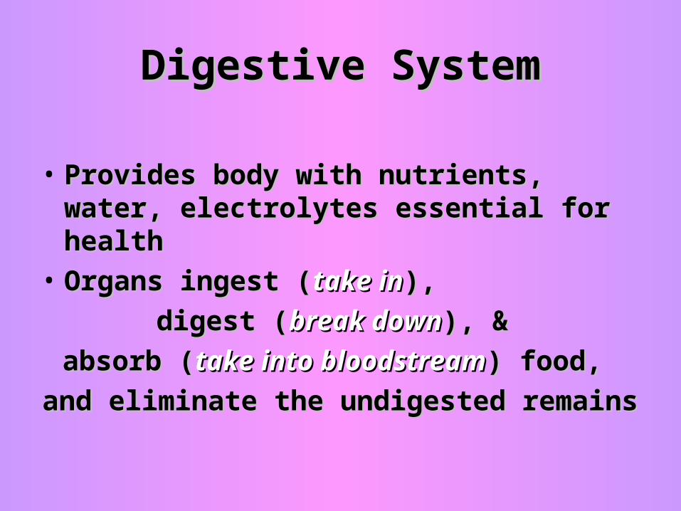
• Provides body with nutrients, water, Provides body with nutrients, water, electrolytes essential for healthelectrolytes essential for health
• Organs ingest (Organs ingest (take intake in), ),
digest (digest (break downbreak down), & ), &
absorb (absorb (take into bloodstreamtake into bloodstream) food, ) food,
and eliminate the undigested remainsand eliminate the undigested remains
Alimentary CanalAlimentary Canal
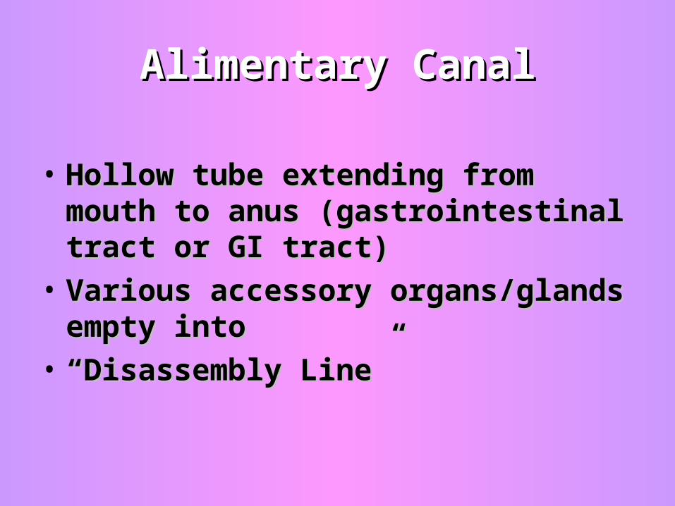
• Hollow tube extending from mouth to Hollow tube extending from mouth to anus (gastrointestinal tract or GI tract)anus (gastrointestinal tract or GI tract)
• Various accessory organs/glands Various accessory organs/glands empty intoempty into
• ““Disassembly Line”Disassembly Line”
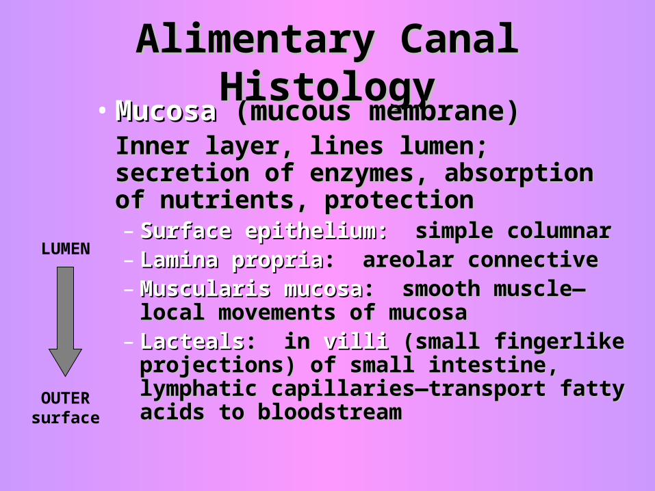
Alimentary Canal HistologyAlimentary Canal Histology• MucosaMucosa (mucous membrane) (mucous membrane)
Inner layer, lines lumen; secretion of Inner layer, lines lumen; secretion of enzymes, absorption of nutrients, enzymes, absorption of nutrients, protectionprotection– Surface epithelium:Surface epithelium: simple columnar simple columnar– Lamina propriaLamina propria: areolar connective : areolar connective – Muscularis mucosaMuscularis mucosa: smooth muscle—: smooth muscle—
local movements of mucosalocal movements of mucosa– LactealsLacteals: in : in villivilli (small fingerlike (small fingerlike
projections) of small intestine, projections) of small intestine, lymphatic capillaries—transport fatty lymphatic capillaries—transport fatty acids to bloodstreamacids to bloodstream
LUMEN
OUTER surface
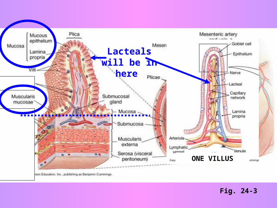
Fig. 24-3
Lacteals will be in here
ONE VILLUS
Alimentary Canal HistologyAlimentary Canal Histology
Fig. 24-3
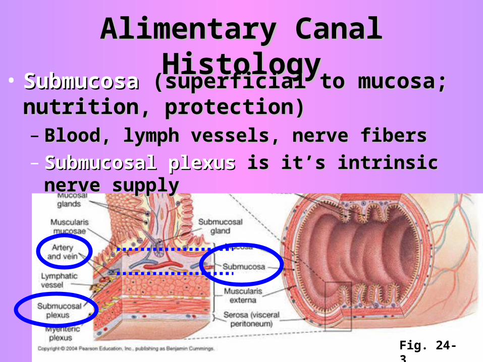
• SubmucosaSubmucosa (superficial to mucosa; (superficial to mucosa; nutrition, protection)nutrition, protection)– Blood, lymph vessels, nerve fibersBlood, lymph vessels, nerve fibers– Submucosal plexusSubmucosal plexus is it’s intrinsic nerve supply is it’s intrinsic nerve supply
Alimentary Canal HistologyAlimentary Canal Histology
Fig. 24-3
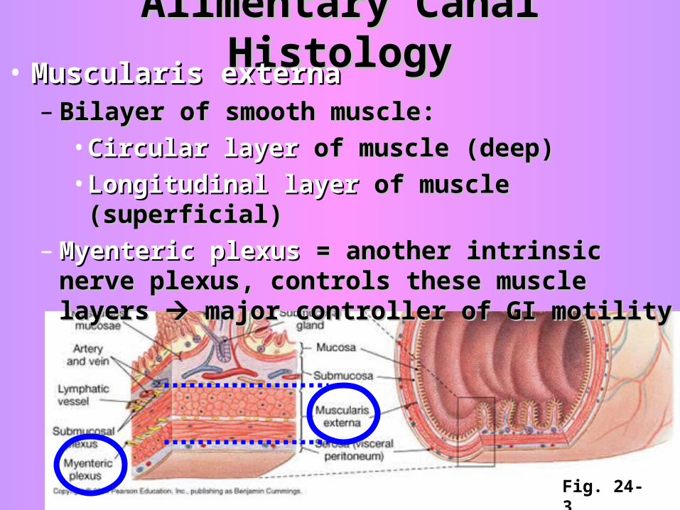
• Muscularis externaMuscularis externa– Bilayer of smooth muscle: Bilayer of smooth muscle:
• Circular layerCircular layer of muscle (deep) of muscle (deep)• Longitudinal layerLongitudinal layer of muscle (superficial) of muscle (superficial)
– Myenteric plexusMyenteric plexus = another intrinsic nerve = another intrinsic nerve plexus, controls these muscle layers plexus, controls these muscle layers major major controller of GI motilitycontroller of GI motility
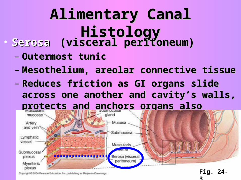
Alimentary Canal HistologyAlimentary Canal Histology• SerosaSerosa (visceral peritoneum)(visceral peritoneum)
– Outermost tunicOutermost tunic– Mesothelium, areolar connective tissueMesothelium, areolar connective tissue– Reduces friction as GI organs slide across one Reduces friction as GI organs slide across one
another and cavity’s walls, protects and another and cavity’s walls, protects and anchors organs alsoanchors organs also
Fig. 24-3
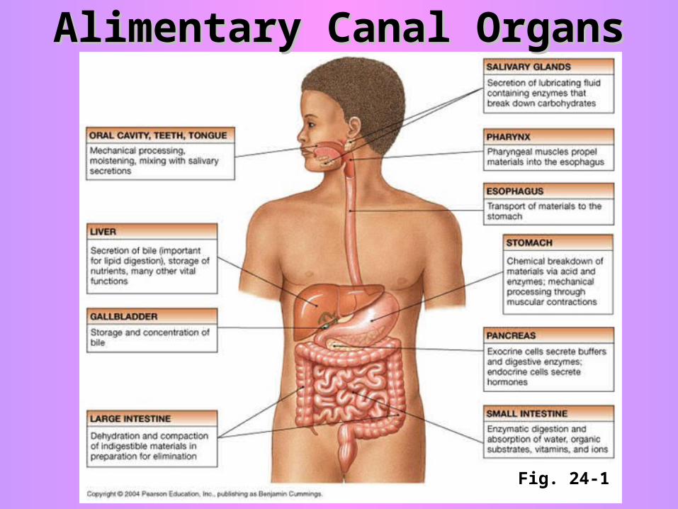
Alimentary Canal OrgansAlimentary Canal Organs
Fig. 24-1
Alimentary Canal OrgansAlimentary Canal Organs
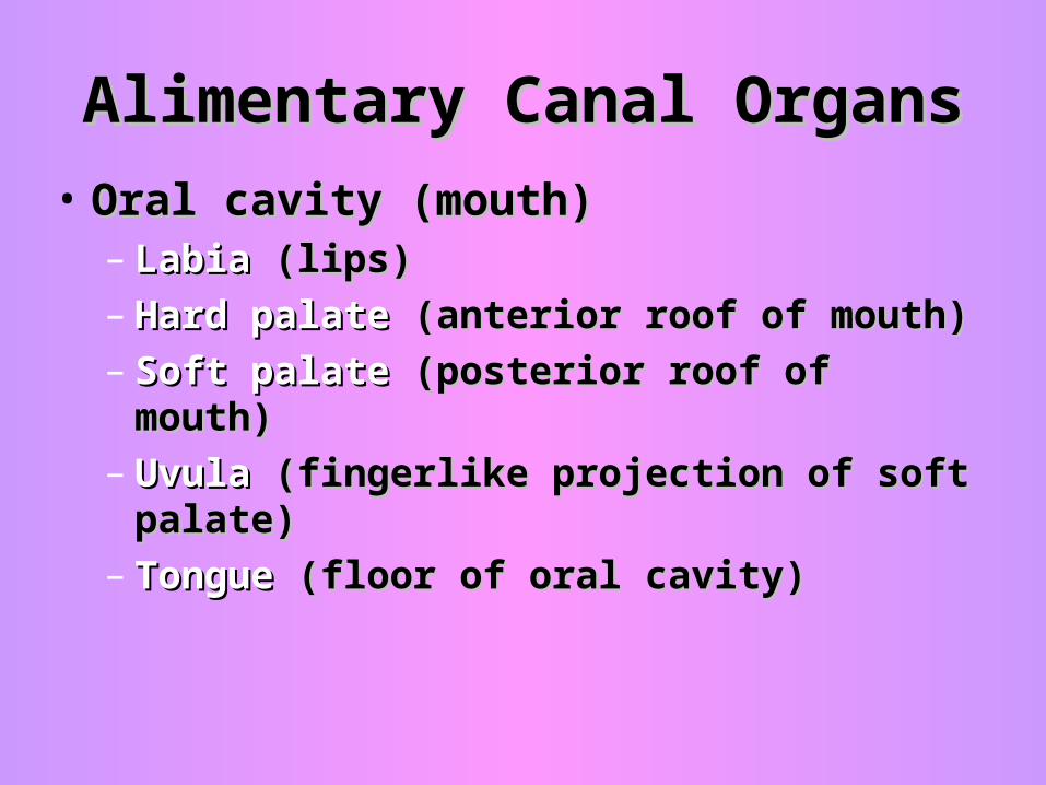
• Oral cavity (mouth)Oral cavity (mouth)– Labia Labia (lips)(lips)– Hard palateHard palate (anterior roof of mouth) (anterior roof of mouth)– Soft palateSoft palate (posterior roof of mouth) (posterior roof of mouth)– Uvula Uvula (fingerlike projection of soft palate)(fingerlike projection of soft palate)– Tongue Tongue (floor of oral cavity)(floor of oral cavity)
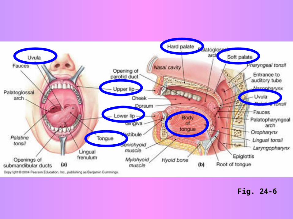
Fig. 24-6
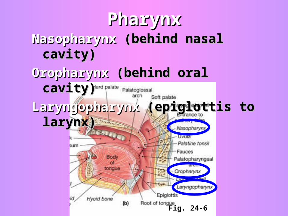
PharynxPharynx
Fig. 24-6
NasopharynxNasopharynx (behind nasal cavity) (behind nasal cavity)
OropharynxOropharynx (behind oral cavity) (behind oral cavity)
Laryngopharynx Laryngopharynx (epiglottis to larynx)(epiglottis to larynx)
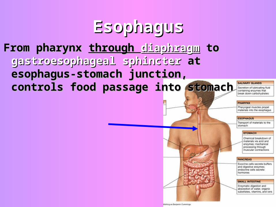
EsophagusEsophagusFrom pharynx From pharynx through through diaphragmdiaphragm to to
gastroesophageal sphinctergastroesophageal sphincter at at esophagus-stomach junction, esophagus-stomach junction, controls food passage into stomachcontrols food passage into stomach
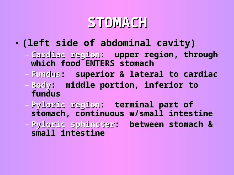
STOMACHSTOMACH• (left side of abdominal cavity)(left side of abdominal cavity)
– Cardiac regionCardiac region: upper region, through : upper region, through which food ENTERS stomachwhich food ENTERS stomach
– FundusFundus: superior & lateral to cardiac: superior & lateral to cardiac– BodyBody: middle portion, inferior to fundus: middle portion, inferior to fundus– Pyloric regionPyloric region: terminal part of stomach, : terminal part of stomach,
continuous w/small intestinecontinuous w/small intestine– Pyloric sphincterPyloric sphincter: between stomach & : between stomach &
small intestinesmall intestine
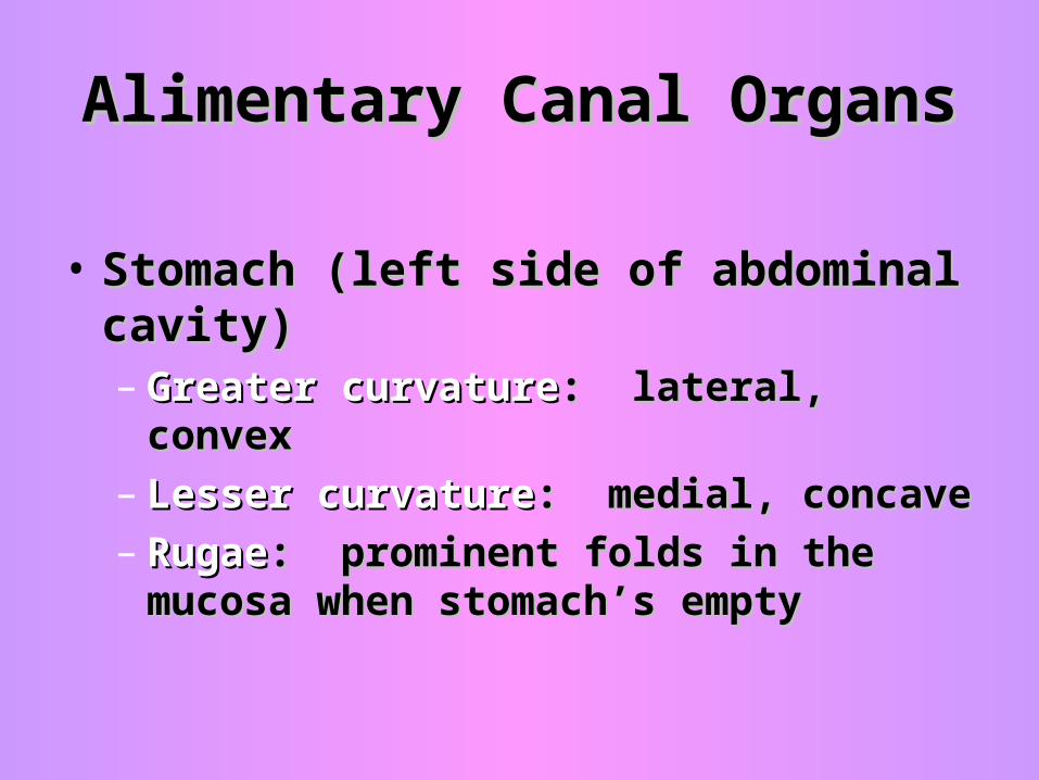
Alimentary Canal OrgansAlimentary Canal Organs
• Stomach (left side of abdominal cavity)Stomach (left side of abdominal cavity)– Greater curvatureGreater curvature: lateral, convex: lateral, convex– Lesser curvatureLesser curvature: medial, concave: medial, concave– RugaeRugae: prominent folds in the mucosa : prominent folds in the mucosa
when stomach’s emptywhen stomach’s empty
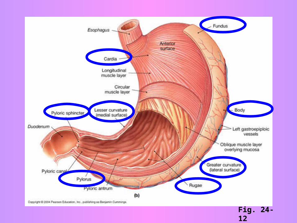
Fig. 24-12
SMALL INTESTINESMALL INTESTINE~2m long~2m long
– DuodenumDuodenum: from pyloric sphincter around : from pyloric sphincter around pancreaspancreas
– JejunumJejunum: umbilical region of abdomen: umbilical region of abdomen– IleumIleum: terminal portion, joins lg intestine: terminal portion, joins lg intestine– Plicae circularisPlicae circularis: like rugae, deep folds: like rugae, deep folds– Ileocecal valveIleocecal valve: between small and large : between small and large
intestineintestine
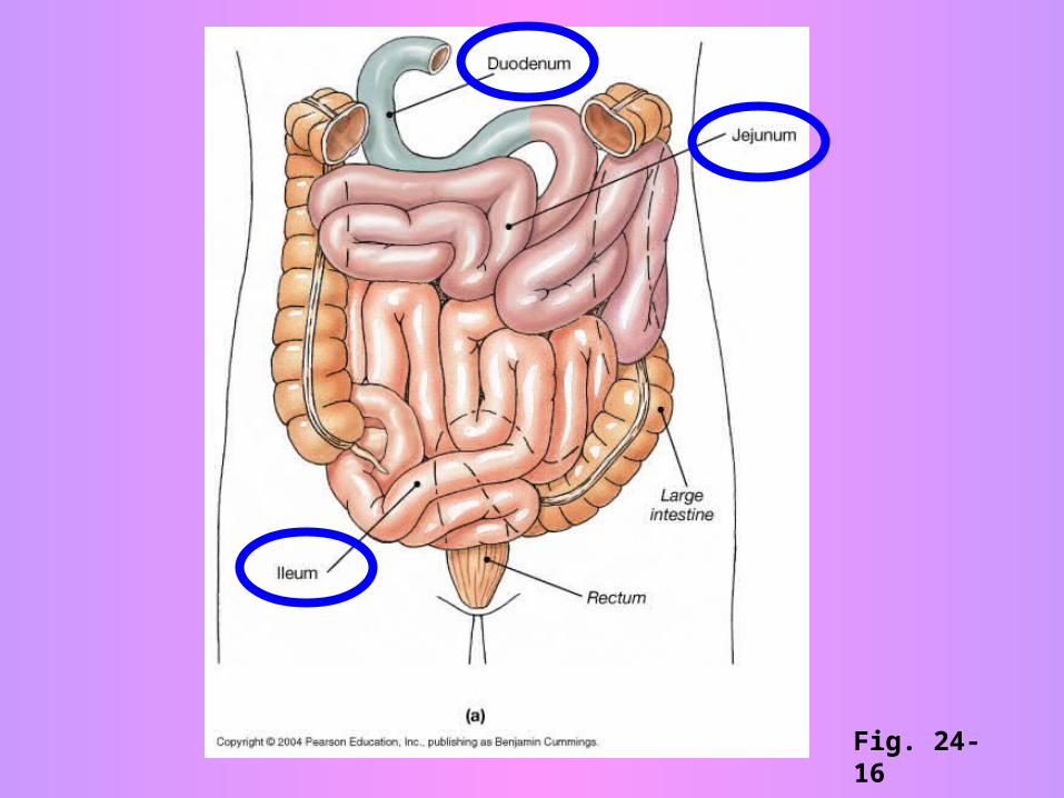
Fig. 24-16
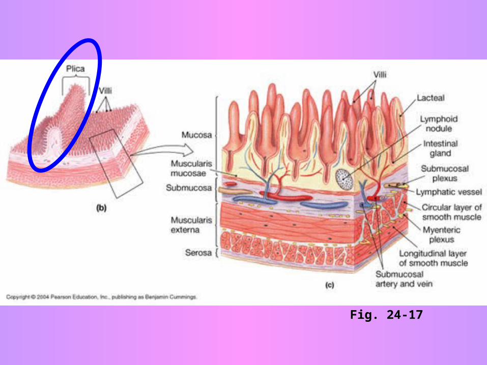
Fig. 24-17
LARGE INTESTINELARGE INTESTINE~1.5m long~1.5m long
Encircles the small intestine on 3 sidesEncircles the small intestine on 3 sides– CecumCecum: 1: 1stst region, expanded pouch region, expanded pouch
– AppendixAppendix: : attached to cecum, ~3.5” attached to cecum, ~3.5” longlong
– Ascending colonAscending colon: up the right side: up the right side
– Transverse colonTransverse colon: across the top: across the top
– Descending colonDescending colon: down the left side: down the left side
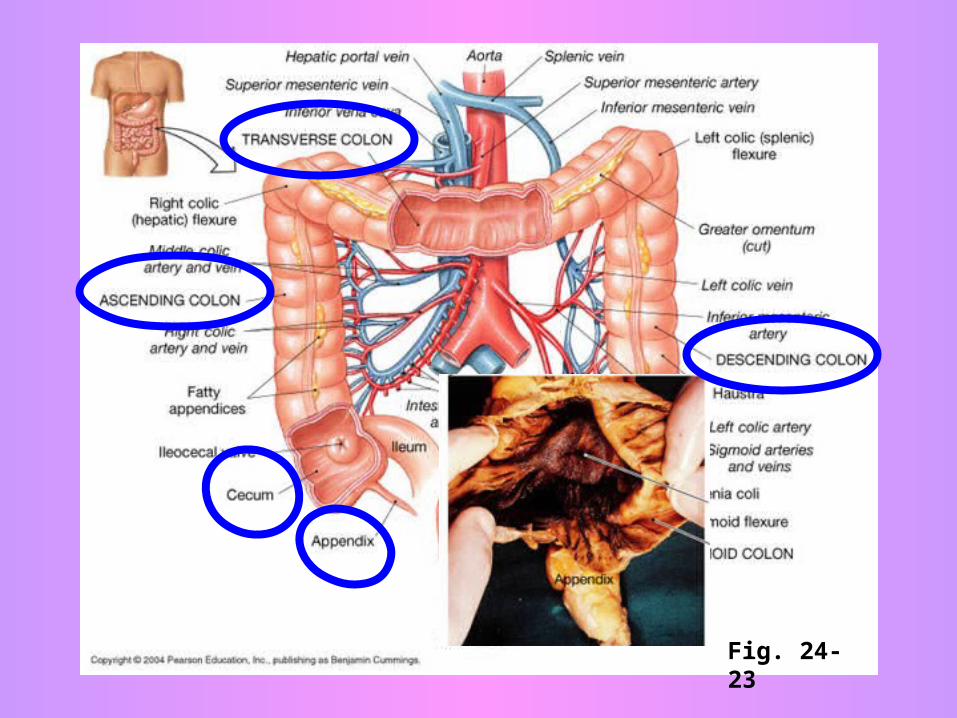
Fig. 24-23
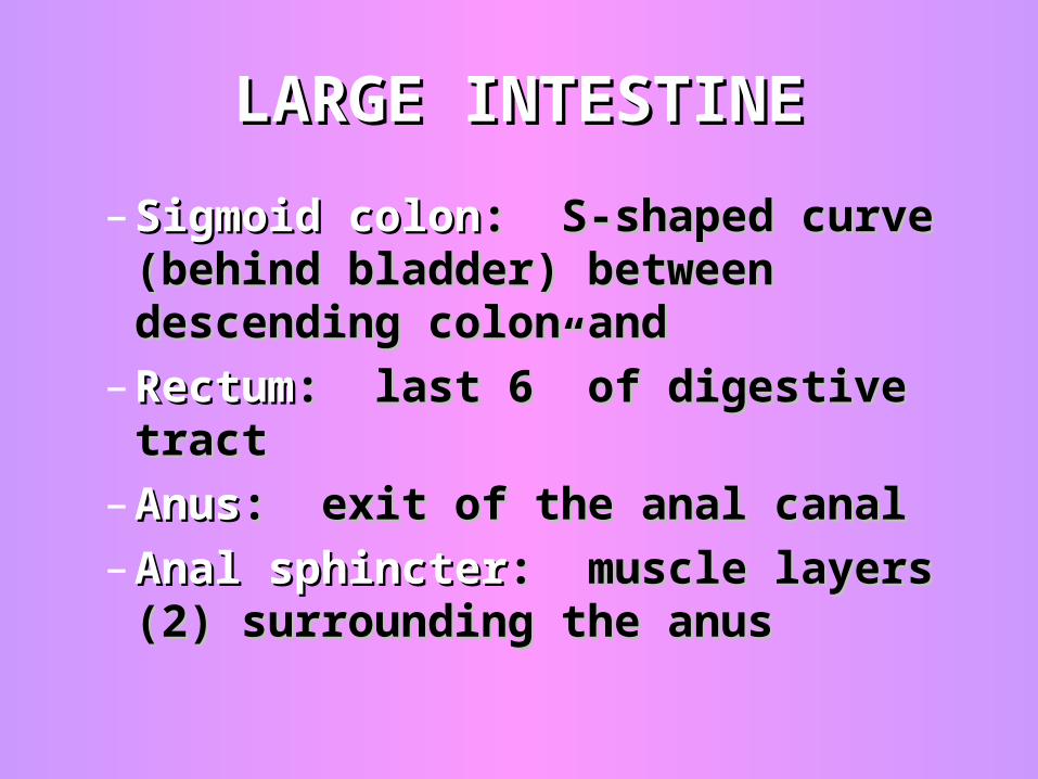
– Sigmoid colonSigmoid colon: S-shaped curve : S-shaped curve (behind bladder) between descending (behind bladder) between descending colon and colon and
– RectumRectum: last 6” of digestive tract: last 6” of digestive tract
– AnusAnus: exit of the anal canal: exit of the anal canal
– Anal sphincterAnal sphincter: muscle layers (2) : muscle layers (2) surrounding the anussurrounding the anus
LARGE INTESTINELARGE INTESTINE
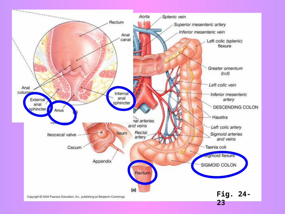
Fig. 24-23
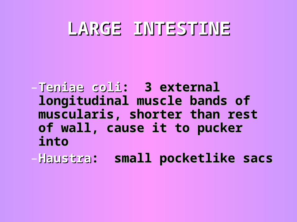
–Teniae coliTeniae coli: 3 external : 3 external longitudinal muscle bands of longitudinal muscle bands of muscularis, shorter than rest of muscularis, shorter than rest of wall, cause it to pucker intowall, cause it to pucker into
–HaustraHaustra: small pocketlike sacs: small pocketlike sacs
LARGE INTESTINELARGE INTESTINE
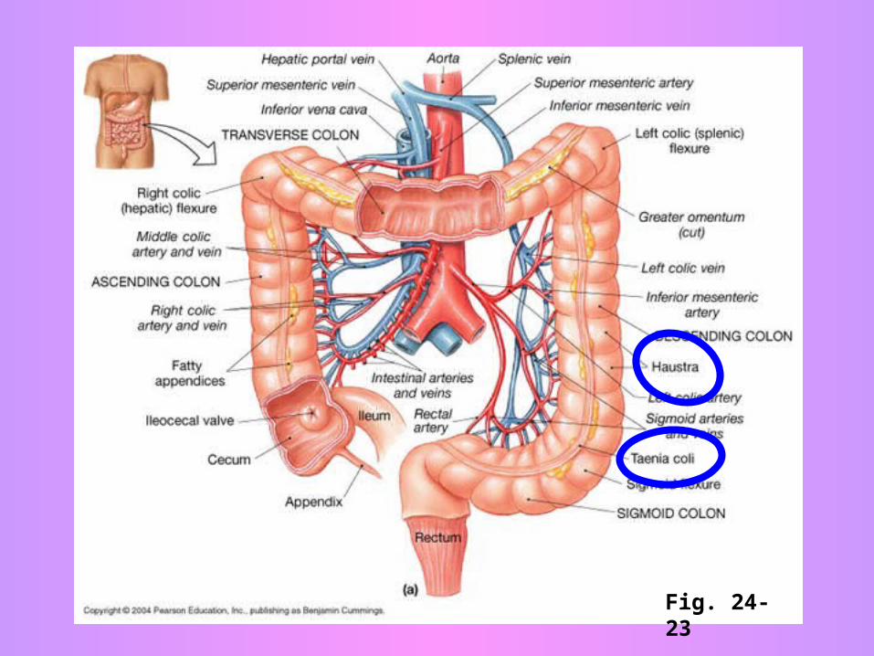
Fig. 24-23
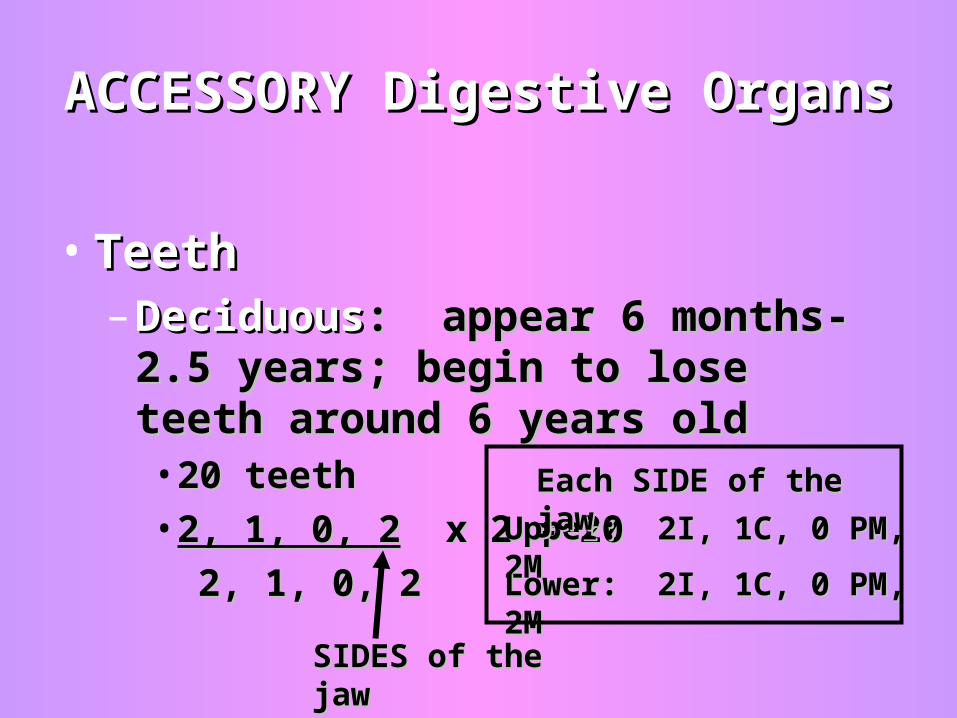
ACCESSORY Digestive OrgansACCESSORY Digestive Organs
• TeethTeeth– DeciduousDeciduous: appear 6 months-2.5 : appear 6 months-2.5
years; begin to lose teeth around 6 years; begin to lose teeth around 6 years oldyears old• 20 teeth 20 teeth • 2, 1, 0, 22, 1, 0, 2 x 2 = 20 x 2 = 20
2, 1, 0, 22, 1, 0, 2
Upper: 2I, 1C, 0 PM, 2MUpper: 2I, 1C, 0 PM, 2M
Lower: 2I, 1C, 0 PM, 2MLower: 2I, 1C, 0 PM, 2M
SIDES of the jawSIDES of the jaw
Each SIDE of the jawEach SIDE of the jaw
TEETHTEETH
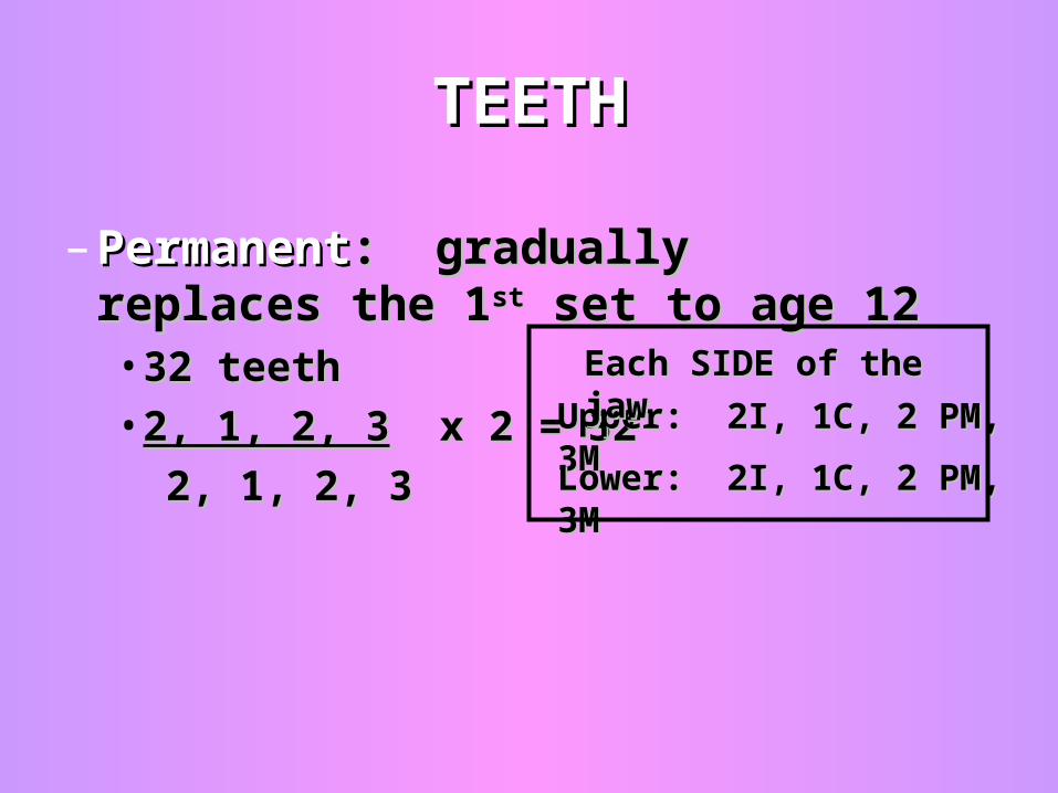
– PermanentPermanent: gradually replaces the 1: gradually replaces the 1stst set to age 12set to age 12• 32 teeth 32 teeth • 2, 1, 2, 32, 1, 2, 3 x 2 = 32 x 2 = 32
2, 1, 2, 32, 1, 2, 3
Upper: 2I, 1C, 2 PM, 3MUpper: 2I, 1C, 2 PM, 3M
Lower: 2I, 1C, 2 PM, 3MLower: 2I, 1C, 2 PM, 3M
Each SIDE of the jawEach SIDE of the jaw
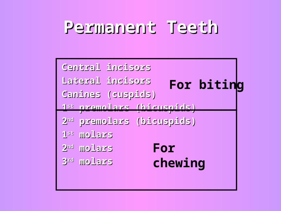
Permanent TeethPermanent Teeth
Central incisorsCentral incisors
Lateral incisorsLateral incisors
Canines (cuspids)Canines (cuspids)
11stst premolars (bicuspids) premolars (bicuspids)
22ndnd premolars (bicuspids) premolars (bicuspids)
11stst molars molars
22ndnd molars molars
33rdrd molars molars
For biting
For chewing
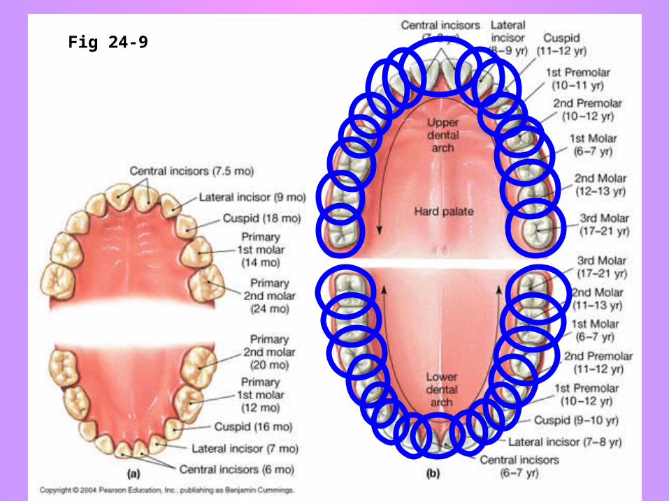
Fig 24-9
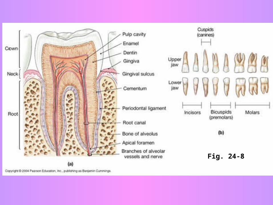
Fig. 24-8
TeethTeeth
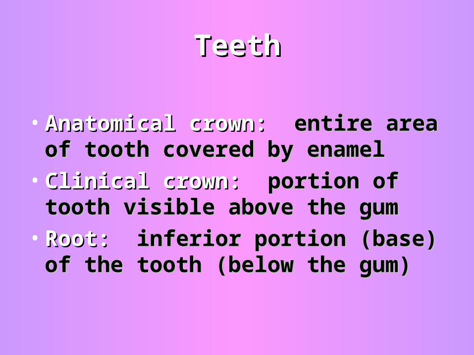
• Anatomical crown: Anatomical crown: entire area of entire area of tooth covered by enameltooth covered by enamel
• Clinical crown: Clinical crown: portion of tooth portion of tooth visible above the gumvisible above the gum
• Root:Root: inferior portion (base) of the inferior portion (base) of the tooth (below the gum)tooth (below the gum)
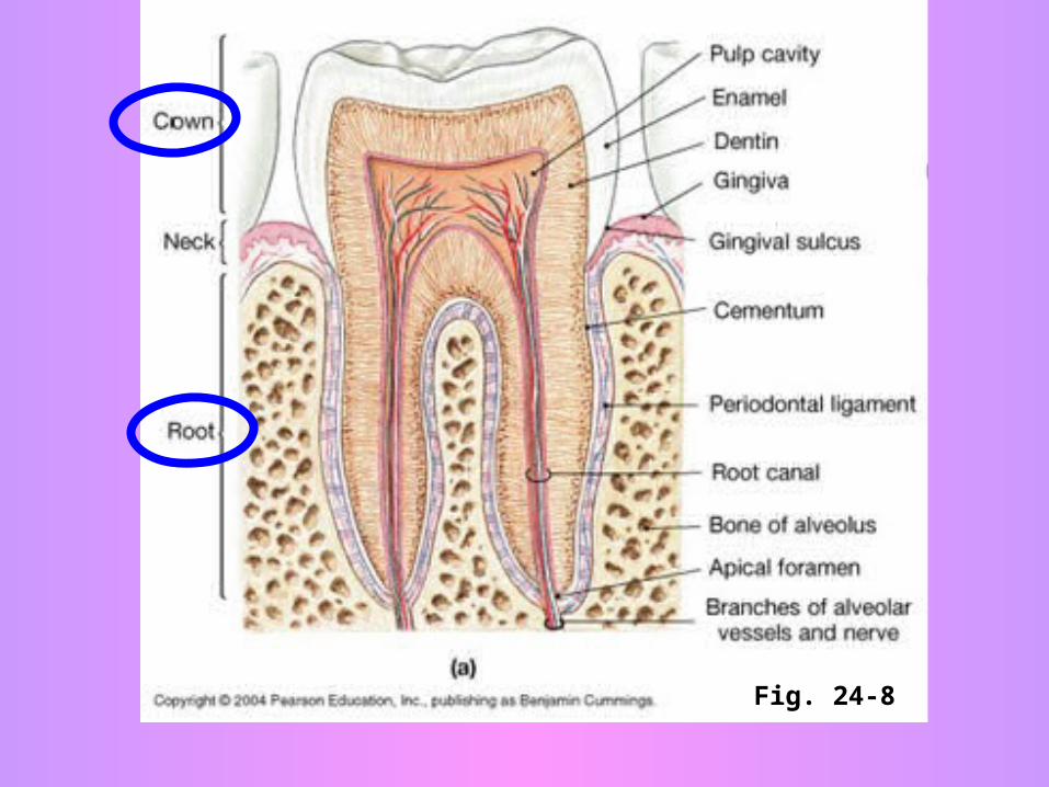
Fig. 24-8
TeethTeeth
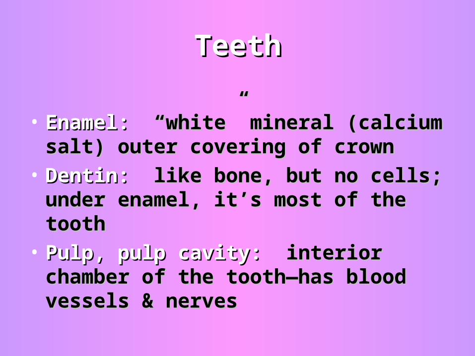
• Enamel: Enamel: “white” mineral (calcium salt) “white” mineral (calcium salt) outer covering of crownouter covering of crown
• Dentin: Dentin: like bone, but no cells; under like bone, but no cells; under enamel, it’s most of the toothenamel, it’s most of the tooth
• Pulp, pulp cavity:Pulp, pulp cavity: interior chamber of interior chamber of the tooth—has blood vessels & nervesthe tooth—has blood vessels & nerves
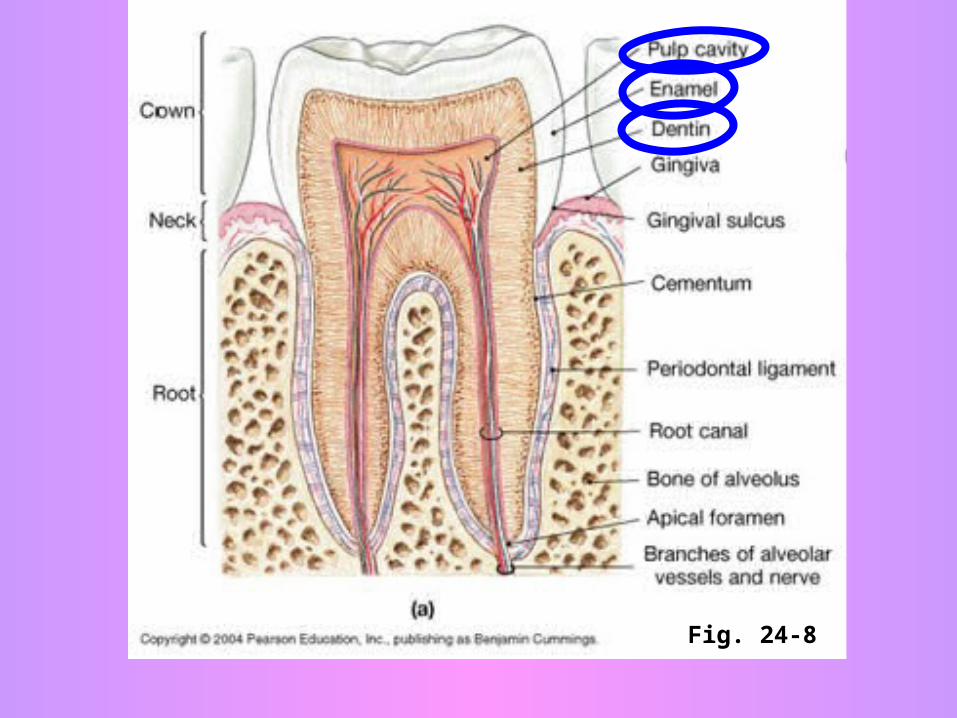
Fig. 24-8
SALIVARY GLANDSSALIVARY GLANDS
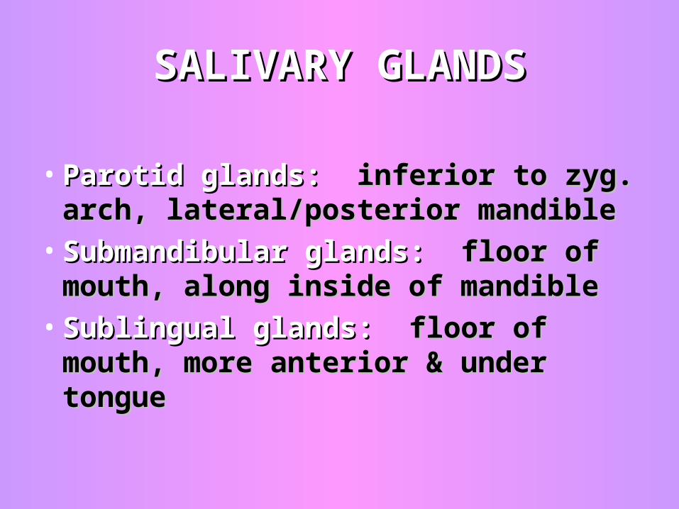
• Parotid glands: Parotid glands: inferior to zyg. inferior to zyg. arch, lateral/posterior mandiblearch, lateral/posterior mandible
• Submandibular glands: Submandibular glands: floor of floor of mouth, along inside of mandiblemouth, along inside of mandible
• Sublingual glands:Sublingual glands: floor of mouth, floor of mouth, more anterior & under tonguemore anterior & under tongue
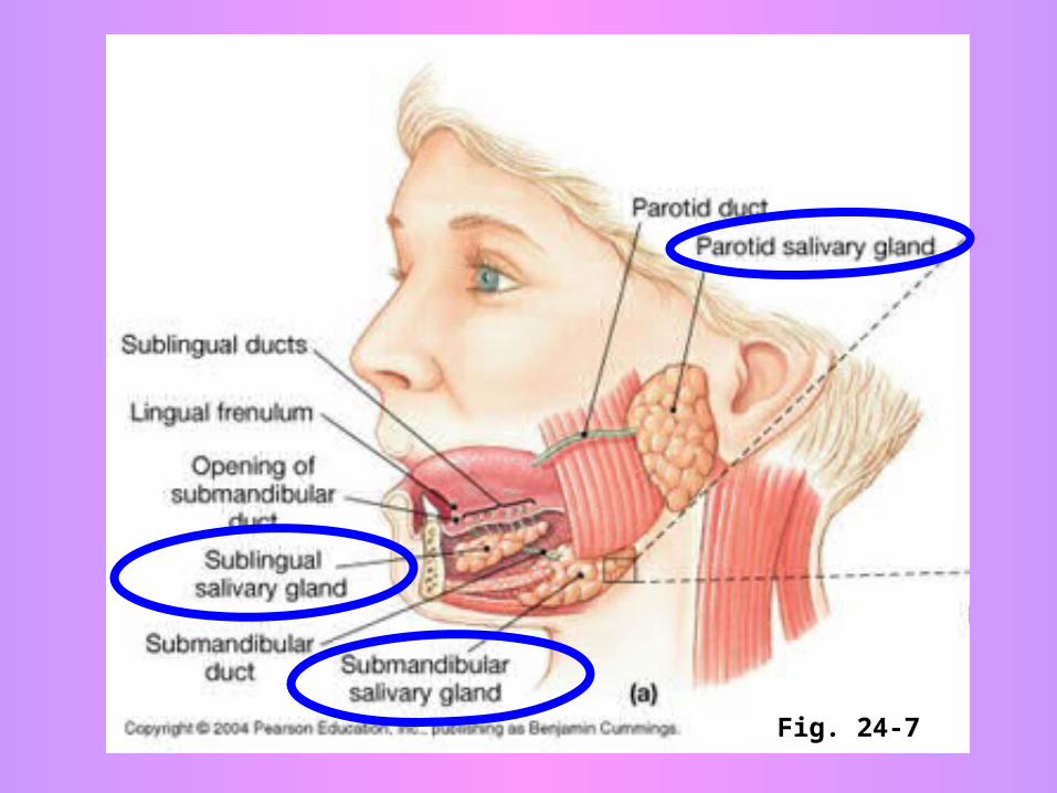
Fig. 24-7
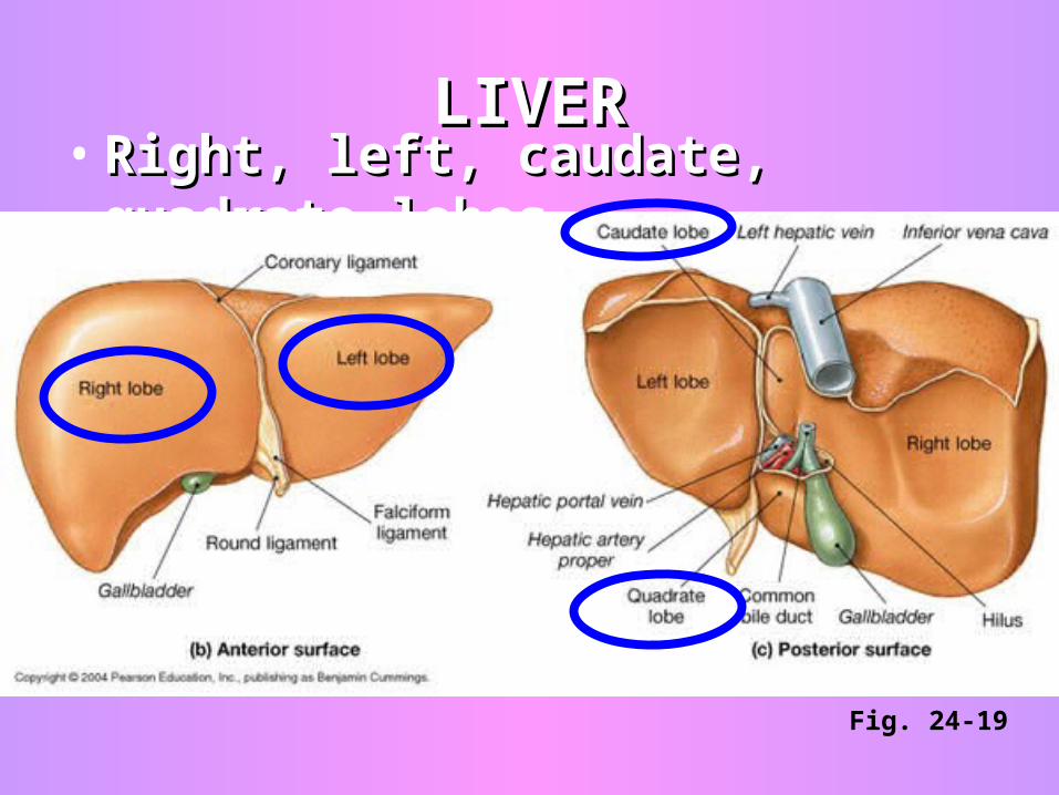
LIVERLIVER• Right, left, caudate, quadrate lobesRight, left, caudate, quadrate lobes
Fig. 24-19
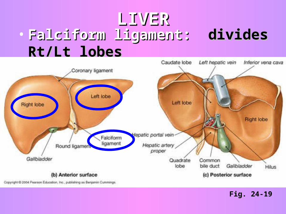
LIVERLIVER• Falciform ligament: Falciform ligament: divides Rt/Lt divides Rt/Lt
lobeslobes
Fig. 24-19
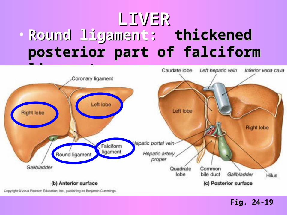
LIVERLIVER• Round ligament: Round ligament: thickened thickened
posterior part of falciform ligamentposterior part of falciform ligament
Fig. 24-19
LIVERLIVER• Hepatic ducts: Hepatic ducts: right, left—collect bile right, left—collect bile
(secreted by liver) from all bile ducts of (secreted by liver) from all bile ducts of lobes, unite to form thelobes, unite to form the
• Common hepatic ductCommon hepatic duct which leaves the which leaves the liver…bile then flows toliver…bile then flows to
• Cystic ductCystic duct which leads to the which leads to the gallbladder gallbladder (stores/concentrates bile)(stores/concentrates bile) ….OR goes to the ….OR goes to the
• Common bile ductCommon bile duct which is formed by which is formed by union of cystic and common hepatic union of cystic and common hepatic ducts--empties into the duodenum (sm ducts--empties into the duodenum (sm intest), intest),
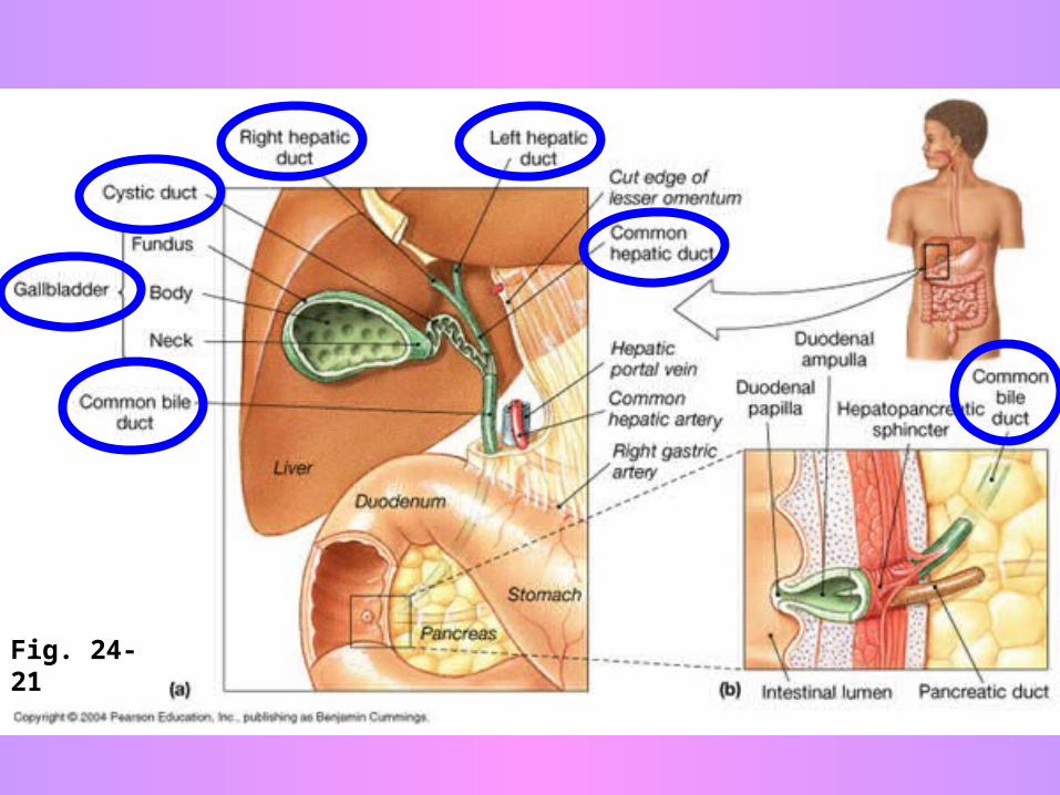
Fig. 24-21
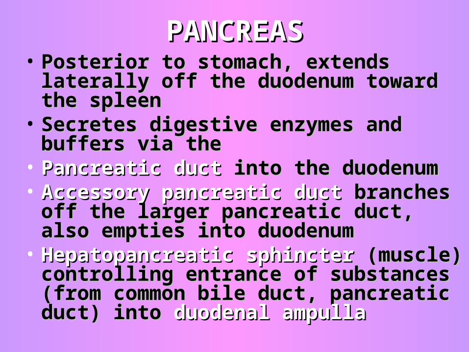
PANCREASPANCREAS• Posterior to stomach, extends laterally off Posterior to stomach, extends laterally off
the duodenum toward the spleenthe duodenum toward the spleen• Secretes digestive enzymes and buffers Secretes digestive enzymes and buffers
via thevia the• Pancreatic duct Pancreatic duct into the duodenuminto the duodenum • Accessory pancreatic ductAccessory pancreatic duct branches off branches off
the larger pancreatic duct, also empties the larger pancreatic duct, also empties into duodenuminto duodenum
• Hepatopancreatic sphincter Hepatopancreatic sphincter (muscle) (muscle) controlling entrance of substances (from controlling entrance of substances (from common bile duct, pancreatic duct) into common bile duct, pancreatic duct) into duodenal ampulladuodenal ampulla
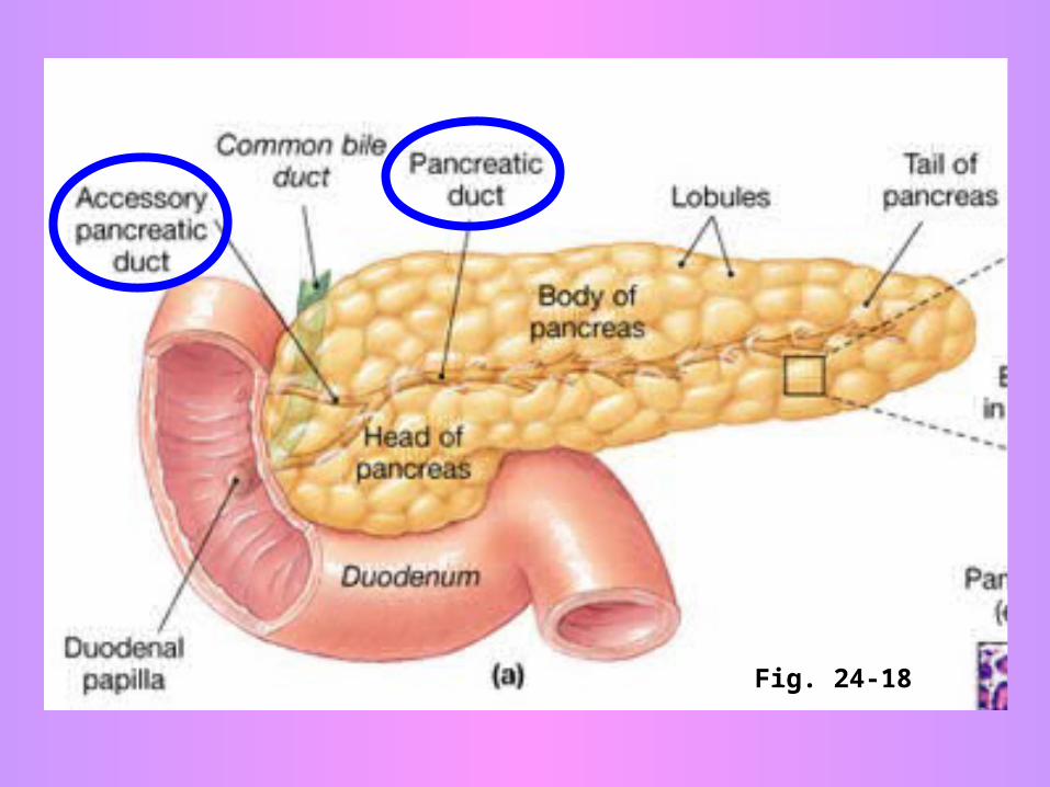
Fig. 24-18
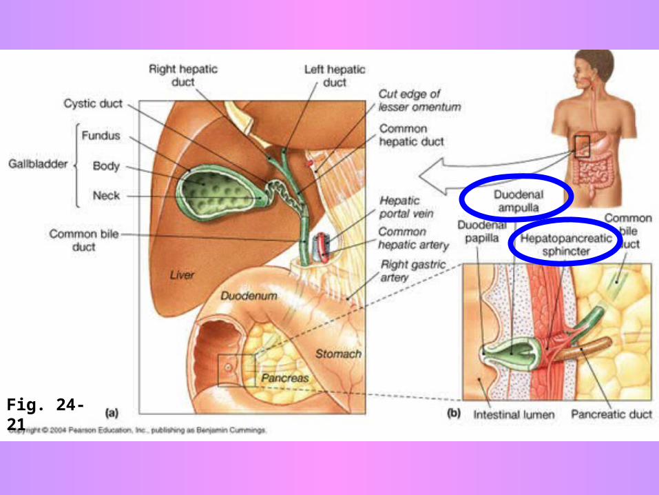
Fig. 24-21
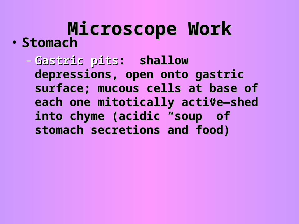
Microscope WorkMicroscope Work• StomachStomach
– Gastric pitsGastric pits: shallow depressions, open : shallow depressions, open onto gastric surface; mucous cells at base onto gastric surface; mucous cells at base of each one mitotically active—shed into of each one mitotically active—shed into chyme (acidic “soup” of stomach chyme (acidic “soup” of stomach secretions and food)secretions and food)
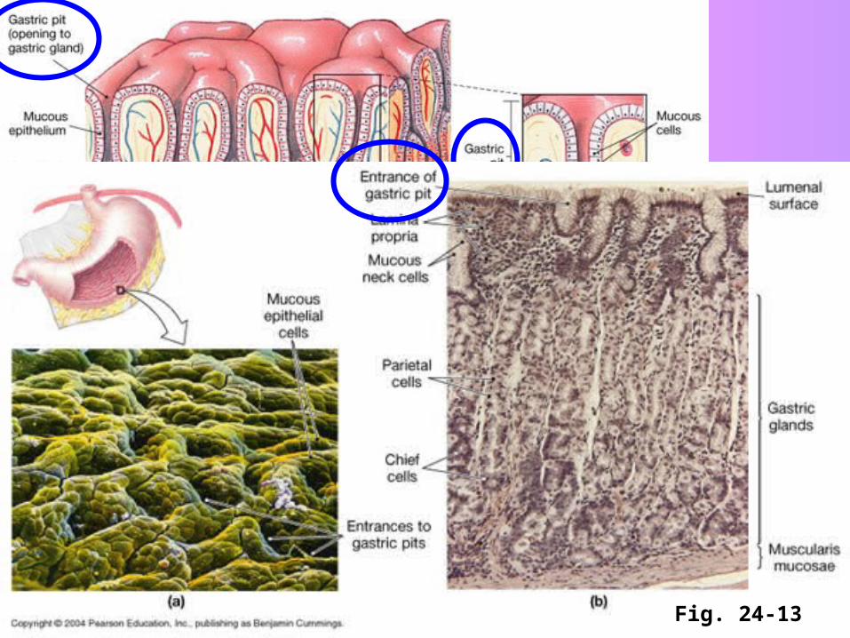
Fig. 24-13Fig. 24-13
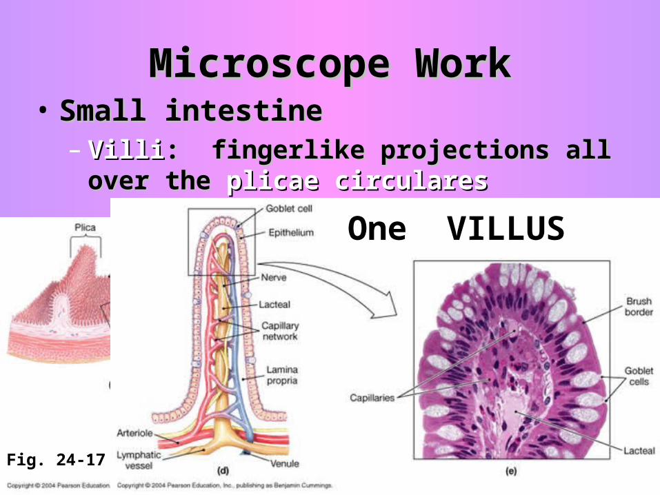
Microscope WorkMicroscope Work• Small intestineSmall intestine
– VilliVilli: fingerlike projections all over the : fingerlike projections all over the plicae circularesplicae circulares
Fig. 24-17
One VILLUS
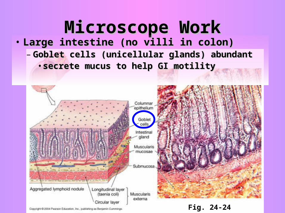
Microscope WorkMicroscope Work• Large intestine (no villi in colon)Large intestine (no villi in colon)
– Goblet cells (unicellular glands) abundantGoblet cells (unicellular glands) abundant• secrete mucus to help GI motilitysecrete mucus to help GI motility
Fig. 24-24