Embed Size (px)
Citation preview
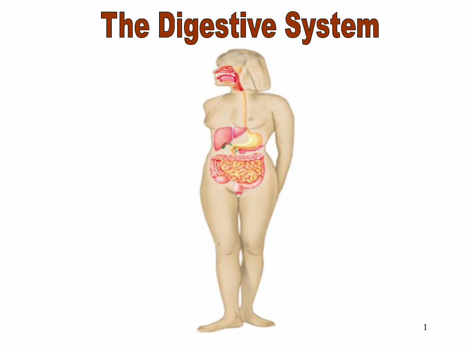
1
The Digestive System
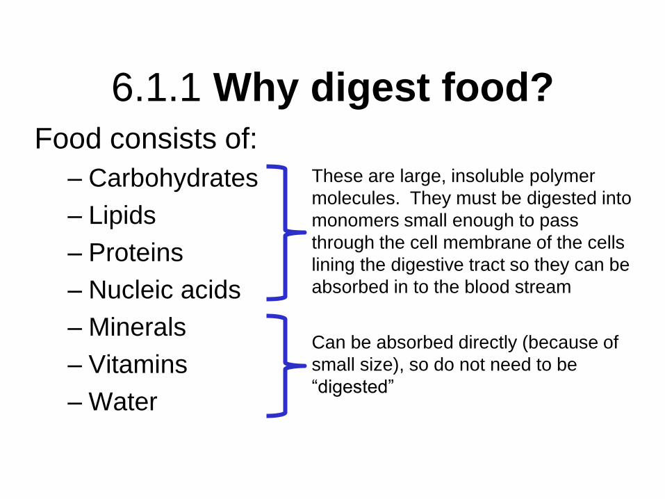
6.1.1 Why digest food? Food consists of:
– Carbohydrates
– Lipids
– Proteins
– Nucleic acids
– Minerals
– Vitamins
– Water
These are large, insoluble polymer
molecules. They must be digested into
monomers small enough to pass
through the cell membrane of the cells
lining the digestive tract so they can be
absorbed in to the blood stream
Can be absorbed directly (because of
small size), so do not need to be
“digested”
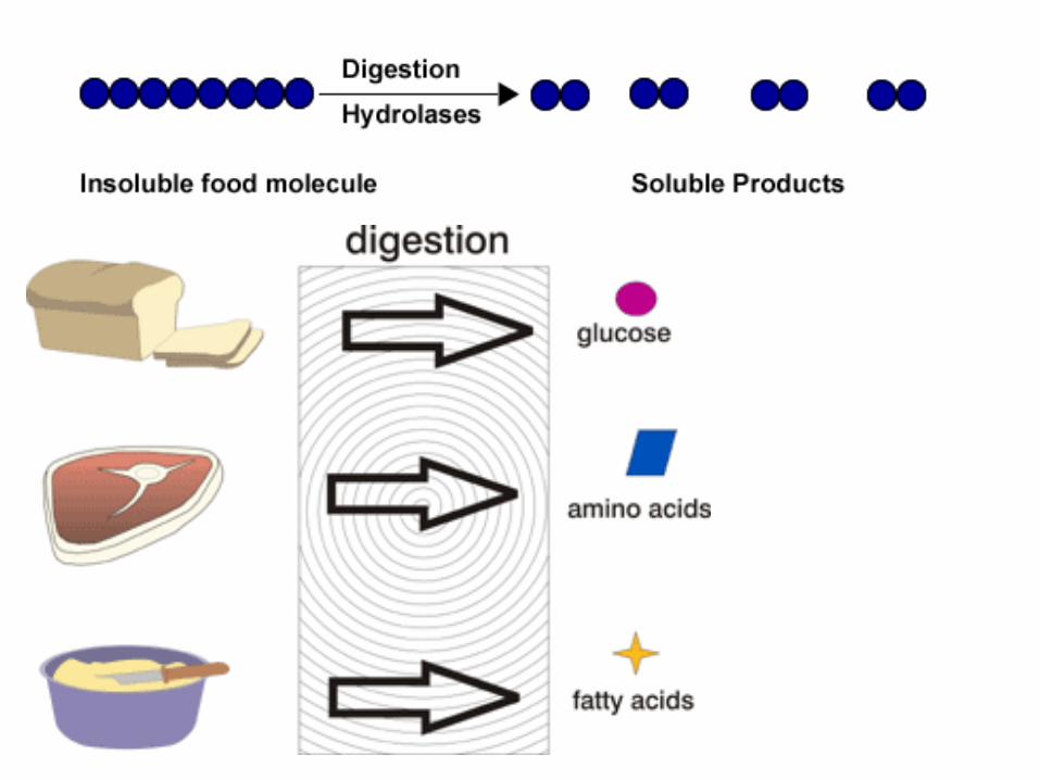

6.1.2 Enzymes and digestion
• Digestion involves hydrolysis of food
molecules
breaking
apart
molecules by
adding water
Hydrolysis of lactose (a
disaccharide) into glucose and
galactose (both monosaccharides)
with the addition of water
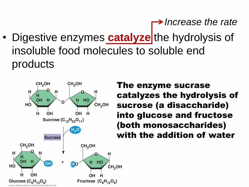
• Digestive enzymes catalyze the hydrolysis of
insoluble food molecules to soluble end
products
Increase the rate
The enzyme sucrase
catalyzes the hydrolysis of
sucrose (a disaccharide)
into glucose and fructose
(both monosaccharides)
with the addition of water
6
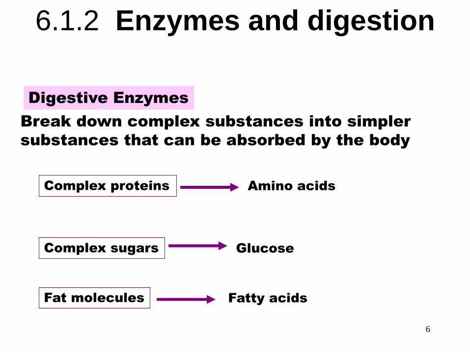
Digestive Enzymes
Break down complex substances into simpler
substances that can be absorbed by the body
Digestive Enzymes
Complex proteins
Complex sugars
Fat molecules
Amino acids
Glucose
Fatty acids
6.1.2 Enzymes and digestion

Digestive enzymes work best at 37⁰ C (body temp)
• Reactions occur faster at
higher temperatures, but the rate of denaturation of enzymes also increases at higher temperatures.
• High temperatures break the bonds important for the tertiary structure of the enzyme.
• This destroys the active sites and therefore makes the enzyme non-functional.
Rapid
denaturation
Temperature (°C)
En
zym
e a
ctivity
Too cold for
the enzyme
to operate
Optimum temperature
for enzyme

6.1.3 Examples of digestive enzymes
Class of
Enzyme
Example Source Substrate Product Optimal
pH
Amylase digest
carbohydrates
Salivary
amylase
Salivary
glands
Starch Maltose
(disaccharide)
7-8
Protease digest proteins
Pepsin Stomach
cells
polypeptides Shorter
polypeptides
2-3
Lipase digest fats
Gastric
lipase
Stomach
cells
triglyceride Glycerol and
3 fatty acids
2-3
9
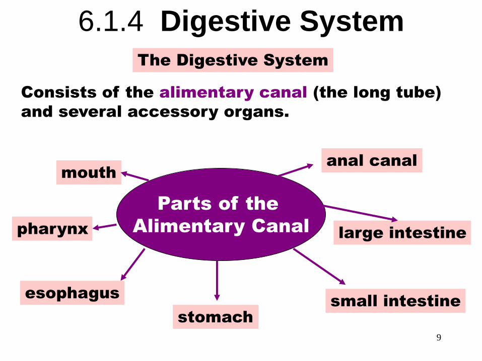
Parts of the Alimentary Canal The Digestive System
Consists of the alimentary canal (the long tube)
and several accessory organs.
Parts of the
Alimentary Canal
mouth
pharynx
esophagus
stomach
anal canal
large intestine
small intestine
6.1.4 Digestive System
10
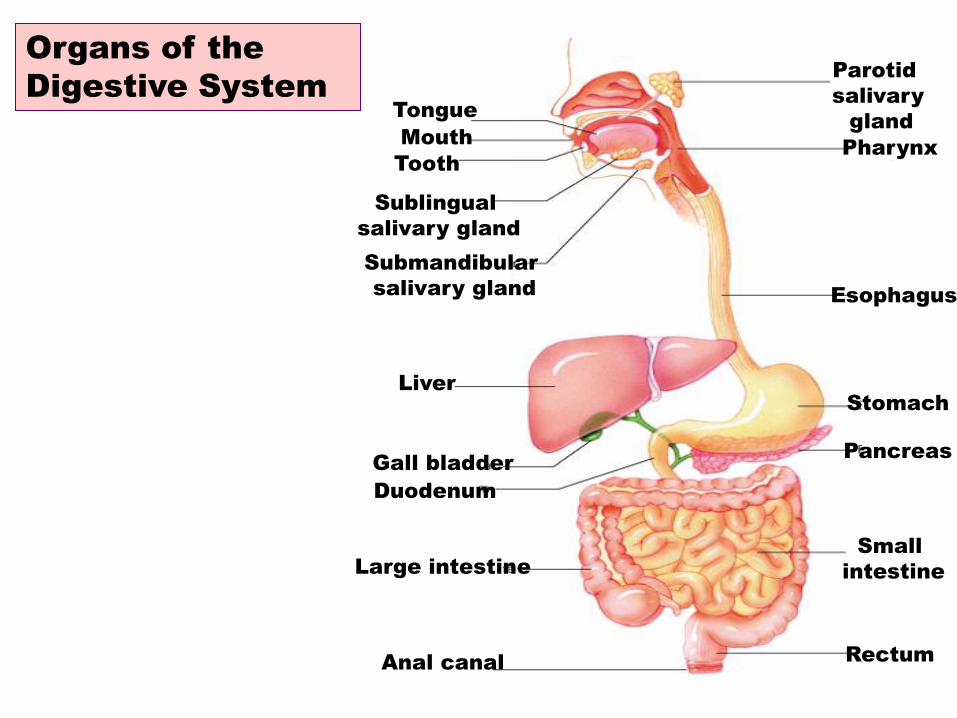
Organs of the Digestive
System
Organs of the
Digestive System Parotid
salivary
gland
Pharynx
Esophagus
Stomach
Pancreas
Small
intestine
Rectum Anal canal
Large intestine
Duodenum
Gall bladder
Liver
Submandibular
salivary gland
Sublingual
salivary gland
Tooth
Tongue
Mouth
11
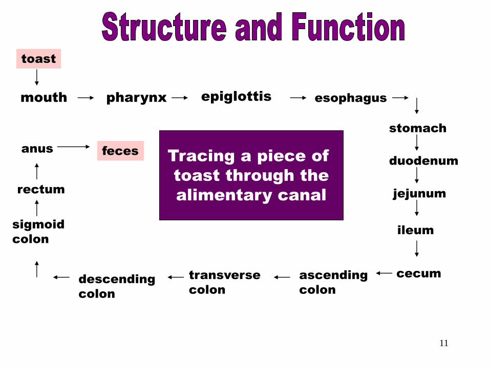
Tracing Toast mouth pharynx epiglottis esophagus
stomach
duodenum
jejunum
ileum
cecum ascending
colon
transverse
colon
descending
colon
sigmoid
colon
rectum
anus feces
toast
Tracing a piece of
toast through the
alimentary canal
Animation of digestive system

6.1.5 Function of the Stomach
1. Mechanical Digestion
• Muscle contractions
break apart and mix
food
• bolus chyme
2. Chemical Digestion
• Enzymes and HCl digest
chyme
Mass of
solid food
Semi-fluid
mass of
partly
digested
food

6.1.5 Function of the Small Intestine
1. Digestion by
enzymes in the
duodenum (first 50
cm)
2. Absorption of
nutrients into the
blood stream (see
6.1.7)
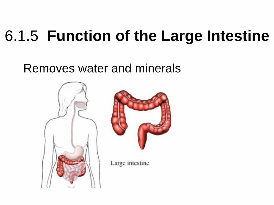
6.1.5 Function of the Large Intestine
Removes water and minerals
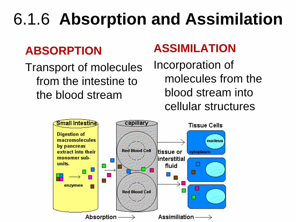
6.1.6 Absorption and Assimilation
ABSORPTION
Transport of molecules
from the intestine to
the blood stream
ASSIMILATION
Incorporation of
molecules from the
blood stream into
cellular structures
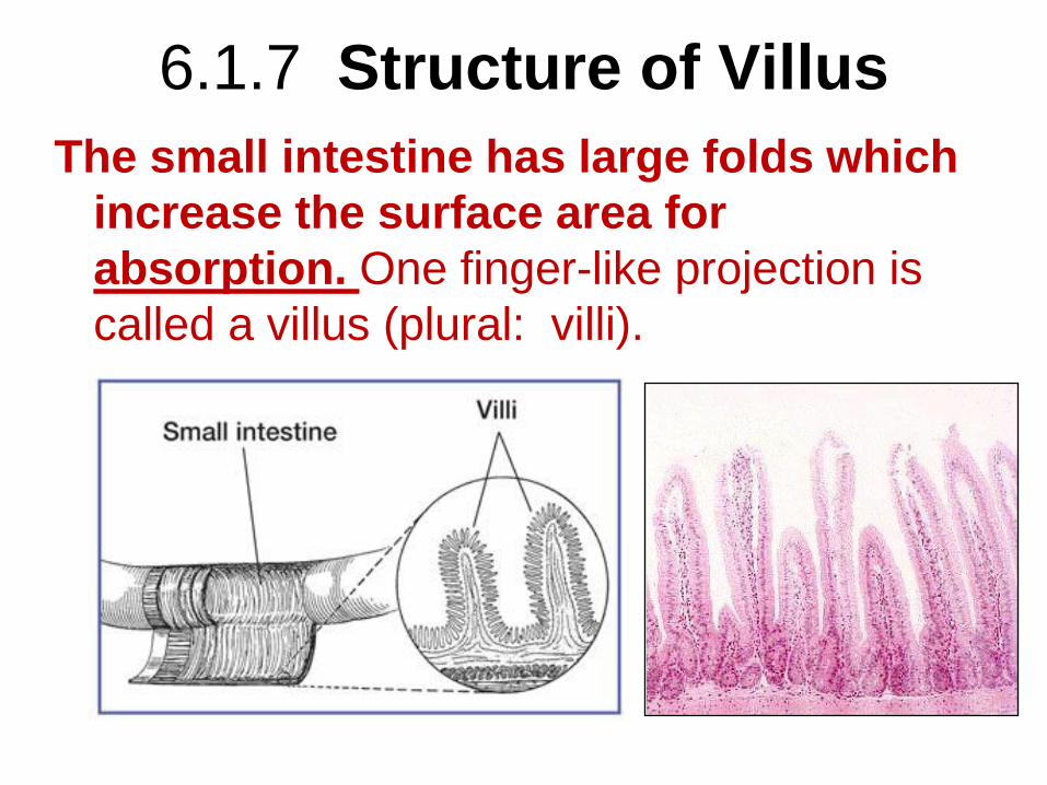
6.1.7 Structure of Villus
The small intestine has large folds which
increase the surface area for
absorption. One finger-like projection is
called a villus (plural: villi).
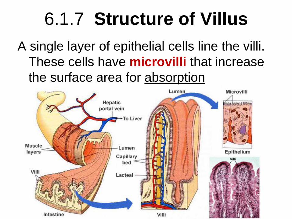
6.1.7 Structure of Villus
A single layer of epithelial cells line the villi.
These cells have microvilli that increase
the surface area for absorption
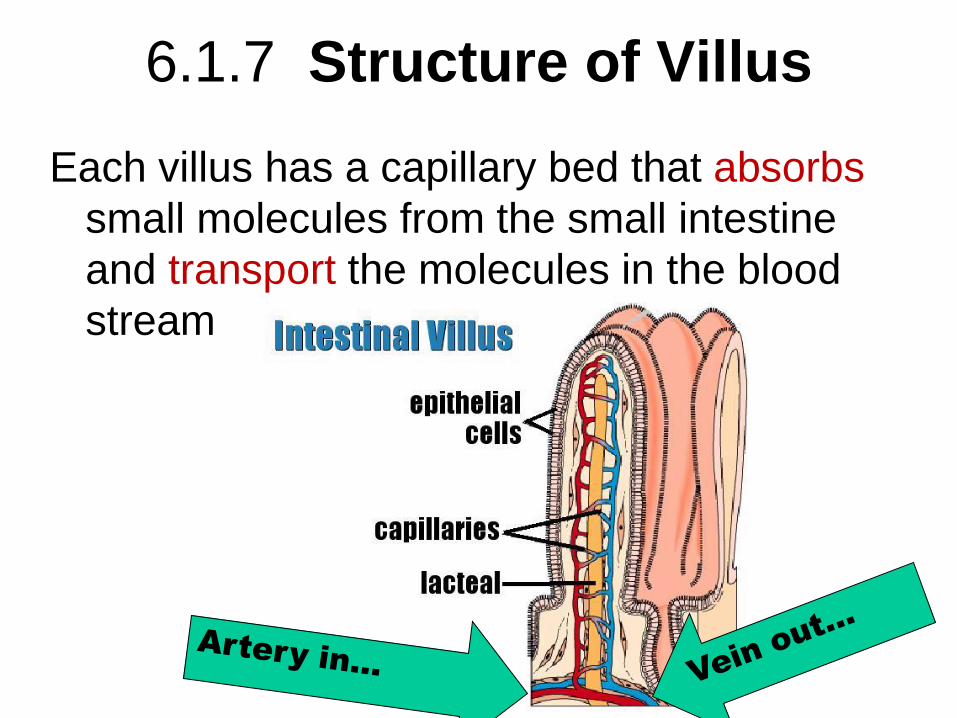
6.1.7 Structure of Villus
Each villus has a capillary bed that absorbs
small molecules from the small intestine
and transport the molecules in the blood
stream
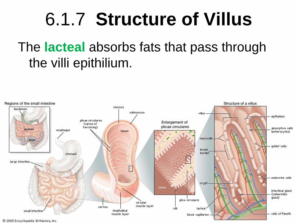
6.1.7 Structure of Villus
The lacteal absorbs fats that pass through
the villi epithilium.

H.2.1 Digestive Juices and Glands
• EXOCRINE glands are different than ENDOCRINE glands
21
Secrete other
stuff (sweat, oil,
wax, enzymes
etc) into ducts
(a pipe or tube)
Secrete
hormones
directly into the
blood stream
Digestive “Juices” are…
• ENZYMES
• Good time to review
• Catalyst
• Lower activation energy
• In the case of digestion, this is catabolic
rxns
22
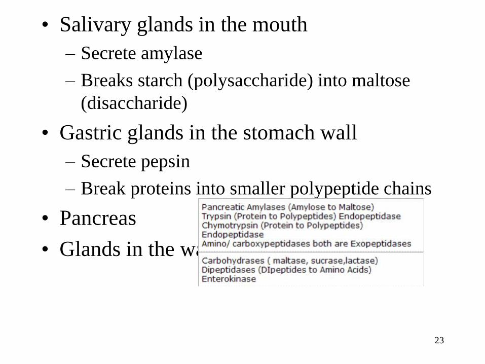
• Salivary glands in the mouth
– Secrete amylase
– Breaks starch (polysaccharide) into maltose
(disaccharide)
• Gastric glands in the stomach wall
– Secrete pepsin
– Break proteins into smaller polypeptide chains
• Pancreas
• Glands in the wall of the small intestine
23
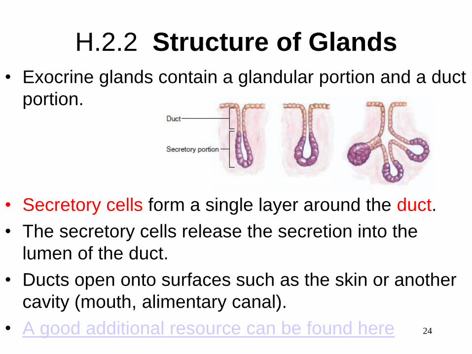
H.2.2 Structure of Glands
24
• Exocrine glands contain a glandular portion and a duct
portion.
• Secretory cells form a single layer around the duct.
• The secretory cells release the secretion into the
lumen of the duct.
• Ducts open onto surfaces such as the skin or another
cavity (mouth, alimentary canal).
• A good additional resource can be found here
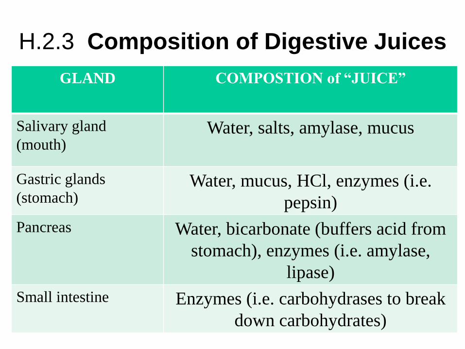
H.2.3 Composition of Digestive Juices
25
GLAND COMPOSTION of “JUICE”
Salivary gland
(mouth) Water, salts, amylase, mucus
Gastric glands
(stomach) Water, mucus, HCl, enzymes (i.e.
pepsin)
Pancreas Water, bicarbonate (buffers acid from
stomach), enzymes (i.e. amylase,
lipase)
Small intestine Enzymes (i.e. carbohydrases to break
down carbohydrates)

H.2.4 Control of Gland Secretions The activities of the digestive system are regulated by
both hormones and neural reflexes.
HORMONES
• The physical presence of food in
the lower region of the stomach
stimulates the endocrine cells
within then stomach wall to
release the protein hormone
“gastrin”.
• Gastrin is released into the blood
and travels back to the heart and
through the arteries only to return
to the digestive system to induce
the secretion of gastric juice.
NEURAL
• Nerves come to the digestive
organs from the brain or the spinal
cord.
• The nervous system is triggered
by the senses (touch, smell, sight,
feeling…)
• The nerves release chemicals
which cause the speed up or
delay of the movement of food
and the production of juices by the
digestive organs.
26
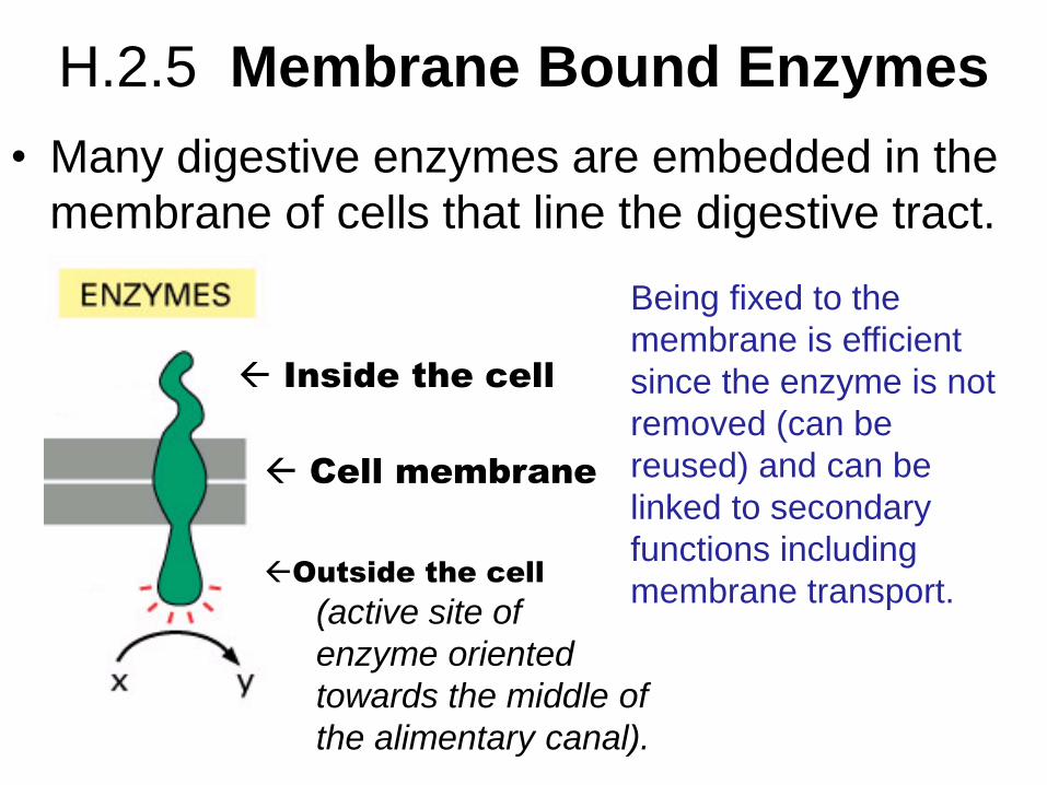
H.2.5 Membrane Bound Enzymes
• Many digestive enzymes are embedded in the
membrane of cells that line the digestive tract.
Inside the cell
Outside the cell
(active site of
enzyme oriented
towards the middle of
the alimentary canal).
Cell membrane
Being fixed to the
membrane is efficient
since the enzyme is not
removed (can be
reused) and can be
linked to secondary
functions including
membrane transport.
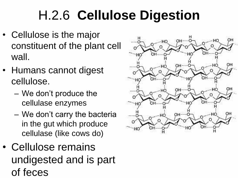
H.2.6 Cellulose Digestion
• Cellulose is the major
constituent of the plant cell
wall.
• Humans cannot digest
cellulose.
– We don’t produce the
cellulase enzymes
– We don’t carry the bacteria
in the gut which produce
cellulase (like cows do)
• Cellulose remains
undigested and is part
of feces
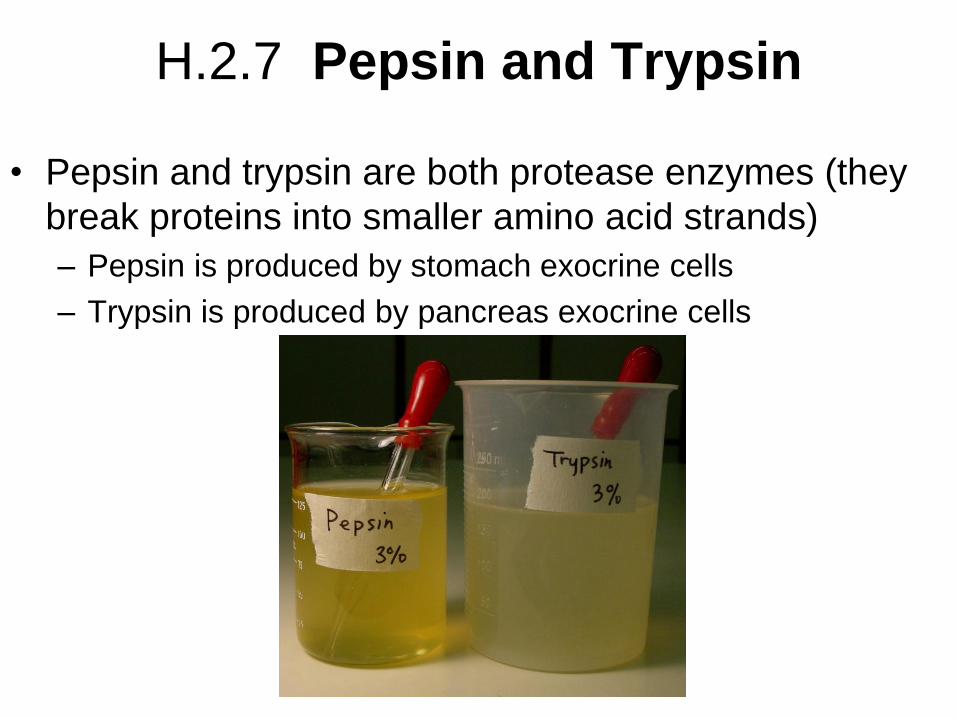
H.2.7 Pepsin and Trypsin
• Pepsin and trypsin are both protease enzymes (they
break proteins into smaller amino acid strands)
– Pepsin is produced by stomach exocrine cells
– Trypsin is produced by pancreas exocrine cells
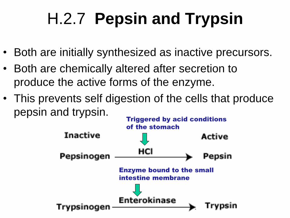
H.2.7 Pepsin and Trypsin
• Both are initially synthesized as inactive precursors.
• Both are chemically altered after secretion to
produce the active forms of the enzyme.
• This prevents self digestion of the cells that produce
pepsin and trypsin.
Triggered by acid conditions
of the stomach
Enzyme bound to the small
intestine membrane

H.2.8 Stomach Ulcers and Cancer
• The stomach has an acidic
environment caused by the
secretion of HCl.
– The acid is a barrier to
infection from
microorganisms ingested with
food.
31
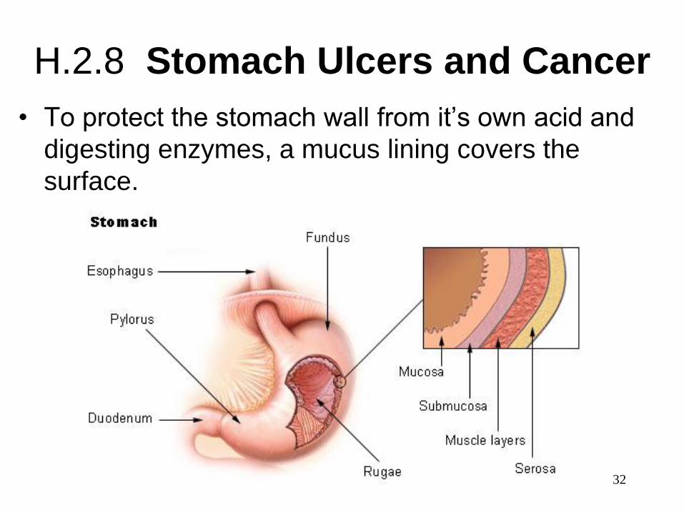
H.2.8 Stomach Ulcers and Cancer
• To protect the stomach wall from it’s own acid and
digesting enzymes, a mucus lining covers the
surface.
32

H.2.8 Stomach Ulcers and Cancer
• Stomach ulcers are areas where the mucus layer
has eroded, leaving the stomach muscle layers
unprotected and exposed to gastric acids and
digestive enzymes.
33
34
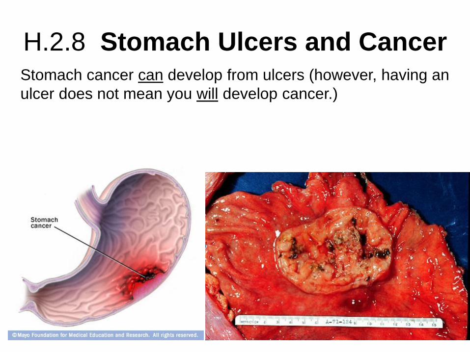
H.2.8 Stomach Ulcers and Cancer Stomach cancer can develop from ulcers (however, having an
ulcer does not mean you will develop cancer.)
H.2.8 Stomach Ulcers and Cancer
• Stomach ulcers occur with an infection of the
bacterium Helicobacter pylori.
• YES! Ulcers are caused by an infectious disease.
• Check out this site for additional information…
– No. Really. Read this information.
35
H.2.9 Lipid digestion in a
hydrophilic medium
36