Embed Size (px)
Citation preview
CLINICAL MICROBIOLOGY REVIEWS, July 2004, p. 553–570 Vol. 17, No. 30893-8512/04/$08.00�0 DOI: 10.1128/CMR.17.3.553–570.2004Copyright © 2004, American Society for Microbiology. All Rights Reserved.
Emerging from Obscurity: Biological, Clinical, and Diagnostic Aspectsof Dientamoeba fragilis
Eugene H. Johnson,1* Jeffrey J. Windsor,2 and C. Graham Clark3
Department of Animal and Veterinary Sciences, College of Agricultural and Marine Sciences, Sultan Qaboos University,Al-Khod 123, Muscat, Sultanate of Oman1; National Public Health Service for Wales, Microbiology Aberystwyth,
Bronglais Hospital, Aberystwyth Ceredigion SY23 1ER, Wales2; and Department of Infectionsand Tropical Diseases, London School of Hygiene and Tropical Medicine,
London WC1E 7HT, England3
INTRODUCTION .......................................................................................................................................................553TAXONOMY ...............................................................................................................................................................554MORPHOLOGY .........................................................................................................................................................554CLINICAL OVERVIEW.............................................................................................................................................556PATHOLOGY..............................................................................................................................................................558TRANSMISSION ........................................................................................................................................................561THERAPY ....................................................................................................................................................................562EPIDEMIOLOGY AND DIAGNOSIS .....................................................................................................................563
Microscopy ...............................................................................................................................................................563Fixatives ...................................................................................................................................................................563Staining ....................................................................................................................................................................564Immunological Diagnosis.......................................................................................................................................565DNA-Based Diagnosis ............................................................................................................................................565
CULTIVATION ...........................................................................................................................................................565General Considerations .........................................................................................................................................565Historical Background ...........................................................................................................................................566Surveys Using Culture ...........................................................................................................................................566Appearance in Culture ...........................................................................................................................................567
CONCLUSIONS .........................................................................................................................................................567ACKNOWLEDGMENT..............................................................................................................................................568REFERENCES ............................................................................................................................................................568
INTRODUCTION
Since the first description of Dientamoeba fragilis by Jeppsand Dobell in 1918 (65) this ameboid organism has escaped theinterest of most clinicians and diagnostic microbiologists. Thisis reflected in a variety of descriptions conferred on the organ-ism, such as: “a neglected cause of diarrhea” (49), “an unusualintestinal pathogen” (12), “an emerging protozoal infection”(129), and “an enigma shrouded in the mysteries of clinicalparasitology” (140).
Jepps and Dobell (65) considered the nucleus of D. fragilis tobe the characteristic feature of the organism, since they ob-served that the predominant form was binucleate, a featurewhich readily differentiated it from other human intestinalamebas. Interestingly, although they had isolated D. fragilisfrom seven persons, six of whom had a history of dysentery orchronic diarrhea, they felt that this observation was of noclinical significance. This conclusion was based on their obser-vation that D. fragilis had a similar mode of nutrition to thenonpathogenic organisms Entamoeba coli and Endolimaxnana, in contrast to Entamoeba histolytica, which was then
considered to be a “tissue parasite.” They proposed that hu-mans were aberrant hosts, in which cysts did not develop, andsuggested that D. fragilis had a true animal host in which it wascapable of encystation. Unfortunately, there is still no evidenceto support the existence of a natural host besides humans norhas a cystic stage of D. fragilis ever been convincingly demon-strated. Furthermore, the lack of a suitable animal model thatis capable of supporting the life cycle of D. fragilis and thatdevelops similar clinical symptoms has severely hindered moredetailed studies of the biology of the organism.
The seeds of doubt concerning the pathogenicity of D. fra-gilis were unfortunately planted by Jepps and Dobell (65) andnourished by Dobell and O’Conner (38) and account at leastpartially for the lingering resistance in many clinical circles toaccept the disease-causing potential of this organism. How-ever, the evidence supporting the pathogenicity of D. fragilis istoo convincing to justify the continued neglect of this parasiteas a cause of diarrhea, abdominal pain, flatulence, fatigue, andanorexia, the symptoms most commonly observed in patientsinfected with this organism (68). Indeed, the organism hasbeen isolated from and associated with clinical symptoms inpatients from numerous countries throughout the world (140).Perhaps the most striking reason to consider D. fragilis a po-tential pathogen is that it can be easily treated and that thegreat majority of patients show significant clinical improve-ment thereafter (35, 49).
* Corresponding author. Mailing address: Department of Animaland Veterinary Sciences, College of Agricultural and Marine Sciences,Sultan Qaboos University, P.O. Box 34, Al-Khod 123, Muscat, Sultan-ate of Oman. Phone: (968) 515-234. Fax: (968) 513-418. E-mail:[email protected].
553
on July 26, 2018 by guesthttp://cm
r.asm.org/
Dow
nloaded from
TAXONOMY
The name Dientamoeba fragilis refers to the fact that it is anenteric ameba with the curious characteristic of being binucle-ate and that it tends to degenerate rapidly after excretion instool (65). It was also classified as an ameba by Chatton (20),who included it in the family Entamoebidae, where it remainedfor the best part of 50 years. However, doubts about its affin-ities were raised by Dobell (36), when he noted the presenceof the “centrodesmus” and the great similarity of D. fragilis atthe light microsopic level to Histomonas meleagridis (see also“Morphology” below). Histomonas was, and is, accepted as atrichomonad flagellate despite its tendency to lose its flagellumin culture or when it invades tissues. Dobell (36) stronglysuggested that Dientamoeba and Histomonas were closelyrelated and that D. fragilis was an aberrant flagellate. Thisrelationship was formalized when Grasse (51) removed Dient-amoeba from the Entamoebidae and created the family Dient-amoebidae to contain these two genera.
Further morphological support for the classification ofD. fragilis within the trichomonad flagellates had to await thetransmission electron microscopy studies by Camp et al. (13),who confirmed Dobell’s observations. Molecular support forthe relationship was first obtained by Dwyer (39, 40), whoshowed substantial antigenic similarities among Dientamoeba,Histomonas, and Trichomonas to the exclusion of Entamoebaspecies.
Another 20-year gap ensued until DNA studies of Dient-amoeba commenced. Phylogenetic analysis of the D. fragilissmall-subunit rRNA gene sequence (115), using the samestrain that had been studied at the morphological level byCamp et al. (13) (strain Bi/pa; ATCC 30948), unequivocallyconfirmed its trichomonad affinities; more recently, analysis ofthe same gene from H. meleagridis confirmed the close andspecific relationship between Histomonas and Dientamoeba(48). The latter investigation hinted at a link between Histo-monas/Dientamoeba and the genus Tritrichomonas, but thisremains to be confirmed.
So where is Dientamoeba classified today? The organismpresently resides in the phylum Parabasala, class Trichomona-dea, family Trichomonadidae, and possibly the subfamily Tri-trichomonadinae (48). The systematics of the parabasalids is,however, in need of revision, and the specifics of Dientamoebaclassification may change. Its affinities to Histomonas and othertrichomonads will not.
At the other end of the taxonomic scale, there are verydifferent questions. How many species of Dientamoeba arethere? 2. Is D. fragilis of human origin a single species?
At the morphological level, amoeboid organisms present aserious problem because there are very few phenotypic char-acteristics on which to base a species diagnosis. Indeed, mor-phology has been superseded by molecular markers in thedescription and identification of species in some genera ofamoeboid organisms, for example Entamoeba (34) and Naegle-ria (31). Although D. fragilis has been found in nonhumanprimates, this diagnosis is based only on morphology (59, 73,83). Whether these organisms represent distinct species withinthe genus awaits further information. However, attempts toinfect a macaque intrarectally with human D. fragilis were
unsuccessful (36). As far as we are aware, no other species inthe genus Dientamoeba have been described.
Some molecular data do exist to address the second ques-tion. Using the approach of examining restriction fragmentlength polymorphisms of the D. fragilis small-subunit rRNAgene (riboprinting), Johnson and Clark (66) identified the ex-istence of two genetically distinct types of D. fragilis among 12isolates from humans. An additional 90 or more isolates havesubsequently been studied using the same method (J. J. Wind-sor, unpublished data). In the latter set of samples only onegenotype has been found. The rarer of the two genotypes wasfound in only two cases; one of these strains happens to be theisolate Bi/pa studied by Camp et al. (13) and Silberman et al.(115). This indicates that the results of studies using isolateBi/pa may not be representative of the species as a whole. Thedegree of sequence divergence between the ribosomal genes ofthe two genotypes is estimated to be approximately 2% (66,94). Whether this constitutes a species level of divergence inprotozoa is a matter for debate, and no consensus exists amongresearchers in the field.
The significance of the existence of two genetically distinctforms of D. fragilis deserves to be investigated further. Is oneform more virulent than the other, and could this possiblycontribute to the differences in clinical perceptions regardingthe organism’s pathogenicity? Results from the largest study sofar have found only one genotype in both symptomatic andasymptomatic individuals (94). The rarity of the second geno-type will make investigation of its role in disease difficult.
MORPHOLOGY
Most of the detailed light microscopic descriptions ofD. fragilis date back to the early and mid-1900s. Using a cameralucida, the parasitologists of the time produced surprisinglydetailed plates (8, 26, 65, 133, 134, 135, 136, 137). There aresubtle differences in the descriptions since some workers pre-pared material direct from feces (57, 133, 134, 135, 136, 137)whereas others used material from culture (36). Dobell (36)considered D. fragilis in feces to be degenerate and thereforenot a true morphological representation.
In direct saline preparations, D. fragilis usually appearsrounded and shows a wide variation in size. In the originaldescription by Jepps and Dobell (65), the size range given was3.5 to 12 �m. Much larger sizes have been found in culture (20to 40 �m) (114). The size range of D. fragilis in culture overlapsthat of E. histolytica, E. hartmanni, and Endolimax nana (102,107). The nuclei of D. fragilis are not visible in saline or iodinepreparations, although food vacuoles or inclusions may beseen. D. fragilis moves by using thin, hyaline, leaf-like pseudo-podia, which are irregularly lobed (Fig. 1) (65). Hakansson(55), examining his own freshly evacuated stool specimen,found rounded trophozoites of D. fragilis. Only after 5 to 10min at room temperature did they recover from this temporary“paralysis” and display the characteristic fan-shaped motility,with lobes and indentations. Unlike Entamoeba trophozoites,no flow of endoplasm into the pseudopodia has been observedin D. fragilis, and while the edge is constantly changing, withsharp points appearing, no progression is seen (55, 56).
Jepps and Dobell (65) found that 80% of D. fragilis tropho-zoites in permanently stained fecal smears were binucleate and
554 JOHNSON ET AL. CLIN. MICROBIOL. REV.
on July 26, 2018 by guesthttp://cm
r.asm.org/
Dow
nloaded from

20% were mononucleate. This percentage can vary consider-ably, even in stool samples taken from the same patient ondifferent days (134). Although seen less frequently, some tro-phozoites have been described with as many as four or fivenuclei (36, 81, 136). The diameter of the nuclei varies from 1 to3 �m but depends largely on the size of the trophozoite (65,
137). Internally, the nuclei appear fragmented, usually contain-ing four to eight granules, without peripheral chromatin (Fig.2) (137). Often, one of the granules is larger than the othersand stains more deeply (8, 65, 133). The binucleate form ofD. fragilis is the typical stage observed. Mononucleated tropho-zoites of D. fragilis are therefore recently divided forms, pro-duced by the process of binary fission, and are slightly smallerthan the binucleates. The division is by simple constriction ofthe cell body. Nuclear division is found only in mononucleatedtrophozoites (36, 135). Dobell (36) described a “connectingthread” which joined the two nuclei together. He termed this a“centrodesmus” and could find no trace of it in mononucleatedorganisms. Wenrich (135) termed this structure a “post-divi-sion desmose,” believing it to arise from an intranucleardivision centre. Dobell, however, thought that this organellepermanently linked the nuclei. Dobell’s review (36) of themorphology of D. fragilis and its comparison to that of theturkey pathogen Histomonas meleagridis was the defining pub-lication of the era and was the first paper to acknowledge theflagellate attributes of D. fragilis and its morphological similar-ities to H. meleagridis.
Curiously, only a handful of papers have been published onthe morphology of D. fragilis on the basis of transmission elec-tron microscopy (13, 91, 112, 113). Camp et al. (13) publisheda comprehensive study of the binucleate stage of D. fragilis
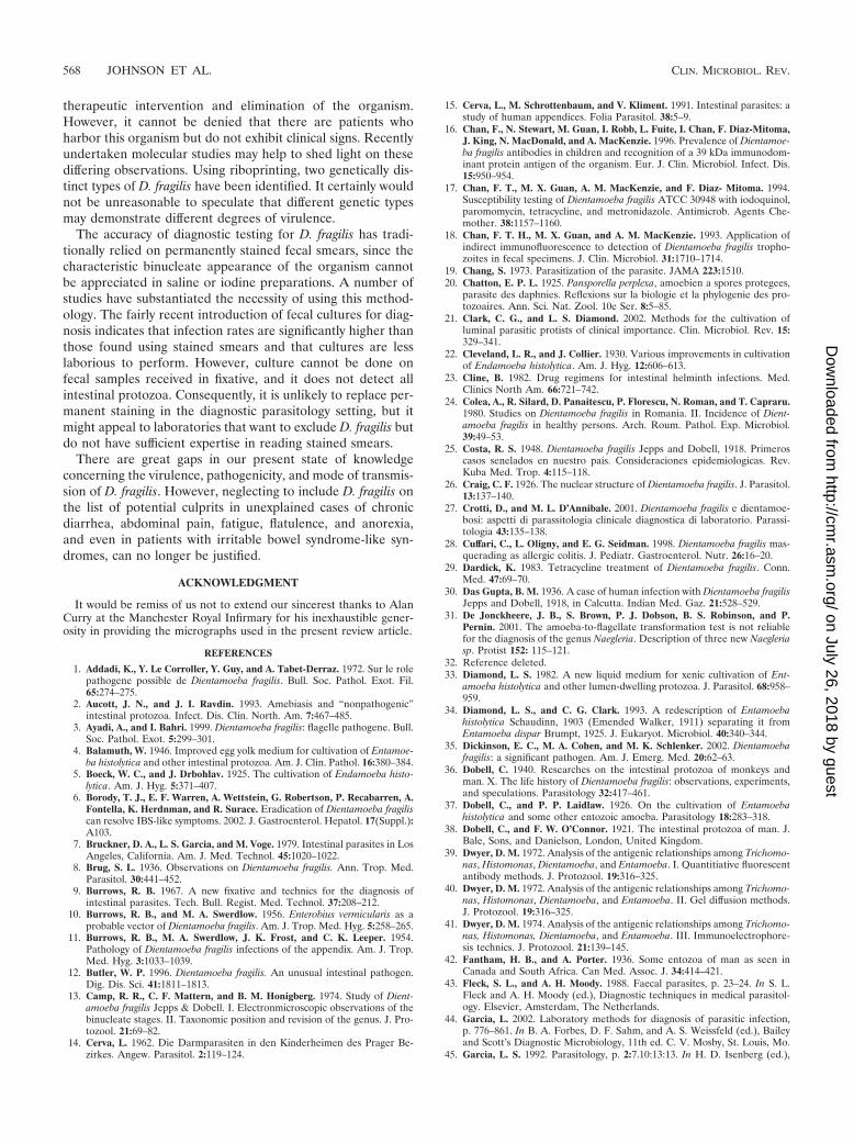
FIG. 1. D. fragilis growing in Robinson’s culture, showing ingestedrice starch and fine leaf-like pseudopodia. magnification, �400.
FIG. 2. Iron-hematoxylin-stained smear of D. fragilis showing pleomorphic trophozoites. Note the characteristic fragmented nuclei and the verysmall mononucleated trophozoite in the center magnification, �1,000.
VOL. 17, 2004 CLINICAL MANIFESTATIONS OF DIENTAMOEBA FRAGILIS 555
on July 26, 2018 by guesthttp://cm
r.asm.org/
Dow
nloaded from

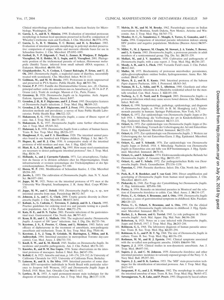
strain Bi/pa. This paper confirmed many of the light micro-scopic observations of the flagellate characteristics of D. fragilisby Dobell (36). An extranuclear spindle is found between thenuclei, originating from polar complexes adjacent to one of thenuclei. These structures are homologous to the atractophoresdescribed in hypermastigotes (61). Parabasal filaments extendlaterally to the external surface of the atractophores. ExtensiveGolgi complexes overlie the filaments and are very similar tothe parabasal apparatus seen in trichomonads and hypermas-tigotes. This spindle is composed of two bundles of approxi-mately 30 to 40 microtubules. One bundle appears at somedistance from the nucleus, whereas the other is juxtanuclearand is often seen in a groove of the nuclear envelope. Thenuclear structure of D. fragilis more closely resembles that oftrichomonad flagellates rather than that of Entamoeba spp.Chromatin bodies or granules are often seen in the nucleo-plasm (Fig. 3 and 4), and the nuclear envelope consists of twomembranes (Fig. 5).
Electron microscopy revealed electron-dense rounded inclu-sions in the cytoplasm that were termed “microbody-like” andwere presumed to be homologous to the paraxostylar granulesof trichomonads (13) (Fig. 3). These inclusions were subse-quently recognized as being hydrogenosomes (82). Digestivegranules are also commonly found in the cytoplasm and maycontain myelin, bacteria, or rice starch (Fig. 3 and 6). D. fragilisfeeds by phagocytosis (Fig. 7), and waste products are releasedfrom the digestive vacuoles by exocytosis (Fig. 8).
CLINICAL OVERVIEW
Eighty-five years after its first description, although D. fra-gilis is accepted as a true pathogen in some countries andinfected patients are treated, it is still struggling to gain accep-tance as a legitimate pathogen in many others. This is a resultof D. fragilis at times being found in patients who exhibitedno apparent clinical symptoms (24) and often being identified
FIG. 3. Electron micrograph of a mononucleated trophozoite of D. fragilis. The nucleus (Nu) and chromatin (Ch) are labeled. Electron-densemicrobody-like inclusions surround the nucleus. Digestive vacuoles (Dv) can be clearly seen, one of which contains an ingested bacterium (B).Magnification, �5,200.
556 JOHNSON ET AL. CLIN. MICROBIOL. REV.
on July 26, 2018 by guesthttp://cm
r.asm.org/
Dow
nloaded from
in patients coharboring other suspect pathogens. Robertson(100) described a young female patient who was admitted tohospital with a history of recurrent attacks of diarrhea andabdominal pain. D. fragilis was isolated from a series of herstool specimens, which also contained E. histolytica, Trichomo-nas spp., and Blastocystis hominis, as well as Endolimax nanaand spirochetes. In the same year, Thomson and Robertson(128) reported a case involving a 38-year-old man who haddysentery. Although D. fragilis was isolated from this patient,other organisms were also found, including Entamoeba coli,Trichuris trichura and B. hominis. Due to the coinfections inthese two patients, there was no sound basis to support aprimary disease-causing role for D. fragilis. D. fragilis was sub-sequently found in stool samples from a healthy 3-year-old girl(125) and in epidemiological studies from Canada (96), En-
gland (64), and South Africa (42), but strong evidence sugges-tive of its pathogenic potential was not presented.
Interestingly, Wenrich et al. (138) reported that 4.3% ofcollege freshmen were infected with D. fragilis in a professionalschool in Philadelphia. This incidence of infection was similarto that of E. histolytica, but, interestingly, more students withD. fragilis had clinical symptoms than did those with E. histo-lytica. Das Gupta (30) also identified D. fragilis in a Bengaliman in India who had intermittent attacks of diarrhea, butHakansson (55) was the first to propose that D. fragilis wasmore than just an innocent commensal organism. He reporteda clinical case involving a 48 year-old physician who had severecolitis. D. fragilis was observed in large numbers at the onset ofthe illness, and variations in its abundance corresponded to theseverity of clinical symptoms. After several treatments with
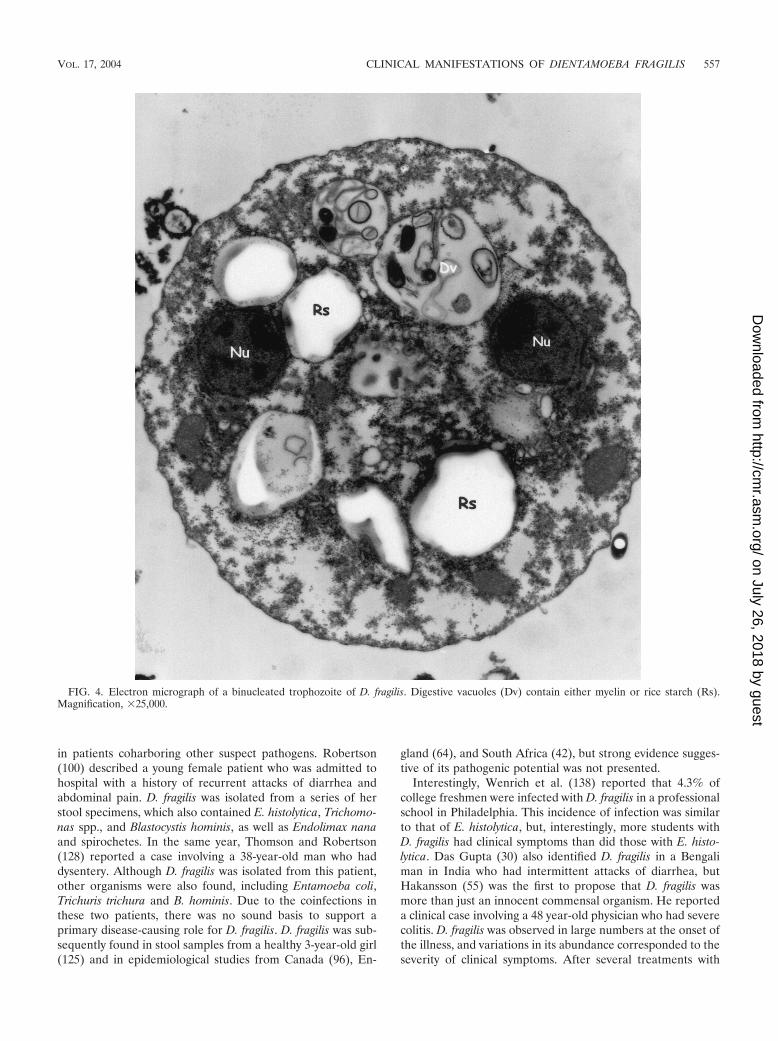
FIG. 4. Electron micrograph of a binucleated trophozoite of D. fragilis. Digestive vacuoles (Dv) contain either myelin or rice starch (Rs).Magnification, �25,000.
VOL. 17, 2004 CLINICAL MANIFESTATIONS OF DIENTAMOEBA FRAGILIS 557
on July 26, 2018 by guesthttp://cm
r.asm.org/
Dow
nloaded from

carbarsone, the patient’s condition improved and the infectionwith D. fragilis disappeared. Subsequently, there have beennumerous reports that have linked D. fragilis infections withclinical symptoms that subsided only after the elimination ofthe organism (25, 35, 56, 72, 80, 99, 103, 104, 137).
Yoeli (147) described nine patients who suffered from acuteintestinal signs such as explosive diarrhea, severe abdominalpain, cramps, nausea, vomiting, mild fever, and general fatigue.In all of these patients, large numbers of D. fragilis organismswere observed in the absence of any other pathogens. Numer-ous reports from many different parts of the world continuedto substantiate the association of D. fragilis with clinical symp-toms, principally abdominal pain, diarrhea, nausea, vomiting,and fatigue (1, 3, 12, 27, 32, 53, 92, 113, 114, 143, 146).
Studies have also demonstrated links between this parasiteand urticaria (146), biliary infections (127), pruritus (116),colitis (111), allergic colitis (28), irritable bowel syndrome (6),and diarrhea in people infected with human immunodeficiencyvirus (75, 78). Of particular significance is the observation thatinfections in children appear to be very common. Keystone et
al. (71) reported that over 8% of 900 children studied in theToronto area were positive for D. fragilis. Also, some authorshave reported that infections in children are more often asso-ciated with clinical symptoms than are infections in adults (3,54, 85, 117); children are also more often reported to exhibitperipheral eosinophilia (2, 93, 97, 98, 117, 134).
Despite the significant number of studies that have incrim-inated D. fragilis as a legitimate enteric pathogen, it is far tooseldom included in the differential diagnostic repertoire ofintestinal pathogens by both practicing physicians and diagnos-tic laboratories in many countries.
PATHOLOGY
In parallel with our poor understanding of the pathogenicityof D. fragilis, only a small number of studies have presentedfindings relevant to the pathological consequences of infec-tions with this organism. The first reported pathological studyperformed on the appendixes of four patients (ranging in agefrom 20 to 28 years of age) infected with D. fragilis showed
FIG. 5. Electron micrograph demonstrating chromatin bodies (Ch) in the nucleus (Nu), surrounded by a double nuclear membrane. Note themicrobody-like (Mb) inclusions, digestive vacuole (Dv), ingested bacteria (B), and Golgi apparatus (Go). Magnification, �15,500.
558 JOHNSON ET AL. CLIN. MICROBIOL. REV.
on July 26, 2018 by guesthttp://cm
r.asm.org/
Dow
nloaded from

distinct similarities (11). In each case there was marked fibrosisof the subserosa of the appendixes and trophozoites were seenin the lumen, with absence of tissue invasion. Of particularrelevance was the observation that in each case there wereingested red blood cells within the D. fragilis organisms. Theauthors considered that this was a hallmark feature of patho-genic potential, since pathogenic amebae such as E. histolyticaingest erythrocytes whereas the nonpathogenic Entamoeba coliand Endolimax nana do not. A critical analysis of these patho-logical findings, however, did not permit D. fragilis to be in-
criminated as the causative agent of the fibrosis because inthree of the four cases the appendixes also contained wormova, larvae, or adults that also could also have potentially beenresponsible for these lesions. In a more extensive study ofappendixes containing D. fragilis, Swerdlow and Burrows (124)examined an additional 11 organs and also reported extensivefibrous connective tissue of the submucosa. They found a va-riety of lesions, ranging from acute suppurative appendicitis tolymphoid hyperplasia to pure fibrosis. They felt that D. fragilisprobably acted as a low-grade irritant, causing a chronic in-
FIG. 6. Electron micrograph of D. fragilis demonstrating digestive vacuoles (Dv) containing myelin. Magnification, �21,000.
VOL. 17, 2004 CLINICAL MANIFESTATIONS OF DIENTAMOEBA FRAGILIS 559
on July 26, 2018 by guesthttp://cm
r.asm.org/
Dow
nloaded from

flammatory reaction that resulted in fibrosis. However, onlythree of the appendixes from this study had pure D. fragilisinfections, making it difficult to conclude that D. fragilis wassolely responsible for the underlying fibrosis.
Shein and Gelb (111) reported finding multiple punctateulcers on endoscopic examination of a female patient who hada history of chronic abdominal pain for 1 year and a suddenproduction of blood-streaked diarrhea. A biopsy of her rec-tum revealed shallow ulcerations with evidence of acute andchronic inflammation. Trichrome staining of mucosal aspiratesrevealed large numbers of D. fragilis organisms in the absenceof any other known pathogens. However, more recentlydescribed pathogens such as the coccidia and microsporidiamight have been overlooked. Three additional studies (92, 110,120) reported inflammatory changes of the rectum and sigmoidcolon in patients with D. fragilis infections. However, many of
these patients had mixed infections and many had no patho-logical abnormalities.
A more recent study (28) reported a case of eosinophiliccolitis in a female patient harboring D. fragilis. The patient wasdocumented as having bovine protein allergy with intermittentepisodes of diarrhea and abdominal pain, despite receiving anappropriate diet. After the patient was treated with iodoquinoland the parasite was eliminated, the symptoms disappeared.
In contrast to the findings of the above studies, Cerva et al.(15) were unable to establish any relationship between thepresence of D. fragilis and any pathological abnormalities inthe appendixes of the patients in their study, nor were they ableto detect red blood cells in any of the D. fragilis organismscontained within the lumen of the appendixes. Certainly, thelack of an animal model hampers our ability to shed light onthe exact pathological manifestations caused by D. fragilis.
FIG. 7. Electron micrograph of D. fragilis exhibiting phagocytosis. Magnification, �6,600.
560 JOHNSON ET AL. CLIN. MICROBIOL. REV.
on July 26, 2018 by guesthttp://cm
r.asm.org/
Dow
nloaded from

TRANSMISSION
The mode of transmission of D. fragilis has remained amystery. Most intestinal protozoa that are transmitted via thefecal-oral route require a cyst stage in order for the organismto survive in the external environment. However, although afew authors have reported pseudocystic, precystic, or cysticstages of D. fragilis (52, 72, 95, 133), it is generally acceptedthat this parasite does not have a cyst form. It is likely that thecysts described by Kofoid (74) were merely rounded-up tro-phozoites. Wenrich (133) also described degenerating forms,which stained more intensely and were thought to be precysticforms or “pseudocysts.” In a later publication, Wenrich (136)found these forms to be no more frequent in older than infresh material and subsequently dismissed the notion of “pseu-docysts”. Piekarski (95) examined iron-hematoxylin-stainedsmears and reported the presence of precystic and cystic forms.However, closer scrutiny of the figures in that paper revealsthat these forms are more likely to be degenerating trophozo-ites. Interestingly, Silard et al. (114) reported possible cysticforms of D. fragilis, with irregular, thick membranes, in cul-
tured preparations examined by phase-contrast microscopy.These forms were not confirmed in permanently stainedsmears. It is still possible that a cyst form might be identified.Despite the larger body of research that has been undertakenon B. hominis, its cyst stage was confirmed only in 1991 (122),79 years after its first description.
The lack of a cyst stage would cast doubt on the possibility ofan effective direct fecal-oral route of transmission. Thomsonand Robertson (128) stated that it appeared unlikely that aninfection could occur via ingestion of the adult form of D. fra-gilis, since the life span of the parasite outside the body is veryshort. Furthermore, they doubted whether D. fragilis couldsurvive the vicissitudes of its journey through the alimentarytract. Indeed, in a most dedicated fashion of scientific pursuit,Dobell (36) swallowed a culture containing thousands of activeand healthy trophozoites of D. fragilis. After 10 years of exam-ining his stools, he was unable to find the organism in a singlesample. His attempts to infect macaques were similarly unsuc-cessful.
Dobell (36) compared D. fragilis with what he felt was its
FIG. 8. Electron micrograph of D. fragilis exhibiting exocytosis. Magnification, �28,500.
VOL. 17, 2004 CLINICAL MANIFESTATIONS OF DIENTAMOEBA FRAGILIS 561
on July 26, 2018 by guesthttp://cm
r.asm.org/
Dow
nloaded from
closest biological relative, Histomonas meleagridis. He pointedout that since H. meleagridis was known not to have a cyst formand was transmitted via the eggs of the nematode Heterakisgallinae, it was highly likely that D. fragilis was also transmittedvia the ova of nematodes, and suggested Trichuris trichiura andAscaris lumbricoides as likely candidates. Circumstantial sup-port for this hypothesis was found in reports by a number ofresearchers who described helminth coinfections in large num-bers of patients infected with D. fragilis (58, 128).
Burrows and Swerdlow (10) agreed with Dobell (36) andWenrich (136) that D. fragilis was probably related to Histo-monas. This was based on the fact that neither organismformed cysts, each was pathogenic to its host to various de-grees, each ingested red blood cells within the host as well asin culture, each showed a lag phase of about 24 h beforemultiplying in cultures, and each is transmitted in the egg of aspecies of roundworm. However, they disagreed with Dobellregarding the intermediate host. Whereas Dobell (36) thoughtthat the intermediate host might be Trichuris or Ascaris, Bur-rows and Swerdlow (10) were convinced that the incriminatingintermediate host was Enterobius vermicularis. This was basedon pathological analyses of 22 appendixes from which D. fra-gilis was isolated. They found a 20-fold greater incidence ofcoinfection with the pinworm E. vermicularis than the calcu-lated expected value. In addition, they observed small amoe-boid bodies, whose nuclei greatly resembled those found inuni-and binucleated D. fragilis, in the eggs of the pinworms.Further circumstantial evidence to support this assumptionwas provided by a number of other studies that showed agreater than expected incidence of coinfections with D. fragilisand E. vermicularis (11, 15, 19, 77, 86, 98, 124, 146). In addition,although Burrows and Swerdlow (10) were unsuccessful intheir attempts to culture D. fragilis from pinworm eggs, Ockert(88, 89) not only successfully infected himself with D. fragilis byingesting eggs of E. vermicularis, taken from a young boy whowas coinfected with D. fragilis, but also successfully infectedtwo other human subjects. It is noteworthy that the reportedincidence of both D. fragilis and pinworms is higher in femalesthan males (15, 53, 146).
Ockert and Schmidt (90) felt that they had found the defin-itive proof which confirmed that Enterobius served as the vec-tor for transmitting D. fragilis. They compared the isoelectricpoints of trophozoites of D. fragilis in culture with ameboid-likecells that they found in the eggs of Enterobius. Interestingly,their nuclei and their cytoplasms had almost identical electro-static charges.
Based on morphological comparisons, Dobell (36) proposedthat Dientamoeba was probably related to Histomonas. Similarassumptions have been expressed by Wenrich (136, 137),Camp et al. (13), and Dwyer (39, 40, 41) as a result of theirstudies. More recently, the specific phylogenetic relationshipbetween Histomonas and Dientamoeba has been determined bythe analysis of small-subunit rRNAs. Both species demonstratereduced G�C contents and longer nucleotide chain lengthscompared to other parabasalids (48). Based on the totality ofthese similarities, it appears plausible to make an assumptionthat both species may also have a common modus of transmis-sion. Certainly, from the standpoint of the parasite’s ability toperpetuate itself and survive in the environment, it would be ofbenefit for D. fragilis to be transmitted by pinworms (23, 132).
Some authors have not been convinced of the role of Ente-robius as the vector of D. fragilis since they were unable toestablish any association between E. vermicularis and D. fragilisin any of their patients (69). Unfortunately, however, it is notalways apparent how, or if, some of the researchers studyingD. fragilis properly screened their patients for pinworms. Notonly do pinworm infections often undergo spontaneous remis-sions, but also, unlike other nematodes that are diagnosed bythe presence of fecal eggs, pinworms are diagnosed only byperforming anal swabs or cellotape smears. These proceduresare often not performed or are undertaken only when there arespecific indications such as anal pruritus. It is therefore likelythat some investigators may not have adequately looked forpinworms. However, it is also possible that other nematodesmight serve as vectors. Indeed, Sukanahaketu (123) foundD. fragilis-like structures in the ova of Ascaris lumbricoides in 38patients in Thailand with D. fragilis infections. He did not findthese structures in patients infected with A. lumbricoides with-out coinfections with D. fragilis.
Attention should be drawn to the fact that our inability toidentify a cyst stage of D. fragilis does not guarantee its non-existence. Indeed, D. fragilis infections are often associatedwith other intestinal parasites. Ayadi and Barri (3) investigated1,497 confirmed D. fragilis cases and found coinfections withE. vermicularis in only 5% but with B. hominis in 40.3% of thecases and with Endolimax nana, Entamoeba coli, and Giardiaintestinalis in 24, 6, and 5.7% of the cases; respectively. Thishigh coincidence of infection with other organisms that aretransmitted via the fecal-oral route suggests the possibility of asimilar mode of transmission for D. fragilis.
THERAPY
There is only a limited amount of information available onthe efficacy of therapeutic agents against this organism. Suc-cessful reported treatments for D. fragilis infections includediphetarsone (70; S. S. Desser and Y. J. Yang, Letter, Can.Med. Assoc. J. 114:290, 293, 1976), tetracycline (29, 69), car-barsone (69, 72), metronidazole (28, 117, 120), iodoquinol (28,79, 116, 117), erythromycin (98) hydroxychinoline (98), paro-momycin (28), and secnidazole (49).
Advancements in our understanding of the effects of proto-zoicidal agents on D. fragilis is technically limited by our in-ability to maintain this organism in axenic cultures. Drug sus-ceptibility testing is consequently undertaken in the presenceof supporting bacteria (17). Accordingly, it is impossible todetermine whether the antimicrobial agent is active againstD. fragilis or the accompanying bacteria supporting its survival.The argument, however, can be made that if bacteria are nec-essary for the survival of D. fragilis, testing should be doneunder conditions which most closely mirror those conditions. Ithas also been difficult to interpret minimal amebecidal in vitroconcentrations of the drugs presently available, since theirconcentrations in the gastrointestinal tract have not been de-termined (17).
Despite the limited number of studies of the treatment of D.fragilis and the still unresolved issues pertaining to in vitrotesting methods, it cannot be denied that a substantial body ofinformation exists indicating that the elimination of D. fragilisfrom symptomatic patients results in clinical improvement. In-
562 JOHNSON ET AL. CLIN. MICROBIOL. REV.
on July 26, 2018 by guesthttp://cm
r.asm.org/
Dow
nloaded from

deed, in a recent study performed in Australia, all 21 patientswith a 2-month to lifelong history of irritable bowel syndromesymptoms (including diarrhea [2 to 15 motions/day], constipa-tion, abdominal cramping, bloating, flatulence, nausea, fatigue,and anorexia) and concurrent D. fragilis infections who weretreated with iodoquinol and doxycycline showed completeelimination of D. fragilis. Clinical improvement was achieved in67% of these patients (6). Some of the patients, however,experienced side effects, including dizziness, headaches, nau-sea, lethargy, and pruritus. These findings, besides demonstrat-ing the effectiveness of this treatment regimen, also suggesteda possible pathogenic role for D. fragilis as a cause of irritablebowel syndrome.
Side effects have also been reported for other therapeuticagents used to treat D. fragilis infections. Transient liver func-tion abnormalities were observed in several patients treatedwith diphetarsone (70). Tetracycline has limited usefulness inchildren because of its well-established deleterious effect ondental development. Presently, iodoquinol and tetracycline arethe most commonly employed medications, but a recent studyfound the antiamebic drug secnidazole to be highly effective.D. fragilis was eradicated in 34 of 35 patients after receiving asingle dose of secnidazole. A second dose was required only forone patient (49). Clearly, however, more work is required toestablish effective and safe therapeutic protocols. In theUnited States, all therapy for D. fragilis is considered investi-gational by the Food and Drug Administration (94).
EPIDEMIOLOGY AND DIAGNOSIS
Studies from a large number of countries have substantiatedthe worldwide distribution of D. fragilis (139). Prevalence rateshave been reported to vary from 0% in Prague (14) to as highas 42% in children in Germany (86, 87). Infection rates areprobably influenced by population density and levels of hy-giene, since rates of infection have been shown to be higher inmental institutions (56), among selected military personnel(105), among parasitology students (119), and in missionaries(127). In a semicommunal group of adults in the United States,an infection rate of 52.5% was reported (79). The diagnosticmethods employed have a profound effect on the successfuldetection of D. fragilis and consequently on the accuracy andinterpretation of such reports. In addition to the level of com-petence of the person evaluating the fecal samples, a numberof other factors influence the diagnostic pursuit. The binucle-ate structure of D. fragilis cannot be appreciated if the sampleis in a saline preparation (142), and so permanently stainedfecal smears should be made (116). Grendon et al. (54) sur-veyed detection methods for D. fragilis in State Public HealthLaboratories in the United States and found that permanentstaining of all stools, rather than only loose and watery ones,resulted in a fivefold increase in the detection rate. The num-bers of D. fragilis organisms shed in feces may vary consider-ably from day to day (146; Desser and Yang, Letter), as is thecase for many intestinal protozoa. Yang and Scholten (145)examined the stool distribution of D. fragilis in one patient andfound that more than twice as many organisms were present inthe last portion evacuated. Also, increasing the number of fecalsamples to three has been reported to increase the detectionrate by over 30% (60). Detection rates have been reported to
double when culture results were compared to stained smearsfrom the same cohort of patients (141). It is clear that accuratediagnosis of D. fragilis requires the use of suitable staining orculture techniques and examination of more than one fecalsample.
Microscopy
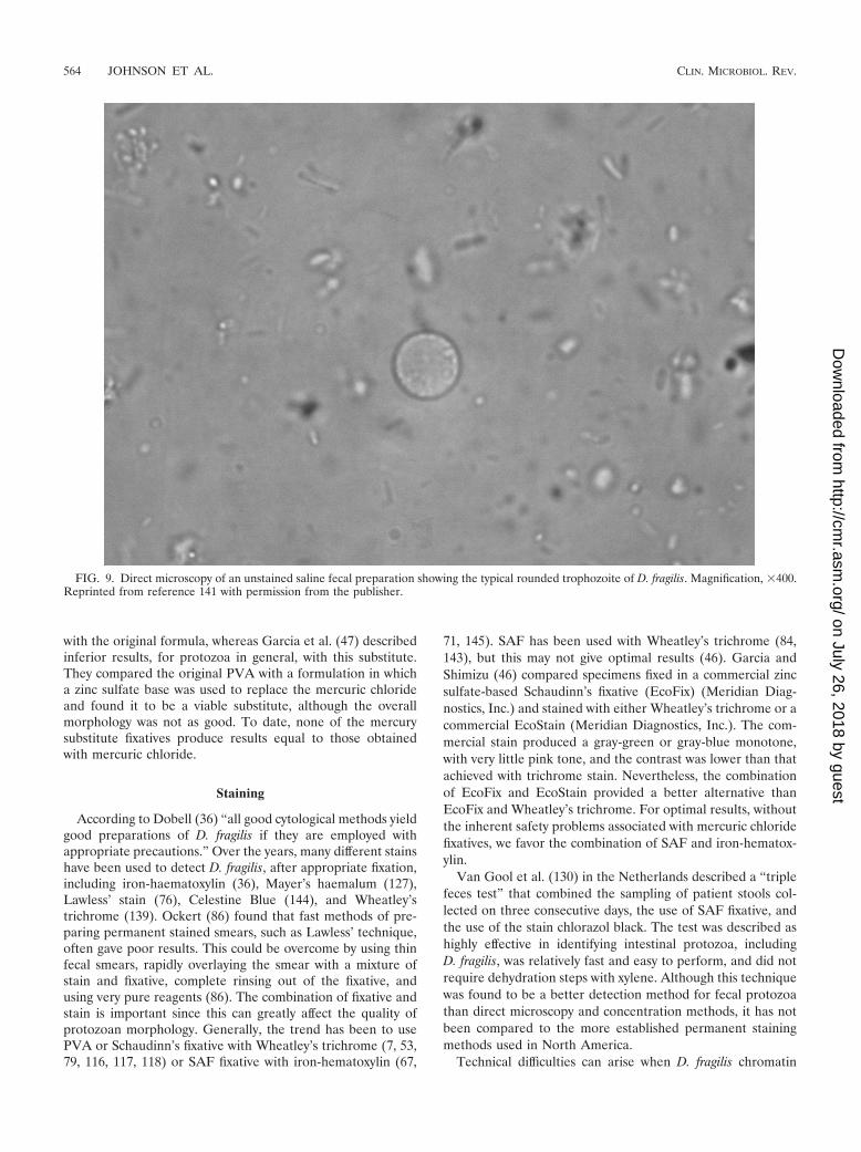
Permanently stained fecal smears are commonly used inNorth America and are appreciated as an essential aid in thediagnosis of intestinal protozoa. In Europe, however, freshunpreserved stool specimens are generally used for examina-tion while stained smears are used in reference centers. Lab-oratories that examine stools by direct microscopy should beaware that D. fragilis trophozoites may be encountered as re-fractile, rounded forms, varying in size from 5 to 15 �m (142)(Fig. 9). The nuclear structure cannot be seen in saline oriodine preparations, and consequently the cells may be dis-missed as artifacts. It is essential that permanent stainedsmears be performed on every stool sample to properly identifytrophozoites of D. fragilis. The crystal violet hematoxylinmethod of Velat et al. (131) can be used to stain D. fragilis andother trophozoites of flagellates in fresh wet preparations. Al-though the preparation of the stain is complex, the method issimple and the results are excellent. Another simple method ofstaining without using specialized fixatives is to air dry a fecalsmear, fix it in industrial methylated spirit, and stain it witheither Giemsa stain (43) or Field’s stain (81). However, it is notpossible to see the typical fragmented nuclei when using thesesimple, rapid methods since the nuclear contents often coa-lesce (86). Much better cytological results can be obtained byusing a suitable fixative (see below) in combination with apermanent staining method. Concentration methods are notgenerally recommended for the recovery of D. fragilis, althoughtrophozoites are sometimes found in concentrated stools(145).
Fixatives
Once D. fragilis trophozoites degenerate, they becomeharder to recognize. Therefore, for optimal results, fecal spec-imens should be placed in a fixative immediately (145). Dobell(36) employed Schaudinn’s fixative followed by staining withHeidenhain’s iron-alum hematoxylin. Generally, all fixativesused for intestinal protozoa are suitable for D. fragilis, theseinclude polyvinyl alcohol (PVA) (50), modified Schaudinn’sfixative (109), phenol-alcohol-formalin (9), and sodium ace-tate-acetic acid-formalin (SAF) (145). SAF has the advantagesthat it is simple to make and relatively nontoxic (compared toother fixatives) and can also be used for concentration meth-ods. The merthiolate-iodine-formalin method (106) is a com-bined fixative and stain technique; however, it is not very stableand does not stain the nuclei of D. fragilis well (146). BothSchaudinn’s fixative and PVA (which is a plastic powder dis-solved in Schaudinn’s fixative) contain mercuric chloride. Con-cerns about safety and problems with the disposal of mercuryled researchers to look for substitutes for mercuric chloridethat are more environmentally friendly (47, 62). Studies usingcopper sulfate (CuSO4) were controversial. Horen (62) foundthat copper sulfate gave results comparable to those obtained
VOL. 17, 2004 CLINICAL MANIFESTATIONS OF DIENTAMOEBA FRAGILIS 563
on July 26, 2018 by guesthttp://cm
r.asm.org/
Dow
nloaded from
with the original formula, whereas Garcia et al. (47) describedinferior results, for protozoa in general, with this substitute.They compared the original PVA with a formulation in whicha zinc sulfate base was used to replace the mercuric chlorideand found it to be a viable substitute, although the overallmorphology was not as good. To date, none of the mercurysubstitute fixatives produce results equal to those obtainedwith mercuric chloride.
Staining
According to Dobell (36) “all good cytological methods yieldgood preparations of D. fragilis if they are employed withappropriate precautions.” Over the years, many different stainshave been used to detect D. fragilis, after appropriate fixation,including iron-haematoxylin (36), Mayer’s haemalum (127),Lawless’ stain (76), Celestine Blue (144), and Wheatley’strichrome (139). Ockert (86) found that fast methods of pre-paring permanent stained smears, such as Lawless’ technique,often gave poor results. This could be overcome by using thinfecal smears, rapidly overlaying the smear with a mixture ofstain and fixative, complete rinsing out of the fixative, andusing very pure reagents (86). The combination of fixative andstain is important since this can greatly affect the quality ofprotozoan morphology. Generally, the trend has been to usePVA or Schaudinn’s fixative with Wheatley’s trichrome (7, 53,79, 116, 117, 118) or SAF fixative with iron-hematoxylin (67,
71, 145). SAF has been used with Wheatley’s trichrome (84,143), but this may not give optimal results (46). Garcia andShimizu (46) compared specimens fixed in a commercial zincsulfate-based Schaudinn’s fixative (EcoFix) (Meridian Diag-nostics, Inc.) and stained with either Wheatley’s trichrome or acommercial EcoStain (Meridian Diagnostics, Inc.). The com-mercial stain produced a gray-green or gray-blue monotone,with very little pink tone, and the contrast was lower than thatachieved with trichrome stain. Nevertheless, the combinationof EcoFix and EcoStain provided a better alternative thanEcoFix and Wheatley’s trichrome. For optimal results, withoutthe inherent safety problems associated with mercuric chloridefixatives, we favor the combination of SAF and iron-hematox-ylin.
Van Gool et al. (130) in the Netherlands described a “triplefeces test” that combined the sampling of patient stools col-lected on three consecutive days, the use of SAF fixative, andthe use of the stain chlorazol black. The test was described ashighly effective in identifying intestinal protozoa, includingD. fragilis, was relatively fast and easy to perform, and did notrequire dehydration steps with xylene. Although this techniquewas found to be a better detection method for fecal protozoathan direct microscopy and concentration methods, it has notbeen compared to the more established permanent stainingmethods used in North America.
Technical difficulties can arise when D. fragilis chromatin
FIG. 9. Direct microscopy of an unstained saline fecal preparation showing the typical rounded trophozoite of D. fragilis. Magnification, �400.Reprinted from reference 141 with permission from the publisher.
564 JOHNSON ET AL. CLIN. MICROBIOL. REV.
on July 26, 2018 by guesthttp://cm
r.asm.org/
Dow
nloaded from

granules are covered with stain deposits, when the nuclearfragmentation is not obvious, or when the majority of tropho-zoites are mononucleated (145). In these circumstances, theycan be confused with trophozoites of Endolimax nana (44, 45,145).
Immunological Diagnosis
Specific immunological tests for D. fragilis are not currentlyavailable. Chan et al. (18) were the first to develop an immu-nofluorescence assay to identify D. fragilis trophozoites in pre-served fecal specimens. They produced anti-D. fragilis anti-serum in rabbits by using the dixenic D. fragilis strain Bi/pa.After absorption of the antiserum with Klebsiella pneumoniaeand Bacteroides vulgatus, the two bacteria present in the cul-ture, it was used in an indirect fluorescent-antibody assay todetect D. fragilis in preserved fecal samples. There were nocross-reactions with any of the 4 species of helminths or 10species of protozoa encountered in their study. The authorsconsidered the indirect fluorescent-antibody assay to be highlyspecific for D. fragilis and were able to identify the organism inseven of nine confirmed positives. Two samples with only verysmall numbers of D. fragilis trophozoites gave questionableresults.
In a later study, Chan et al. (16) employed an immunoblotassay and found that serum samples from patients with con-firmed D. fragilis infections reacted with a 39-kDa D. fragilisprotein. It is unclear what this protein may be, what signifi-
cance it may have in the pathogenesis of the disease, andwhether it has any immunoprophylactic properties.
DNA-Based Diagnosis
Only one study to date has investigated the potential ofdetecting DNA in feces for diagnosis of D. fragilis infection(94) by amplifying a portion of the small-subunit rRNA geneby PCR. However, the sensitivity of this PCR cannot be com-pared directly to microscopy because different samples wereused for the two detection methods. The development of aquick and accurate immunodiagnostic test would be of greatbenefit to the diagnosis of D. fragilis infections.
CULTIVATION
General Considerations
The culture methods used for D. fragilis are xenic, in whichthe parasite is grown in an undefined bacterial flora. The bal-ance required in controlling the bacterial flora while providingfor the needs of the parasite is crucial for the successful cultureof intestinal protozoa (21). The intestinal bacterial flora pro-vides D. fragilis with a food source. In all xenic culture media,rice starch provides the carbohydrate essential for bacterialgrowth. Antibiotics such as erythromycin, penicillin, and strep-tomycin are often used to suppress gram-positive organisms(101, 108, 114). Usually xenic culture media are biphasic, with
FIG. 10. Rounded trophozoites of D. fragilis in Robinson’s culture. Magnification, �400.
VOL. 17, 2004 CLINICAL MANIFESTATIONS OF DIENTAMOEBA FRAGILIS 565
on July 26, 2018 by guesthttp://cm
r.asm.org/
Dow
nloaded from
a slant of agar or egg and a liquid-phase overlay, althoughmonophasic media have also been successfully used. Manydifferent xenic media can be used to grow D. fragilis fromclinical specimens, although we prefer to use Robinson’s me-dium (101).
Historical Background
Some early parasitologists maintained that D. fragilis waseasily isolated and grew abundantly in certain media (22, 36).This is in contrast to the experiences of many present-dayinvestigators (21). Dobell (36) credits Thomson and Robertsonwith the first culture of D. fragilis, using Boeck and Drbohlav’sE. histolytica medium (Locke egg serum) (5). It was heavilycontaminated with Blastocystis hominis, and they were able tomaintain it for only a short period. Dobell and Svensson arecredited with having produced the first culture of D. fragilisfree from other protozoa in 1929 (36).
Dobell (36) used two different biphasic media, one (HSre �S) with a solid slope of inspissated horse serum and the other(Ehs � S) made with an inspissated egg slope. The former gavethe best results when overlaid with dilute egg white in Ringer’sfluid and supplemented with rice starch. Interestingly, Dobell(36) found an optimum growth temperature of 40°C for somestrains of D. fragilis. Cleveland and Collier (22) isolated D.fragilis while attempting to improve their cultivation of E. his-tolytica. The medium they used was Loeffler’s dehydrated beefserum slants covered with fresh horse serum saline.
Balamuth (4) described a monophasic liquid medium con-taining dehydrated egg yolk and liver infusion. This mediumpermitted the growth of amebae (Entamoeba histolytica, Ent-amoeba coli, Iodamoeba butschlii, and Endolimax nana), flagel-lates (Trichomonas spp. and Chilomastix mesnili), and D. fra-gilis. Balamuth used this medium to study the effects of drugs,amebicides, and antibiotics on amebae and D. fragilis. Whileexperimenting on a xenic culture of D. fragilis, Balamuth inad-vertently eliminated all but two of the bacterial species andproduced a dixenic culture. Balamuth designated the sublineBi/pa, and the bacteria were identified as Clostridium perfrin-gens and Aerobacter aerogenes. Klebsiella pneumoniae and Clos-tridium perfringens are now listed as the bacterial species, al-though Chan et al. (18) used this strain and found theanaerobic species to be Bacteroides vulgatus.
Jacobs (63) performed many experiments on the cultivationof D. fragilis but was unable to support its growth withoutviable bacteria. Using crude xenic cultures transplanted intoa medium containing Clostridium perfringens with penicillin,streptomycin, and sulfadiazine, he was able to produce a mon-oxenic culture of D. fragilis. However, all attempts to producean axenic culture (without other organisms) of D. fragilis havefailed to date (17, 18, 21).
Robinson (101) formulated a biphasic medium for the diag-nosis of human parasitic amebae that had a saline agar slopeas the solid phase. The liquid phase was complex and in-cluded Escherichia coli growing in a defined medium (R).Other additives included erythromycin, horse serum, potas-sium phthalate, Bacto Peptone, and rice starch (21). Rob-inson’s medium supported the growth of all intestinal ame-bae including D. fragilis. This medium was used by Johnsonand Clark (66) to grow D. fragilis prior to performing ribo-
printing. Diamond (33) developed a monophasic medium,TYSGM-9 (Trypticase, yeast extract, serum, gastric mucin),which supported the growth of lumen-dwelling protozoa,including D. fragilis.
Surveys Using Culture
The most sensitive method of detecting D. fragilis is by usingculture techniques, compared directly to stained smears (86,141). Although cultivation of intestinal protozoa is not usuallyattempted in routine diagnostic laboratories (21, 140), recentdata have shown that it can be successfully employed outside aresearch environment (141). The use of Robinson’s medium ina small diagnostic laboratory doubled the detection rate from1.3 to 2.6% compared with the rate for trichrome-stainedsmears. It was considered less laborious than staining andrequired a smaller amount of feces, and culture lysates canprovide material for subsequent genotyping. On the negativeside, cultures take 48 h or longer and cannot be used for fecalsamples that are submitted in fixative. They are unlikely toreplace staining methods in diagnostic parasitology laborato-ries because they do not detect all intestinal protozoa. How-ever, cultures may play a role in laboratories that do not havethe expertise to detect D. fragilis in stained smears but want toexclude this parasite. Positive cultures were even obtainedfrom stool samples stored at room temperature or 4°C for 24 h.A previous study (108) had reported that D. fragilis could becultured from feces stored for up to 24 h at room temperaturebut only for 10 h at 4°C.
Ockert (86) reported two studies undertaken in his researchlaboratory where all stool samples were both stained and cul-tured. In the first study, involving 576 children, 3% of thestained smears were positive whereas 35% were positive inculture. In the second study, involving 1,066 persons, 1.97% ofthe stained smears were positive while 39.3% were positive inculture. The medium used was a modification of that of Dobelland Laidlaw (37), using coagulated human serum for the solidphase. The primary cultures were subcultured twice into freshmedium. Of the D. fragilis-positive samples identified, only40% were detected by the primary culture whereas over 80%were detected following the first subculture and the rest weredetected after the second subculture. Silard et al. (114) alsofavored the use of Dobell and Laidlaw medium and isolatedD. fragilis from 2.8% of clinical samples. Using culture over a10-year period in Israel, Talis et al. (127) detected D. fragilis in30,609 (15.2%) of 201,750 specimens. The medium used wasa diphasic egg medium formulated in their laboratory. Usingsamples from a patient with known D. fragilis infection,Sawangjaroen et al. (108) compared three media, modifiedBoeck and Drbohalav (BD) medium, TYSGM-9 (33), andCleveland and Collier medium (22). Modified Boeck and Drbo-halav medium was found to be the most suitable since it wasthe only medium that supported the initial growth and subcul-ture of D. fragilis. The authors then surveyed consecutive sam-ples from patients with diarrhea. A surprisingly low incidenceof 1.5% (4 of 260) was found; however, culture doubled thedetection rate.
566 JOHNSON ET AL. CLIN. MICROBIOL. REV.
on July 26, 2018 by guesthttp://cm
r.asm.org/
Dow
nloaded from
Appearance in Culture
Trophozoites of D. fragilis initially appear as rounded refrac-tile bodies, containing many rice starch granules, in freshlymounted culture preparations after 48 h of incubation (Fig.10). After approximately 10 min at room temperature, theybegin to produce sluggish, small, irregular pseudopodia (108)(Fig. 1). The size of D. fragilis in culture varies considerably,and some workers have described a range of 6 to 40 �m (114).Robinson and Ng (102) found that with experience, this sizerange became a good guide to the presumptive identification ofD. fragilis after addition of a drop of iodine. Moreover, theappearance of circular brown-red forms filled with starchgrains aided this diagnosis. Several intestinal amebae grow wellin culture media used for D. fragilis and also ingest rice starch.Although none of the amebae demonstrate the characteristicmotility of D. fragilis, it is nevertheless recommended that thediagnosis be confirmed by using a suitable staining method(101). Robinson (101) fixed culture-positive amebae in a mix-ture of acetic acid and phosphotungstic acid and stained themwith a hematoxylin stain. Windsor et al. (141) confirmed thepositive amebae by fixing in Schaudinn’s fixative and stainingwith trichrome, whereas Silard et al. (114) preferred a simplermethod of fixing in methanol and staining with Giemsa. Diag-nostic uncertainties can arise for cultures containing granular
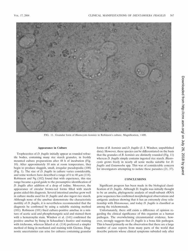
forms of B. hominis and D. fragilis (J. J. Windsor, unpublisheddata). However, these species can be differentiated on the basisthat the granules of B. hominis are distinctly rounded (Fig. 11)whereas D. fragilis simply contains ingested rice starch. Blasto-cystis grows freely in nearly all xenic media suitable for D.fragilis and Entamoeba spp. This was of considerable concernfor investigators attempting to isolate these parasites (21, 37).
CONCLUSIONS
Significant progress has been made in the biological classi-fication of D. fragilis. Although D. fragilis was initially thoughtto be an ameba, phylogenetic analysis of small-subunit rRNAgene sequences has confirmed morphological observations andantigenic analyses showing that it has an extremely close rela-tionship with Histomonas, and today D. fragilis is classified asamong the trichomonads.
Unfortunately, there still exists a difference of opinion re-garding the clinical significance of this organism as a humanpathogen. The overwhelming circumstantial evidence, how-ever, strongly suggests that D. fragilis is a bona fide pathogen.This is based primarily on the observation that there are a largenumber of case reports from many parts of the world thatdescribe patients whose clinical symptoms subsided only after
FIG. 11. Granular form of Blastocystis hominis in Robinson’s culture. Magnification, �400.
VOL. 17, 2004 CLINICAL MANIFESTATIONS OF DIENTAMOEBA FRAGILIS 567
on July 26, 2018 by guesthttp://cm
r.asm.org/
Dow
nloaded from
therapeutic intervention and elimination of the organism.However, it cannot be denied that there are patients whoharbor this organism but do not exhibit clinical signs. Recentlyundertaken molecular studies may help to shed light on thesediffering observations. Using riboprinting, two genetically dis-tinct types of D. fragilis have been identified. It certainly wouldnot be unreasonable to speculate that different genetic typesmay demonstrate different degrees of virulence.
The accuracy of diagnostic testing for D. fragilis has tradi-tionally relied on permanently stained fecal smears, since thecharacteristic binucleate appearance of the organism cannotbe appreciated in saline or iodine preparations. A number ofstudies have substantiated the necessity of using this method-ology. The fairly recent introduction of fecal cultures for diag-nosis indicates that infection rates are significantly higher thanthose found using stained smears and that cultures are lesslaborious to perform. However, culture cannot be done onfecal samples received in fixative, and it does not detect allintestinal protozoa. Consequently, it is unlikely to replace per-manent staining in the diagnostic parasitology setting, but itmight appeal to laboratories that want to exclude D. fragilis butdo not have sufficient expertise in reading stained smears.
There are great gaps in our present state of knowledgeconcerning the virulence, pathogenicity, and mode of transmis-sion of D. fragilis. However, neglecting to include D. fragilis onthe list of potential culprits in unexplained cases of chronicdiarrhea, abdominal pain, fatigue, flatulence, and anorexia,and even in patients with irritable bowel syndrome-like syn-dromes, can no longer be justified.
ACKNOWLEDGMENT
It would be remiss of us not to extend our sincerest thanks to AlanCurry at the Manchester Royal Infirmary for his inexhaustible gener-osity in providing the micrographs used in the present review article.
REFERENCES
1. Addadi, K., Y. Le Corroller, Y. Guy, and A. Tabet-Derraz. 1972. Sur le rolepathogene possible de Dientamoeba fragilis. Bull. Soc. Pathol. Exot. Fil.65:274–275.
2. Aucott, J. N., and J. I. Ravdin. 1993. Amebiasis and “nonpathogenic”intestinal protozoa. Infect. Dis. Clin. North. Am. 7:467–485.
3. Ayadi, A., and I. Bahri. 1999. Dientamoeba fragilis: flagelle pathogene. Bull.Soc. Pathol. Exot. 5:299–301.
4. Balamuth, W. 1946. Improved egg yolk medium for cultivation of Entamoe-ba histolytica and other intestinal protozoa. Am. J. Clin. Pathol. 16:380–384.
5. Boeck, W. C., and J. Drbohlav. 1925. The cultivation of Endamoeba histo-lytica. Am. J. Hyg. 5:371–407.
6. Borody, T. J., E. F. Warren, A. Wettstein, G. Robertson, P. Recabarren, A.Fontella, K. Herdnman, and R. Surace. Eradication of Dientamoeba fragiliscan resolve IBS-like symptoms. 2002. J. Gastroenterol. Hepatol. 17(Suppl.):A103.
7. Bruckner, D. A., L. S. Garcia, and M. Voge. 1979. Intestinal parasites in LosAngeles, California. Am. J. Med. Technol. 45:1020–1022.
8. Brug, S. L. 1936. Observations on Dientamoeba fragilis. Ann. Trop. Med.Parasitol. 30:441–452.
9. Burrows, R. B. 1967. A new fixative and technics for the diagnosis ofintestinal parasites. Tech. Bull. Regist. Med. Technol. 37:208–212.
10. Burrows, R. B., and M. A. Swerdlow. 1956. Enterobius vermicularis as aprobable vector of Dientamoeba fragilis. Am. J. Trop. Med. Hyg. 5:258–265.
11. Burrows, R. B., M. A. Swerdlow, J. K. Frost, and C. K. Leeper. 1954.Pathology of Dientamoeba fragilis infections of the appendix. Am. J. Trop.Med. Hyg. 3:1033–1039.
12. Butler, W. P. 1996. Dientamoeba fragilis. An unusual intestinal pathogen.Dig. Dis. Sci. 41:1811–1813.
13. Camp, R. R., C. F. Mattern, and B. M. Honigberg. 1974. Study of Dient-amoeba fragilis Jepps & Dobell. I. Electronmicroscopic observations of thebinucleate stages. II. Taxonomic position and revision of the genus. J. Pro-tozool. 21:69–82.
14. Cerva, L. 1962. Die Darmparasiten in den Kinderheimen des Prager Be-zirkes. Angew. Parasitol. 2:119–124.
15. Cerva, L., M. Schrottenbaum, and V. Kliment. 1991. Intestinal parasites: astudy of human appendices. Folia Parasitol. 38:5–9.
16. Chan, F., N. Stewart, M. Guan, I. Robb, L. Fuite, I. Chan, F. Diaz-Mitoma,J. King, N. MacDonald, and A. MacKenzie. 1996. Prevalence of Dientamoe-ba fragilis antibodies in children and recognition of a 39 kDa immunodom-inant protein antigen of the organism. Eur. J. Clin. Microbiol. Infect. Dis.15:950–954.
17. Chan, F. T., M. X. Guan, A. M. MacKenzie, and F. Diaz- Mitoma. 1994.Susceptibility testing of Dientamoeba fragilis ATCC 30948 with iodoquinol,paromomycin, tetracycline, and metronidazole. Antimicrob. Agents Che-mother. 38:1157–1160.
18. Chan, F. T. H., M. X. Guan, and A. M. MacKenzie. 1993. Application ofindirect immunofluorescence to detection of Dientamoeba fragilis tropho-zoites in fecal specimens. J. Clin. Microbiol. 31:1710–1714.
19. Chang, S. 1973. Parasitization of the parasite. JAMA 223:1510.20. Chatton, E. P. L. 1925. Pansporella perplexa, amoebien a spores protegees,
parasite des daphnies. Reflexions sur la biologie et la phylogenie des pro-tozoaires. Ann. Sci. Nat. Zool. 10e Ser. 8:5–85.
21. Clark, C. G., and L. S. Diamond. 2002. Methods for the cultivation ofluminal parasitic protists of clinical importance. Clin. Microbiol. Rev. 15:329–341.
22. Cleveland, L. R., and J. Collier. 1930. Various improvements in cultivationof Endamoeba histolytica. Am. J. Hyg. 12:606–613.
23. Cline, B. 1982. Drug regimens for intestinal helminth infections. Med.Clinics North Am. 66:721–742.
24. Colea, A., R. Silard, D. Panaitescu, P. Florescu, N. Roman, and T. Capraru.1980. Studies on Dientamoeba fragilis in Romania. II. Incidence of Dient-amoeba fragilis in healthy persons. Arch. Roum. Pathol. Exp. Microbiol.39:49–53.
25. Costa, R. S. 1948. Dientamoeba fragilis Jepps and Dobell, 1918. Primeroscasos senelados en nuestro pais. Consideraciones epidemiologicas. Rev.Kuba Med. Trop. 4:115–118.
26. Craig, C. F. 1926. The nuclear structure of Dientamoeba fragilis. J. Parasitol.13:137–140.
27. Crotti, D., and M. L. D’Annibale. 2001. Dientamoeba fragilis e dientamoe-bosi: aspetti di parassitologia clinicale diagnostica di laboratorio. Parassi-tologia 43:135–138.
28. Cuffari, C., L. Oligny, and E. G. Seidman. 1998. Dientamoeba fragilis mas-querading as allergic colitis. J. Pediatr. Gastroenterol. Nutr. 26:16–20.
29. Dardick, K. 1983. Tetracycline treatment of Dientamoeba fragilis. Conn.Med. 47:69–70.
30. Das Gupta, B. M. 1936. A case of human infection with Dientamoeba fragilisJepps and Dobell, 1918, in Calcutta. Indian Med. Gaz. 21:528–529.
31. De Jonckheere, J. B., S. Brown, P. J. Dobson, B. S. Robinson, and P.Pernin. 2001. The amoeba-to-flagellate transformation test is not reliablefor the diagnosis of the genus Naegleria. Description of three new Naegleriasp. Protist 152: 115–121.
32. Reference deleted.33. Diamond, L. S. 1982. A new liquid medium for xenic cultivation of Ent-
amoeba histolytica and other lumen-dwelling protozoa. J. Parasitol. 68:958–959.
34. Diamond, L. S., and C. G. Clark. 1993. A redescription of Entamoebahistolytica Schaudinn, 1903 (Emended Walker, 1911) separating it fromEntamoeba dispar Brumpt, 1925. J. Eukaryot. Microbiol. 40:340–344.
35. Dickinson, E. C., M. A. Cohen, and M. K. Schlenker. 2002. Dientamoebafragilis: a significant pathogen. Am. J. Emerg. Med. 20:62–63.
36. Dobell, C. 1940. Researches on the intestinal protozoa of monkeys andman. X. The life history of Dientamoeba fragilis: observations, experiments,and speculations. Parasitology 32:417–461.
37. Dobell, C., and P. P. Laidlaw. 1926. On the cultivation of Entamoebahistolytica and some other entozoic amoeba. Parasitology 18:283–318.
38. Dobell, C., and F. W. O’Connor. 1921. The intestinal protozoa of man. J.Bale, Sons, and Danielson, London, United Kingdom.
39. Dwyer, D. M. 1972. Analysis of the antigenic relationships among Trichomo-nas, Histomonas, Dientamoeba, and Entamoeba. I. Quantitiative fluorescentantibody methods. J. Protozool. 19:316–325.
40. Dwyer, D. M. 1972. Analysis of the antigenic relationships among Trichomo-nas, Histomonas, Dientamoeba, and Entamoeba. II. Gel diffusion methods.J. Protozool. 19:316–325.
41. Dwyer, D. M. 1974. Analysis of the antigenic relationships among Trichomo-nas, Histomonas, Dientamoeba, and Entamoeba. III. Immunoelectrophore-sis technics. J. Protozool. 21:139–145.
42. Fantham, H. B., and A. Porter. 1936. Some entozoa of man as seen inCanada and South Africa. Can Med. Assoc. J. 34:414–421.
43. Fleck, S. L., and A. H. Moody. 1988. Faecal parasites, p. 23–24. In S. L.Fleck and A. H. Moody (ed.), Diagnostic techniques in medical parasitol-ogy. Elsevier, Amsterdam, The Netherlands.
44. Garcia, L. 2002. Laboratory methods for diagnosis of parasitic infection,p. 776–861. In B. A. Forbes, D. F. Sahm, and A. S. Weissfeld (ed.), Baileyand Scott’s Diagnostic Microbiology, 11th ed. C. V. Mosby, St. Louis, Mo.
45. Garcia, L. S. 1992. Parasitology, p. 2:7.10:13:13. In H. D. Isenberg (ed.),
568 JOHNSON ET AL. CLIN. MICROBIOL. REV.
on July 26, 2018 by guesthttp://cm
r.asm.org/
Dow
nloaded from
Clinical microbiology procedures handbook. American Society for Micro-biology, Washington, DC.
46. Garcia, L. S., and R. Y. Shimizu. 1998. Evaluation of intestinal protozoanmorphology in human fecal specimens preserved in EcoFix: comparison ofWheatley’s trichrome stain and EcoStain. J. Clin. Microbiol. 36:1974–1976.
47. Garcia, L. S., R. Y. Shimizu, T. C. Brewer, and D. A. Bruckner. 1983.Evaluation of intestinal parasite morphology in polyvinyl alcohol preserva-tive: comparison of copper sulfate and mercuric chloride bases for use inSchaudinn fixative. J. Clin. Microbiol. 17:1092–1095.
48. Gerbod, D., V. P. Edgcomb, C. Noel, L. Zenner, R. Wintjens, P. Delgado-Viscogliosi, M. E. Holder, M. L. Sogin, and E. Viscogliosi. 2001. Phyloge-netic position of the trichomonad parasite of turkeys, Histomonas melea-gridis (Smith) Tyzzer, inferred from small subunit rRNA sequence. J.Eukaryot. Microbiol. 48:498–504.
49. Girginkardesler, N., S. Coskun, I. Cuneyt Balcioglu, P. Ertan, and U. Z.Ok. 2003. Dientamoeba fragilis, a neglected cause of diarrhea, successfullytreated with secnidazole. Clin. Microbiol. Infect. 9:110–113.
50. Goldman, M., and M. M. Brooke. 1953. Protozoans in stools unpreservedand preserved in PVA fixative. Public Health. Rep. 68:703–706.
51. Grasse, P. P. 1953. Famille des Dientamoebidae Grasse, nov., (E. Chatton,principal author: ordre des amoebiens nus ou Amoebaea), p. 50–54. In P. P.Grasse (ed.), Traite de zoologie. Masson et Cie., Paris, France.
52. Greenway, D. 1928. Dientamoeba fragilis en la Argentina. Arch. Argent.Enferm. Aparato Dig. (Buenos Aires) 3:897.
53. Grendon, J. H., R. F. Digiacomo, and F. J. Frost. 1995. Descriptive featuresof Dientamoba fragilis infections. J. Trop. Med. Hyg. 98:309–315.
54. Grendon, J. H., R. F. Digiacomo, and F. J. Frost. 1991. Dientamoeba fragilisdetection methods and prevalence: a survey of State Public Health Labo-ratories. Public Health Rep. 106:322–325.
55. Hakansson, E. G. 1936. Dientamoeba fragilis, a cause of illness: report ofcase. Am. J. Trop. Med. 16:175–183.
56. Hakansson, E. G. 1937. Dientamoeba fragilis: some further observations.Am. J. Trop. Med. 17:349–362.
57. Halawani, A. E. 1930. Dientamoeba fragilis from a culture of human faeces.Trans. R. Soc. Trop. Med. Hyg. 23:326–327.
58. Haughwout, F. G., and F. S. Horrilleno. 1920. The intestinal animal para-sites found in one hundred sick Filipino children. Philipp. J. Sci. 16:1–73.
59. Hegner, R., and H. J. Chu. 1930. A comparative study of the intestinalprotozoa of wild monkeys and man. Am. J. Hyg. 12:62–108.
60. Hiatt, R. A., E. K. Markell, and E. Ng. 1995. How many stool examinationsare necessary to detect pathogenic intestinal protozoa? Am. J. Trop. Med.Hyg. 53:36–39.
61. Hollande, A., and J. Carruette-Valentin. 1971. Les atractophores, l’induc-tion du fuseau et la division cellulaire chez les Hypermastigines. Etudeinfrastructurale et revision systematique des Trichonymphines et des Spi-rotrichonymphines. Protistologica 7:5–100.
62. Horen, W. P. 1981. Modification of Schaudinn fixative. J. Clin. Microbiol13:204–205.
63. Jacobs, L. 1953. The cultivation of Dientamoeba fragilis. Ann. N. Y. Acad.Sci. 56:1057–1061.
64. Jepps, M. W. 1921. Notes on the intestinal protozoa of 971 men at theUniversity War Hospital, Southampton. J. R. Army Med. Corps 97:366–375.
65. Jepps, M. W., and C. Dobell. 1918. Dientamoeba fragilis n.g., n. sp., newintestinal amoeba from man. Parasitology 10:352–367.
66. Johnson, J. A., and C. G. Clark. 2000. Cryptic genetic diversity in Dient-amoeba fragilis. J. Clin. Microbiol. 38:4653–4654.
67. Kabani, A., G. Cadrain, C. Trevenen, T. Jadavji, and D. L. Church. 1995.Practice guidelines for ordering stool ova and parasite testing in a paedi-atric population. Am. J. Clin. Pathol. 104:272–278.
68. Katz, D. E., and D. N. Taylor. 2001. Parasitic infections of the gastrointes-tinal tract. Gastroenterol. Clin. North Am. 30:797–815.
69. Kean, B. H., and C. L. Malloch. 1966. The neglected ameba: Dientamoebafragilis. A report of 100 “pure” infections. Am. J. Dig. Dis. 11:735–746.
70. Keystone, J. S., E. Proctor, C. Glenn, and L. Mcintyre. 1983. Safety andefficacy of diphetarsone in the treatment of amoebiasis, non-pathogenicamoebiasis and trichuriasis. Trans. R. Soc. Trop. Med. Hyg. 77:84–86.
71. Keystone, J. S., J. Yang, D. Grisdale, M. Harrington, L. Pillon, and R.Andreychuk. 1984. Intestinal parasites in metropolitan Toronto day-carecentres. Can. Med. Assoc. J. 131:733–735.
72. Knoll, E. W., and K. M. Howell. 1945. Studies on Dientamoeba fragilis. Itsincidence and possible pathogenicity. Am. J. Clin. Pathol. 15:178–183.
73. Knowles, R., and B. M. Das Gupta. 1936. Some observations on the intes-tinal protozoa of macaques. Indian J. Med. Res. 24:547–556.
74. Kofoid, C. A. 1923. Amoeba and man, p. 149–174, 219–312. In University ofCalifornia Chronicle for 1923. University of California Press, Berkeley.
75. Lainson, R., and B. A. Da Silva. 1999. Intestinal parasites of some diar-rhoeic HIV-seropositive individuals in North Brazil with particular refer-ence to Isospora belli Wenyon, 1923 and Dientamoeba fragilis Jepps &Dobell, 1918. Mem. Inst. Oswaldo Cruz 94:611–613.
76. Lawless, D. K. 1953. A rapid permanent-mount stain technique for thediagnosis of intestinal protozoa. Am. J. Trop. Med. Hyg. 20:1137–1138.
77. Melvin, D. M., and M. M. Brooke. 1962. Parasitologic surveys on Indianreservations in Montana, South Dakota, New Mexico, Arizona and Wis-consin. Am. J. Trop. Med. Hyg. 11:765–772.
78. Mendez, O. C., G. Szmulewicz, C. Menghi, S. Torres, G. Gonzalez, and C.Gatta. 1994. Comparison of intestinal parasite infestation indexes amongHIV positive and negative populations. Medicina (Buenos Aires) 54:307–310.
79. Millet, V., M. J. Spencer, M. Chapin, M. Stewart, J. A. Yatabe, T. Brewer,and L. S. Garcia. 1983. Dientamoeba fragilis, a protozoan parasite in adultmembers of a semicommunal group. Dig. Dis. Sci. 28:335–339.
80. Mollari, M., and J. V. Anzulovic. 1938. Cultivation and pathogencity ofDientamoeba fragilis, with a case report. J. Trop. Med. Hyg. 41:246–247.
81. Moody, A. H., and S. L. Fleck. 1985. Versatile Field’s stain. J. Clin. Pathol.38:842–843.
82. Muller, M. 1975. Biochemistry of protozoan microbodies: peroxisomes,alpha-glycerophosphate oxidase bodies, hydrogenosomes. Annu. Rev. Mi-crobiol. 29:467–483.
83. Myers, B. J., and R. E. Kuntz. 1968. Intestinal protozoa of the baboonPapio doguera Pucheran, 1856. J. Protozool. 15:363–365.
84. Naiman, H. L., L. Sekla, and W. L. Albritton. 1980. Giardiasis and otherintestinal parasitic infections in a Manitoba residential school for the men-tally retarded. Can. Med. Assoc. J. 122:185–188.
85. Norberg, A., C. E. Nord, and B. Evengard. 2003. Dientamoeba fragilis—aprotozoal infection which may cause severe bowel distress. Clin. Microbiol.Infect. 9:65–68.
86. Ockert, G. 1990. Symptomatology, pathology, epidemiology, and diagnosisof Dientamoeba fragilis, p. 394–410. In B. M. Honigberg (ed.), Tricho-monads parasitic in humans. Springer Publications, New York, N.Y.
87. Ockert, G. 1972. Zur epidemiologie von Dientamoeba fragilis Jepps et Do-bell 1918. 1. Mitteilung: die Verbreitung der art in Kinderkollektiven. J.Hyg. Epidemiol. Microbiol. Immunol. 16:213–221.
88. Ockert, G. 1972. Zur epidemiologie von Dientamoeba fragilis Jepps et Do-bell 1918. 2. Mitteilung: Versuch der Uebertragung der Art mit Enterobius-Eiern. J. Hyg. Epidemiol. Microbiol. Immunol. 16:222–225.
89. Ockert, G. 1975. Zur epidemiologie von Dientamoeba fragilis. 3. Weitere zurUebertragung der Art mit Enterobius-Eiern. J. Hyg. Epidemiol. Microbiol.Immunol. 19:17–21.
90. Ockert, G., and T. Schmidt. 1976. Zur epidemiologie von Dientamoebafragilis Jepps et Dobell. 1918. 4. Mitteilung: Nachweis von Dientamoebafragilis in Enterobius eirn mit hilfe von IEP-bestimmungen. J. Hyg. Epide-miol. Microbiol. Immunol. 20:76–81.
91. Ockert, G., and F. Schneider. 1974. Elektronenmikroskopische Befunde beiDientamoeba fragilis. Z. Gesamte Hyg. 20:555–557.
92. Ockert, G., and U. Schulz. 1972. Zur pathogenetischen Rolle von Dient-amoeba fragilis. Dtsch. Gesundheitsw. 27:1156–1158.
93. Panosian, C. 1988. Parasitic diarrhoea. Infect. Dis. Clin. North Am. 2:685–703.
94. Peek, R., F. R. Reedeker, and T. van Gool. 2004. Direct amplification andgenotyping of Dientamoeba fragilis from human stool specimens. J. Clin.Microbiol. 42:631–635.
95. Piekarski, G. 1948. Zur Frage der Cystenbildung bei Dientamoeba fragilis.Z. Hyg. Infektionskr. 127:496–500.
96. Porter, A. 1934. Remarks on intestinal parasites in Montreal and the rela-tion of Entamoeba histolytica to colitis. Can. Med. Assoc. J. 30:134–137.
97. Preiss, U., G. Ockert, S. Broemme, and A. Otto. 1990. Dientamoeba fragilisinfection, a cause of gastrointestinal symptoms in childhood. Klin. Paediatr.202:120–123.
98. Preiss, U., G. Ockert, S. Broemme, and A. Otto. 1991. On the clinicalimportance of Dientamoeba fragilis infections in childhood. J. Hyg. Epide-miol. Microbiol. Immunol. 35:27–34.
99. Rachet, J., A. Busson, and G. Terrial. 1945. Le role pathogene de ‘Dient-amoeba fragilis’. Arch. Mal. Appar. Dig. Mal. Nutr. 34:196–200.
100. Robertson, A. 1923. Notes on a case infected with Dientamoeba fragilis,Jepps and Dobell, 1917. J. Trop. Med. Hyg. 26:243–244.
101. Robinson, G. L. 1968. The laboratory diagnosis of human parasitic amoe-bae. Trans. R. Soc. Trop. Med. Hyg. 62:285–294.
102. Robinson, G. L., and P. H. T. Ng. 1968. The size of Dientamoeba fragilis inculture. Trans. R. Soc. Trop. Med. Hyg. 62:156–158.
103. Rothman, M. M., and H. J. Epstein. 1941. Clinical symptoms associatedwith the so-called non-pathogenic amoeba. JAMA 116:694–700.
104. Sapero, J. J. 1939. Clinical studies in non-dysenteric amoebiasis. Am. J.Trop. Med. 19:497–541.
105. Sapero, J. J., and C. M. Johnson. 1939. Entamoeba histolytica and otherintestinal parasites: incidence in variously exposed groups of the Navy. U. S.Navy Med. Bull. 37:297–301.
106. Sapero, J. J., and D. K. Lawless. 1953. The ‘MIF’ stain-preservation tech-nique for the identification of intestinal protozoa. Am. J. Trop. Med. Hyg.2:613–619.
107. Sargeaunt, P. G., and J. E. Williams. 1982. The morphology in culture ofthe intestinal amoebae of man. Trans. R. Soc. Trop. Med. Hyg. 76:465–472.
108. Sawangjaroen, N., R. Luke, and P. Prociv. 1993. Diagnosis by faecal culture
VOL. 17, 2004 CLINICAL MANIFESTATIONS OF DIENTAMOEBA FRAGILIS 569
on July 26, 2018 by guesthttp://cm
r.asm.org/
Dow
nloaded from

of Dientamoeba fragilis infections in Australian patients with diarrhoea.Trans. R. Soc. Trop. Med. Hyg. 87:163–165.
109. Scholten, T. 1972. An improved technique for the recovery of intestinalprotozoa. J. Parasitol. 58:633–634.
110. Schulz, U., and G. Ockert. 1971. Darmprotozoen als Ursache intestinalerStoerungen. Dtsch. Med. Wochenschr. 51:1963–1968.
111. Shein, R., and A. Gelb. 1983. Colitis due to Dientamoeba fragilis. Am. J.Gastroenterol. 78:634–636.
112. Silard, R., and B. Burghelea. 1986. Endosymbionts in Dientamoeba fragilistrophozoites resistant to antiprotozoal drugs. Arch. Roum. Pathol. Exp.Microbiol. 45:65–74.
113. Silard, R., B. Burghelea, D. Panaitescu, and V. Burcos. 1984. Ultrastructureof Dientamoeba fragilis: a study of the mononucleated stage. Arch. Roum.Pathol. Exp. Microbiol. 43:87–101.
114. Silard, R., A. Colea, D. Panaitescu, P. Florescu, and N. Roman. 1979.Studies on Dientamoeba fragilis in Romania. I. Dientamoeba fragilis isolatedfrom clinical cases. Problems of diagnosis, incidence, clinical aspects. Arch.Roum. Pathol. Exp. Microbiol. 38:359–372.
115. Silberman, J. D., C. G. Clark, and M. L., Sogin. 1996. Dientamoeba fragilisshares a recent common evolutionary history with the trichomonads. Mol.Biochem. Parasitol. 76:311–314.
116. Spencer, M. J., M. R. Chapin, and L. S. Garcia. 1982. Dientamoeba fragilis:a gastrointestinal protozoan infection in adults. Am. J. Gastroenterol. 77:565–569.
117. Spencer, M. J., L. S. Garcia, and M. R. Chapin. 1979. Dientamoeba fragilis.An intestinal pathogen in children? Am. J. Dis. Child. 133:390–393.
118. Spencer, M. J., V. E. Millet, L. S. Garcia, L. Rhee, and L. Masterson. 1983.Parasitic infections in a pediatric population. Pediatr. Infect. Dis. J. 2:110–113.
119. Stabler, R. M. 1941. Intestinal protozoa in 106 parasitology students.J. Parasitol. 24:90.
120. Steinitz, H. 1973. Klinische Beobachtungen bei Infektionen mit Dientamoe-ba fragilis. Z. Gastroenterol. 11:179–182.
121. Steinitz, H. 1972. The treatment of chronic amoebic infection with metro-nidazole (flagyl). Digestion 6:75–82.
122. Stenzel, D. J., B. Stein, and J. Lengy. 1991. A cyst-like stage of Blastocystishominis. Int. J. Parasitol. 21:613–615.
123. Sukanahaketu, S. 1977. The presence of Dientamoeba fragilis in the Ascarislumbricoides ova: the first report from Thailand. J. Med. Assoc. Thail. 60:256–258.
124. Swerdlow, M. A., and R. B. Burrows. 1955. Dientamoeba fragilis, an intes-tinal pathogen. JAMA 158:176–178.
125. Taliaferro, W. H., and E. R. Becker. 1924. A note on the human intestinalamoeba, Dientamoeba fragilis. Am. J. Hyg. 4:71–74.
126. Talis, B., and B. Stein. 1961. The presence of Dientamoeba fragilis in the gallbladder. Harefueh 61:126.
127. Talis, B., B. Stein, and J. Lengy. 1971. Dientamoeba fragilis in human fecesand bile. Isr. J. Med. Sci. 7:1063–1069.
128. Thomson, J. G., and A. Robertson. 1923. Dientamoeba fragilis, Jepps and
Dobell 1917. A case of human infection in England. J. Trop. Med. Hyg.26:135–136.
129. Van Gool, T., and J. Dankert. 1996. Drie opkomende protozoaire infec-tieziekten in Nederland: Cyclospora-, Dientamoeba-en Microspora-infec-ties. Ned. Tijdschr. Geneeskd. 140:155–160.
130. Van Gool, T., R. Weijts, E. Lommerse, and T. G. Mank. 2003. Triple faecestest: an effective tool for detection of intestinal parasites in routine clinicalpractice. Eur. J. Clin. Microbiol. Infect. Dis. 22:284–290.
131. Velat, C. A., P. P. Weinstein, and G. F. Otto. 1950. A stain for the rapiddifferentiation of the trophozoites of the intestinal amoeba in fresh, wetpreparations. Am. J. Trop. Med. 30:43–51.
132. Warren, K. S., and A. A. Mahmoud. 1975. Algorithms in the diagnosis andmanagement of exotic diseases. V. Enterobiasis. J. Infect. Dis. 132:229–232.
133. Wenrich, D. H. 1936. Studies on Dientamoeba fragilis (protozoa). I. Obser-vations with special reference to nuclear structure. J. Parasitol. 22:76–83.
134. Wenrich, D. H. 1937. Studies on Dientamoneba fragilis (protozoa). II. Re-port of unusual morphology in one case with suggestions as to pathogenic-ity. J. Parasitol. 23:183–196.
135. Wenrich, D. H. 1939. Studies on Dientamoeba fragilis (protozoa). III. Binaryfission with special reference to nuclear division. J. Parasitol. 25:43–45.
136. Wenrich, D. H. 1944. Nuclear structure and nuclear division in Dientamoebafragilis (protozoa). J. Morphol 74:467–491.
137. Wenrich, D. H. 1944. Studies on Dientamoeba fragilis (protozoa). IV. Fur-ther observations, with an outline of present-day knowledge of this species.J. Parasitol. 30:322–337.
138. Wenrich, D. H., R. M. Stabler, and J. H. Arnett. 1935. Entamoeba histolyticaand other intestinal protozoa in 1060 college freshmen. Am. J. Trop. Med.15:331–345.
139. Wheatley, W. B. 1951. A rapid staining procedure for intestinal amoebaeand flagellates. Am. J. Clin. Pathol. 21:990–991.
140. Windsor, J. J., and E. H. Johnson. 1999. Dientamoeba fragilis: the unflagel-lated human falgellate: a review. Br. J. Biomed. Sci 56:293–306.
141. Windsor, J. J., L. MacFarlane, G. Hughes-Thapa, S. K. A. Jones, and T. M.Whiteside. 2003. Detection of Dientamoeba fragilis by culture. Br. J.Biomed. Sci. 60:79–83.
142. Windsor, J. J., and A. M. Rafay. 1997. Laboratory detection of Dientamoebafragilis. Br. J. Biomed. Sci. 54:223–224.
143. Windsor, J. J., A. M. Rafay, A.K. Shenoy, and E. H. Johnson. 1998. Theincidence of Dientamoeba fragilis in faecal samples submitted for routinemicrobiological analysis. Br. J. Biomed. Sci. 55:172–175.
144. Yang, J., and T. Scholten. 1976. Celestin blue B stain for intestinal proto-zoa. Am. J. Clin. Pathol. 65:715–718.
145. Yang, J., and T. Scholten. 1977. A fixative for intestinal parasites permittingthe use of concentration and permanent staining procedures. Am. J. Clin.Pathol. 67:300–304.
146. Yang, J., and T. H. Scholten. 1977. Dientamoeba fragilis: a review with noteson its epidemiology, pathogenicity, mode of transmission, and diagnosis.Am. J. Trop. Med. Hyg. 26:16–22.
147. Yoeli, M. 1955. A report on intestinal disorders accompained by largenumbers of Dientamoeba fragilis. J. Trop. Med. Hyg. 58:38–41.
570 JOHNSON ET AL. CLIN. MICROBIOL. REV.
on July 26, 2018 by guesthttp://cm
r.asm.org/
Dow
nloaded from