Embed Size (px)
Citation preview

Elsevier Editorial System(tm) for The Lancet
Gastroenterology & Hepatology
Manuscript Draft
Manuscript Number: THELANCETGASTROHEP-D-17-00224R1
Title: ICG Fluorescence in Colorectal Surgery: Reviewing the Current
Literature, Applications, and Future Direction
Article Type: Review (Unsolicited)
Keywords: Immunofluorescence, indocyanine green (ICG); fluorescence
angiography; fluorescence imaging; colorectal surgery; colorectal cancer
Corresponding Author: Dr. Deborah Susan Keller, MS, MD
Corresponding Author's Institution: Colorectal Surgical Associates
First Author: Deborah Susan Keller, MS, MD
Order of Authors: Deborah Susan Keller, MS, MD; Takeaki Ishizawa, MD;
Richard Cohen, MD ; Manish Chand, PhD
Manuscript Region of Origin: UNITED KINGDOM
Abstract: Indocyanine green (ICG) fluorescence imaging is a surgical tool
with increasing applications in colorectal surgery. This tool has
received acceptance in various surgical disciplines as a potential method
to enhance surgical field visualization, improve lymph node retrieval,
and decrease anastomotic leaks. In colorectal surgery specifically, small
studies have shown intraoperative fluorescence imaging is a safe and
feasible method to assess anastomotic perfusion, and its use may impact
anastomotic leak rates. Controlled trials are underway to validate these
conclusions. In the meantime, new indications for ICG continue to
develop, including innovative options for detecting and guiding
management of colorectal metastasis to the liver. These advances could
offer great value for surgeons and patients, by improving the accuracy
and outcomes of oncologic resections. The purpose of this article is to
review the literature on the current state of immunofluorescence in colon
and rectal surgery and emerging applications.
1
Indocyanine Green Fluorescence Imaging in Colorectal Surgery: Reviewing the Current
Literature, Applications, and Future Direction
Deborah S Keller, MD, Department of Surgery, Baylor University Medical Center,
Dallas, TX, USA; Department of Surgery, University College London Hospital, NHS
Trust, London, UK
Professor Takeaki Ishizawa, MD, Department of Gastroenterological Surgery, Cancer
Institute Hospital, Japanese Foundation for Cancer Research, Ariake, Japan
Professor Richard Cohen, MD, Department of Surgery, University College London
Hospital, NHS Trust, London, UK
Manish Chand, PhD, Department of Surgery, University College London Hospital, NHS
Trust, London, UK
This paper has not been submitted to another journal, and has not been published
in whole or in part elsewhere previously.
Conflict of Interest: DK, TI, RC, and MC declared no conflicts of interest
Address correspondence to:
Manish Chand, MBBS MBA FRCS PhD
Senior Lecturer and Consultant Surgeon
Department of Surgery, University College London Hospital
250 Euston Rd, Bloomsbury, London NW1 2BU
Tel: 020 3447 5879
Fax: 020 3447 9218
*Manuscript with revisions highlighted

2
ABSTRACT
Indocyanine green (ICG) fluorescence imaging is a surgical tool with increasing
applications in colorectal surgery. This tool has received acceptance in various surgical
disciplines as a potential method to enhance surgical field visualization, improve lymph
node retrieval, and decrease anastomotic leaks. In colorectal surgery specifically, small
studies have shown intraoperative fluorescence imaging is a safe and feasible method to
assess anastomotic perfusion, and its use may impact anastomotic leak rates. Controlled
trials are underway to validate these conclusions. In the meantime, new indications for
ICG continue to develop, including innovative options for detecting and guiding
management of colorectal metastasis to the liver. These advances could offer great value
for surgeons and patients, by improving the accuracy and outcomes of oncologic
resections. The purpose of this article is to review the literature on the current state of
immunofluorescence in colon and rectal surgery and emerging applications.
KEY WORDS
Immunofluorescence, indocyanine green (ICG); fluorescence angiography; fluorescence
imaging; colorectal surgery; colorectal cancer
3
Introduction to Fluorescence Imaging
Fluorescence imaging (FI) is emerging as a major contributor to intraoperative
decision making during surgical procedures. With FI, the tissue of interest is
illuminated with light at an excitation wavelength (750 -800 nm) while observing it
emit fluorescence at longer emission wavelengths (over 800 nm) (1). Indocyanine
green (ICG) is the fluorophore most commonly used in fluorescence imaging. ICG is
a water-soluble, tricarbocyanine dye that binds to blood lipoproteins after
intravenous injection and remains confined in the intravascular compartment until
elimination. It is selectively taken up at the first pass by hepatocytes and excreted
unchanged into the bile. This fluorophore has tissue penetration up to 5mm, a
plasma half-life of 3–5 min, with biliary excretion in 15–20 min, making it ideal for
repeated applications (2). ICG has been used in other clinical applications, such as
determining cardiac output, hepatic function, liver blood flow, and ophthalmic
angiography for several decades (1, 3, 4). Fluorescence imaging with the application
of ICG is an area of new development, and the uses continue to grow in colorectal
surgery. The long-standing safety and efficacy of ICG greatly facilitates its
introduction to new applications in this field (1). To date, the most publicized
application in colorectal surgery has been as fluorescence angiography for perfusion
analysis of the colorectal anastomosis (5). However, the indications continue to
expand, and these advances could offer great value for surgeons and patients, by
improving the visualization, accuracy and outcomes of colorectal resections. The
purpose of this article is to review the literature on the current state of
immunofluorescence in colon and rectal surgery and emerging applications.
4
Search strategy and selection criteria
For this narrative review, a team of surgeons that are subject matter experts in
fluorescence angiography for gastrointestinal and hepatic surgery met to determine
the clinically relevant areas and applications of fluorescence imaging with ICG in
colorectal surgery. The consensus was: review of the available equipment,
fluorescence angiography for anastomotic assessment, ureter visualization,
endoscopic tattooing of colorectal lesions, lymphatic and sentinel lymph node
mapping, and colorectal liver metastasis. Within each defined area, searches using
the PubMed and Medline electronic databases were performed from database
inception to May 2017 for original articles on the use if ICG in the setting of clinical
studies in colorectal surgery. The following search terms were used: “fluorescence
imaging”, “fluorescence angiography”, “Indocyanine green”, and “ICG” with
“colorectal”, “colorectal surgery”, “perfusion”, “lymph node”, “sentinel lymph
node”, “colorectal cancer”, “anastomotic leak”, “ureter” “hepatocellular cancer”,
and “liver metastasis”, and “fluorophores” AND “imaging” AND “surgery”.
Reference lists and specific authors who the team recognized as other subject matter
experts in the field were also hand searched. Articles were included if published in
English and full text was available. Conference proceedings and videos were not
included. Abstracts were initially independently reviewed by 1 author (DK) for
relevance to the defined sections and novelty. Then, full text for the selected articles
was reviewed by all authors, minus the section on metastatic liver lesions, which was
performed by TI. The content was written in a narrative format for full breadth of
the details on the technical aspects, current state, and future application of the field.

5
Fluorescence Angiography: The Technical Details
There are a few options for fluorescent angiography systems for laparoscopic and open
surgery, including the Stryker 1588 AIM Platform (Portage, Michigan, USA),
PINPOINTTM
(Novadaq, Mississauga, Ontario, Canada), the D-Light NIR/ICGTM
(Karl
Storz, Tuttlingen, Germany), IC-View® (Pulsion Medical Systems, Munich, Germany),
PDE-neo SystemTM
(Hamamatsu Photonics K.K., Hamamatsu, Japan), the SPY Elite™
Kit (LifeCell Corporation, Bridgewater, New Jersey, USA), and da Vinci robotic system-
FireflyTM
(Intuitive Surgical Inc., Sunnyvale, California, USA). These systems function
as a conventional laparoscope in white light mode, but can be activated into NIR mode,
where the ICG is visualized as white fluorescence on a black background. The Novadaq
system offers an additional PINPOINT dual display mode, where the green fluorescence
is superimposed over the white-light image, providing a highlighted view of the tissue of
interest (6). Surgeons should understand that the signal detectability in fluorescence
imaging differs greatly among laparoscopic imaging systems and, in general, is inferior to
open imaging systems (7). Studies have demonstrated the feasibility and safety of
intraoperative fluorescence angiography using these systems, with no adverse effects
related to the infusion of ICG (2, 5, 8–18).
Fluorescence Angiography to Assess Anastomotic Perfusion
Anastomotic leaks are a dreaded complication in gastrointestinal surgery. Despite
technical advances in colorectal surgery, the rate of anastomotic leaks has been steady,
reported in 3%-20% of colorectal cases (19, 20). The etiology of anastomotic leaks is

6
multifactorial, and their impact is widespread- adding a significant clinical and economic
burden to the patient and healthcare system, as well as a predisposition to local cancer
recurrence (20–26). The current diagnostic tests- including intraoperative endoscopic
assessment, air leak testing, assessment of anastomotic donuts, measurement of local
tissue oxygenation, CT scan, and water-soluble contrast enema - often fail to establish
an anastomotic leak at stage early enough to allow timely intervention and minimize
morbidity and mortality (24). And while a proximal diverting stoma can minimize the
consequences of a leak, it does not reduce the risk (27). Thus, further study and new
technology are warranted to address the issue of anastomotic leaks.
Perfusion is vital for healing, and inadequate blood flow can result in failure of
anastomotic healing and leak (28). Adequate perfusion of the anastomosis is commonly
confirmed by assessing palpable pulses in the mesentery, lack of bowel discoloration, and
pulsatile bleeding from its cut ends; however, this method of assessment can be
subjective and difficult to quantify.
A valuable tool to visualize perfusion of the bowel anastomosis is fluorescence
angiography. Fluorescence angiography can help confirm anastomotic perfusion by
visualizing the bowel perfusion intraoperatively in real-time. For detection of blood
flow, 2–3 mL of ICG solution (2.5 mg/mL) is injected intravenously during the
operation by the anesthetist. After intravenous injection, ICG is visualized as green
when excited by light in the near-infrared (NIR) spectrum with a NIR camera in 30-

7
60 seconds (29). This assessment allows the surgeon to confirm or revise a proposed
resection margin after ligation of the vascular pedicle and before the anastomosis is
created (5, 8). This interrogation of perfusion before the anastomosis is especially
relevant in non-anatomic resections, where aberrant or altered vascular anatomy can
impair perfusion to the remaining colon (FIGURE 1). In addition, the surgeon can verify
perfusion of the completed anastomosis with an endoscopic mucosal angiography
evaluation of the join (9).
There is a growing body of literature supporting intraoperative assessment of perfusion
impacting intraoperative management and patient outcomes, including anastomotic leak
rate in colorectal surgery. Many studies evaluating immunofluorescence in colorectal
anastomosis are retrospective case series in both laparoscopic and robotic colorectal
resections (2, 5, 8, 10–18). These studies describe the technique as simple, taking
approximately 5 minutes to perform whether laparoscopic or robotic (2, 18), and
providing useful intraoperative information about the vascular perfusion during
minimally invasive colorectal surgery, with promise to reduce anastomotic leak rates.
Boni et al reported results of 107 laparoscopic colorectal resections (40 right colectomies,
10 splenic flexure segmental resections, 35 left colectomies, and 22 anterior resections),
where fluorescence demonstrated an insufficiently perfused bowel margin in 4/107
patients, which was revised before anastomosis; none of these patients had a clinical leak
(10). Gröne et al reported outcomes for consecutive rectal cancer patients who underwent
laparoscopic anterior and lower anterior resection with fluorescence angiography over a
5-months period, finding perfusion imaging influenced surgical decision making in 28%

8
of the patients; there was 1 anastomotic leak (6%, coloanal anastomosis) in all patients
during that period (11). Hellan et al. reported the outcome of perfusion assessment in 40
patients having robotic left-sided colon or rectal resection, where angiography led to a
change in the proposed transection line in 40% of cases; anastomotic leak occurred in 2
patients whose site of transection had been revised (days 15 and 40) (18). Jafari et al
evaluated 16 robotic low anterior resections with ICG perfusion, where the use of
fluorescence angiography resulted in revision of the proximal transection point before
formation of the anastomosis in 19% of patients; only 1 anastomotic leak occurred in the
fluorescence angiography group compared to 4 in a matched control group (8). Ris et al
reported outcomes in 30 consecutive elective minimally invasive colorectal resections (24
left-sided and 6 right-sided resections), where in addition to achieving no postoperative
anastomotic leaks, the authors reported that visualizing the perfusion also encouraged
avoidance of defunctioning stomas in three patients with low anastomoses (2).
With increasing utilization of the tool, the quality and volume of the evidence for
fluorescence angiography in anastomotic assessment continues to grow. Boni et al
completed a case-matched study, comparing 42 fluorescent angiography patients to 38
historic controls undergoing laparoscopic low anterior resection. From fluorescent
angiography-demonstrated hypoperfusion, the planned anastomotic level was revised in
4.7 %. There were 2 anastomotic leaks in the control group, and none in the fluorescent
angiography group (30). While the sample size is too small to power conclusions, they
felt it could lead to a reduction in the anastomotic leakage after total mesorectal
excisions. Kudszus et al performed a case-matched study in 402 colorectal cancer
9
patients, with a subgroup analysis by elective/ emergent case status and age, for the rate
of anastomotic revision for leakage. They found an overall reduction in the absolute
revision rate of the anastomosis of 4% in with fluorescence angiography, with
significantly reduced rate of revision in the subgroup analysis of patients undergoing
elective resections (3.1% vs. 7.7%, p = 0.04, risk of revision reduced by 60%) and in
patients older than 70 years (4.3% vs. 11.9%; p = 0.04, risk of revision reduced by 64%),
supporting that ICG fluorescence may significantly reduce the rate of severe
complications in colorectal surgery (31). Kin et al also performed a case-matched study
in colectomy and proctectomy patient, but concluded that the technology was not
associated with a reduced colorectal anastomotic leak (32). Five percent of patients in the
fluorescent angiography group had a revision of the planned resection margin. In the 173
pairs, they found 7.5% of the fluorescent angiography group developed an anastomotic
leak versus 6.4% of controls. Multivariate analysis demonstrated that level of
anastomosis and surgeon was associated with leaks (32). There were several issues
with this study’s design that could account for the divergent results found in this
work compared to other studies. The study was retrospective, with a historic control
group used for matching. There was also a small overall sample size, and even
smaller number of anastomotic leaks, (n=24), resulting in an underpowered
analysis. With the lack of controlled studies and conflicting data, additional studies are
necessary to determine whether this technology is beneficial for colorectal surgery. A
recent systematic review of 10 cohort studies (916 patient) reported that intraoperative
fluorescent angiography was associated with a reduced risk of anastomotic leakage
(n = 23/693; 3.3 % (95 % CI 1.97-4.63 %) compared with no fluorescent angiography

10
assessment (n = 19/223; 8.5 %; 95 % CI 4.8-12.2 %) (33). In the PILLAR-II
prospective, multicenter, open-label, clinical trial performed at 11 centers across the
United States, 139 patients undergoing left-sided colectomy and anterior resection
had perfusion assessment (5). Fluorescence angiography changed the operative plan
in 8% of patients (n=11). The overall anastomotic leak rate was 1.4%, and there
were no anastomotic leaks in the 11 patients who had a change in operative plan.
While the existing body of literature is promising, we await randomized data to
define the role for fluorescence angiography in routine clinical practice. The
PILLAR III multi-center randomized controlled trial is currently underway, and we
anxiously await the results (ClinicalTrials.gov identifier: NCT02205307) (34).
One limitation that deserves mention in the currently available NIR imaging systems is
the subjective evaluation of fluorescence intensity and lack of quantitative data to
measure tissue perfusion- leaving surgeons with the question, “How green is green?”
Nerup et al aimed to establish a method to quantify perfusion in ICG fluorescence
angiography in an animal model. After marking regions of interest in the stomach,
the authors administered ICG and neutron-activated microspheres, performed NIR
fluorescence, and then sampled the marked tissue to calculate the regional blood
flow (35). They found a strong correlation between regional blood flow and the slope
of the fluorescence curves, with acceptable correlation to predictive algorithms they
developed. While the models show quantification of perfusion is feasible, the clinical
applications of this theoretical work in animal models is pending (35). In 2 separate
single-institution studies in human subjects, the absolute perfusion units were reported

11
as a recommendation of when to resect; the threshold value for absolute perfusion units in
these reports were 18-20 (14, 36). Wada et al aimed to quantify assessment of intestinal
perfusion by reviewing recorded video images and creating a time–fluorescence intensity
curve at the point of bowel transection (37). They found that the fluorescence difference
between maximum and baseline was the most indicative factor for anastomotic leak, with
a sensitivity of 100% and a specificity of 92.5%. Further, no correlation between
anastomotic leak and the time from ICG injection to the first visible fluorescence signal
was found (37). While these studies are promising, controlled trials with larger sample
sizes are needed to standardize and validate the quantitative models. In the meantime, the
adequacy of anastomotic perfusion remains subjective.
Emerging Uses of Fluorescence Angiography in Colorectal Surgery
While developed to assess perfusion, various other uses of fluorescence angiography are
being reported, which compliment the standard of care in colorectal surgery, and could
facilitate improving patient outcomes.
Ureter Visualization
Iatrogenic ureteral injury is a relatively rare but devastating complication in colorectal
surgery, with significant morbidity (38, 39). While rare overall, a higher incidence is
reported with laparoscopic colorectal surgery, especially in rectal cancer resections (40,
41). Preoperative ureteral stenting is an option, but it has not been shown to decrease the
risk of ureteral injury- only helping with early identification of ureteral injuries (42). In
addition, there are complications and costs associated with placing stents, including

12
increased operative time and an increase in length of stay (42, 43). ICG fluorescence of
the ureters has value for facilitating ureter identification, especially in minimally invasive
colorectal surgery (FIGURE 2). The ICG binds proteins on urothelial layer on the ureter,
reversibly staining the inner lining of the ureter, and allowing visualization of the green-
illuminated ureter under NIR fluorescence (44). As ICG fluorescence penetrates tissue,
the intraureteral ICG may be used to localize the entire course of the ureters, even when
obscured in fibrotic tissue. While the intraureteral ICG cannot detect thermal or
devascularization injuries, full-thickness ureteral injuries can be identified cause readily
apparent leakage of ICG into the surgical field (FIGURE 3). Studies have described the
technique, feasibility, and utility for visual identification of the ureters during
laparoscopic (45–47) and robotic surgery using the Firefly® system (44, 48–50). The
protocols described between 0.125-1mg/kg of methylene blue (45, 47) and 10-25mg of
ICG dissolved in 10-mL of sterile water and injected through the open catheter, either as
a bolus or through slow infusion over 5 minutes (44, 48–50). Maximum fluorescence
was reported between 9 and 20 minutes from administration (45). The reported results
describe bilateral ureters fluorescing green in 50-91% when methylene blue was
used, and 100% of patients with ICG (44, 45, 47–50). The fluorescence persisted
through the duration of the operative procedure (mean operative times 121- 171.3
minutes), despite some variation in brightness from the depth of the ureter to the
peritoneal surface across patients (44, 48–50). No intraoperative or postoperative
adverse effects attributable to fluorophore administration were reported (44, 47–
49). Further study will determine the role for this useful adjunct to prevent iatrogenic
ureteral injury during pelvic dissection in colorectal surgery.

13
Endoscopic Tattooing Of Colorectal Lesions
Preoperative endoscopic tattooing of colorectal neoplasms facilitates localization at the
time of surgery, and is crucial for appropriate resection and treatment planning (51).
Tattooing has become especially important in the era of minimally invasive surgery, with
the lack of tactile feedback afforded during resection (52). The correct measurement and
appropriate margins facilitated by precise localization could directly impact recurrence
and survival. India ink has been the standard marking agent, but complications have been
reported, including perforation, peritonitis, abscesses, inflammatory pseudotumors, and
adhesion-related ileus (53–55). ICG has been reported as the endoscopic tattoo agent in
several studies to guide colorectal resections (56–61). In these reports, peritumoral
injection before open or minimally invasive resection using NIR fluorescence to identify
the lesion has been reported to be safe, feasible, and effective. No adverse effects have
been reported and all colorectal lesions were visualized upon open or laparoscopic
resection. The median time for injection of ICG has been reported as 4 days prior to
resection, but the ICG has been described as visible up to 7 days after endoscopic
injection; after 9-10 days, the agent was visible in only 20% of patients (58, 60). A
comparative study was performed by Nagata et al, who endoscopically injected 24
patients with both ICG and India ink injected at separate sites within 4 days of
laparoscopic colorectal surgery (59). They found the NIR fluorescence showed tumor
location clearly and accurately in 100% of patients, while 10 of 24 patients had negative
staining of India ink. The site of ICG injection did not reveal fibrosis, inflammation,
necrosis or microabscesses, while the India Ink injection sites had vasculitis, necrosis,

14
edema, and neutrophilic infiltration of the bowel wall. While large scale, controlled trails
are needed to define the site, concentration, and timing, from the benefits reported, ICG
has value as a preoperative marking agent to identify tumor location.
Lymphatic Mapping and Sentinel Lymph Node Identification
In oncologic resections, sentinel lymph node mapping allows targeted identification and
harvesting of potentially metastatic lymph nodes (62). Taking a clue from breast and
gynecologic malignancies (63, 64), ICG is emerging as a powerful tool for lymphatic
mapping, identification of micrometastatic disease, and focused target nodal assessment
in colorectal cancer. There is the potential that the NIR information could change the
operative course and recommendations for adjuvant therapy postoperatively. A few
studies have reported the ICG immunofluorescence provided valuable information for
finding lymph nodes; if these nodes are present outside of the traditional resection field,
the operative strategy could be changed from this information (61, 65–68). It was noted
that ICG may be may be less reliable due to neoadjuvant therapy in rectal cancers
(61). Near-infrared fluorescence mapping of the pelvic side-wall in low rectal cancer can
also guide the need for extended lymphadenectomy in appropriate patients, and spare
patients with tumor-negative sentinel nodes from the morbidity of a lateral pelvic
sidewall node dissection (67, 69). Intraoperative lymphatic mapping may also help
define the lymphatic course for tumors that have variable drainage or in reoperative
cases, where lymphatic-bearing tissue has been excised (56). The sentinel lymph node
biopsy could have specific utility in transanal resections of rectal cancer, where
identification could change the treatment course to a more radical resection. Arezzo et al
performed a submucosal ICG injection in 3 patients, then used NIR fluorescence to map
15
the mesorectal lymphatics and guide full-thickness resection with any involved lymph
nodes (70). In all 3 patients, the pathologist confirmed presence of excised nodes, lack of
metastasis, and the patients were spared radical surgery. Published studies have described
the accuracy and benefit with this tool. Cahill et al used ICG with intraoperative
fluorescence for real-time identification of the lymphatic drainage and sentinel mesocolic
lymph nodes in 18 patients undergoing laparoscopic surgery for colorectal cancer (56).
They found the fluorescence helped identify the sentinel nodes- whether within the
specimen or outside of the standard resection field- prior to formal dissection, and was
valid to guide their resection. In their series, four of 18 patients had mesocolic sentinel
lymph nodes outside of the traditional resection field identified with ICG
immunofluorescence. Hirche et al evaluated 26 patients with colon cancer with ICG,
then visualized their lymphatic mapping and sentinel lymph nodes with
immunofluorescence, comparing results to the histopathology for clinical feasibility,
detection rate, and sensitivity (66). The found the ICG could have improved accuracy
over conventional methods. Fluorescence imaging identified the sentinel lymph node in
25/ 26 patients (detection rate, 96%); metastatic involvement was found in 9/11 nodal
positive patients by conventional histopathology, a sensitivity of 82% (66). To date, the
rate of lymph node detection has been described from 89% to 98%, with a false negative
rate of 18-67% (71). Nishigori et al reported in 21 patients undergoing laparoscopic
resection for colorectal cancer with intraoperative immunofluorescence, the lymph node
mapping demonstrated 23.5% of patients required modifications in the extent of
lymphadenectomy and 16.7 % required a change in the plan of the intestinal resection
(68). A recent systematic review and meta-analysis review was performed to

16
determine the overall sensitivity and specificity of ICG NIR fluorescence in sentinel
lymph node detection in colorectal cancer. For 12 studies across 248 patients, the
authors reported accuracy rate of 75.7% and pooled sensitivity and specificity rates
of 71% and 84.6%, respectively (72). With further validation and accuracy, ICG
immunofluorescence lymph node mapping could become a valuable tool to guide
tailored, oncologic colorectal resections. However, further controlled trials are needed
studies to determine the true impact and role on outcomes, especially in patients that have
received neoadjuvant therapy.
Detection of Colorectal Peritoneal Carcinomatosis
In carcinomatosis with colorectal cancer, staging and the completeness of the
cytoreductive surgery are important prognostic factors. (73) Fluorescence imaging
may be a tool that facilitates intraoperative assessment of tumoral margins beyond
the present methods of palpation and visual inspection (74). In a proof of concept
study, Filippello et al showed ICG accumulation into the tumor mass of peritoneal
carcinomatosis from colorectal cancer was due to the enhanced permeability and
retention, which could be augmented with bevacizumab to inhibits neoangiogenesis
(74). Barabino et al perfomed a pilot study of 10 patients to validate if ICG could to
detect colorectal carcinomatosis in patients scheduled for cytoreductive surgery
(73). After receiving 0.25 mg/kg of ICG intravenously 24 hours before surgery,
42/58 cancerous lesions were correctly identified (72.4%). However, 12/30 non-
cancerous lesions were also identified, showing the test is sensitive but not specific.
To improve the accuracy, Liberale et al looked at tumor-to-background ratios in
suspected lesions in patients with peritoneal metastases from colorectal cancer
17
undergoing cytoreductive surgery. Patients received 0.25 mg/kg intravenous
intraoperatively, then had a tumor-to-background ratio calculated for all resected
lesions, finding a significant difference betweeb malignant and benign nodules. In
29%, this information modified the surgical decision-making (75). While promising,
further work is needed to improve the accuracy of this application.
ICG to Guide Safe Transanal Total Mesorectal Excision (TaTME)
A new procedure gaining popularity for TME dissection is to perform the procedure
via a combined laparoscopic abdominal and transanal approach: transanal total
mesorectal excision (TaTME) (76, 77). This new technique requires specific training
and distinct pattern recognition from the abdominal approach (78). Consequently,
there is potential to easily enter an incorrect plane, resulting in bleeding, autonomic
nerve injury, and urethral injury – usually of the membranous portion from
inadvertent mobilization of the prostate (79, 80). FI with ICG has been described to
facilitate identifying the transanal TME dissection plane, which could potentially
help avoid injuries, including to the urethra. Dapri et al demonstrated the
intraluminal dissection plane NIR during a single TaTME case, helping to identify
the presacral fascia and correct plane of dissection (81). In a cadaver proof of
concept study, Barnes at al demonstrated the feasibility, with fluorescence to
visualize the urethra specifically, demonstrating the urethra in eight of eight
cadavers (82). Further application of this tool and integration into TaTME training

18
models could improve the safety and complication profile for this new procedure,
especially during the learning curve.
ICG to Confirm Traditional Anatomic Perfusion Models
ICG has also been used to verify blood flow (or lack thereof) in cases of ischemia, and to
demonstrate the watershed areas and marginal arteries. Nowak et al used fluorescence
angiography in 4 emergent cases of mesenteric ischemia, where recoverable, perfused
regions of colon where visualized, and the length of bowel resected was significantly
changed in 1 of the 4 cases (83). Watanabe et al used ICG to evaluate perfusion in
patients who underwent left-sided and anterior resections with the blood flow through the
last sigmoid arterial branch interrupted, leaving the rectosigmoid junction supplied by
only the marginal artery (15). They found only 57.1% had a good anastomosis of the
marginal artery near the rectosigmoid junction, with the remainder having either
diminished, delayed or no fluorescence. Ryu et al describe successful application of
intraoperative ICG fluorescence angiography to confirm a reported watershed area in the
Superior Rectal artery (84). With the new understanding of the mesentery as a continuous
and contiguous structure with avascular areas between arterial trunks, this could provide
useful information to guide precise anatomic resections (85, 86). In a single case,
Atallah et al successfully used the technology for quantitative, real-time adequate
perfusion assessment of an anorectal advancement flap (87). Future studies will no
doubt expand on the use of fluorescence imaging to delineate anatomy and help
guide appropriate resection.
19
Metastatic Colorectal Lesions to the Liver
ICG fluorescence imaging has been used in the field of hepatobiliary surgery for
intraoperative identification of the bile ducts and hepatic tumors for years before its
application in colorectal perfusion assessment (88–90). ICG fluorescence imaging is
emerging as a navigation tool for resecting metastatic hepatic tumors in
laparoscopic hepatectomy. The tool may help surgeons safely and accurately
identify colorectal metastatic lesions and complete laparoscopic hepatectomies,
compensating for the limitations in tactile feedback and ultrasound intraoperative
of the hepatic surfaces (91–94). The accumulation of ICG in metastatic liver tumors is
associated with biliary excretion disorders, which can be caused by morphological
obstruction in the biliary system and/or functional decrease in biliary transport, and
result in accumulation of ICG in the cancerous tissues after preoperative
intravenous administration (95–98) (FIGURE 4). While the tissue permeability of the
fluorescence signal is limited to 5-8mm from the hepatic surfaces, the technique highly
sensitive for detecting small and grossly unidentifiable metastatic lesions in real time,
enhancing the accuracy of liver segmentation for resection and operative staging. (88, 99).
The technical details regarding dose and timing of ICG administration have not yet been
standardized for the intraoperative fluorescence imaging of metastatic hepatic tumors. In
most cases, the ICG was administered for preoperative liver function test at a dose of
0.25-0.5 mg/kg body weight between 1 day and 2 weeks before surgery (91, 100). Since
ICG-fluorescence imaging of hepatic tumors is not a cancer-specific modality, the false
positive rates in the present technique are relatively high (~40%) (99). With this high

20
false positive rate, lesions newly detected by ICG-fluorescence imaging should be
confirmed by other modalities, such as palpation or intraoperative ultrasonography,
before being resected. The false-positive rate can be reduced by not administering ICG on
the day before surgery, especially in patients with decreased liver function from cirrhosis
or preoperative chemotherapy. With the development of cancer-specific fluorophores, the
specificity of intraoperative fluorescence to identify colorectal cancerous tissues can be
improved, and could potentially become standard of care (101).
CONCLUSIONS
ICG fluorescence imaging is a valuable tool that aids decision-making and potentially
improves outcomes in colorectal surgery. While initially introduced as an agent to assess
anastomotic perfusion, the applications of fluorescence angiography continue to evolve.
The current literature demonstrates the tool is safe, feasible, and beneficial in a broad
array of colorectal-related procedures. As we await prospective randomized controlled
trials to define the standards for utilization, the tool continues to have a significant
clinical benefit in minimally invasive colorectal surgery. Future studies will need to
address the fluorophores used in fluorescence imaging. Currently, there are only
two clinically available NIR fluorophores that support image-guided surgery: ICG
(described here) and methylene blue. However, neither provides optimum specificity
and stability for targeted image guidance (102). Therefore, it is of paramount
importance to develop targeted NIR fluorophores for unmet clinical needs. New
fluorophores are in development that are water soluble, biocompatible, have

21
absorption and emission maxima within the desired NIR spectra, and improved
fluorescence performance (103–105). Novel agents are currently being tested in
animal models, and some are used as molecular endoscopically tools, providing an
enhanced, visual picture of the mucosal and molecular targets (106, 107).
Fluorophores could also be used to label antibodies and other biomarkers,
repurposing a therapeutic agent into a diagnostic agent (108–114). Thus, developing
fluorophores with the properties absent in the currently and may contribute
towards the on-going expansion of NIR-fluorescence guidance in colorectal surgery.
With this vast potential for expanded use, studies will need to define the cost-
effectiveness of fluorescence imaging in colorectal surgery. The cost of new
technology can be prohibitive for resource-limited healthcare systems, limiting
widespread application. While fluorescence imaging seems to have a cost benefit in
reducing complications and guiding appropriate resections, studies on the true
value, considering outcomes, costs, and the patient experience, are warranted.
22
Author statement form
Dr. Keller- Contributed to study design, data collection and synthesis of information used,
drafting the manuscript, critical revisions, selecting and approving figures, final approval
of the submitted work
Dr. Ishizawa- Contributed to study design, data collection and synthesis of information
used, drafting the manuscript, critical revisions, selecting and approving figures, final
approval of the submitted work
Mr. Cohen- Contributed to study design, data collection and synthesis of information
used, drafting the manuscript, critical revisions, selecting and approving figures, final
approval of the submitted work
Mr. Chand- Contributed to study design, data collection and synthesis of information
used, drafting the manuscript, critical revisions, selecting and approving figures, final
approval of the submitted work
Declaration of interests and source of funding statements
The authors have no relevant conflicts of interest
The authors received no funding for this work

23
FIGURE LEGEND
Figure 1: Fluorescence Angiography to Assess Anastomotic Perfusion. Using ICG
fluorescence angiography demonstrates a non-perfused segment in bowel that looked
grossly healthy
Figure 2: ICG infusion in the ureter
Figure 3: ICG demonstrating a ureteral injury
Figure 4: Identification of colorectal liver metastasis by ICG-fluorescence imaging during
laparoscopic hepatectomy. a. Fluorescence imaging delineates small subcapsular hepatic
metastasis located in segment 8 (right), which is unidentified by white-light color
imaging (left) or intraoperative ultrasonography; b. Hepatic transection line is set using
fusion-fluorescence imaging of the tumor, which enables superimposition of fluorescence
signals on color images; c. The resected specimen is cut with the use of fluorescence
imaging, and 3 mm-metastatic nodule appears on a cut surface of the specimen (left,
arrow). The tumor shows rim-type fluorescence signals (right).

24
REFERENCES
1. Jarmo T. Alander IK, Aki Laakso, Tommi Pätilä, Thomas Spillmann,Valery V.
Tuchin, Maarit Venermo, and Petri Välisuo. A Review of Indocyanine Green
Fluorescent Imaging in Surgery. International Journal of Biomedical Imaging.
2012;2012:26.
2. Ris F, Hompes R, Cunningham C et al. Near-infrared (NIR) perfusion angiography
in minimally invasive colorectal surgery. Surg Endosc. 2014;28:2221-2226.
3. Owens SL. Indocyanine green angiography. Br J Ophthalmol. 1996;80:263-266.
4. Levesque E, Martin E, Dudau D, Lim C, Dhonneur G, Azoulay D. Current use and
perspective of indocyanine green clearance in liver diseases. Anaesth Crit Care Pain
Med. 2016;35:49-57.
5. Jafari MD, Wexner SD, Martz JE et al. Perfusion assessment in laparoscopic left-
sided/anterior resection (PILLAR II): a multi-institutional study. J Am Coll Surg.
2015;220:82-92.e1.
6. Fengler J. Near-infrared fluorescence laparoscopy--technical description of
PINPOINT® a novel and commercially available system. Colorectal Dis. 2015;17
Suppl 3:3-6.
7. Kono Y, Ishizawa, T., Tani, K., Harada, N., Kaneko, J., Saiura, A., Bandai, Y.,
Kokudo, N. Techniques of fluorescence cholangiography during laparoscopic
cholecystectomy for better delineation of the bile duct anatomy. Medicine.
2015;94:1005.
8. Jafari MD, Lee KH, Halabi WJ et al. The use of indocyanine green fluorescence to
assess anastomotic perfusion during robotic assisted laparoscopic rectal surgery.
Surg Endosc. 2013;27:3003-3008.
9. Sherwinter DA. Transanal near-infrared imaging of colorectal anastomotic
perfusion. Surg Laparosc Endosc Percutan Tech. 2012;22:433-436.
10. Boni L, David G, Dionigi G, Rausei S, Cassinotti E, Fingerhut A. Indocyanine
green-enhanced fluorescence to assess bowel perfusion during laparoscopic
colorectal resection. Surg Endosc. 2016;30:2736-2742.
11. Gröne J, Koch D, Kreis ME. Impact of intraoperative microperfusion assessment
with Pinpoint Perfusion Imaging on surgical management of laparoscopic low rectal
and anorectal anastomoses. Colorectal Dis. 2015;17 Suppl 3:22-28.
12. Kawada K, Hasegawa S, Wada T et al. Evaluation of intestinal perfusion by ICG
fluorescence imaging in laparoscopic colorectal surgery with DST anastomosis.
Surg Endosc. 2017;31:1061-1069.
13. Koh FH, Tan KK. Fluorescent Angiography Used to Evaluate the Perfusion Status
of Anastomosis in Laparoscopic Anterior Resection. Ann Surg Oncol. 2016;23:692.
14. Foppa C, Denoya PI, Tarta C, Bergamaschi R. Indocyanine green fluorescent dye
during bowel surgery: are the blood supply “guessing days” over. Tech Coloproctol.
2014;18:753-758.
15. Watanabe J, Ota M, Suwa Y et al. Evaluation of the intestinal blood flow near the
rectosigmoid junction using the indocyanine green fluorescence method in a
colorectal cancer surgery. Int J Colorectal Dis. 2015;30:329-335.
16. Bae SU, Min BS, Kim NK. Robotic Low Ligation of the Inferior Mesenteric Artery
for Rectal Cancer Using the Firefly Technique. Yonsei Med J. 2015;56:1028-1035.

25
17. Bae SU, Baek SJ, Hur H, Baik SH, Kim NK, Min BS. Intraoperative near infrared
fluorescence imaging in robotic low anterior resection: three case reports. Yonsei
Med J. 2013;54:1066-1069.
18. Hellan M, Spinoglio G, Pigazzi A, Lagares-Garcia JA. The influence of
fluorescence imaging on the location of bowel transection during robotic left-sided
colorectal surgery. Surg Endosc. 2014;28:1695-1702.
19. Vallance A, Wexner S, Berho M et al. A collaborative review of the current
concepts and challenges of anastomotic leaks in colorectal surgery. Colorectal Dis.
2017;19:O1-O12.
20. Chadi SA, Fingerhut A, Berho M et al. Emerging Trends in the Etiology,
Prevention, and Treatment of Gastrointestinal Anastomotic Leakage. J Gastrointest
Surg. 2016;20:2035-2051.
21. Choi HK, Law WL, Ho JW. Leakage after resection and intraperitoneal
anastomosis for colorectal malignancy: analysis of risk factors. Dis Colon Rectum.
2006;49:1719-1725.
22. Hyman NH, Osler T, Cataldo P, Burns EH, Shackford SR. Anastomotic leaks after
bowel resection: what does peer review teach us about the relationship to
postoperative mortality. J Am Coll Surg. 2009;208:48-52.
23. Alves A, Panis Y, Pocard M, Regimbeau JM, Valleur P. Management of
anastomotic leakage after nondiverted large bowel resection. J Am Coll Surg.
1999;189:554-559.
24. Hirst NA, Tiernan JP, Millner PA, Jayne DG. Systematic review of methods to
predict and detect anastomotic leakage in colorectal surgery. Colorectal Dis.
2014;16:95-109.
25. Hammond J, Lim S, Wan Y, Gao X, Patkar A. The burden of gastrointestinal
anastomotic leaks: an evaluation of clinical and economic outcomes. J Gastrointest
Surg. 2014;18:1176-1185.
26. Goto S, Hasegawa S, Hida K et al. Multicenter analysis of impact of anastomotic
leakage on long-term oncologic outcomes after curative resection of colon cancer.
Surgery. 2017
27. Alberts JC, Parvaiz A, Moran BJ. Predicting risk and diminishing the consequences
of anastomotic dehiscence following rectal resection. Colorectal Dis. 2003;5:478-
482.
28. Sheridan WG, Lowndes RH, Young HL. Tissue oxygen tension as a predictor of
colonic anastomotic healing. Dis Colon Rectum. 1987;30:867-871.
29. Cahill RA, Ris F, Mortensen NJ. Near-infrared laparoscopy for real-time intra-
operative arterial and lymphatic perfusion imaging. Colorectal Dis. 2011;13 Suppl
7:12-17.
30. Boni L, Fingerhut A, Marzorati A, Rausei S, Dionigi G, Cassinotti E. Indocyanine
green fluorescence angiography during laparoscopic low anterior resection: results
of a case-matched study. Surg Endosc. 2017;31:1836-1840.
31. Kudszus S, Roesel C, Schachtrupp A, Höer JJ. Intraoperative laser fluorescence
angiography in colorectal surgery: a noninvasive analysis to reduce the rate of
anastomotic leakage. Langenbecks Arch Surg. 2010;395:1025-1030.
32. Kin C, Vo H, Welton L, Welton M. Equivocal effect of intraoperative fluorescence
angiography on colorectal anastomotic leaks. Dis Colon Rectum. 2015;58:582-587.

26
33. Degett TH, Andersen HS, Gögenur I. Indocyanine green fluorescence angiography
for intraoperative assessment of gastrointestinal anastomotic perfusion: a systematic
review of clinical trials. Langenbecks Arch Surg. 2016;401:767-775.
34. Clinicaltrials.gov. A Study Assessing Perfusion Outcomes With PINPOINT®
Near Infrared Fluorescence Imaging in Low Anterior Resection (PILLAR III).
35. Nerup N, Andersen HS, Ambrus R et al. Quantification of fluorescence
angiography in a porcine model. Langenbecks Arch Surg. 2017;402:655-662.
36. Protyniak B, Dinallo AM, Boyan WP, Dressner RM, Arvanitis ML. Intraoperative
indocyanine green fluorescence angiography--an objective evaluation of
anastomotic perfusion in colorectal surgery. Am Surg. 2015;81:580-584.
37. Wada T, Kawada K, Takahashi R et al. ICG fluorescence imaging for quantitative
evaluation of colonic perfusion in laparoscopic colorectal surgery. Surg Endosc.
2017
38. Bothwell WN, Bleicher RJ, Dent TL. Prophylactic ureteral catheterization in colon
surgery. A five-year review. Dis Colon Rectum. 1994;37:330-334.
39. Liguori G, Dobrinja C, Pavan N et al. Iatrogenic ureteral injury during laparoscopic
colectomy: incidence and prevention A current literature review. Ann Ital Chir.
2016;87:446-455.
40. Marcelissen TA, Den Hollander PP, Tuytten TR, Sosef MN. Incidence of Iatrogenic
Ureteral Injury During Open and Laparoscopic Colorectal Surgery: A Single Center
Experience and Review of the Literature. Surg Laparosc Endosc Percutan Tech.
2016;26:513-515.
41. Andersen P, Andersen LM, Iversen LH. Iatrogenic ureteral injury in colorectal
cancer surgery: a nationwide study comparing laparoscopic and open approaches.
Surg Endosc. 2015;29:1406-1412.
42. da Silva G, Boutros M, Wexner SD. Role of prophylactic ureteric stents in
colorectal surgery. Asian J Endosc Surg. 2012;5:105-110.
43. Speicher PJ, Goldsmith ZG, Nussbaum DP, Turley RS, Peterson AC, Mantyh CR.
Ureteral stenting in laparoscopic colorectal surgery. J Surg Res. 2014;190:98-103.
44. Siddighi S, Yune JJ, Hardesty J. Indocyanine green for intraoperative localization
of ureter. Am J Obstet Gynecol. 2014;211:436.e1-2.
45. Yeung TM, Volpi D, Tullis ID et al. Identifying Ureters In Situ Under Fluorescence
During Laparoscopic and Open Colorectal Surgery. Ann Surg. 2016;263:e1-2.
46. Korb ML, Huh WK, Boone JD et al. Laparoscopic Fluorescent Visualization of the
Ureter With Intravenous IRDye800CW. J Minim Invasive Gynecol. 2015;22:799-
806.
47. Al-Taher M, van den Bos J, Schols RM, Bouvy ND, Stassen LP. Fluorescence
Ureteral Visualization in Human Laparoscopic Colorectal Surgery Using Methylene
Blue. J Laparoendosc Adv Surg Tech A. 2016;26:870-875.
48. Lee Z, Moore B, Giusto L, Eun DD. Use of indocyanine green during robot-assisted
ureteral reconstructions. Eur Urol. 2015;67:291-298.
49. Lee Z, Kaplan J, Giusto L, Eun D. Prevention of iatrogenic ureteral injuries during
robotic gynecologic surgery: a review. Am J Obstet Gynecol. 2016;214:566-571.
50. Park H, Farnam RW. Novel Use of Indocyanine Green for Intraoperative, Real-time
Localization of Ureter During Robot-Assisted Excision of Endometriosis. J Minim
Invasive Gynecol. 2015;22:S69.
27
51. Acuna SA, Elmi M, Shah PS, Coburn NG, Quereshy FA. Preoperative localization
of colorectal cancer: a systematic review and meta-analysis. Surg Endosc. 2016
52. Feingold DL, Addona T, Forde KA et al. Safety and reliability of tattooing
colorectal neoplasms prior to laparoscopic resection. J Gastrointest Surg.
2004;8:543-546.
53. Park SI, Genta RS, Romeo DP, Weesner RE. Colonic abscess and focal peritonitis
secondary to india ink tattooing of the colon. Gastrointest Endosc. 1991;37:68-71.
54. Coman E, Brandt LJ, Brenner S, Frank M, Sablay B, Bennett B. Fat necrosis and
inflammatory pseudotumor due to endoscopic tattooing of the colon with india ink.
Gastrointest Endosc. 1991;37:65-68.
55. Gianom D, Hollinger A, Wirth HP. [Intestinal perforation after preoperative colonic
tattooing with India ink]. Swiss Surg. 2003;9:307-310.
56. Cahill RA, Anderson M, Wang LM, Lindsey I, Cunningham C, Mortensen NJ.
Near-infrared (NIR) laparoscopy for intraoperative lymphatic road-mapping and
sentinel node identification during definitive surgical resection of early-stage
colorectal neoplasia. Surg Endosc. 2012;26:197-204.
57. Hammond DC, Lane FR, Mackeigan JM, Passinault WJ. Endoscopic tattooing of
the colon: clinical experience. Am Surg. 1993;59:205-210.
58. Miyoshi N, Ohue M, Noura S et al. Surgical usefulness of indocyanine green as an
alternative to India ink for endoscopic marking. Surg Endosc. 2009;23:347-351.
59. Nagata J, Fukunaga Y, Akiyoshi T et al. Colonic Marking With Near-Infrared,
Light-Emitting, Diode-Activated Indocyanine Green for Laparoscopic Colorectal
Surgery. Dis Colon Rectum. 2016;59:e14-8.
60. Watanabe M, Murakami M, Ozawa Y, Yoshizawa S, Matsui N, Aoki T.
Intraoperative Identification of Colonic Tumor Sites Using a Near-Infrared
Fluorescence Endoscopic Imaging System and Indocyanine Green. Dig Surg. 2017
61. Handgraaf HJ, Boogerd LS, Verbeek FP et al. Intraoperative fluorescence imaging
to localize tumors and sentinel lymph nodes in rectal cancer. Minim Invasive Ther
Allied Technol. 2016;25:48-53.
62. Daskalaki D, Aguilera F, Patton K, Giulianotti PC. Fluorescence in robotic surgery.
J Surg Oncol. 2015;112:250-256.
63. Jewell EL, Huang JJ, Abu-Rustum NR et al. Detection of sentinel lymph nodes in
minimally invasive surgery using indocyanine green and near-infrared fluorescence
imaging for uterine and cervical malignancies. Gynecol Oncol. 2014;133:274-277.
64. Toh U, Iwakuma N, Mishima M, Okabe M, Nakagawa S, Akagi Y. Navigation
surgery for intraoperative sentinel lymph node detection using Indocyanine green
(ICG) fluorescence real-time imaging in breast cancer. Breast Cancer Res Treat.
2015;153:337-344.
65. Boni L, David G, Mangano A et al. Clinical applications of indocyanine green
(ICG) enhanced fluorescence in laparoscopic surgery. Surg Endosc. 2015;29:2046-
2055.
66. Hirche C, Mohr Z, Kneif S et al. Ultrastaging of colon cancer by sentinel node
biopsy using fluorescence navigation with indocyanine green. Int J Colorectal Dis.
2012;27:319-324.
67. Kazanowski M, Al Furajii H, Cahill RA. Near-infrared laparoscopic fluorescence
for pelvic side wall delta mapping in patients with rectal cancer--’PINPOINT’ nodal

28
assessment. Colorectal Dis. 2015;17 Suppl 3:32-35.
68. Nishigori N, Koyama F, Nakagawa T et al. Visualization of Lymph/Blood Flow in
Laparoscopic Colorectal Cancer Surgery by ICG Fluorescence Imaging (Lap-IGFI).
Ann Surg Oncol. 2016;23 Suppl 2:S266-74.
69. Noura S, Ohue M, Seki Y et al. Feasibility of a lateral region sentinel node biopsy
of lower rectal cancer guided by indocyanine green using a near-infrared camera
system. Ann Surg Oncol. 2010;17:144-151.
70. Arezzo A, Arolfo S, Mistrangelo M, Mussa B, Cassoni P, Morino M. Transrectal
sentinel lymph node biopsy for early rectal cancer during transanal endoscopic
microsurgery. Minim Invasive Ther Allied Technol. 2014;23:17-20.
71. Marano A, Priora F, Lenti LM, Ravazzoni F, Quarati R, Spinoglio G. Application
of fluorescence in robotic general surgery: review of the literature and state of the
art. World J Surg. 2013;37:2800-2811.
72. Emile SH, Elfeki H, Shalaby M et al. Sensitivity and specificity of indocyanine
green near-infrared fluorescence imaging in detection of metastatic lymph nodes in
colorectal cancer: Systematic review and meta-analysis. J Surg Oncol. 2017
73. Barabino G, Klein JP, Porcheron J, Grichine A, Coll JL, Cottier M. Intraoperative
Near-Infrared Fluorescence Imaging using indocyanine green in colorectal
carcinomatosis surgery: Proof of concept. Eur J Surg Oncol. 2016;42:1931-1937.
74. Filippello A, Porcheron J, Klein JP, Cottier M, Barabino G. Affinity of Indocyanine
Green in the Detection of Colorectal Peritoneal Carcinomatosis. Surg Innov.
2017;24:103-108.
75. Liberale G, Vankerckhove S, Caldon MG et al. Fluorescence Imaging After
Indocyanine Green Injection for Detection of Peritoneal Metastases in Patients
Undergoing Cytoreductive Surgery for Peritoneal Carcinomatosis From Colorectal
Cancer: A Pilot Study. Ann Surg. 2016;264:1110-1115.
76. Lacy AM, Tasende MM, Delgado S et al. Transanal Total Mesorectal Excision for
Rectal Cancer: Outcomes after 140 Patients. J Am Coll Surg. 2015;221:415-423.
77. Arroyave MC, DeLacy FB, Lacy AM. Transanal total mesorectal excision
(TaTME) for rectal cancer: Step by step description of the surgical technique for a
two-teams approach. Eur J Surg Oncol. 2017;43:502-505.
78. Bernardi MP, Bloemendaal AL, Albert M, Whiteford M, Stevenson AR, Hompes R.
Transanal total mesorectal excision: dissection tips using ‘O’s and ‘triangles’. Tech
Coloproctol. 2016;20:775-778.
79. Penna M, Hompes R, Arnold S et al. Transanal Total Mesorectal Excision:
International Registry Results of the First 720 Cases. Ann Surg. 2017;266:111-117.
80. Atallah S, Mabardy A, Volpato AP, Chin T, Sneider J, Monson JRT. Surgery
beyond the visible light spectrum: theoretical and applied methods for localization
of the male urethra during transanal total mesorectal excision. Tech Coloproctol.
2017
81. Dapri G, Cahill R, Bourgeois P, Liberale G, Galdon Gomez M, Cadière GB.
Peritumoural injection of indocyanine green fluorescence during transanal total
mesorectal excision to identify the plane of dissection - a video vignette.[letter].
Colorectal Dis 2017;19(6):599-600.
82. Barnes TG, Penna M, Hompes R, Cunningham C. Fluorescence to highlight the
urethra: a human cadaveric study. Tech Coloproctol. 2017
29
83. Nowak K, Sandra-Petrescu F, Post S, Horisberger K. Ischemic and injured bowel
evaluation by Fluorescence imaging. Colorectal Dis. 2015;17 Suppl 3:12-15.
84. Ryu S, Yoshida M, Hironori O et al. Intraoperative ICG fluorescence contrast
imaging of the main artery watershed area in colorectal cancer surgery: Report of a
case. Int J Surg Case Rep. 2016;26:176-178.
85. Coffey JC, O’Leary DP. The mesentery: structure, function, and role in disease.
Lancet Gastroenterol Hepatol. 2016;1:238-247.
86. Sehgal R, Coffey JC. Historical development of mesenteric anatomy provides a
universally applicable anatomic paradigm for complete/total mesocolic excision.
Gastroenterol Rep (Oxf). 2014;2:245-250.
87. Atallah SB, Albert MR, deBeche-Adams TC, Izfar S, Larach S. Application of
laser-assisted indocyanine green fluorescent angiography for the assessment of
tissue perfusion of anodermal advancement flaps. Dis Colon Rectum. 2013;56:797.
88. Ishizawa T, Fukushima N, Shibahara J et al. Real-time identification of liver
cancers by using indocyanine green fluorescent imaging. Cancer. 2009;115:2491-
2504.
89. Ishizawa T, Bandai Y, Ijichi M, Kaneko J, Hasegawa K, Kokudo N. Fluorescent
cholangiography illuminating the biliary tree during laparoscopic cholecystectomy.
Br J Surg. 2010;97:1369-1377.
90. Gotoh K, Yamada T, Ishikawa O et al. A novel image-guided surgery of
hepatocellular carcinoma by indocyanine green fluorescence imaging navigation. J
Surg Oncol. 2009;100:75-79.
91. Terasawa M, Ishizawa T, Mise Y et al. Applications of fusion-fluorescence imaging
using indocyanine green in laparoscopic hepatectomy. Surg Endosc. 2017
92. Kudo H, Ishizawa T, Tani K et al. Visualization of subcapsular hepatic malignancy
by indocyanine-green fluorescence imaging during laparoscopic hepatectomy. Surg
Endosc. 2014;28:2504-2508.
93. Uchiyama K, Ueno M, Ozawa S, Kiriyama S, Shigekawa Y, Yamaue H. Combined
use of contrast-enhanced intraoperative ultrasonography and a fluorescence
navigation system for identifying hepatic metastases. World J Surg. 2010;34:2953-
2959.
94. Peloso A, Franchi E, Canepa MC et al. Combined use of intraoperative ultrasound
and indocyanine green fluorescence imaging to detect liver metastases from
colorectal cancer. HPB (Oxford). 2013;15:928-934.
95. Ishizawa T, Masuda K, Urano Y et al. Mechanistic background and clinical
applications of indocyanine green fluorescence imaging of hepatocellular
carcinoma. Ann Surg Oncol. 2014;21:440-448.
96. Harada N, Ishizawa T, Muraoka A et al. Fluorescence navigation hepatectomy by
visualization of localized cholestasis from bile duct tumor infiltration. J Am Coll
Surg. 2010;210:e2-6.
97. Miyata A, Ishizawa T, Kamiya M et al. Photoacoustic tomography of human
hepatic malignancies using intraoperative indocyanine green fluorescence imaging.
PLoS One. 2014;9:e112667.
98. van der Vorst JR, Schaafsma BE, Hutteman M et al. Near-infrared fluorescence-
guided resection of colorectal liver metastases. Cancer. 2013;119:3411-3418.
99. Abo T, Nanashima A, Tobinaga S et al. Usefulness of intraoperative diagnosis of

30
hepatic tumors located at the liver surface and hepatic segmental visualization using
indocyanine green-photodynamic eye imaging. Eur J Surg Oncol. 2015;41:257-264.
100. Barabino G, Porcheron J, Cottier M et al. Improving Surgical Resection of
Metastatic Liver Tumors With Near-Infrared Optical-Guided Fluorescence
Imaging. Surg Innov. 2016;23:354-359.
101. Hiroshima Y, Lwin TM, Murakami T et al. Effective fluorescence-guided surgery
of liver metastasis using a fluorescent anti-CEA antibody. J Surg Oncol.
2016;114:951-958.
102. Jo D, Hyun H. Structure-Inherent Targeting of Near-Infrared Fluorophores for
Image-Guided Surgery. Chonnam Med J. 2017;53:95-102.
103. Daly HC, Sampedro G, Bon C et al. BF2-azadipyrromethene NIR-emissive
fluorophores with research and clinical potential. Eur J Med Chem. 2017;135:392-
400.
104. Sun Y, Ding M, Zeng X et al. Novel bright-emission small-molecule NIR-II
fluorophores for in vivo tumor imaging and image-guided surgery. Chem Sci.
2017;8:3489-3493.
105. Hill TK, Kelkar SS, Wojtynek NE et al. Near Infrared Fluorescent Nanoparticles
Derived from Hyaluronic Acid Improve Tumor Contrast for Image-Guided Surgery.
Theranostics. 2016;6:2314-2328.
106. Burggraaf J, Kamerling IM, Gordon PB et al. Detection of colorectal polyps in
humans using an intravenously administered fluorescent peptide targeted against c-
Met. Nat Med. 2015;21:955-961.
107. Atreya R, Neumann H, Neufert C et al. In vivo imaging using fluorescent
antibodies to tumor necrosis factor predicts therapeutic response in Crohn’s disease.
Nat Med. 2014;20:313-318.
108. Lee JH, Wang TD. Molecular endoscopy for targeted imaging in the digestive tract.
Lancet Gastroenterol Hepatol. 2016;1:147-155.
109. Liu J, Zuo X, Li C et al. In vivo molecular imaging of epidermal growth factor
receptor in patients with colorectal neoplasia using confocal laser endomicroscopy.
Cancer Lett. 2013;330:200-207.
110. Mahmood U, Weissleder R. Near-infrared optical imaging of proteases in cancer.
Mol Cancer Ther. 2003;2:489-496.
111. Zhou J, Joshi BP, Duan X et al. EGFR Overexpressed in Colonic Neoplasia Can be
Detected on Wide-Field Endoscopic Imaging. Clin Transl Gastroenterol.
2015;6:e101.
112. Joshi BP, Zhou J, Pant A et al. Design and Synthesis of Near-Infrared Peptide for in
Vivo Molecular Imaging of HER2. Bioconjug Chem. 2016;27:481-494.
113. Rabinsky EF, Joshi BP, Pant A et al. Overexpressed Claudin-1 Can Be Visualized
Endoscopically in Colonic Adenomas In Vivo. Cell Mol Gastroenterol Hepatol.
2016;2:222-237.
114. Foersch S, Kiesslich R, Waldner MJ et al. Molecular imaging of VEGF in
gastrointestinal cancer in vivo using confocal laser endomicroscopy. Gut.
2010;59:1046-1055.
1
Indocyanine Green Fluorescence Imaging in Colorectal Surgery: Reviewing the Current
Literature, Applications, and Future Direction
Deborah S Keller, MD, Department of Surgery, Baylor University Medical Center,
Dallas, TX, USA; Department of Surgery, University College London Hospital, NHS
Trust, London, UK
Professor Takeaki Ishizawa, MD, Department of Gastroenterological Surgery, Cancer
Institute Hospital, Japanese Foundation for Cancer Research, Ariake, Japan
Professor Richard Cohen, MD, Department of Surgery, University College London
Hospital, NHS Trust, London, UK
Manish Chand, PhD, Department of Surgery, University College London Hospital, NHS
Trust, London, UK
This paper has not been submitted to another journal, and has not been published in
whole or in part elsewhere previously.
Conflict of Interest: DK, TI, RC, and MC declared no conflicts of interest
Address correspondence to:
Manish Chand, MBBS MBA FRCS PhD
Senior Lecturer and Consultant Surgeon
Department of Surgery, University College London Hospital
250 Euston Rd, Bloomsbury, London NW1 2BU
Tel: 020 3447 5879
Fax: 020 3447 9218
*ManuscriptClick here to view linked References
2
ABSTRACT
Indocyanine green (ICG) fluorescence imaging is a surgical tool with increasing
applications in colorectal surgery. This tool has received acceptance in various surgical
disciplines as a potential method to enhance surgical field visualization, improve lymph
node retrieval, and decrease anastomotic leaks. In colorectal surgery specifically, small
studies have shown intraoperative fluorescence imaging is a safe and feasible method to
assess anastomotic perfusion, and its use may impact anastomotic leak rates. Controlled
trials are underway to validate these conclusions. In the meantime, new indications for
ICG continue to develop, including innovative options for detecting and guiding
management of colorectal metastasis to the liver. These advances could offer great value
for surgeons and patients, by improving the accuracy and outcomes of oncologic
resections. The purpose of this article is to review the literature on the current state of
immunofluorescence in colon and rectal surgery and emerging applications.
KEY WORDS
Immunofluorescence, indocyanine green (ICG); fluorescence angiography; fluorescence
imaging; colorectal surgery; colorectal cancer
3
Introduction to Fluorescence Imaging
Fluorescence imaging (FI) is emerging as a major contributor to intraoperative decision
making during surgical procedures. With FI, the tissue of interest is illuminated with light
at an excitation wavelength (750 -800 nm) while observing it emit fluorescence at longer
emission wavelengths (over 800 nm) (1). Indocyanine green (ICG) is the fluorophore
most commonly used in fluorescence imaging. ICG is a water-soluble, tricarbocyanine
dye that binds to blood lipoproteins after intravenous injection and remains confined in
the intravascular compartment until elimination. It is selectively taken up at the first pass
by hepatocytes and excreted unchanged into the bile. This fluorophore has tissue
penetration up to 5mm, a plasma half-life of 3–5 min, with biliary excretion in 15–20
min, making it ideal for repeated applications (2). ICG has been used in other clinical
applications, such as determining cardiac output, hepatic function, liver blood flow, and
ophthalmic angiography for several decades (1, 3, 4). Fluorescence imaging with the
application of ICG is an area of new development, and the uses continue to grow in
colorectal surgery. The long-standing safety and efficacy of ICG greatly facilitates its
introduction to new applications in this field (1). To date, the most publicized application
in colorectal surgery has been as fluorescence angiography for perfusion analysis of the
colorectal anastomosis (5). However, the indications continue to expand, and these
advances could offer great value for surgeons and patients, by improving the
visualization, accuracy and outcomes of colorectal resections. The purpose of this article
is to review the literature on the current state of immunofluorescence in colon and rectal
surgery and emerging applications.

4
Search strategy and selection criteria
For this narrative review, a team of surgeons that are subject matter experts in
fluorescence angiography for gastrointestinal and hepatic surgery met to determine the
clinically relevant areas and applications of fluorescence imaging with ICG in colorectal
surgery. The consensus was: review of the available equipment, fluorescence
angiography for anastomotic assessment, ureter visualization, endoscopic tattooing of
colorectal lesions, lymphatic and sentinel lymph node mapping, and colorectal liver
metastasis. Within each defined area, searches using the PubMed and Medline electronic
databases were performed from database inception to May 2017 for original articles on
the use if ICG in the setting of clinical studies in colorectal surgery. The following search
terms were used: “fluorescence imaging”, “fluorescence angiography”, “Indocyanine
green”, and “ICG” with “colorectal”, “colorectal surgery”, “perfusion”, “lymph node”,
“sentinel lymph node”, “colorectal cancer”, “anastomotic leak”, “ureter” “hepatocellular
cancer”, and “liver metastasis”, and “fluorophores” AND “imaging” AND “surgery”.
Reference lists and specific authors who the team recognized as other subject matter
experts in the field were also hand searched. Articles were included if published in
English and full text was available. Conference proceedings and videos were not
included. Abstracts were initially independently reviewed by 1 author (DK) for relevance
to the defined sections and novelty. Then, full text for the selected articles was reviewed
by all authors, minus the section on metastatic liver lesions, which was performed by TI.
The content was written in a narrative format for full breadth of the details on the
technical aspects, current state, and future application of the field.
Fluorescence Angiography: The Technical Details

5
There are a few options for fluorescent angiography systems for laparoscopic and open
surgery, including the Stryker 1588 AIM Platform (Portage, Michigan, USA),
PINPOINTTM
(Novadaq, Mississauga, Ontario, Canada), the D-Light NIR/ICGTM
(Karl
Storz, Tuttlingen, Germany), IC-View® (Pulsion Medical Systems, Munich, Germany),
PDE-neo SystemTM
(Hamamatsu Photonics K.K., Hamamatsu, Japan), the SPY Elite™
Kit (LifeCell Corporation, Bridgewater, New Jersey, USA), and da Vinci robotic system-
FireflyTM
(Intuitive Surgical Inc., Sunnyvale, California, USA). These systems function
as a conventional laparoscope in white light mode, but can be activated into NIR mode,
where the ICG is visualized as white fluorescence on a black background. The Novadaq
system offers an additional PINPOINT dual display mode, where the green fluorescence
is superimposed over the white-light image, providing a highlighted view of the tissue of
interest (6). Surgeons should understand that the signal detectability in fluorescence
imaging differs greatly among laparoscopic imaging systems and, in general, is inferior to
open imaging systems (7). Studies have demonstrated the feasibility and safety of
intraoperative fluorescence angiography using these systems, with no adverse effects
related to the infusion of ICG (2, 5, 8–18).
Fluorescence Angiography to Assess Anastomotic Perfusion
Anastomotic leaks are a dreaded complication in gastrointestinal surgery. Despite
technical advances in colorectal surgery, the rate of anastomotic leaks has been steady,
reported in 3%-20% of colorectal cases (19, 20). The etiology of anastomotic leaks is
multifactorial, and their impact is widespread- adding a significant clinical and economic
6
burden to the patient and healthcare system, as well as a predisposition to local cancer
recurrence (20–26). The current diagnostic tests- including intraoperative endoscopic
assessment, air leak testing, assessment of anastomotic donuts, measurement of local
tissue oxygenation, CT scan, and water-soluble contrast enema- often fail to establish an
anastomotic leak at stage early enough to allow timely intervention and minimize
morbidity and mortality (24). And while a proximal diverting stoma can minimize the
consequences of a leak, it does not reduce the risk (27). Thus, further study and new
technology are warranted to address the issue of anastomotic leaks.
Perfusion is vital for healing, and inadequate blood flow can result in failure of
anastomotic healing and leak (28). Adequate perfusion of the anastomosis is commonly
confirmed by assessing palpable pulses in the mesentery, lack of bowel discoloration, and
pulsatile bleeding from its cut ends; however, this method of assessment can be
subjective and difficult to quantify.
A valuable tool to visualize perfusion of the bowel anastomosis is fluorescence
angiography. Fluorescence angiography can help confirm anastomotic perfusion by
visualizing the bowel perfusion intraoperatively in real-time. For detection of blood flow,
2–3 mL of ICG solution (2.5 mg/mL) is injected intravenously during the operation by
the anesthetist. After intravenous injection, ICG is visualized as green when excited by
light in the near-infrared (NIR) spectrum with a NIR camera in 30-60 seconds (29). This
assessment allows the surgeon to confirm or revise a proposed resection margin after
7
ligation of the vascular pedicle and before the anastomosis is created (5, 8). This
interrogation of perfusion before the anastomosis is especially relevant in non-anatomic
resections, where aberrant or altered vascular anatomy can impair perfusion to the
remaining colon (FIGURE 1). In addition, the surgeon can verify perfusion of the
completed anastomosis with an endoscopic mucosal angiography evaluation of the join
(9).
There is a growing body of literature supporting intraoperative assessment of perfusion
impacting intraoperative management and patient outcomes, including anastomotic leak
rate in colorectal surgery. Many studies evaluating immunofluorescence in colorectal
anastomosis are retrospective case series in both laparoscopic and robotic colorectal
resections (2, 5, 8, 10–18). These studies describe the technique as simple, taking
approximately 5 minutes to perform whether laparoscopic or robotic (2, 18), and
providing useful intraoperative information about the vascular perfusion during
minimally invasive colorectal surgery, with promise to reduce anastomotic leak rates.
Boni et al reported results of 107 laparoscopic colorectal resections (40 right colectomies,
10 splenic flexure segmental resections, 35 left colectomies, and 22 anterior resections),
where fluorescence demonstrated an insufficiently perfused bowel margin in 4/107
patients, which was revised before anastomosis; none of these patients had a clinical leak
(10). Gröne et al reported outcomes for consecutive rectal cancer patients who underwent
laparoscopic anterior and lower anterior resection with fluorescence angiography over a
5-months period, finding perfusion imaging influenced surgical decision making in 28%
of the patients; there was 1 anastomotic leak (6%, coloanal anastomosis) in all patients

8
during that period (11). Hellan et al. reported the outcome of perfusion assessment in 40
patients having robotic left-sided colon or rectal resection, where angiography led to a
change in the proposed transection line in 40% of cases; anastomotic leak occurred in 2
patients whose site of transection had been revised (days 15 and 40) (18). Jafari et al
evaluated 16 robotic low anterior resections with ICG perfusion, where the use of
fluorescence angiography resulted in revision of the proximal transection point before
formation of the anastomosis in 19% of patients; only 1 anastomotic leak occurred in the
fluorescence angiography group compared to 4 in a matched control group (8). Ris et al
reported outcomes in 30 consecutive elective minimally invasive colorectal resections (24
left-sided and 6 right-sided resections), where in addition to achieving no postoperative
anastomotic leaks, the authors reported that visualizing the perfusion also encouraged
avoidance of defunctioning stomas in three patients with low anastomoses (2).
With increasing utilization of the tool, the quality and volume of the evidence for
fluorescence angiography in anastomotic assessment continues to grow. Boni et al
completed a case-matched study, comparing 42 fluorescent angiography patients to 38
historic controls undergoing laparoscopic low anterior resection. From fluorescent
angiography-demonstrated hypoperfusion, the planned anastomotic level was revised in
4.7 %. There were 2 anastomotic leaks in the control group, and none in the fluorescent
angiography group (30). While the sample size is too small to power conclusions, they
felt it could lead to a reduction in the anastomotic leakage after total mesorectal
excisions. Kudszus et al performed a case-matched study in 402 colorectal cancer
patients, with a subgroup analysis by elective/ emergent case status and age, for the rate

9
of anastomotic revision for leakage. They found an overall reduction in the absolute
revision rate of the anastomosis of 4% in with fluorescence angiography, with
significantly reduced rate of revision in the subgroup analysis of patients undergoing
elective resections (3.1% vs. 7.7%, p = 0.04, risk of revision reduced by 60%) and in
patients older than 70 years (4.3% vs. 11.9%; p = 0.04, risk of revision reduced by 64%),
supporting that ICG fluorescence may significantly reduce the rate of severe
complications in colorectal surgery (31). Kin et al also performed a case-matched study
in colectomy and proctectomy patient, but concluded that the technology was not
associated with a reduced colorectal anastomotic leak (32). Five percent of patients in the
fluorescent angiography group had a revision of the planned resection margin. In the 173
pairs, they found 7.5% of the fluorescent angiography group developed an anastomotic
leak versus 6.4% of controls. Multivariate analysis demonstrated that level of
anastomosis and surgeon was associated with leaks (32). There were several issues with
this study’s design that could account for the divergent results found in this work
compared to other studies. The study was retrospective, with a historic control group used
for matching. There was also a small overall sample size, and even smaller number of
anastomotic leaks, (n=24), resulting in an underpowered analysis. With the lack of
controlled studies and conflicting data, additional studies are necessary to determine
whether this technology is beneficial for colorectal surgery. A recent systematic review
of 10 cohort studies (916 patient) reported that intraoperative fluorescent angiography
was associated with a reduced risk of anastomotic leakage (n = 23/693; 3.3 % (95 % CI
1.97-4.63 %) compared with no fluorescent angiography assessment (n = 19/223; 8.5 %;
95 % CI 4.8-12.2 %) (33). In the PILLAR-II prospective, multicenter, open-label,

10
clinical trial performed at 11 centers across the United States, 139 patients undergoing
left-sided colectomy and anterior resection had perfusion assessment (5). Fluorescence
angiography changed the operative plan in 8% of patients (n=11). The overall
anastomotic leak rate was 1.4%, and there were no anastomotic leaks in the 11 patients
who had a change in operative plan. While the existing body of literature is promising,
we await randomized data to define the role for fluorescence angiography in routine
clinical practice. The PILLAR III multi-center randomized controlled trial is currently
underway, and we anxiously await the results (ClinicalTrials.gov identifier:
NCT02205307) (34).
One limitation that deserves mention in the currently available NIR imaging systems is
the subjective evaluation of fluorescence intensity and lack of quantitative data to
measure tissue perfusion- leaving surgeons with the question, “How green is green?”
Nerup et al aimed to establish a method to quantify perfusion in ICG fluorescence
angiography in an animal model. After marking regions of interest in the stomach, the
authors administered ICG and neutron-activated microspheres, performed NIR
fluorescence, and then sampled the marked tissue to calculate the regional blood flow
(35). They found a strong correlation between regional blood flow and the slope of the
fluorescence curves, with acceptable correlation to predictive algorithms they developed.
While the models show quantification of perfusion is feasible, the clinical applications of
this theoretical work in animal models is pending (35). In 2 separate single-institution
studies in human subjects, the absolute perfusion units were reported as a
recommendation of when to resect; the threshold value for absolute perfusion units in

11
these reports were 18-20 (14, 36). Wada et al aimed to quantify assessment of intestinal
perfusion by reviewing recorded video images and creating a time–fluorescence intensity
curve at the point of bowel transection (37). They found that the fluorescence difference
between maximum and baseline was the most indicative factor for anastomotic leak, with
a sensitivity of 100% and a specificity of 92.5%. Further, no correlation between
anastomotic leak and the time from ICG injection to the first visible fluorescence signal
was found (37). While these studies are promising, controlled trials with larger sample
sizes are needed to standardize and validate the quantitative models. In the meantime, the
adequacy of anastomotic perfusion remains subjective.
Emerging Uses of Fluorescence Angiography in Colorectal Surgery
While developed to assess perfusion, various other uses of fluorescence angiography are
being reported, which compliment the standard of care in colorectal surgery, and could
facilitate improving patient outcomes.
Ureter Visualization
Iatrogenic ureteral injury is a relatively rare but devastating complication in colorectal
surgery, with significant morbidity (38, 39). While rare overall, a higher incidence is
reported with laparoscopic colorectal surgery, especially in rectal cancer resections (40,
41). Preoperative ureteral stenting is an option, but it has not been shown to decrease the
risk of ureteral injury- only helping with early identification of ureteral injuries (42). In
addition, there are complications and costs associated with placing stents, including
increased operative time and an increase in length of stay (42, 43). ICG fluorescence of
12
the ureters has value for facilitating ureter identification, especially in minimally invasive
colorectal surgery (FIGURE 2). The ICG binds proteins on urothelial layer on the ureter,
reversibly staining the inner lining of the ureter, and allowing visualization of the green-
illuminated ureter under NIR fluorescence (44). As ICG fluorescence penetrates tissue,
the intraureteral ICG may be used to localize the entire course of the ureters, even when
obscured in fibrotic tissue. While the intraureteral ICG cannot detect thermal or
devascularization injuries, full-thickness ureteral injuries can be identified cause readily
apparent leakage of ICG into the surgical field (FIGURE 3). Studies have described the
technique, feasibility, and utility for visual identification of the ureters during
laparoscopic (45–47) and robotic surgery using the Firefly® system (44, 48–50). The
protocols described between 0.125-1mg/kg of methylene blue (45, 47) and 10-25mg of
ICG dissolved in 10-mL of sterile water and injected through the open catheter, either as
a bolus or through slow infusion over 5 minutes (44, 48–50). Maximum fluorescence
was reported between 9 and 20 minutes from administration (45). The reported results
describe bilateral ureters fluorescing green in 50-91% when methylene blue was used,
and 100% of patients with ICG (44, 45, 47–50). The fluorescence persisted through the
duration of the operative procedure (mean operative times 121- 171.3 minutes), despite
some variation in brightness from the depth of the ureter to the peritoneal surface across
patients (44, 48–50). No intraoperative or postoperative adverse effects attributable to
fluorophore administration were reported (44, 47–49). Further study will determine the
role for this useful adjunct to prevent iatrogenic ureteral injury during pelvic dissection in
colorectal surgery.

13
Endoscopic Tattooing Of Colorectal Lesions
Preoperative endoscopic tattooing of colorectal neoplasms facilitates localization at the
time of surgery, and is crucial for appropriate resection and treatment planning (51).
Tattooing has become especially important in the era of minimally invasive surgery, with
the lack of tactile feedback afforded during resection (52). The correct measurement and
appropriate margins facilitated by precise localization could directly impact recurrence
and survival. India ink has been the standard marking agent, but complications have been
reported, including perforation, peritonitis, abscesses, inflammatory pseudotumors, and
adhesion-related ileus (53–55). ICG has been reported as the endoscopic tattoo agent in
several studies to guide colorectal resections (56–61). In these reports, peritumoral
injection before open or minimally invasive resection using NIR fluorescence to identify
the lesion has been reported to be safe, feasible, and effective. No adverse effects have
been reported and all colorectal lesions were visualized upon open or laparoscopic
resection. The median time for injection of ICG has been reported as 4 days prior to
resection, but the ICG has been described as visible up to 7 days after endoscopic
injection; after 9-10 days, the agent was visible in only 20% of patients (58, 60). A
comparative study was performed by Nagata et al, who endoscopically injected 24
patients with both ICG and India ink injected at separate sites within 4 days of
laparoscopic colorectal surgery (59). They found the NIR fluorescence showed tumor
location clearly and accurately in 100% of patients, while 10 of 24 patients had negative
staining of India ink. The site of ICG injection did not reveal fibrosis, inflammation,
necrosis or microabscesses, while the India Ink injection sites had vasculitis, necrosis,
edema, and neutrophilic infiltration of the bowel wall. While large scale, controlled trails

14
are needed to define the site, concentration, and timing, from the benefits reported, ICG
has value as a preoperative marking agent to identify tumor location.
Lymphatic Mapping and Sentinel Lymph Node Identification
In oncologic resections, sentinel lymph node mapping allows targeted identification and
harvesting of potentially metastatic lymph nodes (62). Taking a clue from breast and
gynecologic malignancies (63, 64), ICG is emerging as a powerful tool for lymphatic
mapping, identification of micrometastatic disease, and focused target nodal assessment
in colorectal cancer. There is the potential that the NIR information could change the
operative course and recommendations for adjuvant therapy postoperatively. A few
studies have reported the ICG immunofluorescence provided valuable information for
finding lymph nodes; if these nodes are present outside of the traditional resection field,
the operative strategy could be changed from this information (61, 65–68). It was noted
that ICG may be may be less reliable due to neoadjuvant therapy in rectal cancers (61).
Near-infrared fluorescence mapping of the pelvic side-wall in low rectal cancer can also
guide the need for extended lymphadenectomy in appropriate patients, and spare patients
with tumor-negative sentinel nodes from the morbidity of a lateral pelvic sidewall node
dissection (67, 69). Intraoperative lymphatic mapping may also help define the lymphatic
course for tumors that have variable drainage or in reoperative cases, where lymphatic-
bearing tissue has been excised (56). The sentinel lymph node biopsy could have specific
utility in transanal resections of rectal cancer, where identification could change the
treatment course to a more radical resection. Arezzo et al performed a submucosal ICG
injection in 3 patients, then used NIR fluorescence to map the mesorectal lymphatics and
guide full-thickness resection with any involved lymph nodes (70). In all 3 patients, the

15
pathologist confirmed presence of excised nodes, lack of metastasis, and the patients
were spared radical surgery. Published studies have described the accuracy and benefit
with this tool. Cahill et al used ICG with intraoperative fluorescence for real-time
identification of the lymphatic drainage and sentinel mesocolic lymph nodes in 18
patients undergoing laparoscopic surgery for colorectal cancer (56). They found the
fluorescence helped identify the sentinel nodes- whether within the specimen or outside
of the standard resection field- prior to formal dissection, and was valid to guide their
resection. In their series, four of 18 patients had mesocolic sentinel lymph nodes outside
of the traditional resection field identified with ICG immunofluorescence. Hirche et al
evaluated 26 patients with colon cancer with ICG, then visualized their lymphatic
mapping and sentinel lymph nodes with immunofluorescence, comparing results to the
histopathology for clinical feasibility, detection rate, and sensitivity (66). The found the
ICG could have improved accuracy over conventional methods. Fluorescence imaging
identified the sentinel lymph node in 25/ 26 patients (detection rate, 96%); metastatic
involvement was found in 9/11 nodal positive patients by conventional histopathology, a
sensitivity of 82% (66). To date, the rate of lymph node detection has been described
from 89% to 98%, with a false negative rate of 18-67% (71). Nishigori et al reported in
21 patients undergoing laparoscopic resection for colorectal cancer with intraoperative
immunofluorescence, the lymph node mapping demonstrated 23.5% of patients required
modifications in the extent of lymphadenectomy and 16.7 % required a change in the
plan of the intestinal resection (68). A recent systematic review and meta-analysis review
was performed to determine the overall sensitivity and specificity of ICG NIR
fluorescence in sentinel lymph node detection in colorectal cancer. For 12 studies across

16
248 patients, the authors reported accuracy rate of 75.7% and pooled sensitivity and
specificity rates of 71% and 84.6%, respectively (72). With further validation and
accuracy, ICG immunofluorescence lymph node mapping could become a valuable tool
to guide tailored, oncologic colorectal resections. However, further controlled trials are
needed studies to determine the true impact and role on outcomes, especially in patients
that have received neoadjuvant therapy.
Detection of Colorectal Peritoneal Carcinomatosis
In carcinomatosis with colorectal cancer, staging and the completeness of the
cytoreductive surgery are important prognostic factors. (73) Fluorescence imaging may
be a tool that facilitates intraoperative assessment of tumoral margins beyond the present
methods of palpation and visual inspection (74). In a proof of concept study, Filippello et
al showed ICG accumulation into the tumor mass of peritoneal carcinomatosis from
colorectal cancer was due to the enhanced permeability and retention, which could be
augmented with bevacizumab to inhibits neoangiogenesis (74). Barabino et al perfomed a
pilot study of 10 patients to validate if ICG could to detect colorectal carcinomatosis in
patients scheduled for cytoreductive surgery (73). After receiving 0.25 mg/kg of ICG
intravenously 24 hours before surgery, 42/58 cancerous lesions were correctly identified
(72.4%). However, 12/30 non-cancerous lesions were also identified, showing the test is
sensitive but not specific. To improve the accuracy, Liberale et al looked at tumor-to-
background ratios in suspected lesions in patients with peritoneal metastases from
colorectal cancer undergoing cytoreductive surgery. Patients received 0.25 mg/kg
intravenous intraoperatively, then had a tumor-to-background ratio calculated for all
resected lesions, finding a significant difference betweeb malignant and benign nodules.
17
In 29%, this information modified the surgical decision-making (75). While promising,
further work is needed to improve the accuracy of this application.
ICG to Guide Safe Transanal Total Mesorectal Excision (TaTME)
A new procedure gaining popularity for TME dissection is to perform the procedure via a
combined laparoscopic abdominal and transanal approach: transanal total mesorectal
excision (TaTME) (76, 77). This new technique requires specific training and distinct
pattern recognition from the abdominal approach (78). Consequently, there is potential to
easily enter an incorrect plane, resulting in bleeding, autonomic nerve injury, and urethral
injury – usually of the membranous portion from inadvertent mobilization of the prostate
(79, 80). FI with ICG has been described to facilitate identifying the transanal TME
dissection plane, which could potentially help avoid injuries, including to the urethra.
Dapri et al demonstrated the intraluminal dissection plane NIR during a single TaTME
case, helping to identify the presacral fascia and correct plane of dissection (81). In a
cadaver proof of concept study, Barnes at al demonstrated the feasibility, with
fluorescence to visualize the urethra specifically, demonstrating the urethra in eight of
eight cadavers (82). Further application of this tool and integration into TaTME training
models could improve the safety and complication profile for this new procedure,
especially during the learning curve.
ICG to Confirm Traditional Anatomic Perfusion Models

18
ICG has also been used to verify blood flow (or lack thereof) in cases of ischemia, and to
demonstrate the watershed areas and marginal arteries. Nowak et al used fluorescence
angiography in 4 emergent cases of mesenteric ischemia, where recoverable, perfused
regions of colon where visualized, and the length of bowel resected was significantly
changed in 1 of the 4 cases (83). Watanabe et al used ICG to evaluate perfusion in
patients who underwent left-sided and anterior resections with the blood flow through the
last sigmoid arterial branch interrupted, leaving the rectosigmoid junction supplied by
only the marginal artery (15). They found only 57.1% had a good anastomosis of the
marginal artery near the rectosigmoid junction, with the remainder having either
diminished, delayed or no fluorescence. Ryu et al describe successful application of
intraoperative ICG fluorescence angiography to confirm a reported watershed area in the
Superior Rectal artery (84). With the new understanding of the mesentery as a continuous
and contiguous structure with avascular areas between arterial trunks, this could provide
useful information to guide precise anatomic resections (85, 86). In a single case, Atallah
et al successfully used the technology for quantitative, real-time adequate perfusion
assessment of an anorectal advancement flap (87). Future studies will no doubt expand on
the use of fluorescence imaging to delineate anatomy and help guide appropriate
resection.
Metastatic Colorectal Lesions to the Liver
ICG fluorescence imaging has been used in the field of hepatobiliary surgery for
intraoperative identification of the bile ducts and hepatic tumors for years before its

19
application in colorectal perfusion assessment (88–90). ICG fluorescence imaging is
emerging as a navigation tool for resecting metastatic hepatic tumors in laparoscopic
hepatectomy. The tool may help surgeons safely and accurately identify colorectal
metastatic lesions and complete laparoscopic hepatectomies, compensating for the
limitations in tactile feedback and ultrasound intraoperative of the hepatic surfaces (91–
94). The accumulation of ICG in metastatic liver tumors is associated with biliary
excretion disorders, which can be caused by morphological obstruction in the biliary
system and/or functional decrease in biliary transport, and result in accumulation of ICG
in the cancerous tissues after preoperative intravenous administration (95–98) (FIGURE
4). While the tissue permeability of the fluorescence signal is limited to 5-8mm from the
hepatic surfaces, the technique highly sensitive for detecting small and grossly
unidentifiable metastatic lesions in real time, enhancing the accuracy of liver
segmentation for resection and operative staging. (88, 99). The technical details regarding
dose and timing of ICG administration have not yet been standardized for the
intraoperative fluorescence imaging of metastatic hepatic tumors. In most cases, the ICG
was administered for preoperative liver function test at a dose of 0.25-0.5 mg/kg body
weight between 1 day and 2 weeks before surgery (91, 100). Since ICG-fluorescence
imaging of hepatic tumors is not a cancer-specific modality, the false positive rates in the
present technique are relatively high (~40%) (99). With this high false positive rate,
lesions newly detected by ICG-fluorescence imaging should be confirmed by other
modalities, such as palpation or intraoperative ultrasonography, before being resected.
The false-positive rate can be reduced by not administering ICG on the day before
surgery, especially in patients with decreased liver function from cirrhosis or preoperative

20
chemotherapy. With the development of cancer-specific fluorophores, the specificity of
intraoperative fluorescence to identify colorectal cancerous tissues can be improved, and
could potentially become standard of care (101).
CONCLUSIONS
ICG fluorescence imaging is a valuable tool that aids decision-making and potentially
improves outcomes in colorectal surgery. While initially introduced as an agent to assess
anastomotic perfusion, the applications of fluorescence angiography continue to evolve.
The current literature demonstrates the tool is safe, feasible, and beneficial in a broad
array of colorectal-related procedures. As we await prospective randomized controlled
trials to define the standards for utilization, the tool continues to have a significant
clinical benefit in minimally invasive colorectal surgery. Future studies will need to
address the fluorophores used in fluorescence imaging. Currently, there are only two
clinically available NIR fluorophores that support image-guided surgery: ICG (described
here) and methylene blue. However, neither provides optimum specificity and stability
for targeted image guidance (102). Therefore, it is of paramount importance to develop
targeted NIR fluorophores for unmet clinical needs. New fluorophores are in
development that are water soluble, biocompatible, have absorption and emission
maxima within the desired NIR spectra, and improved fluorescence performance (103–
105). Novel agents are currently being tested in animal models, and some are used as
molecular endoscopically tools, providing an enhanced, visual picture of the mucosal and
molecular targets (106, 107). Fluorophores could also be used to label antibodies and
21
other biomarkers, repurposing a therapeutic agent into a diagnostic agent (108–114).
Thus, developing fluorophores with the properties absent in the currently and may
contribute towards the on-going expansion of NIR-fluorescence guidance in colorectal
surgery. With this vast potential for expanded use, studies will need to define the cost-
effectiveness of fluorescence imaging in colorectal surgery. The cost of new technology
can be prohibitive for resource-limited healthcare systems, limiting widespread
application. While fluorescence imaging seems to have a cost benefit in reducing
complications and guiding appropriate resections, studies on the true value, considering
outcomes, costs, and the patient experience, are warranted.

22
Author statement form
Dr. Keller- Contributed to study design, data collection and synthesis of information used,
drafting the manuscript, critical revisions, selecting and approving figures, final approval
of the submitted work
Dr. Ishizawa- Contributed to study design, data collection and synthesis of information
used, drafting the manuscript, critical revisions, selecting and approving figures, final
approval of the submitted work
Mr. Cohen- Contributed to study design, data collection and synthesis of information
used, drafting the manuscript, critical revisions, selecting and approving figures, final
approval of the submitted work
Mr. Chand- Contributed to study design, data collection and synthesis of information
used, drafting the manuscript, critical revisions, selecting and approving figures, final
approval of the submitted work
Declaration of interests and source of funding statements
The authors have no relevant conflicts of interest
The authors received no funding for this work
23
FIGURE LEGEND
Figure 1: Fluorescence Angiography to Assess Anastomotic Perfusion. Using ICG
fluorescence angiography demonstrates a non-perfused segment in bowel that looked
grossly healthy
Figure 2: ICG infusion in the ureter
Figure 3: ICG demonstrating a ureteral injury
Figure 4: Identification of colorectal liver metastasis by ICG-fluorescence imaging during
laparoscopic hepatectomy. a. Fluorescence imaging delineates small subcapsular hepatic
metastasis located in segment 8 (right), which is unidentified by white-light color
imaging (left) or intraoperative ultrasonography; b. Hepatic transection line is set using
fusion-fluorescence imaging of the tumor, which enables superimposition of fluorescence
signals on color images; c. The resected specimen is cut with the use of fluorescence
imaging, and 3 mm-metastatic nodule appears on a cut surface of the specimen (left,
arrow). The tumor shows rim-type fluorescence signals (right).
24
REFERENCES
1. Jarmo T. Alander IK, Aki Laakso, Tommi Pätilä, Thomas Spillmann,Valery V.
Tuchin, Maarit Venermo, and Petri Välisuo. A Review of Indocyanine Green
Fluorescent Imaging in Surgery. International Journal of Biomedical Imaging.
2012;2012:26.
2. Ris F, Hompes R, Cunningham C et al. Near-infrared (NIR) perfusion angiography
in minimally invasive colorectal surgery. Surg Endosc. 2014;28:2221-2226.
3. Owens SL. Indocyanine green angiography. Br J Ophthalmol. 1996;80:263-266.
4. Levesque E, Martin E, Dudau D, Lim C, Dhonneur G, Azoulay D. Current use and
perspective of indocyanine green clearance in liver diseases. Anaesth Crit Care Pain
Med. 2016;35:49-57.
5. Jafari MD, Wexner SD, Martz JE et al. Perfusion assessment in laparoscopic left-
sided/anterior resection (PILLAR II): a multi-institutional study. J Am Coll Surg.
2015;220:82-92.e1.
6. Fengler J. Near-infrared fluorescence laparoscopy--technical description of
PINPOINT® a novel and commercially available system. Colorectal Dis. 2015;17
Suppl 3:3-6.
7. Kono Y, Ishizawa, T., Tani, K., Harada, N., Kaneko, J., Saiura, A., Bandai, Y.,
Kokudo, N. Techniques of fluorescence cholangiography during laparoscopic
cholecystectomy for better delineation of the bile duct anatomy. Medicine.
2015;94:1005.
8. Jafari MD, Lee KH, Halabi WJ et al. The use of indocyanine green fluorescence to
assess anastomotic perfusion during robotic assisted laparoscopic rectal surgery.
Surg Endosc. 2013;27:3003-3008.
9. Sherwinter DA. Transanal near-infrared imaging of colorectal anastomotic
perfusion. Surg Laparosc Endosc Percutan Tech. 2012;22:433-436.
10. Boni L, David G, Dionigi G, Rausei S, Cassinotti E, Fingerhut A. Indocyanine
green-enhanced fluorescence to assess bowel perfusion during laparoscopic
colorectal resection. Surg Endosc. 2016;30:2736-2742.
11. Gröne J, Koch D, Kreis ME. Impact of intraoperative microperfusion assessment
with Pinpoint Perfusion Imaging on surgical management of laparoscopic low rectal
and anorectal anastomoses. Colorectal Dis. 2015;17 Suppl 3:22-28.
12. Kawada K, Hasegawa S, Wada T et al. Evaluation of intestinal perfusion by ICG
fluorescence imaging in laparoscopic colorectal surgery with DST anastomosis.
Surg Endosc. 2017;31:1061-1069.
13. Koh FH, Tan KK. Fluorescent Angiography Used to Evaluate the Perfusion Status
of Anastomosis in Laparoscopic Anterior Resection. Ann Surg Oncol. 2016;23:692.
14. Foppa C, Denoya PI, Tarta C, Bergamaschi R. Indocyanine green fluorescent dye
during bowel surgery: are the blood supply “guessing days” over. Tech Coloproctol.
2014;18:753-758.
15. Watanabe J, Ota M, Suwa Y et al. Evaluation of the intestinal blood flow near the
rectosigmoid junction using the indocyanine green fluorescence method in a
colorectal cancer surgery. Int J Colorectal Dis. 2015;30:329-335.
16. Bae SU, Min BS, Kim NK. Robotic Low Ligation of the Inferior Mesenteric Artery
for Rectal Cancer Using the Firefly Technique. Yonsei Med J. 2015;56:1028-1035.

25
17. Bae SU, Baek SJ, Hur H, Baik SH, Kim NK, Min BS. Intraoperative near infrared
fluorescence imaging in robotic low anterior resection: three case reports. Yonsei
Med J. 2013;54:1066-1069.
18. Hellan M, Spinoglio G, Pigazzi A, Lagares-Garcia JA. The influence of
fluorescence imaging on the location of bowel transection during robotic left-sided
colorectal surgery. Surg Endosc. 2014;28:1695-1702.
19. Vallance A, Wexner S, Berho M et al. A collaborative review of the current
concepts and challenges of anastomotic leaks in colorectal surgery. Colorectal Dis.
2017;19:O1-O12.
20. Chadi SA, Fingerhut A, Berho M et al. Emerging Trends in the Etiology,
Prevention, and Treatment of Gastrointestinal Anastomotic Leakage. J Gastrointest
Surg. 2016;20:2035-2051.
21. Choi HK, Law WL, Ho JW. Leakage after resection and intraperitoneal
anastomosis for colorectal malignancy: analysis of risk factors. Dis Colon Rectum.
2006;49:1719-1725.
22. Hyman NH, Osler T, Cataldo P, Burns EH, Shackford SR. Anastomotic leaks after
bowel resection: what does peer review teach us about the relationship to
postoperative mortality. J Am Coll Surg. 2009;208:48-52.
23. Alves A, Panis Y, Pocard M, Regimbeau JM, Valleur P. Management of
anastomotic leakage after nondiverted large bowel resection. J Am Coll Surg.
1999;189:554-559.
24. Hirst NA, Tiernan JP, Millner PA, Jayne DG. Systematic review of methods to
predict and detect anastomotic leakage in colorectal surgery. Colorectal Dis.
2014;16:95-109.
25. Hammond J, Lim S, Wan Y, Gao X, Patkar A. The burden of gastrointestinal
anastomotic leaks: an evaluation of clinical and economic outcomes. J Gastrointest
Surg. 2014;18:1176-1185.
26. Goto S, Hasegawa S, Hida K et al. Multicenter analysis of impact of anastomotic
leakage on long-term oncologic outcomes after curative resection of colon cancer.
Surgery. 2017
27. Alberts JC, Parvaiz A, Moran BJ. Predicting risk and diminishing the consequences
of anastomotic dehiscence following rectal resection. Colorectal Dis. 2003;5:478-
482.
28. Sheridan WG, Lowndes RH, Young HL. Tissue oxygen tension as a predictor of
colonic anastomotic healing. Dis Colon Rectum. 1987;30:867-871.
29. Cahill RA, Ris F, Mortensen NJ. Near-infrared laparoscopy for real-time intra-
operative arterial and lymphatic perfusion imaging. Colorectal Dis. 2011;13 Suppl
7:12-17.
30. Boni L, Fingerhut A, Marzorati A, Rausei S, Dionigi G, Cassinotti E. Indocyanine
green fluorescence angiography during laparoscopic low anterior resection: results
of a case-matched study. Surg Endosc. 2017;31:1836-1840.
31. Kudszus S, Roesel C, Schachtrupp A, Höer JJ. Intraoperative laser fluorescence
angiography in colorectal surgery: a noninvasive analysis to reduce the rate of
anastomotic leakage. Langenbecks Arch Surg. 2010;395:1025-1030.
32. Kin C, Vo H, Welton L, Welton M. Equivocal effect of intraoperative fluorescence
angiography on colorectal anastomotic leaks. Dis Colon Rectum. 2015;58:582-587.
26
33. Degett TH, Andersen HS, Gögenur I. Indocyanine green fluorescence angiography
for intraoperative assessment of gastrointestinal anastomotic perfusion: a systematic
review of clinical trials. Langenbecks Arch Surg. 2016;401:767-775.
34. Clinicaltrials.gov. A Study Assessing Perfusion Outcomes With PINPOINT® Near
Infrared Fluorescence Imaging in Low Anterior Resection (PILLAR III).
35. Nerup N, Andersen HS, Ambrus R et al. Quantification of fluorescence
angiography in a porcine model. Langenbecks Arch Surg. 2017;402:655-662.
36. Protyniak B, Dinallo AM, Boyan WP, Dressner RM, Arvanitis ML. Intraoperative
indocyanine green fluorescence angiography--an objective evaluation of
anastomotic perfusion in colorectal surgery. Am Surg. 2015;81:580-584.
37. Wada T, Kawada K, Takahashi R et al. ICG fluorescence imaging for quantitative
evaluation of colonic perfusion in laparoscopic colorectal surgery. Surg Endosc.
2017
38. Bothwell WN, Bleicher RJ, Dent TL. Prophylactic ureteral catheterization in colon
surgery. A five-year review. Dis Colon Rectum. 1994;37:330-334.
39. Liguori G, Dobrinja C, Pavan N et al. Iatrogenic ureteral injury during laparoscopic
colectomy: incidence and prevention A current literature review. Ann Ital Chir.
2016;87:446-455.
40. Marcelissen TA, Den Hollander PP, Tuytten TR, Sosef MN. Incidence of Iatrogenic
Ureteral Injury During Open and Laparoscopic Colorectal Surgery: A Single Center
Experience and Review of the Literature. Surg Laparosc Endosc Percutan Tech.
2016;26:513-515.
41. Andersen P, Andersen LM, Iversen LH. Iatrogenic ureteral injury in colorectal
cancer surgery: a nationwide study comparing laparoscopic and open approaches.
Surg Endosc. 2015;29:1406-1412.
42. da Silva G, Boutros M, Wexner SD. Role of prophylactic ureteric stents in
colorectal surgery. Asian J Endosc Surg. 2012;5:105-110.
43. Speicher PJ, Goldsmith ZG, Nussbaum DP, Turley RS, Peterson AC, Mantyh CR.
Ureteral stenting in laparoscopic colorectal surgery. J Surg Res. 2014;190:98-103.
44. Siddighi S, Yune JJ, Hardesty J. Indocyanine green for intraoperative localization
of ureter. Am J Obstet Gynecol. 2014;211:436.e1-2.
45. Yeung TM, Volpi D, Tullis ID et al. Identifying Ureters In Situ Under Fluorescence
During Laparoscopic and Open Colorectal Surgery. Ann Surg. 2016;263:e1-2.
46. Korb ML, Huh WK, Boone JD et al. Laparoscopic Fluorescent Visualization of the
Ureter With Intravenous IRDye800CW. J Minim Invasive Gynecol. 2015;22:799-
806.
47. Al-Taher M, van den Bos J, Schols RM, Bouvy ND, Stassen LP. Fluorescence
Ureteral Visualization in Human Laparoscopic Colorectal Surgery Using Methylene
Blue. J Laparoendosc Adv Surg Tech A. 2016;26:870-875.
48. Lee Z, Moore B, Giusto L, Eun DD. Use of indocyanine green during robot-assisted
ureteral reconstructions. Eur Urol. 2015;67:291-298.
49. Lee Z, Kaplan J, Giusto L, Eun D. Prevention of iatrogenic ureteral injuries during
robotic gynecologic surgery: a review. Am J Obstet Gynecol. 2016;214:566-571.
50. Park H, Farnam RW. Novel Use of Indocyanine Green for Intraoperative, Real-time
Localization of Ureter During Robot-Assisted Excision of Endometriosis. J Minim
Invasive Gynecol. 2015;22:S69.

27
51. Acuna SA, Elmi M, Shah PS, Coburn NG, Quereshy FA. Preoperative localization
of colorectal cancer: a systematic review and meta-analysis. Surg Endosc. 2016
52. Feingold DL, Addona T, Forde KA et al. Safety and reliability of tattooing
colorectal neoplasms prior to laparoscopic resection. J Gastrointest Surg.
2004;8:543-546.
53. Park SI, Genta RS, Romeo DP, Weesner RE. Colonic abscess and focal peritonitis
secondary to india ink tattooing of the colon. Gastrointest Endosc. 1991;37:68-71.
54. Coman E, Brandt LJ, Brenner S, Frank M, Sablay B, Bennett B. Fat necrosis and
inflammatory pseudotumor due to endoscopic tattooing of the colon with india ink.
Gastrointest Endosc. 1991;37:65-68.
55. Gianom D, Hollinger A, Wirth HP. [Intestinal perforation after preoperative colonic
tattooing with India ink]. Swiss Surg. 2003;9:307-310.
56. Cahill RA, Anderson M, Wang LM, Lindsey I, Cunningham C, Mortensen NJ.
Near-infrared (NIR) laparoscopy for intraoperative lymphatic road-mapping and
sentinel node identification during definitive surgical resection of early-stage
colorectal neoplasia. Surg Endosc. 2012;26:197-204.
57. Hammond DC, Lane FR, Mackeigan JM, Passinault WJ. Endoscopic tattooing of
the colon: clinical experience. Am Surg. 1993;59:205-210.
58. Miyoshi N, Ohue M, Noura S et al. Surgical usefulness of indocyanine green as an
alternative to India ink for endoscopic marking. Surg Endosc. 2009;23:347-351.
59. Nagata J, Fukunaga Y, Akiyoshi T et al. Colonic Marking With Near-Infrared,
Light-Emitting, Diode-Activated Indocyanine Green for Laparoscopic Colorectal
Surgery. Dis Colon Rectum. 2016;59:e14-8.
60. Watanabe M, Murakami M, Ozawa Y, Yoshizawa S, Matsui N, Aoki T.
Intraoperative Identification of Colonic Tumor Sites Using a Near-Infrared
Fluorescence Endoscopic Imaging System and Indocyanine Green. Dig Surg. 2017
61. Handgraaf HJ, Boogerd LS, Verbeek FP et al. Intraoperative fluorescence imaging
to localize tumors and sentinel lymph nodes in rectal cancer. Minim Invasive Ther
Allied Technol. 2016;25:48-53.
62. Daskalaki D, Aguilera F, Patton K, Giulianotti PC. Fluorescence in robotic surgery.
J Surg Oncol. 2015;112:250-256.
63. Jewell EL, Huang JJ, Abu-Rustum NR et al. Detection of sentinel lymph nodes in
minimally invasive surgery using indocyanine green and near-infrared fluorescence
imaging for uterine and cervical malignancies. Gynecol Oncol. 2014;133:274-277.
64. Toh U, Iwakuma N, Mishima M, Okabe M, Nakagawa S, Akagi Y. Navigation
surgery for intraoperative sentinel lymph node detection using Indocyanine green
(ICG) fluorescence real-time imaging in breast cancer. Breast Cancer Res Treat.
2015;153:337-344.
65. Boni L, David G, Mangano A et al. Clinical applications of indocyanine green
(ICG) enhanced fluorescence in laparoscopic surgery. Surg Endosc. 2015;29:2046-
2055.
66. Hirche C, Mohr Z, Kneif S et al. Ultrastaging of colon cancer by sentinel node
biopsy using fluorescence navigation with indocyanine green. Int J Colorectal Dis.
2012;27:319-324.
67. Kazanowski M, Al Furajii H, Cahill RA. Near-infrared laparoscopic fluorescence
for pelvic side wall delta mapping in patients with rectal cancer--’PINPOINT’ nodal
28
assessment. Colorectal Dis. 2015;17 Suppl 3:32-35.
68. Nishigori N, Koyama F, Nakagawa T et al. Visualization of Lymph/Blood Flow in
Laparoscopic Colorectal Cancer Surgery by ICG Fluorescence Imaging (Lap-IGFI).
Ann Surg Oncol. 2016;23 Suppl 2:S266-74.
69. Noura S, Ohue M, Seki Y et al. Feasibility of a lateral region sentinel node biopsy
of lower rectal cancer guided by indocyanine green using a near-infrared camera
system. Ann Surg Oncol. 2010;17:144-151.
70. Arezzo A, Arolfo S, Mistrangelo M, Mussa B, Cassoni P, Morino M. Transrectal
sentinel lymph node biopsy for early rectal cancer during transanal endoscopic
microsurgery. Minim Invasive Ther Allied Technol. 2014;23:17-20.
71. Marano A, Priora F, Lenti LM, Ravazzoni F, Quarati R, Spinoglio G. Application
of fluorescence in robotic general surgery: review of the literature and state of the
art. World J Surg. 2013;37:2800-2811.
72. Emile SH, Elfeki H, Shalaby M et al. Sensitivity and specificity of indocyanine
green near-infrared fluorescence imaging in detection of metastatic lymph nodes in
colorectal cancer: Systematic review and meta-analysis. J Surg Oncol. 2017
73. Barabino G, Klein JP, Porcheron J, Grichine A, Coll JL, Cottier M. Intraoperative
Near-Infrared Fluorescence Imaging using indocyanine green in colorectal
carcinomatosis surgery: Proof of concept. Eur J Surg Oncol. 2016;42:1931-1937.
74. Filippello A, Porcheron J, Klein JP, Cottier M, Barabino G. Affinity of Indocyanine
Green in the Detection of Colorectal Peritoneal Carcinomatosis. Surg Innov.
2017;24:103-108.
75. Liberale G, Vankerckhove S, Caldon MG et al. Fluorescence Imaging After
Indocyanine Green Injection for Detection of Peritoneal Metastases in Patients
Undergoing Cytoreductive Surgery for Peritoneal Carcinomatosis From Colorectal
Cancer: A Pilot Study. Ann Surg. 2016;264:1110-1115.
76. Lacy AM, Tasende MM, Delgado S et al. Transanal Total Mesorectal Excision for
Rectal Cancer: Outcomes after 140 Patients. J Am Coll Surg. 2015;221:415-423.
77. Arroyave MC, DeLacy FB, Lacy AM. Transanal total mesorectal excision
(TaTME) for rectal cancer: Step by step description of the surgical technique for a
two-teams approach. Eur J Surg Oncol. 2017;43:502-505.
78. Bernardi MP, Bloemendaal AL, Albert M, Whiteford M, Stevenson AR, Hompes R.
Transanal total mesorectal excision: dissection tips using ‘O’s and ‘triangles’. Tech
Coloproctol. 2016;20:775-778.
79. Penna M, Hompes R, Arnold S et al. Transanal Total Mesorectal Excision:
International Registry Results of the First 720 Cases. Ann Surg. 2017;266:111-117.
80. Atallah S, Mabardy A, Volpato AP, Chin T, Sneider J, Monson JRT. Surgery
beyond the visible light spectrum: theoretical and applied methods for localization
of the male urethra during transanal total mesorectal excision. Tech Coloproctol.
2017
81. Dapri G, Cahill R, Bourgeois P, Liberale G, Galdon Gomez M, Cadière GB.
Peritumoural injection of indocyanine green fluorescence during transanal total
mesorectal excision to identify the plane of dissection - a video vignette.[letter].
Colorectal Dis 2017;19(6):599-600.
82. Barnes TG, Penna M, Hompes R, Cunningham C. Fluorescence to highlight the
urethra: a human cadaveric study. Tech Coloproctol. 2017

29
83. Nowak K, Sandra-Petrescu F, Post S, Horisberger K. Ischemic and injured bowel
evaluation by Fluorescence imaging. Colorectal Dis. 2015;17 Suppl 3:12-15.
84. Ryu S, Yoshida M, Hironori O et al. Intraoperative ICG fluorescence contrast
imaging of the main artery watershed area in colorectal cancer surgery: Report of a
case. Int J Surg Case Rep. 2016;26:176-178.
85. Coffey JC, O’Leary DP. The mesentery: structure, function, and role in disease.
Lancet Gastroenterol Hepatol. 2016;1:238-247.
86. Sehgal R, Coffey JC. Historical development of mesenteric anatomy provides a
universally applicable anatomic paradigm for complete/total mesocolic excision.
Gastroenterol Rep (Oxf). 2014;2:245-250.
87. Atallah SB, Albert MR, deBeche-Adams TC, Izfar S, Larach S. Application of
laser-assisted indocyanine green fluorescent angiography for the assessment of
tissue perfusion of anodermal advancement flaps. Dis Colon Rectum. 2013;56:797.
88. Ishizawa T, Fukushima N, Shibahara J et al. Real-time identification of liver
cancers by using indocyanine green fluorescent imaging. Cancer. 2009;115:2491-
2504.
89. Ishizawa T, Bandai Y, Ijichi M, Kaneko J, Hasegawa K, Kokudo N. Fluorescent
cholangiography illuminating the biliary tree during laparoscopic cholecystectomy.
Br J Surg. 2010;97:1369-1377.
90. Gotoh K, Yamada T, Ishikawa O et al. A novel image-guided surgery of
hepatocellular carcinoma by indocyanine green fluorescence imaging navigation. J
Surg Oncol. 2009;100:75-79.
91. Terasawa M, Ishizawa T, Mise Y et al. Applications of fusion-fluorescence imaging
using indocyanine green in laparoscopic hepatectomy. Surg Endosc. 2017
92. Kudo H, Ishizawa T, Tani K et al. Visualization of subcapsular hepatic malignancy
by indocyanine-green fluorescence imaging during laparoscopic hepatectomy. Surg
Endosc. 2014;28:2504-2508.
93. Uchiyama K, Ueno M, Ozawa S, Kiriyama S, Shigekawa Y, Yamaue H. Combined
use of contrast-enhanced intraoperative ultrasonography and a fluorescence
navigation system for identifying hepatic metastases. World J Surg. 2010;34:2953-
2959.
94. Peloso A, Franchi E, Canepa MC et al. Combined use of intraoperative ultrasound
and indocyanine green fluorescence imaging to detect liver metastases from
colorectal cancer. HPB (Oxford). 2013;15:928-934.
95. Ishizawa T, Masuda K, Urano Y et al. Mechanistic background and clinical
applications of indocyanine green fluorescence imaging of hepatocellular
carcinoma. Ann Surg Oncol. 2014;21:440-448.
96. Harada N, Ishizawa T, Muraoka A et al. Fluorescence navigation hepatectomy by
visualization of localized cholestasis from bile duct tumor infiltration. J Am Coll
Surg. 2010;210:e2-6.
97. Miyata A, Ishizawa T, Kamiya M et al. Photoacoustic tomography of human
hepatic malignancies using intraoperative indocyanine green fluorescence imaging.
PLoS One. 2014;9:e112667.
98. van der Vorst JR, Schaafsma BE, Hutteman M et al. Near-infrared fluorescence-
guided resection of colorectal liver metastases. Cancer. 2013;119:3411-3418.
99. Abo T, Nanashima A, Tobinaga S et al. Usefulness of intraoperative diagnosis of

30
hepatic tumors located at the liver surface and hepatic segmental visualization using
indocyanine green-photodynamic eye imaging. Eur J Surg Oncol. 2015;41:257-264.
100. Barabino G, Porcheron J, Cottier M et al. Improving Surgical Resection of
Metastatic Liver Tumors With Near-Infrared Optical-Guided Fluorescence
Imaging. Surg Innov. 2016;23:354-359.
101. Hiroshima Y, Lwin TM, Murakami T et al. Effective fluorescence-guided surgery
of liver metastasis using a fluorescent anti-CEA antibody. J Surg Oncol.
2016;114:951-958.
102. Jo D, Hyun H. Structure-Inherent Targeting of Near-Infrared Fluorophores for
Image-Guided Surgery. Chonnam Med J. 2017;53:95-102.
103. Daly HC, Sampedro G, Bon C et al. BF2-azadipyrromethene NIR-emissive
fluorophores with research and clinical potential. Eur J Med Chem. 2017;135:392-
400.
104. Sun Y, Ding M, Zeng X et al. Novel bright-emission small-molecule NIR-II
fluorophores for in vivo tumor imaging and image-guided surgery. Chem Sci.
2017;8:3489-3493.
105. Hill TK, Kelkar SS, Wojtynek NE et al. Near Infrared Fluorescent Nanoparticles
Derived from Hyaluronic Acid Improve Tumor Contrast for Image-Guided Surgery.
Theranostics. 2016;6:2314-2328.
106. Burggraaf J, Kamerling IM, Gordon PB et al. Detection of colorectal polyps in
humans using an intravenously administered fluorescent peptide targeted against c-
Met. Nat Med. 2015;21:955-961.
107. Atreya R, Neumann H, Neufert C et al. In vivo imaging using fluorescent
antibodies to tumor necrosis factor predicts therapeutic response in Crohn’s disease.
Nat Med. 2014;20:313-318.
108. Lee JH, Wang TD. Molecular endoscopy for targeted imaging in the digestive tract.
Lancet Gastroenterol Hepatol. 2016;1:147-155.
109. Liu J, Zuo X, Li C et al. In vivo molecular imaging of epidermal growth factor
receptor in patients with colorectal neoplasia using confocal laser endomicroscopy.
Cancer Lett. 2013;330:200-207.
110. Mahmood U, Weissleder R. Near-infrared optical imaging of proteases in cancer.
Mol Cancer Ther. 2003;2:489-496.
111. Zhou J, Joshi BP, Duan X et al. EGFR Overexpressed in Colonic Neoplasia Can be
Detected on Wide-Field Endoscopic Imaging. Clin Transl Gastroenterol.
2015;6:e101.
112. Joshi BP, Zhou J, Pant A et al. Design and Synthesis of Near-Infrared Peptide for in
Vivo Molecular Imaging of HER2. Bioconjug Chem. 2016;27:481-494.
113. Rabinsky EF, Joshi BP, Pant A et al. Overexpressed Claudin-1 Can Be Visualized
Endoscopically in Colonic Adenomas In Vivo. Cell Mol Gastroenterol Hepatol.
2016;2:222-237.
114. Foersch S, Kiesslich R, Waldner MJ et al. Molecular imaging of VEGF in
gastrointestinal cancer in vivo using confocal laser endomicroscopy. Gut.
2010;59:1046-1055.
Reviewers' comments:
Reviewer #1:
Introduction
1. The introduction essentially revolves around anastomotic leakage which gives an
impression that the article is basically discussing this problem whereas the article is about
ICG fluorescence and its uses in colorectal surgery which include, but not limited to,
perfusion assessment. I propose to start the introduction with a background on ICG itself,
its structure, development, uses in other medical fields then gradually proceed to discuss
its primary indication in colorectal surgery highlighting anastomotic leakage in this
context.
Per the reviewer’s preference, the introduction and initial sections have been completely
restructured, and these focused, additional details have been added into the text.
2. First paragraph: "the current diagnostic tests often fail to…" it would be useful to
enumerate these current diagnostic tests to the reader.
Per the reviewer’s request, the details of intraoperative endoscopic assessment, air leak
testing, assessment of anastomotic donuts, measurement of local tissue oxygenation, CT
scan, and water-soluble contrast enema have been added into the text.
3. Second paragraph: "Adequate perfusion of the anastomosis is commonly
confirmed by the assessing palpable pulses in the mesentery", I assume it would read as
"commonly confirmed by assessing palpable pulses in the mesentery"
This has been corrected.
Technical details
4. Please clarify the meaning of the abbreviation "ICG" at the first appearance in the
text as you did in the abstract.
This has been expanded.
5. Last sentence reads "Studies have demonstrated the feasibility and safety of
intraoperative fluorescence angiography using these systems, with no adverse effects
related to the infusion of ICG", however none of these studies were cited to support this
sentence.
Per the reviewer’s request, references were added here.
Fluorescence Angiography to Assess Anastomotic Perfusion
6. Page 7, line 4: the felt or they felt?
This has been corrected.
7. While all studies reported that ICG flouresence effectively managed to reduce
anastomotic leakage; Kim et al (30) were the only authors who concluded that this
technology was not associated with a reduced leak rate, is there something unique with
this particular study regarding patients' characteristics, indications for or type of surgery
*Reply to Reviewers Comments

performed that led to this contradiction?
The article by Kin et al was divergent from the rest of the literature. There were several
issues with this study’s design that could account for the divergent results found in this
work compared to other studies. The study was retrospective, with a historic control
group used for matching. There was also a small overall sample size, and even smaller
number of anastomotic leaks, (n=24), resulting in an underpowered analysis. This has
been added to the text.
8. Since the PILLAR II trial is the only RCT included, it would be helpful to extend
the discussion on its outcome. Also, the registered protocol of the PILLAR III trial could
be cited as well in the text.
PILLAR II was a prospective trial, but not RCT. Additional details of the PILLAR II
outcomes are under the section for “Fluorescence Angiography to Assess Anastomotic
Perfusion”. This has been highlighted for the reviewer’s easy review. PILLAR III is an
RCT. The registered protocol for PILLAR III was added, as requested.
9. The subjective assessment of flouresence intensity is indeed a shortcoming, a
recent study employed quantitative assessment of flouresence angiography in animal
model (N Nerup et al. Langenbecks Arch Surg 402 (4), 655-662) and concluded that
quantitative assessment is not only feasible but also easy to perform, perhaps the authors
may like to refer to this trial in their review.
This animal model study has been added into the text as requested.
Ureter Visualization
10. Six studies evaluating the utility of ICG flouresence in the visual identification of
the ureters during Laparoscopic or robotic colectomy were cited, however no mention of
the outcome was made. Some information needs to be displayed such as: was the ureter
clearly identified in all patients? Time elapsed between ICG injection and visualization of
the ureter, and the incidence of ureteral injury in each study.
Details for these studies have been added to the text.
Lymphatic mapping and sentinel lymph node identification
Some recent publications on the role of ICG flouresence in sentinel LN mapping could be
added to this section as:
11. A recent meta-analysis reported the median and pooled sensitivity and specificity
rates of ICG flouresence in sentinel LN mapping in colorectal cancer (Emile SH et al, J
Surg Oncol. 2017 Jun 1. doi: 10.1002/jso.24701), this meta-analysis emphasized the
importance of standardizing the technique of ICG flouresence to achieve optimal
outcomes.
Per request, the reviewer’s work was included in the text.
12. Watanabe and colleagues evaluated lymph flow pattern in splenic flexural cancers
using ICG flouresence (Watanabe et al, Int J Colorectal Dis. 2017 Feb;32(2):201-207.
doi: 10.1007/s00384-016-2669-4).
ICG to Confirm Traditional Anatomic Perfusion Models

This article is cited in the Anatomic model section
13. I suggest to move this section to be included to the previous section discussing
anastomotic perfusion since both sections reflect the same utility (perfusion assessment).
We thank the author for his suggestion. This section details visualization of the ureters-
not perfusion assessment, while the prior details anastomotic perfusion assessment.
Primary and Metastatic Liver Lesions
14. This section details the use of ICG flouresence in hepatic malignancies overall
which is not really pertinent to the topic of the review. Since the review is all about the
uses of ICG in colorectal surgery, a discussion of the utility of ICG-FI in detecting
colorectal liver metastasis would be more appropriate and relevant whilst highlighting its
mechanism referring to its original indication in primary hepatic malignancies.
This section has seen revised and focused, as suggested.
Additional comments
15. Please add a section on other emerging clinical applications of ICG flouresence in
colorectal surgery that were not discussed in this review such as:
1. Detection of colorectal peritoneal carcinomatosis (Flippelo et al, Surg Innov. 2017
Apr;24(2):103-108. doi: 10.1177/1553350616681897), (Librale et al, Ann Surg. 2016
Dec;264(6):1110-1115), (Barabino et al, Eur J Surg Oncol. 2016 Dec;42(12):1931-1937.
doi: 10.1016/j.ejso.2016.06.389).
2. Application of ICG-FI in transanal TME to identify the plane of dissection (Dapri
et al, Colorectal Dis. 2017 Jun;19(6):599-600. doi: 10.1111/codi.13698) and to highlight
the urethra (Barnes et al, Tech Coloproctol. 2017 May 30. doi: 10.1007/s10151-017-
1615-y).
3. Assessment of perfusion of anodermal/anorectal advancement flap (Atallah et al,
Dis Colon Rectum. 2013 Jun;56(6):797. doi: 10.1097/DCR.0b013e31828e1b71), (Turner
et al, Dis Colon Rectum. 2016 Jul;59(7):700. doi: 10.1097/DCR.0000000000000640).
4. A revolutionary, yet still experimental, application of ICG-FI is in the assessment
of anal blood supply in anorectal auto-transplantation (Araki et al, PLoS One.
2012;7(9):e44310. doi: 10.1371/journal.pone.0044310)
Per the reviewer’s comment, sections have been added or references incorporated into
existing sections.
16. One of the shortcomings of ICG-flouresence systems is their high cost that can be
prohibitive for resource-limited communities, and hence limits its wide application. This
should be discussed in the manuscript with emphasis on the cost-effectiveness of this
technology as demonstrated in the literature.
This limitation has been added to the conclusions.
Reviewer #2: Although on page 4 of their manuscript the authors describe multiple
commercially available systems for ICG imaging their illustrations focus strictly upon the
equipment available by one manufacturer. The only way that this manuscript would be
suitable for publication is if the authors offered images from a variety of manufacturers to
give a better balance. For example, figures 1 and 2 do not lend any particular importance
or help clarify comprehension of the subject, but seem almost like marketing for the
Stryker company.
Thank you for the suggestion- the company specific images were removed.
[Editor's note: we agree, please remove the images of the specific systems from the
manuscript]
This was removed- thank you!
Reviewer #3:
1. Firstly, with reference to the originality of the study, it does not stand out from other
reviews published so far on the same subject. The introduction and the description of the
technical details are correct, but somewhat brief. Since it is a review of the literature, I
believe that it would be appropriate to explain initially all the aspects that will be
analyzed in the article, and not only the suture failure, which, although important, is not
the only issue at hand, since other applications and their general use in colorectal surgery
are also discussed. Introduction provide a background for the paper but don´t state the
specific purpose (only named it in the abstract).
This article synthesizes all current and emerging applications of ICG fluorescence in
colorectal surgery imaging into a detailed narrative review, a unique addition to the
literature to the best of our knowledge.
The purpose was added to the introduction, with explanation that the review will describe
popular current and emerging applications.
2. Then it goes on to speak directly about the uses of the ICG without a section of
material and methods. A section of material and methods should be presented, explaining
how the literature review was carried out, which databases were used, what type of
articles they analyzed and their number, as well as inclusion and exclusion
criteria. Reference to a minimum number of patients in the series collected, design of the
studies analyzed, deadline for antiquity, literature only in English or not, etc. (Meta-
analyses, systematic reviews, review articles, randomised controlled trials and non-
randomised comparative studies including controlled clinical trials and cohort studies
were included or not?). It would be useful also to know how the Quality Assessment of
the included studies was performed.
We thank the author for his suggestion. A “Search Strategy and Selection Criteria” was
included at the end of the paper. This was a narrative review, not a systematic review or
meta-analysis, where the included studies are graded, but the methods were detailed as
requested.
[Editor's note: this should be included as a Search Strategy and Selection Criteria panel at
the end of the paper.]
After explaining the context of the study, it goes on to explain one by one the uses of

ICG,
3. Fluorescence Angiography to Assess Anastomotic Perfusion
In this section an adequate summary of the current situation of the use of ICG to asses
anastomotic perfusion. The studies to which they refer are current and relevant. They also
refer to future studies such as PILLAR III and also talk about new methods to evaluate
the use of indocyanine green objectively and not subjectively as has been done so far.
We thank the author for their comment that this section is adequate. There are no specific
points to address.
4. Emerging Uses of Fluorescence Angiography in Colorectal Surgery
This section discusses the use of ICG in ureter visualization, endoscopic tattooing of
colorectal lesions, lymphatic mapping and sentinel lymph node identification, anatomic
perfusion models and primary and metastatic liver lesions. The review of each topic is
detailed, identifying in each case relevant literature on the subject, explaining sufficiently
recent and innovative studies that support the objective to be addressed.
Maybe in this section could have been added a reference to the use in the future of
fluorescent agents such as icg in what is believed to be going to revolutionize the role of
the fluorescence imaging system in oncologic surgery, the discovery of a contrast agent
that binds to tumor-specific targets. To date, new fluorophores have been developed.
They are characterized by improved fluorescent properties and can be conjugated to
specific ligands. These contrast agents have already been tested in animals. The next step
is to transfer the achieved results to human clinical trials.
Discussion on the current and future state of fluorophores was added to the text.
[Editor's note: we have published a review on molecular imaging in the digestive tract
(http://www.thelancet.com/journals/langas/article/PIIS2468-1253(16)30027-9/fulltext)
and a research article on molecular fluorescence guided surgery of peritoneal metastases
of colorectal origin (http://www.thelancet.com/journals/langas/article/PIIS2468-
1253(16)30082-6/fulltext), which may be of use while researching this last component]
Thank you for the provided references- they were used in the revision.
5. The conclusions are short but clear, although they do not bring any novelty to highlight
Additional text considering cost studies and the requested items on other fluorophores
was added for novelty.
Editorial comments:
1. Please add a conflict of interest statement that matches the declarations on your
author statement forms, which were submitted with your first draft. Authors should be
referred to by their initials in this section. If there are none, then please state "The authors
declared no conflicts of interest" or "The other authors declared no conflicts of interest".
This has been added. The authors declare no conflicts of interest
2. We require confirmation that the paper has not been submitted to another journal, and
has not been published in whole or in part elsewhere previously.

This paper has not been submitted to another journal, and has not been published in
whole or in part elsewhere previously. A statement to this effect has been added to the
cover page.
3. Please check with your co-authors, and confirm, that all names are spelt correctly, and
affiliations listed correctly. We cannot guarantee that we will be able to correct names
and affiliations after publication of your article.
This is correct, thank you
4. Please provide: one preferred degree qualification per author and indicate any full
professors; affiliation details (department, institute, city, state, country) for each author;
full institutional correspondence address for corresponding author.
This is completed
5. Please supply the photographic figures as high-resolution TIF format. For more
information, see download.thelancet.com/flatcontentassets/authors/artwork-
guidelines.pdf.
The images have been reformatted per the guidelines.
6. Please supply a section entitled "Search strategy and selection criteria". This should
state clearly the sources (databases, journals, or book reference lists, etc) of the material
covered and the criteria used to include or exclude studies. Please state which search
terms, languages and date ranges were used.
This has been added, thank you for allowing us to revise.
--- End of editorial comments ---

Figure 1Click here to download high resolution image

Figure 2Click here to download high resolution image
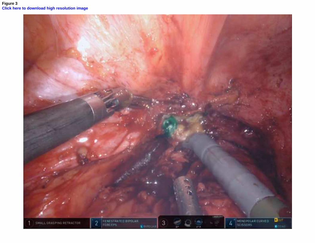
Figure 3Click here to download high resolution image

Figure 4Click here to download high resolution image