Embed Size (px)
Citation preview

Elsevier Editorial System(tm) for The American Journal of Emergency Medicine
Manuscript Draft
Manuscript Number: AJEM9012R1
Title: Assessing Response to Changing Plasma:Red Cell Ratios in a Bleeding Trauma Patient
Article Type: Case Report
Corresponding Author: Dr. Homer Tien, MD, MSc
Corresponding Author's Institution: Sunnybrook Health Sciences Centre
First Author: Homer Tien, MD, MSc
Order of Authors: Homer Tien, MD, MSc; Sandro Scarpellini, MD, PhD; Jeannie Callum, MD;
Lorraine Tremblay, MD, PhD; Sandro B Rizoli, MD, PhD
Abstract: N/A
Response to Reviewers: April 4, 2009
Douglas White MD, MPH, MBA
Medical College of Virginia/VCU, Richmond
Dear Dr. White:
Thank you very much for sending your reviewer’s comments. We found them very useful and
revised our manuscript accordingly. We are pleased to re-submit our article: Assessing Response
to Changing Plasma:Red Cell Ratios in a Bleeding Trauma Patient for consideration by the
American Journal of Emergency Medicine as a Case Report.

With regard to Reviewer#1’s comments:
i) We changed the title and are resubmitting the manuscript as a case report.
ii) We removed the Abstract.
iii) For methods, we added quite a bit to methods. First, we explicitly stated that “we assessed the
dose:response of increasing the ratio of plasma to red blood cells transfused …” as recommended
by the reviewer. To help clarify the measurements, we then explicitly stated that we were
measuring INR, PTT, thrombolelastograms and clotting factor levels at four different time points,
and we labeled those consistently throughout the manuscript (Points A, B, C, D).
iv) As suggested, we then reported the results of all measures of coagulation at these four points in
time. We included these time points in all three figures.
v) We revised our conclusions in the Discussion section. Specifically, we stated that increasing
the plasma:RBC ratio appeared to correct the hemostatic mechanism as measured in our case
report.
vi) We removed the term “paradigm shift” in the Introduction, with regards to damage control
resuscitation.
vii) We outlined Methods before the Case Report.
viii) Limitations: we included a discussion that changes in the measurements of coagulation in this
patient are only associated with a change in FFP:RBC ratios, and do not demonstrate cause/effect.
We added a statement that a randomized controlled trial is required to definitively demonstrate
efficacy of this approach in reversing coagulopathy.
ix) We added the missing references.
We did keep the headings, however. Because there is a technical component to describing how
the clotting factor assays and thromboelastography are done, we felt that the case report was

better organized with the headings. Reviewer#1 only suggested that we change the order of
“Methods” and “Case Report”.
Thank you again for forwarding the comments. Thank you also for re-considering our
manuscript.
HC Tien, MD MSc FRCSC FACS
Lieutenant Colonel, Canadian Forces
Assistant Professor
Universitiy of Toronto

April 4, 2009
Douglas White MD, MPH, MBAMedical College of Virginia/VCU, Richmond
Dear Dr. White:
Thank you very much for sending your reviewer’s comments. We found them very useful and revised our manuscript accordingly. We are pleased to re-submit our article: Assessing Response to Changing Plasma:Red Cell Ratios in a Bleeding Trauma Patient for consideration by the American Journal of Emergency Medicine as a Case Report.
With regard to Reviewer#1’s comments:
i) We changed the title and are resubmitting the manuscript as a case report.
ii) We removed the Abstract.
iii) For methods, we added quite a bit to methods. First, we explicitly stated that “we assessed the dose:response of increasing the ratio of plasma to red blood cells transfused …” as recommended by the reviewer. To help clarify the measurements, we then explicitly stated that we were measuring INR, PTT, thrombolelastograms and clotting factor levels at four different time points, and we labeled those consistently throughout the manuscript (Points A, B, C, D).
iv) As suggested, we then reported the results of all measures of coagulation at these four points in time. We included these time points in all three figures.
v) We revised our conclusions in the Discussion section. Specifically, we stated that increasing the plasma:RBC ratio appeared to correct the hemostatic mechanism as measured in our case report.
vi) We removed the term “paradigm shift” in the Introduction, with regards to damage control resuscitation.
vii) We outlined Methods before the Case Report.
viii) Limitations: we included a discussion that changes in the measurements of coagulation in this patient are only associated with a change in FFP:RBC ratios, and do not demonstrate cause/effect. We added a statement that a randomized controlled trial is required to definitively demonstrate efficacy of this approach in reversing coagulopathy.
* Cover Letter

ix) We added the missing references.
We did keep the headings, however. Because there is a technical component to describing how the clotting factor assays and thromboelastography are done, we felt that the case report was better organized with the headings. Reviewer#1 only suggested that we change the order of “Methods” and “Case Report”.
Thank you again for forwarding the comments. Thank you also for re-considering our manuscript.
HC Tien, MD MSc FRCSC FACSLieutenant Colonel, Canadian ForcesAssistant Professor Universitiy of Toronto

1 2 3 4 5 6 7 8 9 10 11 12 13 14 15 16 17 18 19 20 21 22 23 24 25 26 27 28 29 30 31 32 33 34 35 36 37 38 39 40 41 42 43 44 45 46 47 48 49 50 51 52 53 54 55 56 57 58 59 60 61 62 63 64 65
Case Report
Title: Assessing Response to Changing Plasma:Red Cell Ratios in a Bleeding Trauma Patient
Authors:
Homer C. Tien1, 4
Sandro Scarpellini2
Jeannie Callum3
Lorraine Tremblay4,5
Sandro Rizoli4,5
From:
1. Canadian Forces Health Services
2. Acute and Trauma Surgery, Faculty of Medicine of Ribeirao Preto, University of São Paulo, Brazil
3. Department of Medicine, Sunnybrook Health Sciences Centre
4. Tory Regional Trauma Centre and the Department of Surgery, Sunnybrook Health Sciences Centre
5. Department of Critical Care Medicine, Sunnybrook Health Sciences Centre
Corresponding author:
LCol. Homer TienSunnybrook Health Sciences CentreH186 – 2075 Bayview AvenueToronto, ONCanada, M4N 3M5Phone: 416-480-5850 Fax: 416-480-5851Email: [email protected]
Financial support: Defence Research and Development Canada; Canadian Forces Health Services
Running Head: Plasma Transfusion and Trauma
* Title Page

1 2 3 4 5 6 7 8 9 10 11 12 13 14 15 16 17 18 19 20 21 22 23 24 25 26 27 28 29 30 31 32 33 34 35 36 37 38 39 40 41 42 43 44 45 46 47 48 49 50 51 52 53 54 55 56 57 58 59 60 61 62 63 64 65
Introduction
Damage control resuscitation (DCR) is a novel resuscitation strategy that may increase
survivability of combat casualties [1, 2]. One important aspect of DCR is transfusing
fresh frozen plasma (FFP) and packed red blood cells (PRBCs) in a 1:1 ratio to patients at
risk for coagulopathy. DCR has also been adopted by many civilian trauma centres [3-9],
despite the lack of supporting prospective trials. DCR is only supported by retrospective
studies which may have been affected by survivorship bias [10], and other studies have
found no benefit [11, 12]. Caution is needed in recommending DCR, considering the
potential risks arising from increased FFP use [3-6, 10]. We report a massively
transfused gunshot victim, whose case highlights a possible novel method for assessing a
patient’s response to different FFP:PRBC transfusion ratios.
Methods
The patient was enrolled in a prospective study on thromboelastography in trauma
patients. In brief, two extra 1.8mL tubes with 0.109M trisodium citrate additive were
drawn each time routine coagulation tests were obtained. One tube was centrifuged, the
plasma was frozen, and then sent to an outside laboratory (Haemostasis Reference
Laboratory, Hamilton, Canada) to perform clotting factor activity assays [13, 14]. From
the second tube, thromboelastography (TEG®) was performed on whole blood within 30
minutes of collection. We assessed the dose:response of increasing plasma:red cell
transfusions on INR (international normalized ratio), PTT (partial thrombolplastin time),
thromboelastograms, clotting factor assays and the clinical situation at four different time
points during the first 20 hours of this patient’s hospital admission. The study was
Blinded Manuscript

1 2 3 4 5 6 7 8 9 10 11 12 13 14 15 16 17 18 19 20 21 22 23 24 25 26 27 28 29 30 31 32 33 34 35 36 37 38 39 40 41 42 43 44 45 46 47 48 49 50 51 52 53 54 55 56 57 58 59 60 61 62 63 64 65
approved by our institutional review ethics board, with delayed consent (within 48 hours)
where required. The patient’s family also consented to having his case reported.
Clotting Factor assays were done as follow: extrinsic factor assays were performed
by mixing patient plasma with plasma controls known to be deficient in factors II, V, VII
or X (Precision BioLogics). The degree of correction of the Prothrombin Time (PT)
(Dade-Behring Innovin) is proportional to the factor activity. Similarly, intrinsic factor
assays were done by mixing patient plasma with plasma known to be deficient in factors
VIII, IX, XI or XII (precision BiolLogicis). Again, the degree of correction of the partial
thromboplastin time (PTT) (Date-Behring Actin FSL) is proportional to the factor
activity. Clotting factor activity was considered critically low if any factor level was
below 30%, which is often cited as the threshold level for hemostasis [15, 16]. In
calculating transfusion ratios, we assumed that five units of platelets is equivalent to one
unit of FFP [2].
The TEG® 5000 Hemostasis Analyser (Haemoscope Corporation, Illinois, USA), was
used to produce thromboelastograms. TEG® can be useful in assessing coagulation
status in trauma patients [17-21], and the basic TEG® principles have been previously
described [22, 23]. One mL of whole citrated blood was mixed with buffered stabilizers
and a blend of phospholipids (Kaolin®). A 340 uL sample was then warmed and mixed
with calcium chloride. Measurements were made for no less than 40 minutes

1 2 3 4 5 6 7 8 9 10 11 12 13 14 15 16 17 18 19 20 21 22 23 24 25 26 27 28 29 30 31 32 33 34 35 36 37 38 39 40 41 42 43 44 45 46 47 48 49 50 51 52 53 54 55 56 57 58 59 60 61 62 63 64 65
Case
A 26 year-old male arrived to our trauma room with a transpelvic gunshot wound and
pulseless electrical activity. Resuscitation commenced with crystalloid (2 liters) and 2
units of uncross-matched blood. He went directly to the operating room for a
laparotomy, where his right internal iliac artery and vein were ligated for bleeding. He
was massively transfused intra-operatively, and was hypothermic (30.80C). The
abdomen was packed and temporarily closed.
Re-warming and further transfusions were administered in the intensive care unit
(ICU). Transfusions were aimed at normalizing standard laboratory coagulation tests and
hemodynamic indices according to our institutional massive transfusion protocol.
Platelet transfusions were aimed at restoring platelet count to above 50x109/L, FFP to
keep International Normalized Ratio (INR) below 1.5, and cryoprecipitate to keep
fibrinogen above 0.8g/L. PRBC transfusions were aimed at normalizing tissue perfusion
and at maintaining hemoglobin levels above 70g/L. He initially continued to bleed,
averaging 500mL/hour from his abdominal dressing. A second laparotomy failed to
identify surgical bleeding. His coagulopathy was finally controlled seven hours later and
abdominal bleeding stopped.

1 2 3 4 5 6 7 8 9 10 11 12 13 14 15 16 17 18 19 20 21 22 23 24 25 26 27 28 29 30 31 32 33 34 35 36 37 38 39 40 41 42 43 44 45 46 47 48 49 50 51 52 53 54 55 56 57 58 59 60 61 62 63 64 65
Results
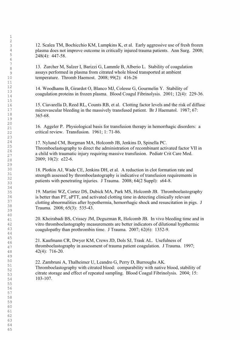
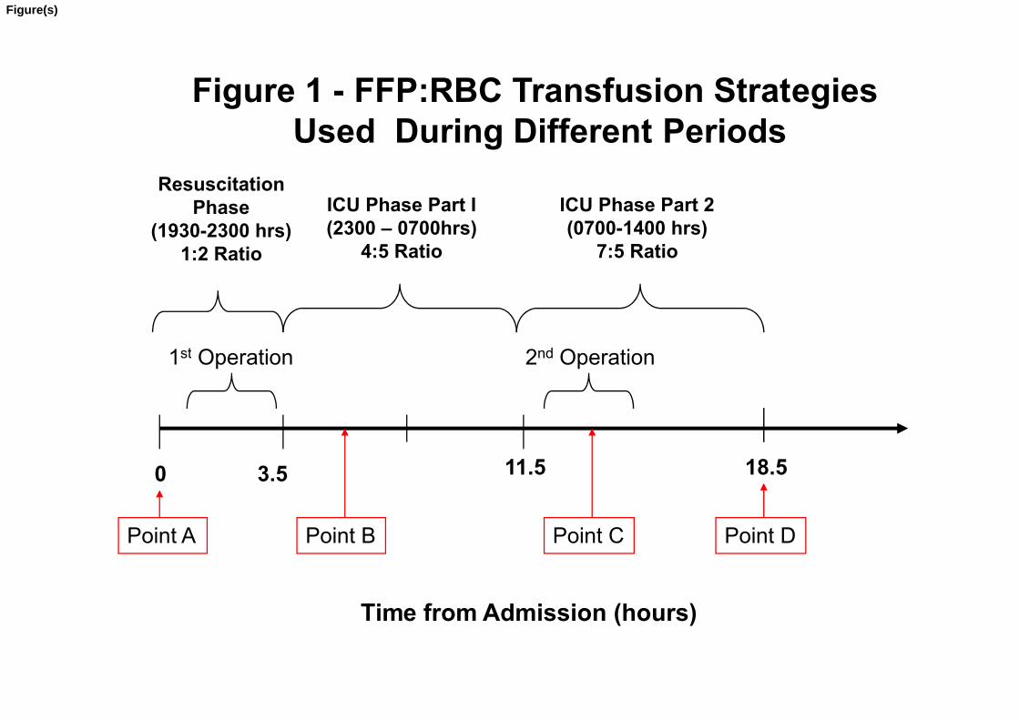
During the first 20 hour period in hospital, three distinct phases of care were evident:
the Resuscitation phase, ICU phase 1 and ICU phase 2 (See Figure 1). The Resuscitation
phase included treatment administered in the trauma room and operating room, and lasted
for 3.5 hours. During this time, the patient received FFP:PRBC in a 1:2 ratio. The
second phase (ICU phase 1) consisted of the overnight resuscitation that occurred in the
ICU, and lasted for eight hours. During this phase, the patient received FFP:PRBC in a
4:5 ratio. The final phase (ICU phase 2) phase commenced with the arrival of the day-
time ICU staff and lasted for seven hours. During this phase, the patient received
FFP:PRBC in a 7:5 ratio. Also, during this period, the patient underwent a bedside
laparotomy to look for missed bleeding.
We obtained the full complement of coagulation testing at four different time points
during this 20 hour period. Testing at Time Point A assessed the patient’s status on
arrival in the trauma room at the beginning of the Resuscitation Phase. Laboratory
testing at Time Point B occurred two hours after arrival into the ICU after the first
surgery (ICU Phase 1). Testing at Time C occurred 2 hours after the arrival of the day-
time ICU staff (ICU phase 2), during the second-look laparotomy. Testing at Time D
occurred at the end of this study’s observation period, 18.5 hours after arrival in the
trauma room.

1 2 3 4 5 6 7 8 9 10 11 12 13 14 15 16 17 18 19 20 21 22 23 24 25 26 27 28 29 30 31 32 33 34 35 36 37 38 39 40 41 42 43 44 45 46 47 48 49 50 51 52 53 54 55 56 57 58 59 60 61 62 63 64 65
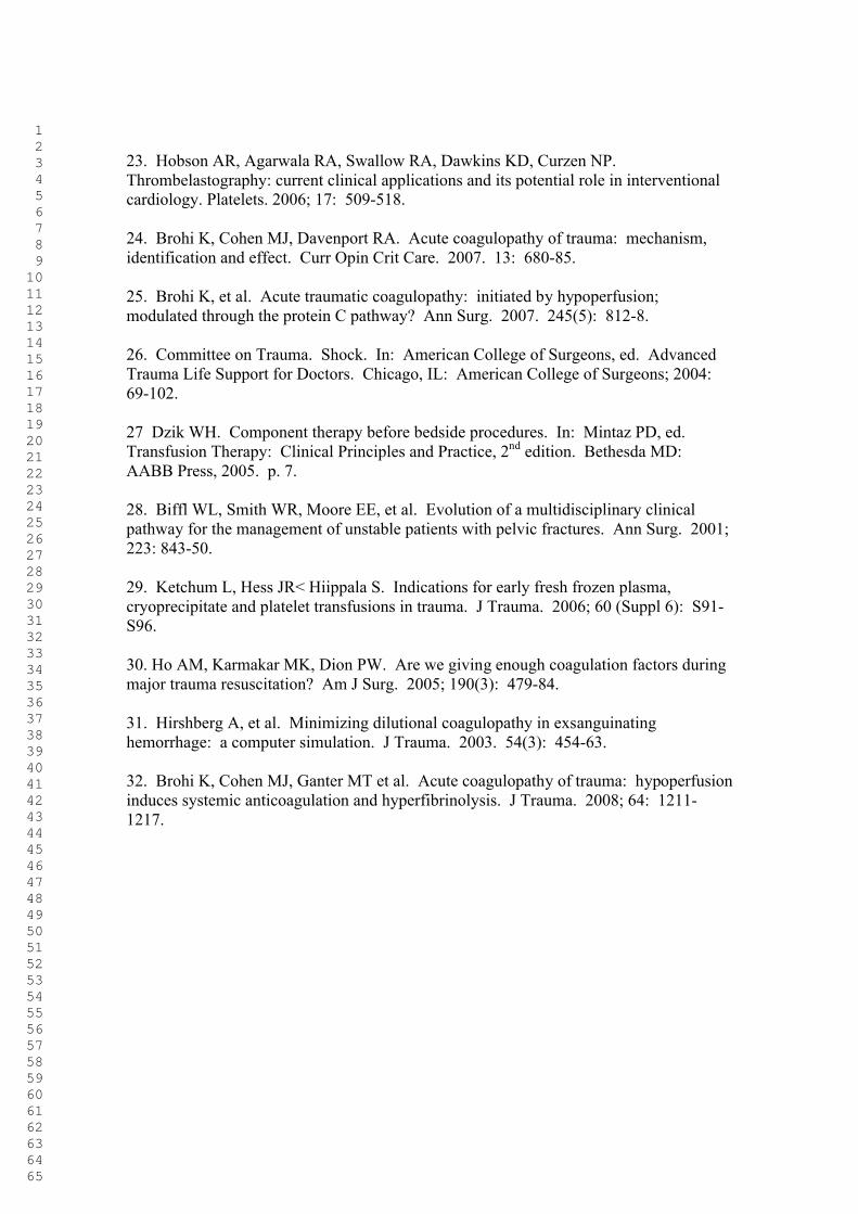
Time Point A: On arrival to the trauma room, the patient’s INR was greater than 13, and
PTT was greater than 150 seconds. His platelet count was 16 (x109/ L) and his
fibrinogen level was 0.5 grams/L. His TEG® (Figure 2) showed almost no clot
formation and all clotting factor activity was critically low (Figure 3).
Resuscitation Phase: During this phase, he was transfused 8 units of FFP, 15 units of
platelets, and 20 units PRBCs (FFP:PRBC of 1:2). As well, he received 8 units of
cryoprecipitate, 2.4 mg of recombinant activated factor VII (rFVIIa), 5 liters of isotonic
crystalloid and 1 liter of colloid.
Time Point B: Testing at this time point reflected the coagulation response of the patient
to the 1:2 resuscitation strategy used in the trauma room and operating room
(Resuscitation Phase). His INR had normalized to 1.2 and clotting factor VII activity
corrected (Figure 3). This was likely a direct response to the recombinant activated
factor VII given in the operating room. However, PTT remained elevated at 100 seconds
and his TEG® showed no clotting activity (Figure 2). As well, all other factors remained
critically low (Figure 3). His hemoglobin had fallen to 43 g/L .
ICU Phase 1: During this phase, he received 12 units of FFP, 20 units of platelets, and
23 units of PRBCs (FFP:PRBC of 4:5). He also received 3 liters of isotonic crystalloid, 1
liter of colloid, and 24 units of cryoprecipitate. His temperature was corrected to 35.80C.

1 2 3 4 5 6 7 8 9 10 11 12 13 14 15 16 17 18 19 20 21 22 23 24 25 26 27 28 29 30 31 32 33 34 35 36 37 38 39 40 41 42 43 44 45 46 47 48 49 50 51 52 53 54 55 56 57 58 59 60 61 62 63 64 65
Time Point C: This time point reflected the coagulation response of the patient to the
4:5 transfusion strategy used during ICU Phase 1. His INR and factor VII activity
remained normal, likely because of residual rFVIIa activity. However, his PTT remained
elevated (50.4 seconds). His TEG® still showed hypocoagulability (Figure 2), and
clotting factor V remained at critical levels (5%). Most importantly, his hemoglobin
dropped to 23 g/L. A second laparotomy failed to identify surgical bleeding.
ICU Phase 2: This phase started with the arrival of the day-time ICU staff, and lasted
seven hours. During this period, he received 16 units FFP, 30 units of platelets, and 18
units of PRBCs (FFP:PRBC of 7:5). He also received 16 units of cryoprecipitate, and
more rFVIIa (9.6 mg).
Time Point D: Testing occurred 7 hours into ICU phase 2, and reflected the coagulation
response to the 7:5 transfusion strategy adopted during ICU phase 2. His TEG®
corrected to within normal limits (Figure 2). INR was 1.15 and PTT was 42.4 seconds.
Also, all clotting factor activity was above 30% activity (Figure 3), with the exception
was factor V (13%). His hemoglobin stabilized and bleeding stopped. The patient had a
difficult ICU course, and unfortunately, died a month later from multiple organ failure
after a septic event.

1 2 3 4 5 6 7 8 9 10 11 12 13 14 15 16 17 18 19 20 21 22 23 24 25 26 27 28 29 30 31 32 33 34 35 36 37 38 39 40 41 42 43 44 45 46 47 48 49 50 51 52 53 54 55 56 57 58 59 60 61 62 63 64 65
Discussion
We described the changes in coagulation measures that occurred in association with
changes to FFP:PRBC transfusion ratios in an exsanguinating trauma patient. This
patient initially had critically low clotting factor activity, TEG® evidence of
coagulopathy and abnormal standard coagulation parameters. These did not correct until
almost 20 hours after admission. Correction occurred after FFP and PRBCs were
transfused at a 7:5 ratio. Only at this point did clinical hemostasis also occur.
Clotting factor V activity was the only coagulation parameter that remained difficult
to correct. One possible reason for this is activation of protein C. Brohi and colleagues
reported that shock leads to activation of protein C via thrombomodulin expression, and
subsequent down-regulation of factor V [24, 25]. For this patient, Factor V only
corrected to 13% activity, but bleeding did stop. Although 20-30% activity level is
considered the threshold for hemostatic activity [15, 16], we have no data on the level
required for hemostasis in patients with massive injuries.
Standard resuscitation practice [26] withholds plasma transfusions until after infusing
crystalloid and red cells. The need for FFP transfusion is assessed by laboratory tests.
When required, four units of FFP are transfused at a time so as to raise clotting factor
activity by 20-30% [27]. However, retrospective studies [28, 29] and mathematical
models [30, 31] suggest this approach is inadequate for exsanguinating trauma patients.
We showed that increasing FFP:PRBC transfusion ratios appeared to correct the

1 2 3 4 5 6 7 8 9 10 11 12 13 14 15 16 17 18 19 20 21 22 23 24 25 26 27 28 29 30 31 32 33 34 35 36 37 38 39 40 41 42 43 44 45 46 47 48 49 50 51 52 53 54 55 56 57 58 59 60 61 62 63 64 65
hemostatic mechanisms in this coagulopathic, hemorrhaging trauma patient. Evidence of
correction included improvement of almost all clotting factor activity to above 30%
normal activity, normalization of thromboelastography, and normalization of INR and
PTT laboratory parameters. Most importantly, the patient stopped bleeding clinically.
Limitations
We only demonstrated an association between a high FFP:PRBC ratio and correction
of coagulopathy in an exsanguinating patient. One interpretation is that our patient was
initially under-resuscitated with FFP, and would have benefited from a DCR approach
from the outset. Another distinct possibility, however, is that a separate pathologic
process such as hypo-perfusion caused coagulopathy [32]. However, as this process was
reversed by resuscitation, clotting factor activity became easier to correct. In either case,
clotting factor activity remains a useful measure of the adequacy of resuscitation with
fresh frozen plasma.
A second limitation is that the critically low levels of factor activity observed in this
case may be due to laboratory error, as clotting factors may have degraded. This is
unlikely, however. Many studies have reported on the stability of clotting factors in
citrated blood despite transportation at ambient temperatures and despite freezing [13,
14]. These studies also studied factor V activity, which remained stable for up to 24
hours at ambient temperatures in citrated whole blood. Randomized controlled trials are
required to definitively assess the efficacy of high FFP:PRBC ratios in controlling
coagulopathy in exsanguinating trauma patients.
1 2 3 4 5 6 7 8 9 10 11 12 13 14 15 16 17 18 19 20 21 22 23 24 25 26 27 28 29 30 31 32 33 34 35 36 37 38 39 40 41 42 43 44 45 46 47 48 49 50 51 52 53 54 55 56 57 58 59 60 61 62 63 64 65
Conflicts of Interest
SR receives salary support from Novo Nordisk.
Acknowledgements
We would like to acknowledge the Surgeon General of the Canadian Forces Health Services (BGen H. Jaeger) for her support of this project, and the financial support from a Military Health Services Research Grant from the Canadian Forces Health Services. As well, we would like to acknowledge the support of Defence Research and Development Canada (DRDC) for their financial support as well.
Both HT and SR had full access to all data in this study, and take responsibility for the integrity of the data and for the accuracy of its analysis.
SR receives salary support from a combined partnership of the Canadian Institute of Health Research (CIHR) and from Novo Nordisk.

1 2 3 4 5 6 7 8 9 10 11 12 13 14 15 16 17 18 19 20 21 22 23 24 25 26 27 28 29 30 31 32 33 34 35 36 37 38 39 40 41 42 43 44 45 46 47 48 49 50 51 52 53 54 55 56 57 58 59 60 61 62 63 64 65
References
1. Holcomb JB, Jenkins D, Rhee P, et al. Damage Control Resuscitation: Directly Addressing the Early Coagulopathy of Trauma. J Trauma. 2007; 62: 307-310.
2. Borgman M, Spinella P, Perkins M, et al. The ratio of blood products transfused affects mortality in patients receiving massive transfusions at a combat support hospital. J Trauma. 2007; 63: 805-813.
3. Gonzalez EA, Moore FA, Holcomb JB, et al. Fresh frozen plasma should be given earlier to patients requiring massive transfusion. J Trauma. 2007; 62: 112-119.
4. Gunter OL Jr, Au BK, Isbell JM, Mowery NT, Young PP, Cotton BA. Optimizing Outcomes in Damage Control Resuscitation: Identifying Blood Product Ratios Associated with Improved Survival. J Trauma. 2008; 65: 527-34.
5. Cotton BA, Gunter OL, Isbell J, et al. Damage control hematology: the impact of a trauma exsanguination protocol on survival and blood product utilization. J Trauma. 2008. 64(5): 1177-83.
6. Moore FA, Nelson T, McKinley BA, et al. Is there a role for aggressive use of fresh frozen plasma in massive transfusion of civilian trauma patients ? Am J Surg. 2008; 196(6): 958-58.
7. Sperry JL, Ochoa JB, Gunn SR, et al. An FFP:PRBC transfusion ratio >/= 1:1.5 is associated with a lower risk of mortality after massive transfusion. J Trauma. 2008; 65(5): 986-93.
8. Holcomb JB, Wade CE, Michalek JE, et al. Increased plasma and platelet to red blood cell ratios improves outcome in 466 massively transfused civilian trauma patients. Ann Surg. 2008; 248(3): 447-58.
9. Maegele M, Lefering R, Paffrath T, et al. Red-blood-cell to plasma ratios transfused during massive transfusion are associated with mortality in severe multiple injury: a retrospective analysis from the Trauma Registry of the Deutsche Gesellschaft für Unfallchirurgie. Vox Sang. 2008; 95(2): 112-9.
10. Dzik S, Callum J, Haspel R. Journal Club. Transfus Med Reviews. 2008; 22(2): 174.
11. Zehtabchi S, Nishijima KD. Impact of transfusion of fresh-frozen plasma and packed red blood cells in a 1:1 ratio on survival of emergency department patients with severe trauma. Acad Emerg Med. 2009 March 14 [epub ahead of print].
1 2 3 4 5 6 7 8 9 10 11 12 13 14 15 16 17 18 19 20 21 22 23 24 25 26 27 28 29 30 31 32 33 34 35 36 37 38 39 40 41 42 43 44 45 46 47 48 49 50 51 52 53 54 55 56 57 58 59 60 61 62 63 64 65
12. Scalea TM, Bochicchio KM, Lumpkins K, et al. Early aggressive use of fresh frozen plasma does not improve outcome in critically injured trauma patients. Ann Surg. 2008; 248(4): 447-58.
13. Zurcher M, Sulzer I, Barizzi G, Lammle B, Alberio L. Stability of coagulation assays performed in plasma from citrated whole blood transported at ambient temperature. Thromb Haemost. 2008; 99(2): 416-26
14. Woodhams B, Girardot O, Blanco MJ, Colesse G, Gourmelin Y. Stability of coagulation proteins in frozen plasma. Blood Coagul Fibrinolysis. 2001; 12(4): 229-36.
15. Ciavarella D, Reed RL, Counts RB, et al. Clotting factor levels and the risk of diffuse microvascular bleeding in the massively transfused patient. Br J Haematol. 1987; 67: 365-68.
16. Aggeler P. Physiological basis for transfusion therapy in hemorrhagic disorders: a critical review. Transfusion. 1961; 1: 71-86.
17. Nylund CM, Borgman MA, Holcomb JB, Jenkins D, Spinella PC. Thromboelastography to direct the administration of recombinant activated factor VII in a child with traumatic injury requiring massive transfusion. Pediatr Crit Care Med. 2009; 10(2): e22-6.
18. Plotkin AJ, Wade CE, Jenkins DH, et al. A reduction in clot formation rate and strength assessed by thromboelastography is indicative of transfusion requirements in patients with penetrating injuries. J Trauma. 2008; 64(2 Suppl): s64-8.
19. Martini WZ, Cortez DS, Dubick MA, Park MS, Holcomb JB. Thromboelastography is better than PT, aPTT, and activated clotting time in detecting clinically relevant clotting abnormalities after hypothermia, hemorrhagic shock and resuscitation in pigs. J Trauma. 2008; 65(3): 535-43.
20. Kheirabadi BS, Crissey JM, Deguzman R, Holcomb JB. In vivo bleeding time and in vitro thromboelastography measurements are better indicators of dilutional hypthermic coagulopathy than prothrombin time. J Trauma. 2007; 62(6): 1352-9.
21. Kaufmann CR, Dwyer KM, Crews JD, Dols SJ, Trask AL. Usefulness of thromboelastography in assessment of trauma patient coagulation. J Trauma. 1997; 42(4): 716-20.
22. Zambruni A, Thalheimer U, Leandro G, Perry D, Burroughs AK. Thromboelastography with citrated blood: comparability with native blood, stability of citrate storage and effect of repeated sampling. Blood Coagul Fibrinolysis. 2004; 15: 103-107.
1 2 3 4 5 6 7 8 9 10 11 12 13 14 15 16 17 18 19 20 21 22 23 24 25 26 27 28 29 30 31 32 33 34 35 36 37 38 39 40 41 42 43 44 45 46 47 48 49 50 51 52 53 54 55 56 57 58 59 60 61 62 63 64 65
23. Hobson AR, Agarwala RA, Swallow RA, Dawkins KD, Curzen NP. Thrombelastography: current clinical applications and its potential role in interventional cardiology. Platelets. 2006; 17: 509-518.
24. Brohi K, Cohen MJ, Davenport RA. Acute coagulopathy of trauma: mechanism, identification and effect. Curr Opin Crit Care. 2007. 13: 680-85.
25. Brohi K, et al. Acute traumatic coagulopathy: initiated by hypoperfusion; modulated through the protein C pathway? Ann Surg. 2007. 245(5): 812-8.
26. Committee on Trauma. Shock. In: American College of Surgeons, ed. Advanced Trauma Life Support for Doctors. Chicago, IL: American College of Surgeons; 2004: 69-102.
27 Dzik WH. Component therapy before bedside procedures. In: Mintaz PD, ed. Transfusion Therapy: Clinical Principles and Practice, 2nd edition. Bethesda MD: AABB Press, 2005. p. 7.
28. Biffl WL, Smith WR, Moore EE, et al. Evolution of a multidisciplinary clinical pathway for the management of unstable patients with pelvic fractures. Ann Surg. 2001; 223: 843-50.
29. Ketchum L, Hess JR< Hiippala S. Indications for early fresh frozen plasma, cryoprecipitate and platelet transfusions in trauma. J Trauma. 2006; 60 (Suppl 6): S91-S96.
30. Ho AM, Karmakar MK, Dion PW. Are we giving enough coagulation factors during major trauma resuscitation? Am J Surg. 2005; 190(3): 479-84.
31. Hirshberg A, et al. Minimizing dilutional coagulopathy in exsanguinating hemorrhage: a computer simulation. J Trauma. 2003. 54(3): 454-63.
32. Brohi K, Cohen MJ, Ganter MT et al. Acute coagulopathy of trauma: hypoperfusion induces systemic anticoagulation and hyperfibrinolysis. J Trauma. 2008; 64: 1211-1217.
1st Operation
Resuscitation Phase
(1930-2300 hrs)1:2 Ratio
ICU Phase Part I(2300 – 0700hrs)
4:5 Ratio
2nd Operation
ICU Phase Part 2(0700-1400 hrs)
7:5 Ratio
Figure 1 - FFP:RBC Transfusion Strategies Used During Different Periods
0 3.5 11.5 18.5
Time from Admission (hours)
1st Operation 2nd Operation
Point A Point B Point C Point D
Figure(s)

Figure 2 – Thromboelastography Results
Time Point A
FFP:PRBCOf 1:2
Time Point B
FFP:PRBCOf 4:5
Time Point C
FFP:PRBCOf 7:5
Time Point D
Figure(s)
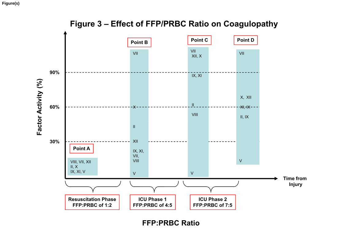
Figure 3 – Effect of FFP/PRBC Ratio on Coagulopathy
Fac
tor
Act
ivit
y(%
)
VII
X
VIIXII, X
IX, XI
II
VIII
VII
X, XII
XI, IX
II, IX
60%
90%
Point B Point C Point D
FFP:PRBC Ratio
Time fromInjury
Fac
tor
Act
ivit
y
30%
Resuscitation PhaseFFP:PRBC of 1:2
II
XII
IX, XI, VII, VIII
V V
VVIII, VII, XIIII, XIX, XI, V
ICU Phase 1FFP:PRBC of 4:5
ICU Phase 2FFP:PRBC of 7:5
Point A
Figure(s)