Embed Size (px)
Citation preview

J A C C : C A R D I O V A S C U L A R I N T E R V E N T I O N S V O L . 1 0 , N O . 9 , 2 0 1 7
ª 2 0 1 7 B Y T H E AM E R I C A N C O L L E G E O F C A R D I O L O G Y F O U N D A T I O N
P U B L I S H E D B Y E L S E V I E R
I S S N 1 9 3 6 - 8 7 9 8 / $ 3 6 . 0 0
h t t p : / / d x . d o i . o r g / 1 0 . 1 0 1 6 / j . j c i n . 2 0 1 6 . 1 2 . 2 8 0
STRUCTURAL
Elevated Mitral Valve Pressure GradientAfter MitraClip Implantation DeterioratesLong-Term Outcome in Patients WithSevere Mitral Regurgitation andSevere Heart Failure
Michael Neuss, MD, Thomas Schau, MD, Akihiro Isotani, MD, Markus Pilz, Maren Schöpp, MD, Christian Butter, MDABSTRACT
Fro
gra
La
rec
eq
Ma
OBJECTIVES This single-center study was performed to analyze the effect of an increased transvalvular gradient after
the MitraClip (MC) (Abbott Laboratories, Abbott Park, Illinois) procedure on patient outcome during follow-up.
BACKGROUND Percutaneous transcatheter repair of the mitral valve with the MC device has been established as a
novel technique for patients with severe mitral regurgitation and high surgical risk. This study investigated the influence
of an increased pressure gradient after MC implantation on the long-term outcome of patients.
METHODS A total of 268 patients were enrolled, who received MC implantation between April 2009 and July 2014 in
our institution (75 � 9 years of age, 68% men, weight 76 � 15 kg, median N-terminal pro–B-type natriuretic peptide
3,696 [interquartile range: 1,989 to 7,711] pg/ml, left ventricular ejection fraction 39 � 16%, log European System for
Cardiac Operative Risk Evaluation score 20% [interquartile range: 12% to 33%]). Pressure in the left atrium and left
ventricle were measured during the procedure using fluid-filled catheters. The pressure gradients over the mitral valve
were determined simultaneously invasively and echocardiographically directly after MC deployment. A Kaplan-Meier
analysis was performed and correlated with the pressure gradients. We used a combined primary endpoint: all-cause-
mortality, left ventricular assist device, mitral valve replacement, and redo procedure.
RESULTS The Kaplan-Meier-analysis showed a significantly poorer long-term-outcome in the case of an invasively
determined mitral valve pressure gradient (MVPG) in excess of 5 mm Hg at implantation for the combined endpoint
(p ¼ 0.001) and for all-cause mortality (p ¼ 0.018). For the echocardiographically determined MVPG the cutoff value
was 4.4 mm Hg. Propensity score matching was used to balance baseline differences between the groups. In a Cox model
the increased residual MVPG >5 mm Hg was a significant outcome predictor in univariate and multivariate analysis
(hazard ratio: 2.3; 95% confidence interval: 1.4 to 3.8; p ¼ 0.002, multivariate after adjustment for N-terminal
pro–B-type natriuretic peptide, age, and remaining mitral regurgitation).
CONCLUSIONS It is recommended that the quality of the implantation result be analyzed carefully and repositioning of the
MC be considered in the case of an elevated pressure gradient over the mitral valve. (J Am Coll Cardiol Intv 2017;10:931–9)
© 2017 by the American College of Cardiology Foundation.
m the Heart Center Brandenburg in Bernau, Fontane University Brandenburg, Bernau, Germany. Dr. Neuss has received travel
nts and lecture honoraria from Abbott Laboratories. Drs. Schau, Isotani, and Schöpp have received travel grants from Abbott
boratories. Mr. Pilz has reported that he has no relationships relevant to the contents of this paper to disclose. Dr. Butter has
eived travel grants, consulting honoraria, and lecture honoraria from Abbott Laboratories. Drs. Neuss and Schau contributed
ually to this work.
nuscript received January 4, 2016; revised manuscript received October 31, 2016, accepted December 16, 2016.

ABBR EV I A T I ON S
AND ACRONYMS
CI = confidence interval
HR = hazard ratio
MC = MitraClip
MR = mitral regurgitation
MVOA = mitral valve opening
area
MVPG = mitral valve pressure
gradient
MS = mitral stenosis
NT-proBNP = N-terminal
pro–B-type natriuretic peptide
Neuss et al. J A C C : C A R D I O V A S C U L A R I N T E R V E N T I O N S V O L . 1 0 , N O . 9 , 2 0 1 7
Mitral Stenosis After MitraClip Implantation M A Y 8 , 2 0 1 7 : 9 3 1 – 9
932
M itraClip (MC) (Abbott Labora-tories, Abbott Park, Illinois) is anew percutaneous transcatheter
therapy of mitral valve (MV) repair for pa-tients with severe mitral regurgitation (MR).Safety and feasibility of the therapy in com-parison to standard surgical treatment wasestablished in the EVEREST (EndovascularValve Edge-to-Edge Repair Study) II trial.This trial demonstrated also similar mortalityin comparison to surgery during 5 years offollow-up (1,2). Compared to the EVEREST IItrial, patients treated in Europe on averagehad a higher surgical risk, more frequently
functional than degenerative MR, and valvemorphology considered unsuitable for treatmentwithin the EVEREST II trial (3–7). Some publicationsand registries suggest improvement of clinical andechocardiographic parameters in such patients notamenable to cardiac surgery (8,9).
SEE PAGE 940
First papers report that the long-term outcome ofpatients may depend on the grade of the remainingMR (10). The relevance of a remaining transvalvulargradient over the MV after the procedure on the long-term outcome is completely uncertain. It was the aimof this study to investigate how the MV pressuregradient (MVPG) influences the long-term outcome ofpatients after MC implantation.
METHODS
STUDY POPULATION. This retrospective studyincluded 268 consecutive patients with severe MRwho underwent MC implantation between March2009 and April 2014 in our heart center. A patient flowchart is given in Figure 1. All patients were evaluatedby an interdisciplinary heart team for MC implanta-tion and referred to interventional treatment due tohigh surgical risk (mostly European System for Car-diac Operative Risk Evaluation score >20% or othersevere comorbidities). Patients had symptomaticheart failure (New York Heart Association functionalclass III or IV) despite established optimal medicaltherapy. All patients gave written consent. Thisretrospective study was performed according toethical guidelines of our institution.
PROCEDURES. The MC implantation procedure hasbeen described previously (1,2). All procedures wereperformed using the 24-F CDS01 or CDS02 MC device(Abbott Vascular, Santa Clara, California) followingthe standard instruction for use.
ECHOCARDIOGRAPHIC MEASUREMENT. Transthoracicand transesophageal echocardiography were performedby experienced sonographers using commerciallyavailable ultrasound systems (Vivid 7 and Vivid E9,GE Medical Systems, Milwaukee, Wisconsin; and Phi-lips IE 33, Royal Philips Electronics, Amsterdam, theNetherlands). The echocardiographic loops recordedduring the procedure were retrospectively read andanalyzed for this study by an experienced board-certified echocardiographer independently of the im-plantation procedure and the treatment process ofthe patients.
Initial MR was graded comprehensively using semi-quantitative methods measuring the color Doppler regur-gitation area, assessment of vena contractawidth, and thequantitative method of the proximal isovelocity surfacearea according to the guideline of the American Society ofEchocardiography using 4 MR grades (11). After the inter-vention, MR severity was assessed with the techniquepreviously reported (12). The MR was determined 1 or 2days after the implantation procedure before discharge bytransthoracic echocardiography. Mitral stenosis (MS) wasevaluated by recording the transmitral mean pressuregradient calculated from the continuous Doppler wave-form in transesophageal echocardiography during theimplantation procedure simultaneously to invasiveMVPGmeasurement directly after clip deployment (13).
MV orifice area (MVOA) was traced at the level ofthe leaflet tip in maximum opening during diastoleusing transgastric view or in orthogonal flexi-slices ofmidtransesophageal 3-dimensional views, if avail-able. Because 3-dimensional transesophageal echo-cardiography was provided by GE Medical Systemsonly at the end of 2012, we decided to use mainly2-dimensional echocardiography and to confirm thesedata by 3-dimensional echocardiography, if available.
INVASIVE MEASUREMENT OF TRANSMITRAL PRESSURE
GRADIENT. For the invasive measurement of thetransmitral pressure gradient a 5-F pigtail-catheter(Merit Medical Systems, South Jordan, Utah) wasplaced in the apex of the left ventricle through a 6-Fsheath in the left radial artery and the left ventricu-lar pressure was measured using a pressure trans-ducer (Medex, Smiths Medical, Ashford, UnitedKingdom). In the first patients the left atrial pressurewas determined by connecting the MC steerableguide catheter to a fluid filled pressure line that wasconnected to a pressure transducer. In some patientsthe left atrial pressure could not be reliably measuredusing this approach. Data from these patients werenot used for data analysis (Figure 1). Later during ourstudy a 4-F pigtail catheter was introduced into theleft atrium in parallel to the MC steerable guide
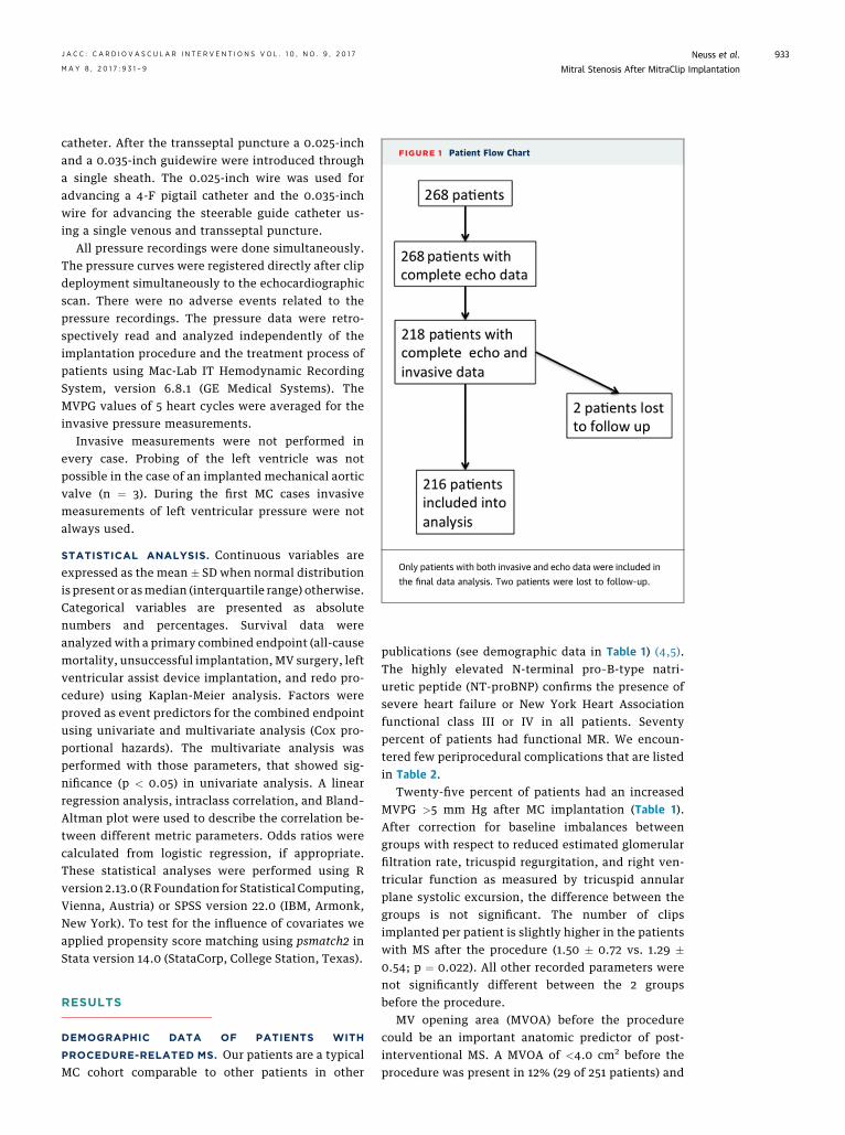
FIGURE 1 Patient Flow Chart
Only patients with both invasive and echo data were included in
the final data analysis. Two patients were lost to follow-up.
J A C C : C A R D I O V A S C U L A R I N T E R V E N T I O N S V O L . 1 0 , N O . 9 , 2 0 1 7 Neuss et al.M A Y 8 , 2 0 1 7 : 9 3 1 – 9 Mitral Stenosis After MitraClip Implantation
933
catheter. After the transseptal puncture a 0.025-inchand a 0.035-inch guidewire were introduced througha single sheath. The 0.025-inch wire was used foradvancing a 4-F pigtail catheter and the 0.035-inchwire for advancing the steerable guide catheter us-ing a single venous and transseptal puncture.
All pressure recordings were done simultaneously.The pressure curves were registered directly after clipdeployment simultaneously to the echocardiographicscan. There were no adverse events related to thepressure recordings. The pressure data were retro-spectively read and analyzed independently of theimplantation procedure and the treatment process ofpatients using Mac-Lab IT Hemodynamic RecordingSystem, version 6.8.1 (GE Medical Systems). TheMVPG values of 5 heart cycles were averaged for theinvasive pressure measurements.
Invasive measurements were not performed inevery case. Probing of the left ventricle was notpossible in the case of an implanted mechanical aorticvalve (n ¼ 3). During the first MC cases invasivemeasurements of left ventricular pressure were notalways used.
STATISTICAL ANALYSIS. Continuous variables areexpressed as the mean � SD when normal distributionis present or asmedian (interquartile range) otherwise.Categorical variables are presented as absolutenumbers and percentages. Survival data wereanalyzed with a primary combined endpoint (all-causemortality, unsuccessful implantation, MV surgery, leftventricular assist device implantation, and redo pro-cedure) using Kaplan-Meier analysis. Factors wereproved as event predictors for the combined endpointusing univariate and multivariate analysis (Cox pro-portional hazards). The multivariate analysis wasperformed with those parameters, that showed sig-nificance (p < 0.05) in univariate analysis. A linearregression analysis, intraclass correlation, and Bland–Altman plot were used to describe the correlation be-tween different metric parameters. Odds ratios werecalculated from logistic regression, if appropriate.These statistical analyses were performed using Rversion 2.13.0 (R Foundation for Statistical Computing,Vienna, Austria) or SPSS version 22.0 (IBM, Armonk,New York). To test for the influence of covariates weapplied propensity score matching using psmatch2 inStata version 14.0 (StataCorp, College Station, Texas).
RESULTS
DEMOGRAPHIC DATA OF PATIENTS WITH
PROCEDURE-RELATED MS. Our patients are a typicalMC cohort comparable to other patients in other
publications (see demographic data in Table 1) (4,5).The highly elevated N-terminal pro–B-type natri-uretic peptide (NT-proBNP) confirms the presence ofsevere heart failure or New York Heart Associationfunctional class III or IV in all patients. Seventypercent of patients had functional MR. We encoun-tered few periprocedural complications that are listedin Table 2.
Twenty-five percent of patients had an increasedMVPG >5 mm Hg after MC implantation (Table 1).After correction for baseline imbalances betweengroups with respect to reduced estimated glomerularfiltration rate, tricuspid regurgitation, and right ven-tricular function as measured by tricuspid annularplane systolic excursion, the difference between thegroups is not significant. The number of clipsimplanted per patient is slightly higher in the patientswith MS after the procedure (1.50 � 0.72 vs. 1.29 �0.54; p ¼ 0.022). All other recorded parameters werenot significantly different between the 2 groupsbefore the procedure.
MV opening area (MVOA) before the procedurecould be an important anatomic predictor of post-interventional MS. A MVOA of <4.0 cm2 before theprocedure was present in 12% (29 of 251 patients) and

TABLE 1 Demographic and Procedural Data of Patients With Normal MVPG and
Increased MVPG > 5 mm Hg After MC Implantation
MVPG #5 mm HgAfter MC
MVPG >5 mm HgAfter MC p Value
n 150 (75) 50 (25)
Age, yrs 77 � 10 76 � 10 0.55
Female/male 48 (33)/96 (67) 19 (40)/28 (60) 0.328
Weight, kg 75 � 15 77 � 24 0.528
Height, cm 169 � 9 168 � 7 0.641
BMI, kg/m2 25 � 4 26 � 4 0.149
BSA, kg*m 1.84 (1.72–2.00) 1.9 (1.79–2.01) 0.195
EuroSCORE I, % 22 (11–27) 22 (13–31) 0.977
EuroSCORE II, % 10 (4–13) 10 (4–15) 0,495
NYHA functional class, %
I 5 (3.3) 0 0.250
II 105 (70) 40 (83)
II 40 (26.7) 8 (17)
NT-proBNP, pg/ml 6,100 (1,845–6,486) 6,950 (2,068–9,375) 0.485
LVEF, % 39 � 17 37 � 15 0.604
eGFR, ml/min 53 (38–66) 47 (33–57) 0.105
MVOA, cm2 5.8 (4.7–7.3) 6.1 (5.4–6.9) 0.609
DMR/FMR 52 (35)/98 (65) 15 (30)/35 (70) 0.416
DMR without rupture 34 (65) 7
DMR with rupture 18 (35) 5
VC width, mm 6.75 � 1.56 6.6 � 1.43 0.542
EROA, cm2 0.46 � 0.14 0.38 � 0.12 0.056
PISA, cm 0.77 � 0.14 0.71 � 0.17 0.029
Regurgitant volume, ml 71.52 � 9.18 65.27 � 19.7 0.086
Clips implanted 1.29 � 0.54 1.50 � 0.72 0.022
CAD 100 (71) 32 (72) 1.000
COPD 28 (19) 16 (32) 0.170
Previous cardiac surgery 35 (23) 12 (30) 0.629
Values are n (%), mean � SD, or median (interquartile range).
BMI ¼ body mass index; BSA ¼ body surface area; CAD ¼ coronary artery disease; COPD ¼ chronic obstructivepulmonary disease; DMR ¼ degenerative mitral regurgitation; eGFR ¼ estimated glomerular filtration rate;EROA ¼ effective regurgitant orifice area; EuroSCORE ¼ European System for Cardiac Operative Risk Evaluationscore; FMR ¼ functional mitral regurgitation; LVEF ¼ left ventricular ejection fraction; MC ¼ MitraClip; MVOA ¼mitral valve opening area; MVPG ¼ mitral valve pressure gradient; NT-proBNP ¼ N-terminal pro–B-typenatriuretic peptide; NYHA ¼ New York Heart Association; PISA ¼ proximal isovelocity surface area; VC ¼ venacontracta.
Neuss et al. J A C C : C A R D I O V A S C U L A R I N T E R V E N T I O N S V O L . 1 0 , N O . 9 , 2 0 1 7
Mitral Stenosis After MitraClip Implantation M A Y 8 , 2 0 1 7 : 9 3 1 – 9
934
these patients had a significantly higher initial MVPGcompared to those with a larger MVOA (5.0 mm Hg vs.3.4 mm Hg; p < 0.001). An MVOA #4.0 cm2 before theprocedure predicted the development of MS (MVPG
TABLE 2 Procedural Complications in the Entire Cohort
Pericardial tamponade 1 (0.5)
Retroperitoneal hematoma 2 (1.0)
Requiring transfusion 2 (1.0)
Requiring operation and transfusion 1 (0.5)
Femoral arteriovenous fistula 2 (1.0)
Requiring operation 1 (0.5)
Pneumothorax after insertion of central venous line 1 (0.5)
Anemia requiring transfusion, bleeding unclear 1 (0.5)
Partial clip detachment before discharge 2 (1.0)
Values are n (%).
>5 mm Hg) after the procedure (odds ratio: 3.41;p ¼ 0.003). Fourteen of 29 patients (48%) with anMVOA #4.0 cm2 had MS after procedure, whereasonly 21% of patients with a larger mitral orificedeveloped MS (p ¼ 0.017).
There was a significant linear correlation betweenMVOA and MVPG after MC implantation in alinear regression analysis. The following linearregression equation was determined: MVPG ¼ 4.4 –
(0.21 $ MVOA) (r2 ¼ 0.31; p ¼ 0.008).
CORRELATION BETWEEN ECHOCARDIOGRAPHIC
AND INVASIVE MEASUREMENTS OF MVPG. Theintraclass correlation between the echocardio-graphically and invasively determined MVPG ismoderate (Pearson-correlation coefficient, r ¼ 0.539)(Figure 2). In the Bland–Altman plot the mean differ-ence between echocardiographic and invasive mea-surements is 0.6. Additionally, the limits ofagreement (–2.2 and 3.6) may be relevant and showsome difference, which may be caused by changedinflow pattern after MC implantation with double ormore orifices.
LONG-TERM OUTCOME. The Kaplan-Meier-analysesof the entire cohort and some subgroups are shownin Figures 2A to 2D. The combined endpoint wasmainly determined by all-cause mortality, whichwas 22% after 1 year and 30% after 2 years (Figure 2A).Few patients received a surgical MV replacement,an implantation of a left ventricular assist device,or a redo procedure during follow-up (<5% after2 years).
The residual MR after MC implantation has a largeimpact on the long-term outcome (Figure 2B). Smallremaining MR grade #1þ has the best long-termoutcome. The outcome of MR grade 2þ is slightlypoorer. Residual MR grade >2þ predicts higher mor-tality rates, which increases further with higher MRgrades.
Procedure-related MS with an invasively measuredMVPG >5 mm Hg predicted a significantly poorerlong-term outcome (Figures 3A and 3B). For theinvasively determined MVPG the cutoff value of5 mm Hg was found by repetitive log-rank testing asminimum p value. The all-cause mortality as sec-ondary endpoint was also poorer in the case of post-procedural MVPG >5 mm Hg (p ¼ 0.018).
For the echocardiographically determined MVPG acutoff value of 4.4 mm Hg was determined by repet-itive log-rank testing (Figure 3D). Patients with ahigher post-procedural MVPG had a significantlypoorer long-term outcome (p ¼ 0.018). The all causesurvival showed also a trend toward a poorer

FIGURE 2 Intraclass Correlation and Bland–Altman Plot Between MVPG Measured in Echocardiography and Invasively
11.00
10.00
9.00
8.00
7.00
6.00
5.00
.00 2.00 4.00 8.00 10.006.00-4
-20
24
2 4 6 8
MVPG (Echocardiographic Measurement)
MVP
G (I
nvas
ive
Mea
sure
men
t)
mean (mmHg)
Diff
eren
ce (m
mH
g)
A B
(A) The graph shows a significant intraclass correlation between the echocardiographically and invasively determined mitral valve pressure
gradient (MVPG). (B) The Bland–Altman plot shows a significant interaction between echocardiographically and invasively determined
pressure gradient.
FIGURE 3 Kaplan-Meier Survival Analysis of the Long-Term Outcome After MC Implantation
(A) The graph shows the long-term outcome after MitraClip (MC) (Abbott Laboratories) implantation according to a combined endpoint (black line), all-cause death
(red line), mitral valve replacement (MVR) (green line), left ventricular assist device (LVAD) or heart transplantation (HTX) (blue line), and redo procedure (purple
line) for the entire patient cohort treated. (B) The graph shows the long-term outcome after MC implantation as a function of residual mitral regurgitation (MR) after
the procedure. The higher the degree of residual mitral regurgitation is, the worse the prognosis is.
J A C C : C A R D I O V A S C U L A R I N T E R V E N T I O N S V O L . 1 0 , N O . 9 , 2 0 1 7 Neuss et al.M A Y 8 , 2 0 1 7 : 9 3 1 – 9 Mitral Stenosis After MitraClip Implantation
935
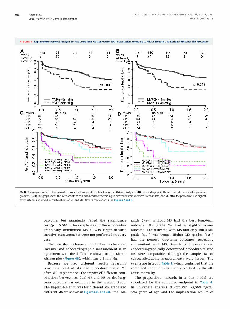
FIGURE 4 Kaplan-Meier Survival Analysis for the Long-Term Outcome After MC Implantation According to Mitral Stenosis and Residual MR After the Procedure
(A, B) The graph shows the freedom of the combined endpoint as a function of the (A) invasively and (B) echocardiographically determined transvalvular pressure
gradient. (C, D) The graph shows the freedom of the combined endpoint according to different extents of mitral stenosis (MS) and MR after the procedure. The highest
event rate was observed in combinations of MS and MR. Other abbreviations as in Figures 2 and 3.
Neuss et al. J A C C : C A R D I O V A S C U L A R I N T E R V E N T I O N S V O L . 1 0 , N O . 9 , 2 0 1 7
Mitral Stenosis After MitraClip Implantation M A Y 8 , 2 0 1 7 : 9 3 1 – 9
936
outcome, but marginally failed the significancetest (p ¼ 0.062). The sample size of the echocardio-graphically determined MVPG was larger becauseinvasive measurements were not performed in everycase.
The described difference of cutoff values betweeninvasive and echocardiographic measurement is inagreement with the difference shown in the Bland–Altman plot (Figure 4B), which was 0.6 mm Hg.
Because we had different results regardingremaining residual MR and procedure-related MSafter MC implantation, the impact of different com-binations between residual MR and MS on the long-term outcome was evaluated in the present study.The Kaplan-Meier curves for different MR grade anddifferent MS are shown in Figures 3C and 3D. Small MR
grade (#1þ) without MS had the best long-termoutcome. MR grade 2þ had a slightly pooreroutcome. The outcome with MS and only small MRgrade (#1þ) was worse. Higher MR grades (>2þ)had the poorest long-term outcomes, especiallyconcomitant with MS. Results of invasively andechocardiographically determined procedure-relatedMS were comparable, although the sample size ofechocardiographic measurements were larger. Theevents are listed in Table 3, which confirmed that thecombined endpoint was mainly reached by the all-cause mortality.
The proportional hazards in a Cox model arecalculated for the combined endpoint in Table 4.In univariate analysis NT-proBNP >8,000 pg/ml,>74 years of age and the implantation results of

TABLE 3 Events of the Combined Endpoint for Different MR Grade and
Different MVPG Groups
MR Grade
1þ (n ¼ 56) 2þ (n ¼ 72) 3þ (n ¼ 11) 1þ (n ¼ 20) >1þ (n ¼ 23)
MVPG #5 #5 #5 >5 >5
Death 11 (20) 24 (33) 4 (36) 5 (25) 12 (52)
MVR 2 (4) 2 (3) 2 (18) 2 (10) 2 (8)
LVAD 2 (4) 0 1 (9) 1 (5) 0 (0)
J A C C : C A R D I O V A S C U L A R I N T E R V E N T I O N S V O L . 1 0 , N O . 9 , 2 0 1 7 Neuss et al.M A Y 8 , 2 0 1 7 : 9 3 1 – 9 Mitral Stenosis After MitraClip Implantation
937
MVPG >5 mm Hg and MR grade >2þ were event pre-dictors for the combined endpoint. In multivariateanalysis the procedure-related MS with MVPG >5mm Hg (invasive measurement) was a significantevent predictor for the combined endpoint (hazardratio [HR]: 2.3; 95% confidence interval [CI]: 1.4 to 3.8;p < 0.002) adjusted for MR grade >2þ at discharge(HR: 3.7; 95% CI: 1.8 to 7.7; p < 0.001) and NT-proBNP>8.000pg/ml (HR: 1.8; 95% CI: 1.1 to 2.9; p ¼ 0.23).
Values are n (%).
LVAD ¼ left ventricular assist device; MR ¼ mitral regurgitation; MVPG ¼ mitral valve pressuregradient; MVR ¼ mitral valve replacement.
TABLE 4 Outcome Prediction for the Combined Endpoint of Clinical and
Functional Parameters After MC Implantation (Cox Proportional Hazards Model)
Univariate AnalysisMultivariate Analysis,
Optimized Model
HR (95% CI) p Value HR (95% CI) p Value
NT-proBNP >8,000 pg/ml 1.8 (1.2–2.8) 0.006 1.8 (1.1–2.9) 0.023
>74 yrs of age 1.6 (1.1–2.5) 0.021
Post-MC implantation
MVPG >5 mm Hg 2.1 (1.3–3.4) 0.003 2.3 (1.4–3.8) 0.002
MR grade >2þ at discharge 4.3 (2.5–7.5) <0.001 3.7 (1.8–7.7) <0.001
CI ¼ confidence interval; HR ¼ hazard ratio; MR ¼ mitral regurgitation; other abbreviations as in Table 1.
DISCUSSION
Our results demonstrate that a post-procedural MSafter MC implantation has a negative impact on thelong-term outcome of patients. A cutoff value wasfound at 5 mm Hg for invasively and 4.4 mm Hg forechocardiographically determined MVPG.
In the EVEREST II or other trials, MS was notdefined as a failed procedure and therefore few dataare available (1,7,14). The frequency of MS atdischarge was previously reported to occur in 31% to35% of patients (10,15–17) and is comparable to thedata of the present study (25%).
Baseline MVOA #4.0 cm2 was found as a significantpredictor of MS after MC implantation. Interestingly,patients with 2 or more clips had significantlyelevated MVPG and more frequently MS after theprocedure, which differs from the report of Biaggiet al. (13). Because baseline MVPG was different(2.2 mm Hg vs. 1.0 mm Hg) between our report andthe Biaggi et al. (13) report, patients’ background andbaseline MV condition might have been different.
The relevant impact of remaining MR and theworse outcome with increasing MR was recentlypublished (7,16) and can be confirmed with the data ofthe present study.
In daily routines it is a common problem for theinterventional team to accept a higher MVPG forbetter MR reduction during a MC implantationprocedure. Our results give some guidance for thisfrequent problem. In accordance with our results wedo not accept an elevated MVPG >5 mm Hg becauseaccording to our data the long-term outcome ofprocedure-related MS is poorer than MR grade #2þ.MC are no longer deployed in our institution insuch cases. In cases of doubt test clipping wasperformed without clip deployment and patientswere hemodynamically challenged using an eleva-tion of heart rate or cardiac output using a combi-nation of atropine and orciprenaline or dobutamine.In our hands we see more options for the medicaltreatment of MR than for the medical treatment ofMS and try to avoid the deployment of clips if a
procedure-related MS would be the result. In rarecases we removed clips before deployment to avoidthe creation of MS. Due to different cutoff valuesfor invasively (5.0 mm Hg) and echocardio-graphically (4.4 mm Hg) determined transmitralpressure gradients we believe that both techniquesare relevant and should be performed to monitorthe implantation.
The relevance of MS in the present study is inaccordance with the literature. A recently publishedstudy reported the impact of remaining MV area aftermitral valvotomy in cases of severe rheumatic MS(17). Little is known about the relevance of MS aftersurgical MV repair. Patients with higher MV gradientshad worse quality of life after mitral annuloplasty(18). Recently published reports suggest that MVrepair for rheumatic MR is associated with a signifi-cant rate of valve failure and reoperation (19).Bertrand et al. (20) recently stressed the relevance ofeffective MV area after restrictive MV annuloplastyfor secondary MR for the long-term outcome.However, the relevance of MS in conjunction with MChas to our knowledge never been described before.
STUDY LIMITATIONS. This is a single-center studywith a limited number of patients. An initial learningcurve might have affected the results and biased the
PERSPECTIVES
WHAT IS KNOWN? The treatment with the MC
device is a safe and efficient way of treating patients
with symptomatic MR and high surgical risk. In terms
of residual MR the results are inferior to surgical
mitral repair.
WHAT IS NEW? In this single-center analysis we
report on the follow-up of 218 patients treated with
the MC device. Transvalvular mitral gradient was
measured invasively and echocardiographically after
deployment of the clip. A transvalvular gradient of
>4.4 mm Hg in echo or >5.0 mm Hg invasively
predicted a significantly worse outcome during
follow-up.
WHAT IS NEXT? Data from registries and
randomized studies should be analyzed whether the
adverse effect of an increased transvalvular gradient
can be confirmed in larger patient groups. Until
further data are available, increased transvalvular
gradients should be avoided.
Neuss et al. J A C C : C A R D I O V A S C U L A R I N T E R V E N T I O N S V O L . 1 0 , N O . 9 , 2 0 1 7
Mitral Stenosis After MitraClip Implantation M A Y 8 , 2 0 1 7 : 9 3 1 – 9
938
patient selection. Due to the retrospective design, thedata sets are not completely available for all patients.
CONCLUSIONS
Increased MVPG deteriorates the long-term outcomeafter MC therapy and is a significant event predictor forpoorer long-term outcome and increased all-causemortality. A procedure-related MS has a poor prog-nosis. It is therefore recommended to check the qualityof the implantation result carefully and to considerrepositioning of the MC in case of a slightly elevatedpressure gradient over the MV. Patients with procedure-related MS should be followed frequently and cardiacsurgery should be discussed on a nonurgent basis.
ACKNOWLEDGMENTS The authors would like tothank our study nurses Daniela Bettin, Olga Hecht,and Martina Ninnemann for their dedicated supportat patient follow-up visits and data acquisition.
ADDRESS FOR CORRESPONDENCE: Dr. MichaelNeuss, Heart Center Brandenburg in Bernau,Ladeburger Straße 17, 16321 Bernau, Germany. E-mail:[email protected].
RE F E RENCE S
1. Mauri L, Garg P, Massaro JM, et al. The EVERESTII trial: design and rationale for a randomizedstudy of the evalve MitraClip system comparedwith mitral valve surgery for mitral regurgitation.Am Heart J 2010;160:23–9.
2. Mauri L, Foster E, Glower DD, et al., EVEREST IIInvestigators. 4-year results of randomizedcontrolled trial of percutaneous repair versussurgery for mitral regurgitation. J Am Coll Cardiol2013;62:317–28.
3. Boekstegers P, Hausleiter J, Baldus S, et al.Percutaneous interventional mitral regurgitationtreatment using the Mitra-Clip system. Clin ResCardiol 2014;103:85–96.
4. Franzen O, Baldus S, Rudolph V, et al. Acuteoutcomes of MitraClip therapy for mitral regur-gitation in high-surgical-risk patients: emphasison adverse valve morphology and severe leftventricular dysfunction. Eur Heart J 2010;31:1373–81.
5. Tamburino C, Ussia GP, Maisano F, et al.Percutaneous mitral valve repair with the Mitra-Clip system: acute results from a real worldsetting. Eur Heart J 2010;31:1382–9.
6. Rudolph V, Knap M, Franzen O, et al. Echocar-diographic and clinical outcomes of MitraCliptherapy in patients not amenable to surgery. J AmColl Cardiol 2011;58:2190–5.
7. Maisano F, Franzen O, Baldus S, et al.Percutaneous mitral valve interventions in thereal world: early and 1-year results from theACCESS-EU, a prospective, multicenter, non-randomized post-approval study of the MitraCliptherapy in Europe. J Am Coll Cardiol 2013;62:1052–61.
8. Pleger ST, Schulz-Schönhagen M, Geis N, et al.One year clinical efficacy and reverse cardiacremodeling and reduced ejection fraction afterMitraClip implantation. Eur J Heart Fail 2013;15:919–27.
9. Neuss M, Schau T, Schoepp M, et al. Patientselection criteria and midterm clinicaloutcome for MitraClip therapy in patients withsevere mitral regurgitation and severecongestive heart failure. Eur J Heart Fail 2013;15:786–95.
10. Toggweiler S, Zuber M, Surder D, et al. Two-year outcomes after percutaneous mitral valverepair with the MitraClip system: durability of theprocedure and predictors of outcome. Open Heart2014;1:e000056.
11. Zoghbi WA, Enriquenz-Sarano M, Foster E,et al. Recommendations for evaluation ofthe severity of native valvular regurgitationwith two-dimensional and Doppler echocardi-ography. J Am Soc Echocardiogr 2003;16:777–802.
12. Foster E, Wasserman HS, Gray W, et al.Quantitative assessment of severity of mitralregurgitation by serial echocardiography ina multicenter clinical trial of percutaneousmitral valve repair. Am J Cardiol 2007;100:1577–83.
13. Biaggi P, Felix C, Gruner C, et al. Assessment ofmitral valve area during percutaneous mitral valverepair using the MitraClip system: comparison ofdifferent echocardiographic methods. Circ Car-diovasc Imaging 2013;6:1032–40.
14. Baldus S, Schillinger W, Franzen O, et al.,German Transcatheter Mitral Valve Intervention(TRAMI) investigators. MitraClip therapy indaily clinical practice: initial results from theGerman transcatheter mitral valve interventions(TRAMI) registry. Eur J Heart Fail 2012;14:1050–5.
15. Boelage-van Dijk B, van Riel AC, de Bruin-Bon RH, et al. Mitral inflow patterns after Mitra-Clip implantation at rest and during exercise. J AmSoc Echocardiogr 2014;27:24–31.
16. Van Riel ACMJ, Boerlage-van Dijk K, de Bruin-Bon RHACM, et al. Percutaneous mitral valverepair preserves right ventricular function. J AmSoc Echocardiogr 2014;27:1098–106.
17. Sharma J, Goel PK, Pandey CM, et al. Inter-mediate outcomes of rheumatic mitral stenosis

J A C C : C A R D I O V A S C U L A R I N T E R V E N T I O N S V O L . 1 0 , N O . 9 , 2 0 1 7 Neuss et al.M A Y 8 , 2 0 1 7 : 9 3 1 – 9 Mitral Stenosis After MitraClip Implantation
939
post-balloon mitral valvotomy. Asian CardiovascThorac Ann 2015;23:923–30.
18. Measana TG, Lam BK, Can V, et al. Clinicalevaluation of functional mitral stenosis aftermitral valve repair for degenerative disease:potential affect on surgical strategy. J ThoracCardiovasc Surg 2013;146:1418–23.
19. Waikittipong S. Mitral valve repair forrheumatic mitral regurgitation: mid-term re-sults. Asian Cardiovasc Thorac Ann 2015;23:658–64.
20. Bertrand PB, Verbrugge FH, Verhaert D, et al.Mitral valve area during exercise after restrictivemitral valve annuloplasty: importance of diastolic
anterior leaflet tethering. J Am Coll Cardiol 2015;65:452–61.
KEY WORDS MitraClip, mitralregurgitation, mitral stenosis,transcatheter mitral valve repair