Embed Size (px)
Citation preview
Ehlers-Danlos Syndromes
Overview for primary care
providers
Roberto Mendoza-Londono MD, MSc, FCCMG, FRCPC
Medical Director-EDS service, Hospital for Sick Children/UHN
Interim Division Head, Genetics, HSC
Outline
Introduction to connective tissue disorders
Disorders associated to hypermobility
Hypermobile type EDS (hEDS)
Prevalence
Diagnostic criteria
Other associated symptoms and complications
Management goals
The EDS clinic at HSC/ UHN
Question and answer period
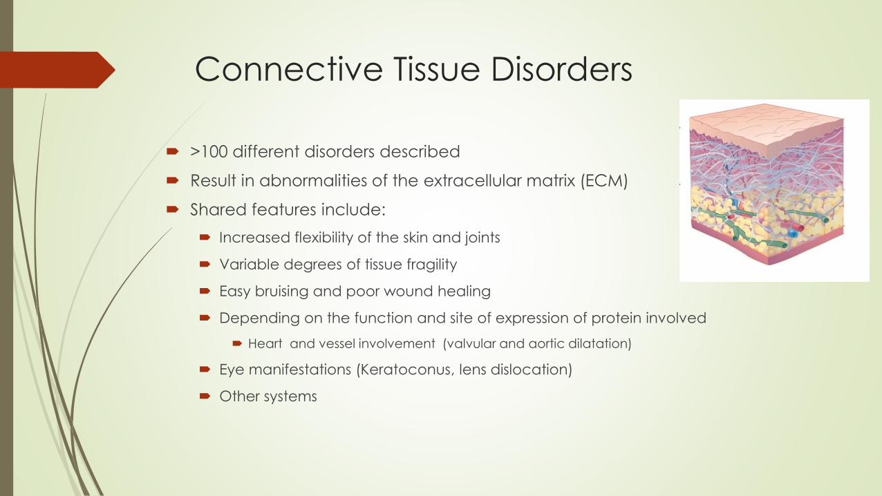
Connective Tissue Disorders
>100 different disorders described
Result in abnormalities of the extracellular matrix (ECM)
Shared features include:
Increased flexibility of the skin and joints
Variable degrees of tissue fragility
Easy bruising and poor wound healing
Depending on the function and site of expression of protein involved
Heart and vessel involvement (valvular and aortic dilatation)
Eye manifestations (Keratoconus, lens dislocation)
Other systems
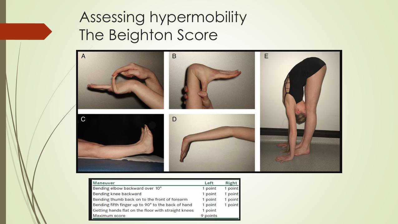
Assessing hypermobility
The Beighton Score
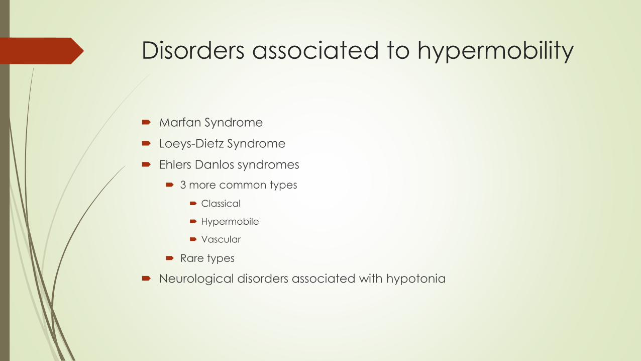
Disorders associated to hypermobility
Marfan Syndrome
Loeys-Dietz Syndrome
Ehlers Danlos syndromes
3 more common types
Classical
Hypermobile
Vascular
Rare types
Neurological disorders associated with hypotonia
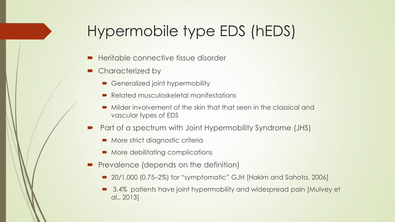
Hypermobile type EDS (hEDS)
Heritable connective tissue disorder
Characterized by
Generalized joint hypermobility
Related musculoskeletal manifestations
Milder involvement of the skin that that seen in the classical and
vascular types of EDS
Part of a spectrum with Joint Hypermobility Syndrome (JHS)
More strict diagnostic criteria
More debilitating complications
Prevalence (depends on the definition)
20/1,000 (0.75–2%) for “symptomatic” GJH [Hakim and Sahota, 2006]
3.4% patients have joint hypermobility and widespread pain [Mulvey et
al., 2013]
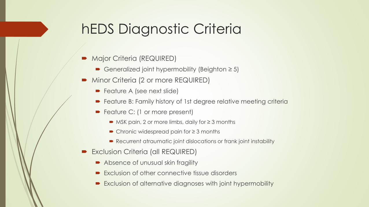
hEDS Diagnostic Criteria
Major Criteria (REQUIRED)
Generalized joint hypermobility (Beighton ≥ 5)
Minor Criteria (2 or more REQUIRED)
Feature A (see next slide)
Feature B: Family history of 1st degree relative meeting criteria
Feature C: (1 or more present)
MSK pain, 2 or more limbs, daily for ≥ 3 months
Chronic widespread pain for ≥ 3 months
Recurrent atraumatic joint dislocations or frank joint instability
Exclusion Criteria (all REQUIRED)
Absence of unusual skin fragility
Exclusion of other connective tissue disorders
Exclusion of alternative diagnoses with joint hypermobility
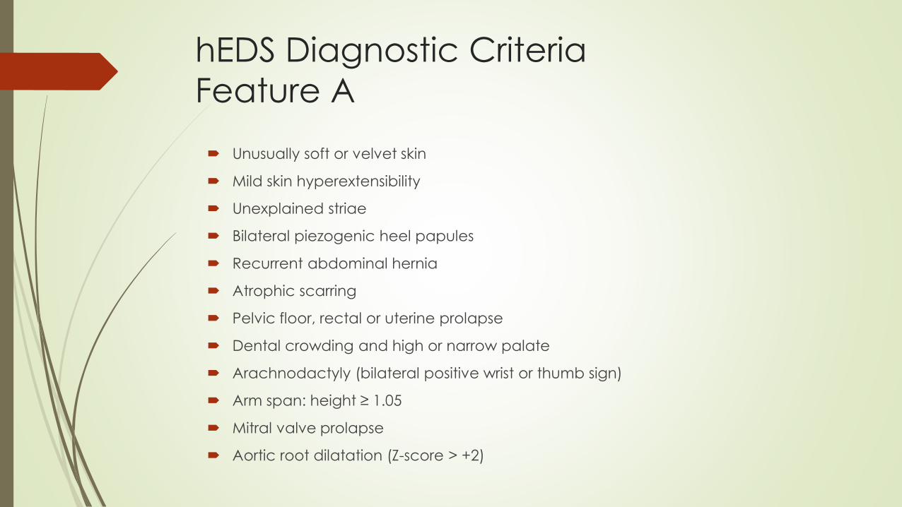
hEDS Diagnostic Criteria
Feature A
Unusually soft or velvet skin
Mild skin hyperextensibility
Unexplained striae
Bilateral piezogenic heel papules
Recurrent abdominal hernia
Atrophic scarring
Pelvic floor, rectal or uterine prolapse
Dental crowding and high or narrow palate
Arachnodactyly (bilateral positive wrist or thumb sign)
Arm span: height ≥ 1.05
Mitral valve prolapse
Aortic root dilatation (Z-score > +2)
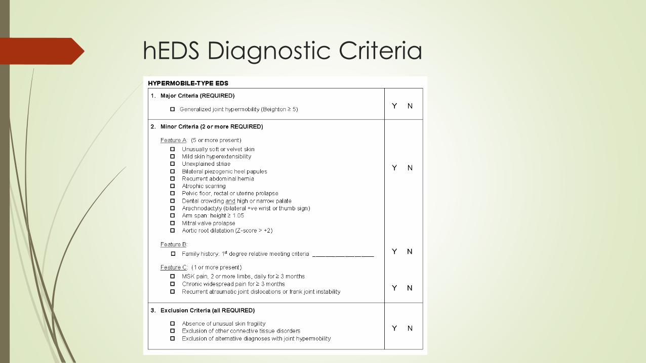
hEDS Diagnostic Criteria
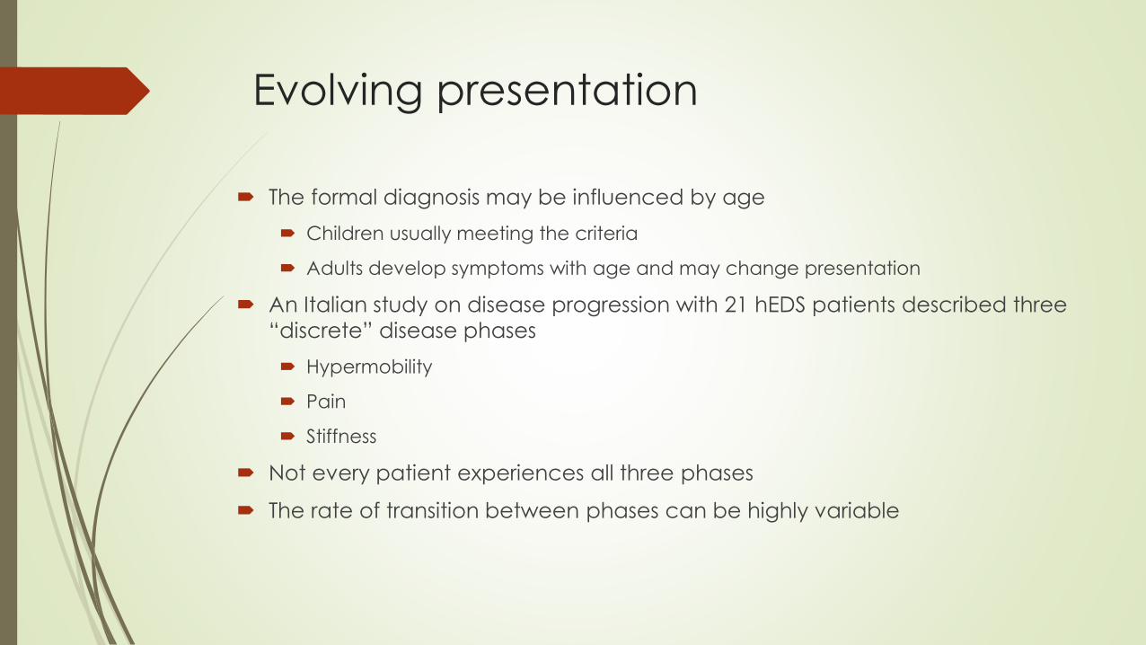
Evolving presentation
The formal diagnosis may be influenced by age
Children usually meeting the criteria
Adults develop symptoms with age and may change presentation
An Italian study on disease progression with 21 hEDS patients described three
“discrete” disease phases
Hypermobility
Pain
Stiffness
Not every patient experiences all three phases
The rate of transition between phases can be highly variable
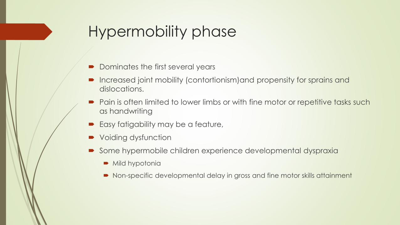
Hypermobility phase
Dominates the first several years
Increased joint mobility (contortionism)and propensity for sprains and
dislocations.
Pain is often limited to lower limbs or with fine motor or repetitive tasks such
as handwriting
Easy fatigability may be a feature,
Voiding dysfunction
Some hypermobile children experience developmental dyspraxia
Mild hypotonia
Non-specific developmental delay in gross and fine motor skills attainment
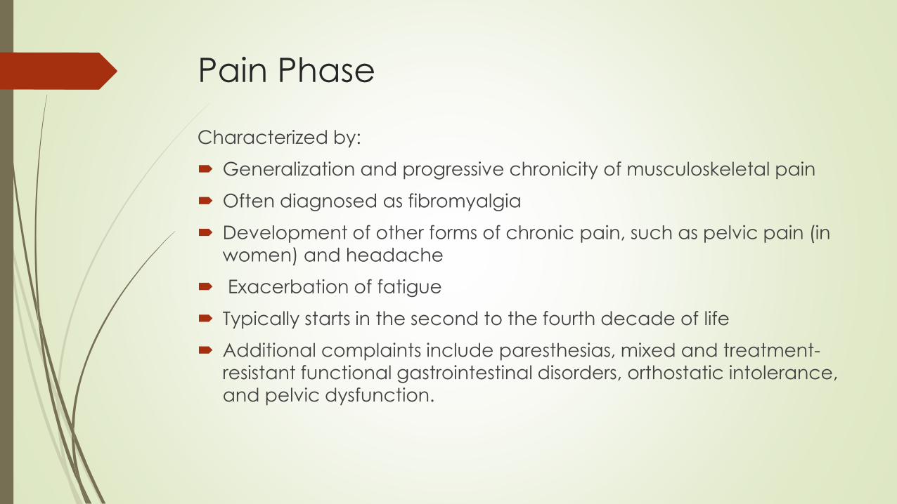
Pain Phase
Characterized by:
Generalization and progressive chronicity of musculoskeletal pain
Often diagnosed as fibromyalgia
Development of other forms of chronic pain, such as pelvic pain (in
women) and headache
Exacerbation of fatigue
Typically starts in the second to the fourth decade of life
Additional complaints include paresthesias, mixed and treatment-
resistant functional gastrointestinal disorders, orthostatic intolerance,
and pelvic dysfunction.
Stiffness Phase
Generalized reduction of joint mobility
Significant reduction in functionality due to the
combination of:
Disabling symptoms (e.g., pain and fatigue)
Motor limitations
Reduced muscle mass and weakness
Defective proprioception
Prior injuries
Arthritis
hEDS and Pain
Specific underlying cause (s) and mechanism(s) of pain in hEDS, are not
well understood
Acute and chronic pain are common
Nociceptive pain directly related to affected muscles, joints, and
connective tissue
Neuropathic pain, characterized by allodynia and/or typical quality
descriptors, such as electrical, burning, numb, or tingling.
Anatomic imaging and functional electrodiagnostic studies are often
negative
Skin biopsy may reveal reduction of intradermal nerve fiber density,
suggestive of small fiber neuropathy
Central sensitization, generalized hyperalgesia, chronic regional pain
syndrome, and similar systemic or regional pathogenic mechanisms
contribute in later stages [
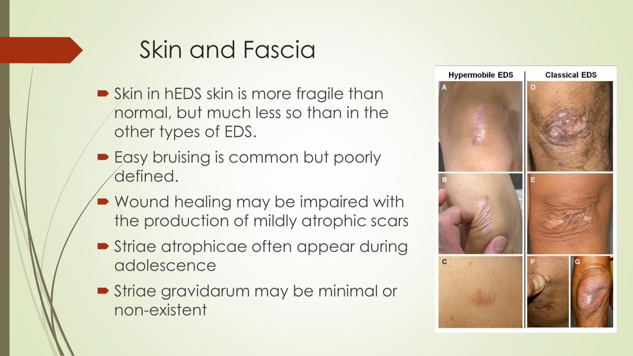
Skin and Fascia
Skin in hEDS skin is more fragile than
normal, but much less so than in the
other types of EDS.
Easy bruising is common but poorly
defined.
Wound healing may be impaired with
the production of mildly atrophic scars
Striae atrophicae often appear during
adolescence
Striae gravidarum may be minimal or
non-existent
Other connective tissue weakness
Cerebrospinal fluid (CSF) leaks are a possible cause of orthostatic
headaches.
Failure of musculotendinous support
The diaphragm
hiatal hernia
The pelvic floor
Uterine/rectal prolapse, rectocele, cystocele, and/or enterocele
Fascial weakness can lead to hernias in the inguinal, femoral, or
umbilical areas or at sites of previous surgical incisions
Fatigue in hEDS
Chronic, debilitating fatigue is common in hEDS
Multifactorial
Pain
Sleep disturbance
Dysautonomia
Medications
Allergies.
Decreases muscle control and coordination
Inhibits physical activity
May increase risk for injury
Mental fatigue leads to impaired cognition and memory
Cardiovascular Involvement
Mild dilation of the aortic root may develop
Unlikely to progress
Baseline echocardiography is not recommended
Postural Orthostatic Tachycardia (POTS) and orthostatic intolerance
are common
Head-up tilt test may or may not establish a specific etiology
Often does not affect therapeutic decision-making
Mitral valve prolapse (MVP)
Frequency of 28–67% among hEDS patients
Increased prevalence of mitral and tricuspid insufficiency has also been
reported
Gastrointestinal Disorders
Wide range of functional complaints in adults
Link between a congenital laxity of the soft connective tissue and
gut diseases is still unclear
Functional Features may be observed in 1/3 to 3/4 of the patients
Gastroesophageal reflux/Heartburn
Recurrent abdominal pain and bloating
Irritable bowel syndrome
Constipation or Diarrhea
Dysphagia
Dysautonomia
Orthostatic hypotension
POTS
Uncategorized orthostatic intolerance
Increase of the physiological heart rate variability
Greater blood pressure fall during Valsalva maneuver
Lower initial systolic blood pressure
Cardiovascular dysautonomia contributes to
Atypical chest pain
Neurological secondary manifestations
Spine Hypermobility
Joint hyperlaxity may affect cranio-cervical junction
Cranio-vertebral instability
Chiaru type I
gait disturbance
Numbness and tingling of the hands and feet
Dizziness
Dysphagia
Speech difficulties
Postural kyphosis
Scoliosis acquired and flexible
Gynecologic Issues
Mucosal problems in genital area
Heavy menstrual bleeding (menorrhagia)
Painful intercourse
Pelvic Dysfunction
Urinary incontinence
Pelvic organ prolapse
Sensory and emptying abnormalities.
Mast Cell Activation Disorder
Characterized by:
Increased number of mast cells
Increased mast cell mediators (e.g., histamine, tryptase)
Clinical symptoms of MCAS include
Flushing
Pruritis
Hypotension
Asthma
Diarrhea
Abdominal bloating
Cramping
Psychiatric co-morbidities
Psychological dysfunction and emotional problems are comon
Depression
Anxiety
Affective disorder
Low self-confidence
Negative thinking
Hopelessness
May exacerbate the other symptoms
Management of hEDS
Assessment is based on symptoms
Musculoskeletal symptoms should be approached conservatively
Physical therapy, education, and pacing are paramount
Frank joint instability should be evaluated by orthopedics
Symptoms of orthostatic intolerance, tachycardia with palpitations, and/or
near-syncope
Treated conservatively by fluid and salt intake
Education and the appropriate exercise.
Syncope should be evaluated further by specialists such as neurology or
cardiology for concerns of arrhythmia, seizure disorder, cardiomyopathy
Management of hEDS
Includes :
Treatment of acute/emergency manifestations
Attenuation of chronic symptoms
Primary and secondary prevention of acute and chronic
complications
As many patients with hEDS have multiple symptoms, a coordinated
effort is required as other specialists are incorporated into the
medical team.
The approach should be holistic focusing on the complications, the desire(s) of the patient, QoL and functionality, as well as the
psychological aspects.
The EDS clinic
Vision:
Improving the lives of patients with EDS through collaborative clinical care and
driving best practices, knowledge translation, innovation and research.
Mission:
To support patients and families living with EDS by providing timely diagnosis,
coordination of medical care, and expertise in the treatment and management of
EDS both internally at SickKids and UHN, and through external partnerships and
collaboration.
Referral system:
Paediatric patients can be referred through the Ambulatory Referral Management
System (ARMS) at https://www.sickkids.ca/referralsystem/
Adult patients can be referred by faxing (416) 340-3792, the following form:
http://www.uhn.ca/MCC/Health_Professionals/Referrals/Documents/Ehlers_Danl
os_Syndrome_Clinic_Referral_Form.pdf#search=eds%20program

Questions and comments?