Embed Size (px)
Citation preview

EGFR, CD10 and proliferation marker Ki67expression in ameloblastoma: possible role inlocal recurrenceAbdel-Aziz and Amin
Abdel-Aziz and Amin Diagnostic Pathology 2012, 7:14http://www.diagnosticpathology.org/content/7/1/14 (2 February 2012)

RESEARCH Open Access
EGFR, CD10 and proliferation marker Ki67expression in ameloblastoma: possible role inlocal recurrenceAzza Abdel-Aziz* and Maha M Amin
Abstract
Background: Ameloblastoma is an odontogenic neoplasm characterized by local invasiveness and tendencytowards recurrence.
Aims: Studying the role played by EGFR, CD10 and Ki67 in the recurrence of ameloblastoma.
Methods: This study was carried out on 22 retrospective cases of mandibular ameloblastoma from the period fromJan 2002 to Jan 2008 with follow up period until Jan 2011 (3 to 8 years follow up peroid). Archival materials wereobtained from pathology department, Mansoura university. Paraffin sections of tumor tissue from all cases weresubmitted for routine H&E stains and immunohistochemistry using EGFR, CD10 and Ki67 monoclonal antibodies.Statistical analysis using of clinical data for all patients, tumor type, EGFR, CD10 and Ki67 expression in relation torecurrence were evaluated.
Results: Among the 22 cases, 10 cases were males and 12 were females with sex ratio 1:1.2. Age ranged from 34to 59 years old with a mean age 44.18 year. Five cases showed local recurrence within studied period and provedby biopsy. No statistically significant relation was found between local recurrence and patient age, tumor size,tumor type, EGFR expression. There was a significant relation between CD10 expression as well as Ki67 labellingindex and recurrence (P value = 0.003, 0.000 respectively).
Conclusion: Evaluation of CD10 and Ki67 status together with conventional histological evaluation can help inproviding more information about the biologic behavior of the tumor, while EGFR could be a target of anexpanding class of anticancer therapies.Since ameloblastomas are EGFR-positive tumors, anti-EGFR agents could be considered to reduce the size of largetumors and to treat unresectable tumors that are in close proximity to vital structures.
Virtual Slides: The virtual slide(s) for this article can be found here:http://www.diagnosticpathology.diagnomx.eu/vs/1902106905645651
Keywords: Ameloblastoma, EGFR, CD10, Ki67 and recurrence
IntroductionAmeloblastoma is the most common odontogenic neo-plasm that accounts for about 1% of all oral tumors [1]and arises from the epithelium of the dental laminaaffecting mainly the posterior mandible (80%) and to alesser extent the posterior maxilla (20%). It usually affectsadults in the 4th - 5th decades of life [2]. Ameloblastomais generally benign, grows slowly and is not associated
with symptoms until it becomes large. However, it islocally aggressive and displays a strong tendency to recurespecially if not adequately removed [3,4] and evenmetastasize in rare conditions[5]. It is important to assessthe type according to recent WHO classification (solid/multicystic, unicystic, desmoplastic and peripheral), loca-lization, and size of the tumors as well as age of thepatient [6]. There are two basic histopathologic patterns;the follicular and plexiform without clinical relevance [7].The epidermal growth factor receptor (EGFR) is a
transmembrane receptor tyrosine kinase comprising an* Correspondence: [email protected] Department, Faculty of medicine, Mansoura University, Egypt
Abdel-Aziz and Amin Diagnostic Pathology 2012, 7:14http://www.diagnosticpathology.org/content/7/1/14
© 2012 Abdel-Aziz and Amin; licensee BioMed Central Ltd. This is an Open Access article distributed under the terms of the CreativeCommons Attribution License (http://creativecommons.org/licenses/by/2.0), which permits unrestricted use, distribution, andreproduction in any medium, provided the original work is properly cited.
extracellular ligand binding domain, a transmembranedomain, and an intracellular tyrosine kinase domain [8].Binding of epidermal growth factor results in EGFRdimerization and subsequent activation of the intrinsictyrosine kinase activity. Phosphorylated EGFR concomi-tantly triggers downstream mitogenic signaling via boththe MAPK and PI3K pathways [9].Expression of epidermal growth factor receptor
(EGFR) regulates proliferation of both normal and neo-plastic cells [10,11]. It has been observed in normalepithelia, including the oral mucosa, and might provideepigenetic control of odontogenesis [12].CD10 (common acute lymphoblastic leukemia antigen,
CALLA) is a 100-kDa transmembrane glycoprotein, alsoknown as neutral endopeptidase (NEP), involved in thecleavage and inactivation of certain peptide hormonesimportant for signal transduction [13]. CD10 isexpressed by a variety of normal cell types, includinglymphoid precursor cells, germinal center B lymphocytesand some epithelial cells as gastric mucosa [14]. First,CD10 was reported in relation to lymphoid neoplasms.However, its expression is also reported in malignantepithelial neoplasm and melanoma [15]. Although CD10expression is observed in neoplastic cells, there arereports of its expression in stromal cells. Moreover,there are cumulative data indicating that CD10 expres-sion by stromal cells is involved in carcinogenesis and itis supposed to be a novel prognostic factor in somemalignant tumors [14].Identification of proliferating activities in tumors may
be useful to predict their biological behavior. Ki-67 pro-tein is a nuclear non-histone protein which is requiredfor maintaining the cell cycle. Ki-67 is expressed by pro-liferating cells in all phases of the active cell cycle (G1,S, G2 and M phase) but is absent in resting (G0) cells[16]. Ki-67 has been used to determine the proliferationrate of many tumors, including ameloblastomas [17].For a better understanding of the aggressive behavior
of ameloblastomas, their expression of growth factorreceptors, metalloproteinases and their proliferativeactivity have been investigated using immunohistochem-ical methods. So, the aim of the present study is evalua-tion of EGFR, CD10 expression and Ki-67 labelled indexin ameloblastoma and their relation to recurrence.
Materials and methodsThis retrospective study was carried out on mandibularameloblastoma specimens received in the pathologydepartment from the period from Jan 2002 to Jan 2008.Follow up data were retrieved from patient’s files for atleast 3 years duration. Each specimen was coded andpatient’s name was not shown for ethical reasons. Ageof the patients, sex, tumor size, site and recurrence wererevised.
Ethical approval of this study was not required by ourinstitution as this study was based on retrospective ana-lysis dealing with archival paraffin slides and blocks, notrelated to patient’s privacy, impairment or treatment.
HistopathologySections of 4 um thickness have been cut from formalinfixed paraffin embedded blocks of archival ameloblas-toma tissues for routine H&E, others were prepared oncharged slides for immunohistochemistry. Examinationof three tumor slides from each specimen were done onan Olympus CX31 light microscope. Pictures wereobtained by a PC-driven digital camera (Olympus E-620).The computer software (Cell*, Olympus Soft ImagingSolution GmbH) allowed morphometric analysis to beperformed.
ImmunohistochemistryImmunohistochemical analysis for EGFR, CD10 and KI67with a labelled streptavidin- biotin-peroxidase complextechnique was performed on tumor sections. The antibo-dies used were monoclonal antibody against EGFR (cloneH11-DAKO, DakoCytomation, Carpinteria, CA, USA), at1:25 dilution, CD10 (Santa Cruz Biotechnology Inc., sc-19993, dilution 1: 50) and Ki-67 (clone MIB-1, N1633,Dako Corporation, Carpinteria, CA, USA, RTU). Detectionkit used was high sensitive kit (DakoCytomation envision+dual link system peroxidase code K4061) using DAB aschromogene. EGFR immunostaining required antigenretrieval with 0.2% trypsin, CD10 and Ki67 immunostain-ing required pretreatment with 1 mM EDTA (at pH 8.0)for 20 minutes in microwave oven. Proper positive andnegative controls were performed. Normal oral mucosawas used as positive control for EGFR, tonsils for Ki67and CD10. As a negative control, sections were stainedwithout the addition of a primary antibody.
Immunohistochemical AnalysisAs for the immunohistochemistry assessment, Slideswere scanned by X40 magnification. Ten cellular areasselected (i.e. the so-called hot spots) and evaluated atX400 magnification by two pathologists.Assessment of EGFRBoth membranous and cytoplasmic staining of EGFRwere evaluated. The proportion of stained cells andstaining intensity were combined to assess the immuno-histochemical staining according to previous records[18-20]. Staining intensity was evaluated on a semi-quantitative three-point scale: 0–no staining, 1–weakand 2–strong staining. The final EGFR staining scorewas calculated by multiplying the percentage of posi-tively stained tumor cells by the staining intensity.Accordingly, the highest score for a given tumor wouldbe equal to 2.
Abdel-Aziz and Amin Diagnostic Pathology 2012, 7:14http://www.diagnosticpathology.org/content/7/1/14
Page 2 of 7

Assessment of CD10Stromal CD10 was scored according to similar systemsuggested by Iwaya et al, [21]. and Zu et al. [22]. as fol-low 0, equivalent to the negative control; 1, weak cyto-plasmic stain; 2, moderate stain; 3, intense stain. Thepercentage of stained cells was also scored on a semi-quantitative 4-point scale as: 0, < 10%; 1, 10-25%; 2, 25-50%; 3, > 50%. Then, combining the score of stainingintensity and percentage of stained cells: a score of 0-1was -, 2 was +, 3-4 was ++ and 5-6 was +++.Assessment of Ki67Ki67 labeling index was done by calculating the ratio ofpositive nuclei in relation to total number of neoplasticnuclei in 10 HPFs. The labeling index (number of posi-tive tumor cells/total number of tumor cells expressedas a percentage) was calculated in every specimen [23].
Statistical analysisAll parameters included age, tumor size, pathologic type,EGFR, CD10 expression in stromal cells and ki67 label-ing index with recurrence were evaluated by statisticalanalysis. The statistical analysis of data was done byusing statistical package for social science (SPSS) pro-gram version 14. Descriptive statistics were done. Thepresented data was asymmetrical. One Way Anova test(for EGFR, Ki67) and Chi square test (for the remainingdata including CD10) were performed to determine sig-nificance of the relations. Survival analysis of recurrencefree survival was done by Kaplan-Meier analysis and logrank test was used for comparison between groups.Probability (p) values < 0.05 were considered significant.
ResultsThis study was carried out on retrospective cases man-dibular ameloblastoma received in the pathology depart-ment from the period from Jan. 2002 to Jan. 2008 withfollow up period until Jan. 2011 (minimal 3 years followup).
Clinical characteristicsTen cases were males and 12 were females with sexratio 1:1.2. Age ranged from 34 to 59 years old withmean age 44.18 ± 6.97. Five cases (22.7%) showed localrecurrence, four of them recurred 3-5 years after resec-tion and one case recurred 2 years after
Pathology resultsSixteen cases were follicular and six cases were plexi-form subtype. As shown in table (1), all specimensdemonstrated EGFR-positively stained tumor cells.Staining was membranous and cytoplasmic, both periph-eral and central cells were stained (Figure 1). Two cases(9.1%) exhibited the maximum score of 2 and 20(90.9%) scored between 0.2 and 1.8. Stromal CD10
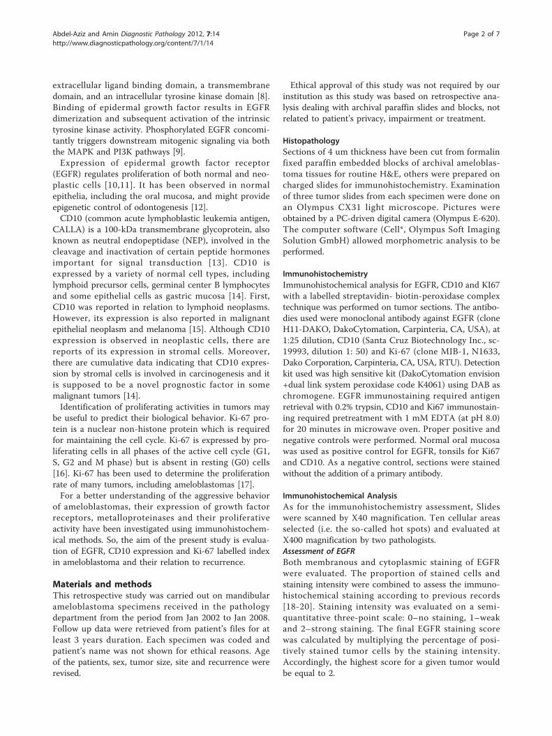
immunostaining was negative in one case, 11 cases were(+), 7 cases were (++), 3 cases were (+++) (Figure 2).Regarding Ki67 labeling index, 17 cases showed lowindex with mean 8.29 ± 3.15 and 5 cases showed highindex with mean 19 ± 2.12 (Figure 3).Table (2) showed no statistically significant association
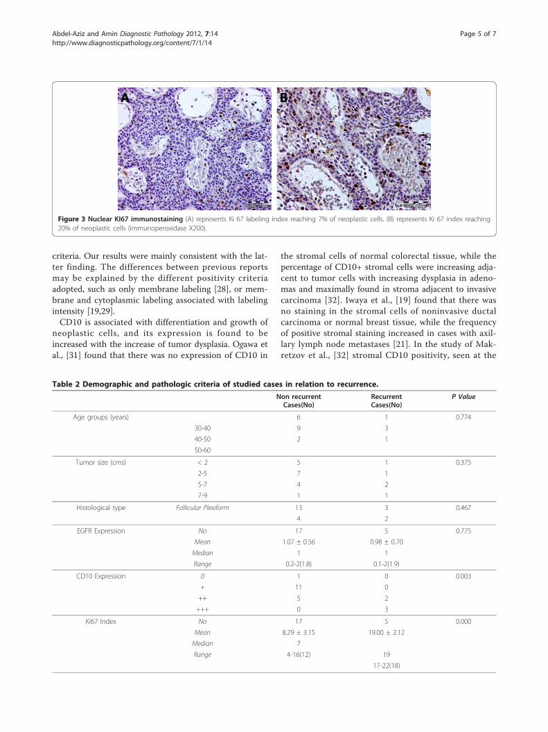
between local recurrence and patient age (p = 0.774),tumor size (p = 0.375), tumor types (p = 0.467), andEGFR score (p = 0.774). There was a statistically signifi-cant relation between stromal CD10 and recurrence(P = 0.003); the stronger expression is associated withrecurrence. Statistically significant relation was foundbetween Ki67 labeling index and recurrence (P < 0.001);cases with recurrent ameloblastoma showed higherindex (mean 19 ± 2.12) in contrast to non-recurrentcases (mean 8.29 ± 3.15).During the follow-up period, three cases having short
recurrence free survival (RFS) (2-3 years) showedmarked stromal CD10 expression (+++) and higher Ki67labeling indices (19-22). On the other hand, cases withlonger RFS (5, 7 years) showed moderate stromal CD10expression (++) and less Ki67 labeling indices (17, 18).A statistically significant decrease in Patient’s RFS wasassociated with stromal CD10 expression (P < 0.001 by
Table 1 Summary of clinicopathologic finding of thestudied cases.
No Age Sex Size(cm)
Pathologicaltype
EGFR CD10
Ki67 Recurrence
1 46 M 5 Plexiform 0.4 + 9 -
2 37 F 4 Plexiform 0.9 ++ 6 -
3 34 M 4 Tubular 1.6 + 6 -
4 38 M 3 Tubular 1 ++ 17 +
5 48 F 4 Plexiform 2 +++ 20 +
6 42 M 5 Plexiform 1.6 + 5 -
7 48 M 3 Plexiform 1.2 + 7 -
8 52 F 6 Tubular 0.2 ++ 18 +
9 47 M 4 Plexiform 0.5 ++ 4 -
10 45 F 6 Plexiform 1.3 ++ 16 -
11 46 M 5 Plexiform 0.3 +++ 22 +
12 45 F 6 Plexiform 1.5 +++ 19 +
13 59 M 3 Tubular 1.8 - 11 -
14 35 F 4 Plexiform 1 ++ 9 -
15 43 F 3 Tubular 1.5 + 10 -
16 39 F 4 Plexiform 0.8 + 6 -
17 49 M 5 Plexiform 2 + 6 -
18 36 F 2 Plexiform 1.2 + 7 -
19 59 M 4 Tubular 0.2 + 9 -
20 46 F 5 Plexiform 1.4 + 14 -
21 42 F 2 Plexiform 2 + 7 -
22 36 F 4 Plexiform 0.6 ++ 9 -
Abdel-Aziz and Amin Diagnostic Pathology 2012, 7:14http://www.diagnosticpathology.org/content/7/1/14
Page 3 of 7

Log-Rank test). The mean RFS was 8 ± 0.1 in casesshowing mild stromal CD10 expression, 7.4 ± 0.43 incases showing moderate stromal CD10 expression, 2.7 ±0.33 in cases showing mild stromal CD10 expression(Figure 4). A statistically significant decrease in Patient’sRFS was associated with Ki67 labeling index (P < 0.001by Log-Rank test). The mean RFS was 8 ± 0.1 in caseswith Ki67 index < 10, 5.1 ± 0.96 in cases with Ki67index > 10 (Figure 5).
DiscussionAmeloblastomas are locally invasive and destructivebenign odontogenic tumors that arise from rests of thedental lamina. Their recurrence rate is high [3,4] evenfor patients that undergo surgical excision of the tumoreven in excised tumors with free safety margin. Theimmunohistochemistry can describe the biological dif-ferences of these tumor types [24]. This study was car-ried out to elucidate the relationship between theexpression of EGFR, CD10 and Ki-67 labelled index andameloblastoma recurrence using clinical and pathologi-cal data.
Normal EGFR signaling plays an essential role inorgan development, repair and in the regulation of cellsurvival. Aberrant signaling can be the result of EGFRoverexpression by EGFR gene amplification or muta-tions with ligand-independent tyrosine kinase activitywhich could result in uncontrolled cell division; a pre-disposition for cancer [25,26]. EGFR upregulationappeared to be selectively expressed in a number oftumors as glioblastomas and lung cancer [27].In the current study, all ameloblastomas exhibited
EGFR immunoexpression with no identified relation torecurrence. Previous studies in the literature evaluatedEGFR expression in ameloblastomas [19,28,29], andtheir results were divergent. Shrestha et al. [28] claimedthat of the 23 cases of examined solid ameloblastomas,none demonstrated EGFR expression. However, Li et al.[29] reported that EGFR was detected in all six of theircases of ameloblastoma. Ueno et al., [30] examined 39cases of solid ameloblastoma and EGFR expression wasfound in 30 (88%). Vered et al., [19] reported that allspecimens were EGFR-positive using membranous, orboth membranous and cytoplasmic staining as positivity
Figure 1 EGFR immunostaining (A) represents membranous and cytoplasmic EGFR expression in basal and stellate reticulum like cells. (B)represents membranous and cytoplasmic EGFR expression in focus of squamous differentiation (immunoperoxidase X200).
Figure 2 Stromal CD10 immunostaining (A) represents cytoplasmic and nuclear CD10 expression of mild intensity in about 15% of stromalcells. (B) represents CD10 expression of moderate to strong intensity in about 70% of stromal cells (immunoperoxidase X200).
Abdel-Aziz and Amin Diagnostic Pathology 2012, 7:14http://www.diagnosticpathology.org/content/7/1/14
Page 4 of 7
criteria. Our results were mainly consistent with the lat-ter finding. The differences between previous reportsmay be explained by the different positivity criteriaadopted, such as only membrane labeling [28], or mem-brane and cytoplasmic labeling associated with labelingintensity [19,29].CD10 is associated with differentiation and growth of
neoplastic cells, and its expression is found to beincreased with the increase of tumor dysplasia. Ogawa etal., [31] found that there was no expression of CD10 in
the stromal cells of normal colorectal tissue, while thepercentage of CD10+ stromal cells were increasing adja-cent to tumor cells with increasing dysplasia in adeno-mas and maximally found in stroma adjacent to invasivecarcinoma [32]. Iwaya et al., [19] found that there wasno staining in the stromal cells of noninvasive ductalcarcinoma or normal breast tissue, while the frequencyof positive stromal staining increased in cases with axil-lary lymph node metastases [21]. In the study of Mak-retzov et al., [32] stromal CD10 positivity, seen at the
Figure 3 Nuclear KI67 immunostaining (A) represents Ki 67 labeling index reaching 7% of neoplastic cells. (B) represents Ki 67 index reaching20% of neoplastic cells (immunoperoxidase X200).
Table 2 Demographic and pathologic criteria of studied cases in relation to recurrence.
Non recurrentCases(No)
RecurrentCases(No)
P Value
Age groups (years) 6 1 0.774
30-40 9 3
40-50 2 1
50-60
Tumor size (cms) < 2 5 1 0.375
2-5 7 1
5-7 4 2
7-9 1 1
Histological type Follicular Plexiform 13 3 0.467
4 2
EGFR Expression No 17 5 0.775
Mean 1.07 ± 0.56 0.98 ± 0.70
Median 1 1
Range 0.2-2(1.8) 0.1-2(1.9)
CD10 Expression 0 1 0 0.003
+ 11 0
++ 5 2
+++ 0 3
Ki67 Index No 17 5 0.000
Mean 8.29 ± 3.15 19.00 ± 2.12
Median 7
Range 4-16(12) 19
17-22(18)
Abdel-Aziz and Amin Diagnostic Pathology 2012, 7:14http://www.diagnosticpathology.org/content/7/1/14
Page 5 of 7

invasive front, was associated with higher tumor grade,and decreased survival in breast carcinoma, suggestingtumor-stromal interactions. In a study of CD10 in oralsquamous cell carcinoma, it was found that CD10 posi-tivity in stromal cells was an indicator of worse prog-nosis; a significant correlation was found with lymphnode metastases, local recurrences, and histologic grade[33].Ameloblastoma is a tumor that shows heterogeneous
expression of CD10 [34]. Most recurrent tumors stronglyexpress CD10 and could be a marker for aggressive beha-vior. Our data demonstrated that patients with tumorsstrongly express CD10 in the peritumoral stromal cellswere more prone to local recurrence after resection withuninvolved cut margins. This is similar to the results in
studies done by Lezzi et al., [35] and Masloub et al., [36].It is possible then that the function of CD10 is employedprimarily in invasion of extracellular matrix. The pre-sence of CD10+ stromal cells may signify the aggressive-ness of tumor. Similarly, Bilalovic et al., [37] reportedmetastatic behavior of melanoma cases with peritumoralCD10 positive stain.Assessment of cell proliferation in many types of
tumors is important together with histologically basedtumor classification and has potential relevance as anindicator of tumor behavior, treatment response andrelapse [38]. The results of this study showed that cellu-lar proliferative activity as assessed by Ki67 labelingindices varied within recurrent and non-recurrent casesof ameloblastoma. There was a significant relationbetween labeling index of nuclear proliferation markerki67 and recurrence of ameloblastoma. This is in con-cordance with Hirayama et al., [39] who found a highproliferative activity in recurrent ameloblastoma. In thisstudy, the assessment of the cellular proliferation markerwas shown to be reliable and reproducible.All the above-mentioned immunohistochemical data
indicated that the immunoexpression of CD10 and Ki67labeling index may be good predictor for recurrence inameloblastoma.
ConclusionOur data demonstrated that CD10-positive tumors withhigh Ki67 index were associated with high recurrencerate, while EGFR expression was not predictive for prog-nosis in ameloblastomas but may render such tumorscandidate for the new targeted anti-EGFR treatmentmodalities. Moreover, we hope formulation of a targettherapy against CD10 positive cells, including monoclo-nal antibody mediated-delivery of chemotherapy. Clini-cal surveys with larger study cohorts will be needed toverify our findings.
Authors’ contributionsAA contributed to study design, collecting data, analysis, writing themanuscript, performed statistical analysis and in deciding to submit themanuscript for publication. MMA contributed to revision and approval offinal and revised manuscript draft, and in deciding to submit the manuscriptfor publication. All authors read and approved the final manuscript.
Competing interestsThe authors declare that they have no competing interests.
Received: 5 December 2011 Accepted: 2 February 2012Published: 2 February 2012
References1. Melrose R: Benign epithelial odontogenic tumors. Semin Diagn Pathol
2002, 16:271-87.2. Philipsen H, Reichart P, Nikai H, Takata T, Kudo Y: Peripheral
ameloblastoma: biological profile based on 160 cases from theliterature. Oral Oncol 2001, 37:17-27.
Figure 4 Kaplan-Meier survival analysis for recurrence freesurvival (years) in relation to stromal CD10. (One case withnegative stromal CD10 was excluded from the curve).
Figure 5 Kaplan-Meier survival analysis for recurrence freesurvival (years) in relation to Ki67 labeling index.
Abdel-Aziz and Amin Diagnostic Pathology 2012, 7:14http://www.diagnosticpathology.org/content/7/1/14
Page 6 of 7
3. Regezi J: Odontogenic cysts, odontogenic tumors, fibroosseous, andgiant cell lesions of the jaws. Mod Pathol 2001, 15:331-41.
4. Pinheiro J, Freitas V, Moretti A, Jorge AG, Jaeger RG: Local invasiveness ofameloblastoma. Role played by matrix metallo-proteinases andproliferative activity. Histopathology 2004, 45:65-72.
5. Gilijamse M, Leemans CR, Winters HA, Schulten EA, Van der Waal I:Metastasizing ameloblastoma. Int J Oral Maxillofac Surg 2007, 36:462-64.
6. World Health Organization Classification of Tumours. In Pathology andGenetics. Head and Neck Tumours. Edited by: Barnes L, Eveson J, Reichart P,Sidransky D. Lyon: IARC Press; 2005:.
7. Gardner DG, Heikinheimo K, Shear M, Philipsen HP, Colemen H:ameloblastoma. In World Health Organization Classification of Tumors.Pathology and Genetics of Head and Neck Tumors. Edited by: Leon Barnes,John W Eveson, Peter Reichart, David Sidransky. Published by IARCPressLyon, France; 2005:296.
8. Oda K, Matsuoka Y, Funahashi A, Kitano H: A comprehensive pathwaymap of epidermal growth factor receptor signaling. Mol Syst Biol 2005,1:2005.010.
9. Sorensen OE, Tapa DR, Roupé KM, Valore EV, Sjobring U, Roberts AA,Schmidtchen A, Ganz T: Injury-induced innate immune response inhuman skin mediated by transactivation of the epidermal growth factorreceptor. J Clin Invest 2006, 116(7):1878-1885.
10. Baselga J, O’Dwyer PJ, Thor AD, Vokes EE, Weiner LM: Epidermal growthfactor receptor: potential target for antitumor agents. Dallas, TX: TheCenter for Biomedical Continuing Education; 2000, 1-24.
11. Jost M, Kari C, Rodeck U: The EGF receptor–an essential regulator ofmultiple epidermal functions. Eur J Dermatol 2000, 10:505-10.
12. Agaram NP, Collins BM, Barnes L, Lomago D, Aldeeb D, Swalsky P,Finkelstein S, Hunt JL: Molecular analysis to demonstrate thatodontogenic keratocysts are neoplastic. Arch Pathol Lab Med 2004,128:313-7.
13. Goo YA, Goodlett DR, Pascal LE, Worthington KD, Vessella RL, True LD,Liu AY: Stromal mesenchymal cell genes of the human prostate andbladder. BMC Urology 2005, 5:17.
14. Huang W, Zhou X, Chen J, Zhang LH, Meng K, Ma HH, Lu ZF: CD10-positive stromal cells in gastric carcinoma: Correlation with invasion andmetastasis. Japanese J of Clini Oncol 2005, 35(5):245-50.
15. Kanitakis J, Narvaez D, Claudy A: Differential expression of the CD10antigen (neutral endopeptidase) in primary versus metastatic malignantmelanomas of the skin. Melanoma Res 2002, 12(3):241-4.
16. O’Leary TJ, Frisman DM: Antigenes. In Advanced diagnostic methods inpathology: Principles, practice and protocols Saunders Edited by: O’Leary TJ ,1 2003, 35-91.
17. Sandra F, Mitsuyasu T, Nakamura N, Shiratsuchi Y, Ohishi M:Immunohistochemical evaluation of PCNA and Ki-67 in ameloblastoma.Oral Oncology 2001, 37:193-198.
18. Partridge M, Gullick WJ, Langdon JD, Sherriff M: Expression of epidermalgrowth factor receptor on oral squamous cell carcinoma. Br J OralMaxillofac Surg 1988, 26:381-9.
19. Vered M, Shohat I, Buchner A: Epidermal growth factor receptorexpression in ameloblastoma. Oral Oncol 2003, 39:138-43.
20. de Vicente JC, Torre-Iturraspe A, Gutiérrez AM, Lequerica-Fernández P:Immunohistochemical comparative study of the odontogenickeratocysts and other odontogenic lesions. Med Oral Patol Oral Cir Bucal2010, 15(5):e709-15.
21. Iwaya K, Ogawa H, Izumi M, Kuroda M, Kuroda M, Mukai K: Stromalexpression of CD10 in invasive breast carcinoma: a new predictor ofclinical outcome. Virchow Arch 2002, 440:589-93.
22. Zu X, Tang Z, Li Y, Gao N, Din J, Qi L: Vascular endothelial growth factor-Cexpression in bladder transitional cell cancer and its relationship tolymph node metastasis. BJU Int 2006, 98(5):1090-3.
23. Bologna-Molina R, Mosqueda-Taylor A, Lopez-Corella E, Almeida OP,Carrasco-Daza D, Garcia-Vazquez F, Farfan-Morales JE, Irigoyen-Camacho ME,Damián-Matsumura : Syndecan-1 (CD138) and Ki-67 expression indifferent subtypes of ameloblastomas. Oral Oncol 2008, 44(8):805-11.
24. Pinheiro JJ, Freitas VM, Moretti AI, Jorge AG, Jaeger RG: Local invasivenessof ameloblastoma. Role played by matrix metalloproteinases andproliferative activity. Histopathol 2004, 45(1):65-72.
25. Lynch TJ, Bell DW, Sordella R, Gurubhagavatula S, Okimoto RA,Brannigan BW, Harris PL, Haserlat SM, Supko JG, Haluska FG, Louis DN,Christiani DC, Settleman J, Haber DA: Activating mutations in the
epidermal growth factor receptor underlying responsiveness of non-small-cell lung cancer to gefitinib. N. Engl. J. Med 2004, 350(21):2129-39.
26. Zhang H, Berezov A, Wang Q, Zhang G, Drebin J, Murali R, Greene MI: ErbBreceptors: from oncogenes to targeted cancer therapies. J Clin Invest2007, 117(8):2051-8.
27. Kuan CT, Wikstrand CJ, Bigner DD: EGF mutant receptor VIII as amolecular target in cancer therapy. Endocr Relat Cancer 2001, 8(2):83-96.
28. Shrestha P, Yamada K, Higashiyama H, Takagi H, Mori M: Epidermal growthfactor receptor in odontogenic cysts and tumors. J Oral Pathol Med 1992,21:314-7.
29. Li T, Browne RM, Matthews JB: Expression of epidermal growth factorreceptors by odontogenic jaw cysts. Virchows Archiv A Pathol Anat 1993,423:137-44.
30. Ueno S, Miyagawa T, Kaji R, Mushimoto K, Shirasu R: Immunohistochemicalinvestigation of epidermal growth factor receptor expression inameloblastomas. J Pathol 1994, 173:33-8.
31. Ogawa H, Iwaya K, Izumi M, Kuroda M, Serizawa H, Koyanagi Y, Mukai K:Expression of CD10 by stromal cells during colorectal tumordevelopment. Hum Pathol 2002, 33:806-11.
32. Makretzov NA, Hayes M, Carter BA, Dabiri S, Gilks CB, Huntsman DG:Stromal CD10 expression in invasive breast carcinoma correlates withpoor prognosis, estrogen receptor negativity, and high grade. ModPathol 2007, 20(1):84-89.
33. Piattelli A, Fioroni M, Iezzi G, Perritti V, Stellini E, Piattelli M, Rubini C: CD10expression in stromal cells of oral cavity squamous cell carcinoma: aclinic and pathologic correlation. Oral Dis 2006, 12:301-4.
34. Freedland SJ, Seligson DB, Liu AY, Pantuck AJ, Paik SH, Horvath S,Wieder JA, Zisman A, Nguyen D, Tso CL, Palotie AV, Belldegrun AS: Loss ofCD10 (neutral endopeptidase) is a frequent and early event in humanprostate cancer. Prostate 2003, 55:71-80.
35. Lezzi G, Piattelli A, Rubini C, Artese L, Goteri G, Artese L, Goteri G, Fioroni M,Carinci F: CD10 expression in stromal cells of ameloblastoma variants.Oral Surg Oral Med Oral Pathol Oral Radiol Endod 2008, 105(2):206-9.
36. Masloub SM, Abdel-Azim AM, Abd Elhamid ES: CD10 and osteopontinexpression in dentigerous cyst and ameloblastoma. Diagnostic Pathology2011, 6:44.
37. Sandstad B, Golouh R, Nesland JM, Selak I, Torlakovic EE: CD10 proteinexpression in tumor and stromal cells of malignant melanoma isassociated with tumor progression. Modern Pathology 2004, 17:1251-58.
38. Tsai ST, Jin YT: Proliferating cell nuclear antigen (PCNA) expression in oralsquamous cell carcinomas. J Oral Pathol Med 1995, 24:313-5.
39. Hirayama T, Hamada T, Hasui K: Immunohistochemical analysis of cellproliferation and suppression of ameloblastoma with special referenceto plexiform and follicular ameloblastoma. Acta Histochem Cytochem2004, 37:391-8.
doi:10.1186/1746-1596-7-14Cite this article as: Abdel-Aziz and Amin: EGFR, CD10 and proliferationmarker Ki67 expression in ameloblastoma: possible role in localrecurrence. Diagnostic Pathology 2012 7:14.
Submit your next manuscript to BioMed Centraland take full advantage of:
• Convenient online submission
• Thorough peer review
• No space constraints or color figure charges
• Immediate publication on acceptance
• Inclusion in PubMed, CAS, Scopus and Google Scholar
• Research which is freely available for redistribution
Submit your manuscript at www.biomedcentral.com/submit
Abdel-Aziz and Amin Diagnostic Pathology 2012, 7:14http://www.diagnosticpathology.org/content/7/1/14
Page 7 of 7