Embed Size (px)
Citation preview
Role of Se+Zn in Regeneration (Ki67) Following Pb Toxicity in Wistar rat.
B.A Falana, O.M Ogundele, F.I Duru, A.A.Osinubi, D.T Falode
Introduction
• Lead poisoning is a serious health [reblem in the developing countries, (WHO 2002)
• Causes reproductive toxicity via the suppression of spermatogenesis and Androgenesis in males Apostoli and Catalini 2011), (Benoff et
al., 2000; Akinloye et al 2006 ), Fukushima et al., (2005), Yi Lu ( 2009),
Salgado et al (2010).
Introduction
• This study addresses the effect of Pb toxicity on the germinal epithelium and the proliferative effect of selenium and zinc.
Overall aim
• The aim of this study is to determine
the effect of selenium and zinc on
lead-induced testicular damage in
Sprague-Dawley Rats.

The Specific objectives
• The specific objective of this study are to determine the effect of
(a) Selenium and zinc supplementation on the activities of Ki 67 (a proliferation marker) following treatment with lead
(b) Selenium and zinc on the activities of P53 and CaD in lead induce-testicular damage

Statement of Problem
• Lead poisoning in Nigeria (Doctors without borders 2010)
• Occupational and Environmental Health
• Developed and Developing Countries
• Acute and Chronic exposure

Background
• The Germinal epithelium
• Blood-testis barrier
• Lead, Selenium and zinc ( Antioxidants)
Methods
• 60F1 generation of Wistar rats were divided
into four groups 1-4
• Group 1: Normal saline control
• Group 2: Pb only
• Group 3: Pb+Se+Zn
• Group 4: Se+Zn
Histology
• Embedded tissues were sectioned to
obtainn 7 microns thickness for
routine H&E (Kareema et al., 2012)

Immunohistochemistry
• Paraffin wax embedded sections were mounted
on a glass slide in preparation for antigen
retrieval.
• Immersed in Urea overnight and placed in
microwave for 45 mins
• Primary and secondary antibodies application
( Biotinylated goat serum for 1hr. And 1%
Bovine serum albumin)

Results

Fig. 1(a-d): General morphology of the germinal epithelium of wild type adult wistar rats (stained with Hematoxylin and Eosin) (a) Control, (b) 100 mg kg-1 Pb-Acetate, (c) Pb-Acetate+Se+Zn and (D) Se+Zn only, Degeneration was most prominent in the lead treatment group, cell proliferation is most prominent in the Se+Zntreatment group. (‡) represents regions of cell proliferation and (†) represents regions of cellular degeneration, (n) represents normal cells of the epithelium, (BM) basement membrane, arrow head indicates the lumen of the seminiferous tubule (Scale bar is 30 μm

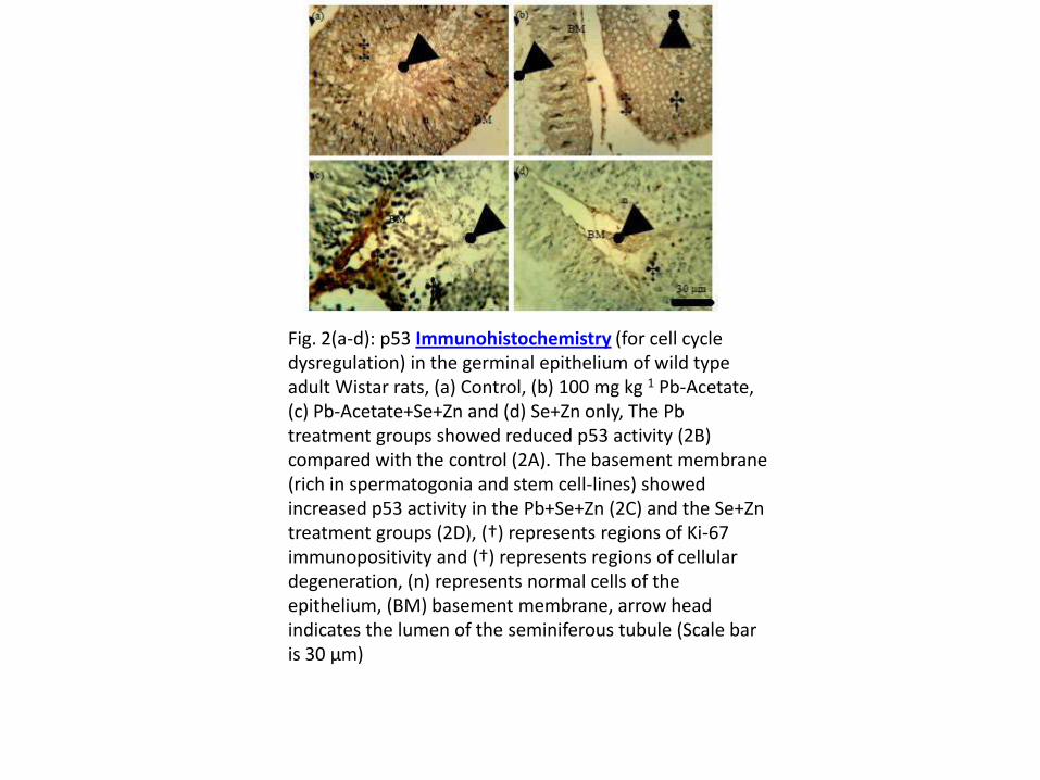
Fig. 2(a-d): p53 Immunohistochemistry (for cell cycle dysregulation) in the germinal epithelium of wild type adult Wistar rats, (a) Control, (b) 100 mg kg 1 Pb-Acetate, (c) Pb-Acetate+Se+Zn and (d) Se+Zn only, The Pbtreatment groups showed reduced p53 activity (2B) compared with the control (2A). The basement membrane (rich in spermatogonia and stem cell-lines) showed increased p53 activity in the Pb+Se+Zn (2C) and the Se+Zntreatment groups (2D), (†) represents regions of Ki-67 immunopositivity and (†) represents regions of cellular degeneration, (n) represents normal cells of the epithelium, (BM) basement membrane, arrow head indicates the lumen of the seminiferous tubule (Scale bar is 30 μm)

Fig. 3(a-d): Cathepsin D (CAD) immunohistochemistryin the germinal epithelium of wild type adult Wistarrats, (a) Control, (b) 100 mg kg 1 Pb-Acetate, (c) Pb-Acetate+Se+Zn and (D) Se+Zn only. CAD activity is high in the BM of the control (3a), in the BM and degenerating cells of the lead treated group (3b), in the BM of Pb+Se+Zn and it is widely diffused in the epithelial cells of the Se+Zn treated group (3d). (‡) represents regions of CAD immunopositivity and (†) represents regions of cellular degeneration, (n) represents normal cells of the epithelium, (BM) basement membrane, arrow head indicates the lumen of the seminiferous tubule (Scale bar is 30 μm)


Fig. 4(a-d): Ki-67 Immunohistochemistry (for cell proliferation) in the germinal epithelium of wild type adult Wistar rats, (a) Control, (b) 100 mg kg-1 Pb-Acetate, (c) Pb-Acetate+Se+Zn and (d) Se+Zn only. Cell proliferation marked by Ki-67 immunopositivityis higher in the Se+Zn treatment group (Fig. 4d), followed by the control (4a). (‡) represents regions of Ki-67 immunopositivity and (†) represents regions of cellular degeneration, (n) represents normal cells of the epithelium, (BM) basement membrane, arrow head indicates the lumen of the seminiferous tubule
(Scale bar is 30 μm)

Literature review
• Se+Zn supplementation attenuates lead-
induced reproductive toxicity in rats (Falana and
Oyeyipo 2012)
• Primary mechanism of Pb Toxicity is majorly
induction of oxidative stress via inhibition of
allosteric sites in metalloenzyme ALP, CcOx: Kumari et al 2013, Musatoy and Robinson 2012)

Literature review
• Molecular oxygen reacts with electrons from the reduction of food substances to generate ROS
• The primary role of ROS is peroxidation of lipids (Humphrey et al.,2012)
• The first response involes production of mtNOS to counter the no that is formed from the reaction of ROS wth nitrogen containing groups (Ekici et al., 2012)

Literature review
• This study evaluates the role of Pb as
an agent capable of inducing
degeneration via cell cycle proteins
as well as role of selenium and zinc
2.25mg/kg BW as agents capable of
reducing such toxicity by evaluation
of proliferation

Literature review
• Degeneration and toxicity can be accessed
through mitochondria and cytoplasmic pathways
by measuring immunohistochemically the level of
expression of P53 (a 53 Kda Tumor supressor
protein that is usually activated in the Gap phases
of the cell cycle to proof read the genome for
errors such as cleavage and deletions which are
induced by Pb toxicity
( National Toxicology programme 2007)

Literature review
• P53 is a nucleolase that will digest the genetic materialsin case of such errors ( Matteo et al 1995
• The Mitochondrial pathway can be tracked via the caspase 3 and 9 syytems through the P21 shunt to Cathepsin D(CAD) ( Zeng et al)
Discussion
• The cytoplasmic pathway was observed to be the most predominant in lead toxicity, although oxidative stress is primary, it is important to distinguish the resultant forms of cell death in the germinal epithelium
• Apoptotic pathway was predominant as seen in the lead treated group 2( Fig 1b,2b,3b, and 4b), although necrosis was observed in the sertolicells closer to the BM

Discussion
• The expression of P53 was found to
be prominent in the BM region
• This suggests alteration in membrane
integrity and in essence structural
conformation of the BM and the
junctional complexes that forms the
basis of the barrier.
Discussion
• Although toxicity was observed in group treated with Pb the Se and Zn(Se+Zn),it was observed that greatly reduced.
• The observed cellular changes were limited to slight enlargement in cell size with no obvious cytorchitectural alteration thus conforming that at moderate doses Se+Zn can reduce the toxicity of Pbeither by functioning as co-factors to activate radical scavengers or by functioning as as a competitor to reduce the ability of Pb to alter the allosteric sites in oxidative enzymes (Fig 1c,2c,3c and 4c)

Discussion
• A second control was set upto confirm the proliferative effect of se+zn trace on normal germinal epithelium without toxicity.
• It was observed that the se+zn induced cell proliferation (fig 1d), and ki67 immunohistochemistry figure 4d is similar to the findings of Chen et al., 2013

Discussion
• Cathepsin D and P53 expression were also greatly reduced showing that neither the cytoplasmic or mitochondria pathway was activated nor oxidative stress induced and if at all induced was not significant enough to cause ant obvious structural damage
Discussion
• The proliferative effect of (Se+Zn) inki67 studies where theSe+Zn (fig 4d) was more immuno positive for the protein than the control (4a)

Summary and Conclusion
• Pb toxicity can follow a mitochondria pathway (CAD) or a cytoplasmic pathway involving P53, the most predominant form of cell death is apoptosis which can result from both pathways.
• Se+Zn treatment improves proliferation and counters Pb toxicity by substitution, activation of enzymes and Growth factors, Endothelial factors, and radical scavengers.

Thanks
• Thanks for listening.

References
• Apostoli P and Catalini, S (2011). Metal ions Affecting Reproduction and Development. Met ions life sci.8: 263-303
• Adamopoulos DA., Pappa A, Nicopoulou, S. et al., (1996). Seminal volume and total sperm number trends in men attending sub-fertility clinics in the greater Athens area during the period 1977-1993. Reprod.,9, 1936-1941
• Atar D, Backx,PH, Appel MM. et al .,(1995). Excitation-Transcription coupling mediated by zinc influx through voltage-dependent calcium chanels.J.Biol.Chem, 270,2473-2477
• Becker, S. and Berhane, K (1997) A met-analysis of 61 sperm count studies revisited. Fertil.steril., 67,1103-1108
• Bennof, S, Jabbob, A and Hurley, R (2000).Male fertility and environmental exposure to lead and cadmium. Human reproduction update (6) 2:107-121
• Jenny.P. glusker, Amy K. Kats and Charles W. Bock (1999).Metal ions in biological system .The Rigaku Journal 16(2):1-10

References
• Benin AL, Sargent JD., DaltonM, et al., (1999).High Concentrations of
Heavy metals in Neighborhoods near Ore smelters in Northern
Mexico.Environ.Health Perspect.,107,279-284
• Buchancova, J.,Knizkova, M.,Hyllova, D.et al (1994) Content of selected
trace elements (Al, As,Cd,Cu,Fe,Hg,Mn,Ni,Pb,Zn) in blood urine, hair of
blood donors without occupational exposure to theses metals.Cent
Eur.J.Public health,2,82-87
• Carlsen, E.,Giwercman, A.,Keiding,Net al (1992). Evidence for decreasing
quality of semen during the past 50 years.Br.Med.J.305, 609-613
• Falana, B.A and Oyeyipo, I.P (2012). Selenium and Zinc Attenuate Lead-
induced reroductive toxicity in male Sprague-Dawley Rats. Research
Journal of Medical Sciences 6(2):66-70
• Fisch, H and Goluboff E.T (1996).Geographic Variations in Sperm Counts:
A Potential Cause o Bias in Studies of Semen Quality. Fertil.Steril.65,1044-
1046

References
• Fukushima et al., (2005). Effects of Male Reproductive Toxicant on Gene Expression in Rat Testes.J.Toxicol.Sci. 30(3):195-206
• Hidiroglou M, and Knipfel J.E (1984). Zinc in Mammalian Sperm: A Review.J.Diary. Sc.i 67:1147-1156
• Glusker, JP, Kats AK, and Bock C.W (1999).Metal ions in Biological Systems. The Rigaku Journal 16(2):1-10
• Matzui MM, and Lamb DJ. Genetic Dissection of Mammalian Fertility Pathways.Nat.med 8 suppl:533-540
• Markku Saaranen (1990). Glutathione Peroxidase and Some Metal ions in Male Reproductive System 69,(5): 453-454
• Merker HJ and Gunther T.(1997). Testis Damage Induced by Zinc deficiency in Rat. J.Trace element 11:19-22

References
• Miura T, Ando A, Miura C, Yamauchi K .(2002). Comparative Studies
Between Invivo and Invitro Spermatogenesis of Japanesse Eel(Anguina
japonica). Proc Natl Acad Sci USA 88:5774-5778
• Miura T, Higuchi M, Ozaki Y, Ohta ,T and Miura C.(2006) .Progestin is an
essential factor for the initiation of meiosis in spermatogenic cell of the
eel.Proc. Natl .Acad.Sci .USA 103:7333-7338
• Miura T, Yamauchi K, Takahashi H, and Nagahama, Y (1991) Hormonal
Induction of all the Stages of Spermatogenesis invitro in the Male Japanese
Eel (Anguilla japonica) Proc. Natl .Acad .Sci. USA ,88:5774-5778
• Morisawa M, and Mohri H. (1972).Heavy Metals and Spermatozoon
Motility. I. distribution of iron, zinc, and copper in sea urchin spermatozoa.
Exp.Cell.Res.70:311-316
• Salgado, E.N et al.,(2010) Metal Templated design of protein interfaces.
Proc.Natl. Acad .Sci .USA ,107:1827-1832

References
Ogunlesi Modupe(2009) Determination of the concentration of zinc and vitamin c in oyster and some medicinal plants used to correct male factor fertility. Journal of Natural Product 2:89-97.Yamamoto et al.,(2005). Protein expression analysis of
rat testes induced testicular toxicity with several reproductive toxicants. The Journal of Toxicological Sciences 30(2):111-126Yi Lu (2009). Metal ions as matchmakers for protein.
Current Issue 107(5):234-239.Xiao X, Mruk DD, Cheng FL, Cheng CY (2012). C-Src and
C-Yes are two unlikely partners of spermatogenesis and their roles in blood-testes barrier dynamics. Adv Exp Med Biol. 763: 295-317.


![KI67 et indications de chimiothérapie Nuclear antigen KI67 ... tissu, et évalue la fraction des cellules en phase S [1]. ... mitotique, les principes méthodologiques de mesure du](https://img.dokumen.tips/doc/110x75/5be26a9c09d3f20f518c5382/ki67-et-indications-de-chimiotherapie-nuclear-antigen-ki67-tissu-et-evalue.jpg)