Embed Size (px)
Citation preview
BMJ Evidence- Based Medicine Month 2020 | volume 0 | number 0 | 1
Effectiveness of tests to detect the presence of SARS- CoV-2 virus, and antibodies to SARS- CoV-2, to inform COVID-19 diagnosis: a rapid systematic review
David Jarrom ,1 Lauren Elston,1 Jennifer Washington,1 Matthew Prettyjohns,1 Kimberley Cann,1,2 Susan Myles,1 Peter Groves1
Evidence synthesis
► Additional material is published online only. To view please visit the journal online (http:// dx. doi. org/ 10. 1136/ bmjebm- 2020- 111511).
1Health Technology Wales, Velindre NHS Trust, Cardiff, UK2Local Public Health Team, Cwm Taf Morgannwg University Health Board, Abercynon, UK
Correspondence to: Dr David Jarrom, Health Technology Wales, Velindre NHS Trust, Cardiff, UK; david. jarrom@ wales. nhs. uk
10.1136/bmjebm-2020-111511
To cite: Jarrom D, Elston L, Washington J, et al. BMJ Evidence- Based Medicine Epub ahead of print: [please include Day Month Year]. doi:10.1136/bmjebm-2020-111511
© Author(s) (or their employer(s)) 2020. No commercial re- use. See rights and permissions. Published by BMJ.
AbstractObjectives We undertook a rapid systematic review with the aim of identifying evidence that could be used to answer the following research questions: (1) What is the clinical effectiveness of tests that detect the presence of severe acute respiratory syndrome coronavirus 2 (SARS- CoV-2) to inform COVID-19 diagnosis? (2) What is the clinical effectiveness of tests that detect the presence of antibodies to the SARS- CoV-2 virus to inform COVID-19 diagnosis?Design and setting Systematic review and meta- analysis of studies of diagnostic test accuracy. We systematically searched for all published evidence on the effectiveness of tests for the presence of SARS- CoV-2 virus, or antibodies to SARS- CoV-2, up to 4 May 2020, and assessed relevant studies for risks of bias using the QUADAS-2 framework.Main outcome measures Measures of diagnostic accuracy (sensitivity, specificity, positive/negative predictive value) were the main outcomes of interest. We also included studies that reported influence of testing on subsequent patient management, and that reported virus/antibody detection rates where these facilitated comparisons of testing in different settings, different populations or using different sampling methods.Results 38 studies on SARS- CoV-2 virus testing and 25 studies on SARS- CoV-2 antibody testing were identified. We identified high or unclear risks of bias in the majority of studies, most commonly as a result of unclear methods of patient selection and test conduct, or because of the use of a reference standard that may not definitively diagnose COVID-19. The majority were in hospital settings, in patients with confirmed or suspected COVID-19 infection. Pooled analysis of 16 studies (3818 patients) estimated a sensitivity of 87.8% (95% CI 81.5% to 92.2%) for an initial reverse- transcriptase PCR test. For antibody tests, 10 studies reported diagnostic accuracy outcomes: sensitivity ranged from 18.4% to 96.1% and specificity 88.9% to 100%. However, the lack of a true reference standard for SARS- CoV-2 diagnosis makes it challenging to assess the true diagnostic accuracy of these tests. Eighteen studies reporting different sampling methods suggest that for virus tests, the type of sample obtained/type of tissue sampled could influence test accuracy. Finally, we searched for, but did not identify, any evidence
on how any test influences subsequent patient management.Conclusions Evidence is rapidly emerging on the effectiveness of tests for COVID-19 diagnosis and management, but important uncertainties about their effectiveness and most appropriate application remain. Estimates of diagnostic accuracy should be interpreted bearing in mind
Summary box
What is already known about this subject?
► Tests for the presence of the SARS- CoV-2 virus, and antibodies to the virus, are being deployed rapidly and at scale as part of the global response to COVID-19.
► At the outset of this work (March 2020), no high- quality evidence reviews on the effectiveness of SARS- CoV-2 virus or antibody tests were available.
► High- quality evidence reviews are required to help decision- makers deploy and interpret these tests effectively.
What are the new findings? ► Here, we synthesise evidence on the diagnostic accuracy of all known tests for SARS- CoV-2, as well as tests for antibodies to SARS- CoV-2.
► We also systematically summarise evidence on the influence of tissue sample site on virus test detection rates and the influence of test timing relative to disease course on antibody detection. The results suggest that both these factors could influence test results.
► We conclude that evidence on SARS- CoV-2 virus and antibody tests is nascent and significant uncertainties remain in the evidence base regarding their clinical and public health application. We also note that potential risks of bias exist within many of the available studies.
on February 7, 2022 by guest. P
rotected by copyright.http://ebm
.bmj.com
/B
MJ E
BM
: first published as 10.1136/bmjebm
-2020-111511 on 1 October 2020. D
ownloaded from

BMJ Evidence- Based Medicine Month 2020 | volume 0 | number 0 | 2
the absence of a definitive reference standard to diagnose or rule out COVID-19 infection. More evidence is needed about the effectiveness of testing outside of hospital settings and in mild or asymptomatic cases. Implementation of public health strategies centred on COVID-19 testing provides opportunities to explore these important areas of research.
IntroductionIn December 2019, a novel coronavirus was discovered in Wuhan, China, which has since spread rapidly across the world. This virus was named severe acute respiratory syndrome coronavirus 2 (SARS- CoV-2), and the disease that it causes, COVID-19. Early on in the pandemic, the World Health Organization (WHO stated that testing for the virus should be considered for symptomatic patients on the basis of the suspicion and likelihood of COVID-19, as well as in those who are asymptomatic or minimally sympto-matic but who have been in contact with confirmed cases.1 More recently, WHO highlighted the importance of testing for disease surveillance, to limit the spread of the disease and to manage COVID-19 risk during attempts to restore normal economic and social functioning.2 Furthermore, the Organization for Economic Co- operation and Development have identified the potential importance of testing when combined with effective contact tracing in suppressing local outbreaks of COVID-19 as well as in determining individuals who have been previously infected who may safely re- integrate into work and healthcare environments.3
Tests for COVID-19 fall into two broad groups: tests that detect the presence of SARS- CoV-2 virus and tests that detect the pres-ence of antibodies to SARS- CoV-2. Tests for the presence of virus usually use methods that recognise and amplify SARS- CoV-2 viral nucleic acid, such as reverse- transcriptase polymerase chain reaction (RT- PCR) or isothermal amplification. SARS CoV-2 virus testing is usually done in a specialised laboratory setting using respiratory samples, such as nasopharyngeal swabs, but near- patient tests have also been developed. SARS CoV-2 antibody testing (also called serology testing) is done on blood or serum samples and tests have been developed both for analysis in a labo-ratory and a near- patient setting. Since antibodies are produced as part of the body’s immune response to infection, serology tests may be useful to identify ongoing, recovering (convalescent) or previous SARS- CoV-2 infection.
The validation and application of the different tests for COVID-19, whether for individual clinical decision- making or population- based public health strategies, is dependent on the accuracy and performance of these tests. The purpose of this review is to iden-tify, appraise and summarise the published evidence on the diag-nostic performance and effectiveness of SARS- CoV-2 virus and antibody tests in the diagnosis and management of current or previous COVID-19. The review also explores the influence of a range of factors on test outcomes, such as the timing of testing relative to first diagnosis/symptom onset, sampling methods, and whether testing is laboratory based or done at point of care.
MethodsWe systematically searched for evidence to answer the following questions:1. What is the clinical effectiveness of tests that detect the
presence of the SARS- CoV-2 virus to inform COVID-19 diagnosis?
2. What is the clinical effectiveness of tests that detect the presence of antibodies to the SARS- CoV-2 virus to inform COVID-19 diagnosis?Searching and screening for both questions was undertaken
based on one search strategy. Initial scoping- level evidence searches were conducted using online databases set up to aggre-gate COVID-19–specific evidence.4–6
Based on the results of these, a specific search strategy (online supplementary appendix 1; developed and run by JW) was used to capture published evidence on SARS- CoV-2 diagnostics. The databases searched were Medline, Embase, Cochrane Library, International Network of Agencies for Health Technology Assess-ment (INAHTA) database and Open Grey, to include all evidence published up to 4 May 2020. The sources included in the Health Technology Wales COVID-19 Evidence Digest7 were hand- searched for relevant evidence and key stakeholders in Wales contacted for any published or unpublished data of relevance to this review. Because this was a rapid review, the protocol was not prospec-tively published.
Articles were included that studied any test to detect the pres-ence of SARS- CoV-2, or antibodies to SARS- CoV-2, in people suspected of having recent or ongoing infection, and reported detection rates, influence of test result on changes in patient management or diagnostic accuracy. For the latter outcome, we included studies that used any suitable reference standard method of diagnosis (we excluded studies that used CT scan results alone as a reference standard). The detailed criteria used to select evidence are provided in online supplemental appendix 1. The following data were extracted from all studies deemed relevant: study design; number of centres and their location(s); dates of enrolment; inclusion/exclusion criteria; number of patients included; age and sex of included patients; test type; test target; test supplier or manufacturer; reference standard; outcome data for each relevant outcome reported. We used the QUADAS-2 tool to assess risk of bias and applicability of relevant articles.8 Two authors (DJ and LE) screened studies, extracted data and carried out QUADAS-2 assessments; results were checked by a third author (KC) and any disagreements were resolved by consensus.
Meta- analysis of diagnostic accuracy outcomes (sensitivity and/or specificity) was conducted only for suitable studies that reported numbers of true and false- positive and false- negative results validated against a suitable reference standard. Pooled estimates were calculated for diagnostic accuracy outcomes using a random- effects bivariate binomial model in MetaDTA V.1.25.9
Summary box
How might it impact on clinical practice in the foreseeable future?
► In a rapidly developing pandemic, the widespread use of testing is an essential element in the development of effective public health strategies, but it is important to acknowledge the gaps and limitations that exist in the current evidence base and that, where possible, these should be addressed in future studies.
► In particular, more evidence is needed on the performance of point- of- care or near- patient tests compared with their laboratory equivalents, and results of testing in people with no or minimal symptoms in community- based settings need further analysis.
on February 7, 2022 by guest. P
rotected by copyright.http://ebm
.bmj.com
/B
MJ E
BM
: first published as 10.1136/bmjebm
-2020-111511 on 1 October 2020. D
ownloaded from

BMJ Evidence- Based Medicine Month 2020 | volume 0 | number 0 | 3
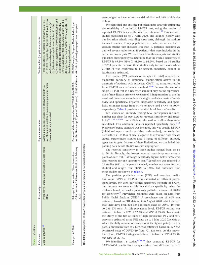
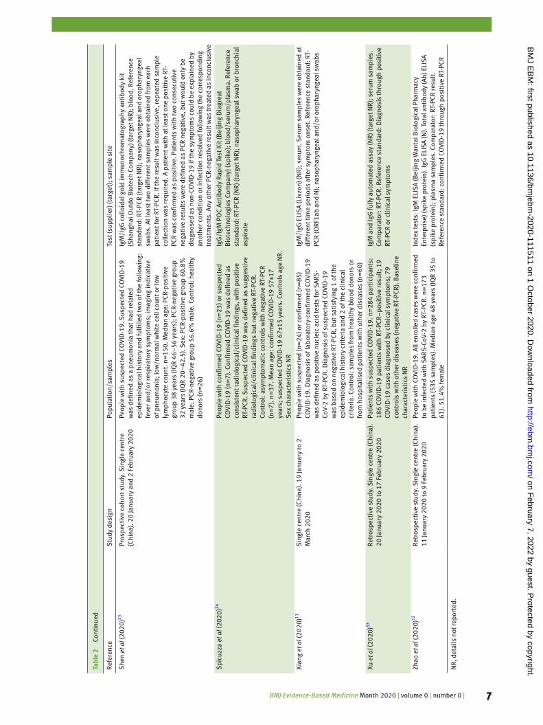
ResultsFigure 1 summarises articles included and excluded at each stage, and reasons that studies were excluded. A total of 13 677 unique articles were screened for eligibility, of which 13 285 were excluded after reading the title and abstract because they did not meet our inclusion criteria. The full text of the remaining 392 arti-cles were read and checked for eligibility, and a further 329 were excluded. Of the remaining 63 relevant articles, 38 studied virus tests and 25 studied antibody tests. Tables 1 and 2 summarise the design and characteristics of studies reporting diagnostic accuracy outcomes for virus and antibody tests, respectively. Characteris-tics of studies that reported other outcomes are reported in online supplemental appendix 2.
All the articles that reported on virus detection were based on the detection of amplified viral SARS- CoV-2 nucleic acid sequences. Most studies used laboratory- based RT- PCR tests conducted using standard in- house or commercially available reagents and equipment, although in some cases, assay details were not reported. The RT- PCR primer used (ie, which part of the viral RNA is targeted and amplified) varied between studies, although again in some cases, primer details were not reported. In addition to RT- PCR, we identified five studies reporting the diag-nostic performance of isothermal amplification assays.
The antibody tests studied used a range of different assay methods to detect one or more antibody type (different immuno-globulin classes and/or antibody targeted). In seven of the studies, tests were laboratory based (ELISA).10–16 We identified 17 studies using assays (lateral- flow immunoassay (LFIA); chemiluminescent
immunoassay (CLIA); colloidal gold immunochromatographic assay (GICA)) that could be suitable for point- of- care use,13 17–32 but the tests were not applied at point of care, or it was not clearly reported that the test had been applied at point of care, in 14 of these studies. In two studies, the type of assay was unclear.33 34
The reliability and applicability of each study’s conduct and reporting was assessed using the QUADAS-2 tool.8 Virus tests and antibody tests were assessed separately and summary judgements are shown in figure 2; signalling questions used and judgements per study are shown in online supplemental appendix 3.
For virus tests, most studies were judged to be of high or unclear risk of bias regarding patient selection, either because patients were selected for the study in a way that could have introduced bias (11% of studies) or because the method of patient selection was unclear (56% of studies). Risk of bias regarding how the index test was conducted or interpreted was judged to be high or unclear for 14% and 44% of studies, respectively, either because aspects of how the tests were conducted were unclear or because tests were not conducted in a uniform manner. For the 12 studies that included a reference standard, we judged the risk of bias to be unclear in 42% and to be high in 8%, largely because not all tests were compared against a uniform reference standard or some details of the reference standard were uncertain.
For antibody tests, the method of patient selection was judged to be unclear for 36% of studies and high for a further 52%. There was an unclear and high risk of bias regarding how the index test was conducted or interpreted in 72% and 12% of studies, respec-tively. For the 19 studies that included a reference standard, 42%
Figure 1 Summary of study selection.
on February 7, 2022 by guest. P
rotected by copyright.http://ebm
.bmj.com
/B
MJ E
BM
: first published as 10.1136/bmjebm
-2020-111511 on 1 October 2020. D
ownloaded from

BMJ Evidence- Based Medicine Month 2020 | volume 0 | number 0 | 4
Tabl
e 1
Char
acte
rist
ics
of in
clud
ed s
tudi
es re
port
ing
diag
nost
ic a
ccur
acy
outc
omes
for v
irus
test
s
Refe
renc
eS
tudy
des
ign
Popu
lati
on/s
ampl
esTe
st (s
uppl
ier)
(tar
get)
; sam
ple
site
Kim
et a
l (20
20)35
Syst
emat
ic re
view
and
met
a- an
alys
is. 6
8 st
udie
s w
ere
incl
uded
: 19
stud
ies
(n=
1502
) rep
orte
d on
RT
- PCR
. Ini
tial
sea
rch
in M
edlin
e an
d Em
base
from
1
Dec
embe
r 201
9 to
16
Mar
ch 2
020.
The
sea
rch
was
up
date
d to
3 A
pril
2020
Stu
dies
on
COVI
D-1
9 th
at re
port
ed th
e di
agno
stic
sen
siti
vity
an
d/or
spe
cific
ity
of c
hest
CT
scan
s an
d/or
RT-
PCR
assa
ysIn
dex
test
s: in
itia
l RT-
PCR
test
(tar
get v
arie
d am
ong
stud
ies)
; nas
opha
ryng
eal s
wab
, thr
oat s
wab
or
sput
um. R
efer
ence
sta
ndar
d: re
peat
ed R
T- PC
R te
sts.
RT
- PCR
resu
lts
wer
e ex
trac
ted
wit
hin
14 d
ays
of
sym
ptom
ons
et
Bae
k et
al (
2020
)36D
esig
n/va
lidat
ion
stud
y (s
ampl
es u
sed
colle
cted
re
tros
pect
ivel
y). K
orea
, num
ber o
f cen
tres
not
cle
arPa
tien
ts w
ith
COVI
D-1
9. n
=14
. No
dem
ogra
phic
det
ails
re
port
edRT
- LAM
P as
say
(dev
elop
ed in
- hou
se);
(N p
rim
er);
na
sal s
wab
s
Fang
et a
l (20
20)68
Retr
ospe
ctiv
e ca
se s
erie
s. S
ingl
e ce
ntre
(Chi
na).
19
Janu
ary
2020
to 4
Feb
ruar
y 20
20Pe
ople
wit
h ev
entu
al c
onfir
med
dia
gnos
is o
f CO
VID
-19
infe
ctio
n w
ho h
ad a
n RT
- PCR
test
and
CT
scan
wit
hin
3 da
ys
or le
ss. E
vent
ual c
onfir
med
dia
gnos
is is
def
ined
as
thro
ugh
repe
ated
RT-
PCR
t est
ing
of n
egat
ive
pati
ents
, unt
il a
posi
tive
te
st is
rece
ived
. n=
51. M
edia
n ag
e 45
yea
rs (I
QR
39 to
55
year
s). 2
9 m
en:2
2 w
omen
Inde
x te
st: i
niti
al R
T- PC
R (S
hang
hai Z
J Bio
- Tec
h)
(pri
mer
not
spe
cifie
d); t
hroa
t or s
putu
m s
ampl
es.
Refe
renc
e st
anda
rd: e
vent
ual c
onfir
med
dia
gnos
is
thro
ugh
RT- P
CR
Fang
et a
l (20
20)43
Retr
ospe
ctiv
e ca
se s
erie
s. S
ingl
e ce
ntre
(Chi
na).
Ja
nuar
y 20
20 to
Feb
ruar
y 20
20 (s
peci
fic d
ates
not
sp
ecifi
ed)
Peop
le w
ith
COVI
D-1
9. n
=32
(8 IC
U p
atie
nts;
24
non-
ICU
pa
tien
ts).
Age
rang
e 35
to 5
4 ye
ars.
Sex
not
repo
rted
RT- P
CR (N
R) (p
rim
er n
ot s
peci
fied)
; fro
m n
asal
sw
abs,
bl
ood,
faec
al, u
rine
, sal
iva
and
tear
sam
ples
Har
ring
ton
et a
l (20
20)37
Pros
pect
ive
case
ser
ies.
Fiv
e ce
ntre
s (U
SA)
Sym
ptom
atic
pat
ient
s m
eeti
ng c
urre
nt c
rite
ria
for d
iagn
osis
of
COVI
D-1
9. n
=52
4. D
emog
raph
ic d
etai
ls n
ot re
port
edIs
othe
rmal
am
plifi
cati
on (I
D N
OW
CO
VID
-19
assa
y (A
bbot
t)) (
RdRp
); n
asal
sw
abs.
Ref
eren
ce s
tand
ard
was
RT-
PCR
(Abb
ott R
ealT
ime
SARS
- CoV
-2 a
ssay
pe
rfor
med
on
the
Abbo
tt m
2000
sys
tem
)
He
et a
l (20
20)69
Retr
ospe
ctiv
e ca
se s
erie
s. S
ingl
e ce
ntre
(Chi
na).
10
Janu
ary
2020
to 2
8 Fe
brua
ry 2
020
Hos
pita
lised
pat
ient
s w
ith
susp
ecte
d CO
VID
-19
who
un
derw
ent h
igh-
reso
luti
on c
hest
CT
and
real
- tim
e RT
- PCR
. n=
82. M
edia
n ag
e 52
yea
rs (r
ange
8 to
74
year
s). 4
9 m
ales
RT- P
CR (B
GI G
enom
ics)
; (pr
imer
NR)
; nas
opha
ryng
eal
swab
, oro
phar
ynge
al s
wab
, end
otra
chea
l asp
irat
e or
br
onch
oalv
eola
r lav
age.
Ref
eren
ce s
tand
ard:
eve
ntua
l co
nfir
med
dia
gnos
is th
roug
h RT
- PCR
Lee
et a
l (20
20)70
Retr
ospe
ctiv
e ca
se s
erie
s. S
ingl
e ce
ntre
(Sin
gapo
re).
U
p to
29
Febr
uary
202
0 (s
tart
dat
e no
t rep
orte
d)Pa
tien
ts a
dmit
ted
to h
ospi
tal w
ith
susp
ecte
d CO
VID
-19
infe
ctio
n. n
=70
. Dem
ogra
phic
s no
t rep
orte
dRT
- PCR
(in-
hous
e or
A*S
TAR
Fort
itud
e Ki
t (Ac
cele
rate
Te
chno
logi
es))
; (N
, Orf
1ab)
; nas
opha
ryng
eal s
wab
s
Lu e
t al (
2020
)38D
esig
n/va
lidat
ion
stud
y. C
hina
. Dat
es o
f sam
plin
g no
t rep
orte
dPa
tien
ts w
ith
susp
ecte
d CO
VID
-19
adm
itte
d to
hos
pita
l and
qu
aran
tine
d. n
=56
. Dem
ogra
phic
s no
t rep
orte
dIn
dex
test
: RT-
LAM
P (in
- hou
se a
ssay
) (N
); th
roat
sw
abs.
Ref
eren
ce s
tand
ard:
RT-
PCR
(Life
Rive
r Bio
)
She
n et
al (
2020
)62Re
tros
pect
ive
case
ser
ies.
Sin
gle
cent
re (C
hina
).22
Janu
ary
to 1
8 Fe
brua
ry 2
020
Sub
ject
s ju
dged
at h
igh
risk
of S
ARS
- CoV
-2 in
fect
ion.
n=
5630
. M
edia
n ag
e 51
yea
rs (I
QR
36–
63).
Mal
e 26
31 (4
6.7%
)RT
- PCR
(Sha
ngha
i Hui
rui B
iote
chno
logy
); (O
rf1a
b, N
);
thro
at s
wab
s
Yan
et a
l (20
20)39
Dev
elop
men
t/va
lidat
ion
stud
y. C
entr
e N
R. D
ates
NR
Pati
ents
wit
h pn
eum
onia
and
sus
pect
ed S
ARS
- CoV
-2 in
fect
ion.
n=
130
spec
imen
s. C
hara
cter
isti
cs N
RRT
- LAM
P (L
oopa
mp
RNA
ampl
ifica
tion
kit
; Loo
pam
p Re
al- t
ime
Turb
idim
eter
, bot
h Ei
ken
Chem
ical
, Tok
yo,
Japa
n, u
sed
to p
erfo
rm a
nd m
onit
or th
e RT
- LAM
P re
acti
on) (
Orf
1ab
and
spik
e). R
efer
ence
sta
ndar
d:
RT- P
CR. S
ampl
ing
from
sw
abs
(not
spe
cifie
d) a
nd
bron
choa
lveo
lar l
avag
e flu
id
Cont
inue
d
on February 7, 2022 by guest. P
rotected by copyright.http://ebm
.bmj.com
/B
MJ E
BM
: first published as 10.1136/bmjebm
-2020-111511 on 1 October 2020. D
ownloaded from
BMJ Evidence- Based Medicine Month 2020 | volume 0 | number 0 | 5
were judged to have an unclear risk of bias and 16% a high risk of bias.
We identified one existing published meta- analysis estimating the sensitivity of an initial RT- PCR test, using the results of repeated RT- PCR tests as the reference standard.35 This included studies published up to 3 April 2020, and aligned closely with our inclusion criteria regarding virus tests, although the authors included studies of any population size, whereas we elected to exclude studies that included less than 10 patients, meaning we omitted seven studies (total 46 patients) that were included in the earlier meta- analysis. We used data from this analysis and studies published subsequently to determine that the overall sensitivity of RT- PCR is 87.8% (95% CI 81.5% to 92.2%), based on 16 studies of 3818 patients. Because these studies only included cases where COVID-19 was confirmed to be present, specificity cannot be legitimately estimated.
Five studies (972 patients or samples in total) reported the diagnostic accuracy of isothermal amplification assays in the diagnosis of patients with suspected COVID-19, using test results from RT- PCR as a reference standard.36–40 Because the use of a single RT- PCR test as a reference standard may not be representa-tive of true disease presence, we deemed it inappropriate to use the results of these studies to derive a single pooled estimate of sensi-tivity and specificity. Reported diagnostic sensitivity and speci-ficity estimates range from 74.7% to 100% and 87.7% to 100%, respectively. Table 3 provides a detailed breakdown of results.
Ten studies on antibody testing (757 participants included; number not clear for two studies) reported sensitivity and speci-ficity11 12 17 18 20–24 33 or sufficient information to allow these to be calculated. Two additional studies reported specificity only.10 19 Where a reference standard was included, this was usually RT- PCR (initial and repeats until a positive confirmation); one study that used either RT- PCR or clinical diagnosis to determine final disease status. Furthermore, studies used a range of different antibody types and targets. Because of these limitations, we concluded that pooling data across studies was not appropriate.
The reported sensitivity in these studies ranged from 18.4% to 96.1%. Notably, the lowest reported sensitivity was using a point- of- care test,17 although sensitivity figures below 50% were also reported for one laboratory test.20 Specificity was reported in 12 studies (682 participants included; number not clear for two studies) and ranged from 88.9% to 100%. Full outcomes from these studies are shown in table 4.
The positive predictive value (PPV) and negative predic-tive value (NPV) of RT- PCR was estimated at different preva-lence levels. We used our pooled sensitivity estimate of 87.8%, and because we were unable to calculate specificity using the evidence found, we used a previously published estimate of 98.0% for specificity.41 Prevalence estimates were based on data from Public Health England (PHE).42 A prevalence rate of 3.0% was estimated based on PHE data up to 6 August 2020, which showed that there have been 308 134 confirmed cases of COVID-19 from 10 236 970 tests. At this prevalence level, RT- PCR testing was estimated to have a PPV of 57.7% and NPV of 99.6%. To estimate the utility of the test at times of high prevalence, PPV and NPV were also estimated using PHE data up to 1 May 2020 (the date at which the daily number of cases was at its highest point). On this date, a prevalence rate of 24.6% was estimated based on 177 454 confirmed cases of COVID-19 from 721 124 tests. At this preva-lence level, RT- PCR testing was estimated to have a PPV of 93.5% and NPV of 96.1%.
We identified 18 studies30 43–59 that compared RT- PCR for SARS- CoV-2 results from samples taken from different parts of Re
fere
nce
Stu
dy d
esig
nPo
pula
tion
/sam
ples
Test
(sup
plie
r) (t
arge
t); s
ampl
e si
te
Zhen
et a
l (20
20)40
Pros
pect
ive
com
para
tive
eva
luat
ion.
Sin
gle
cent
re
(USA
)Sy
mpt
omat
ic p
atie
nts
susp
ecte
d of
hav
ing
COVI
D-1
9.
n=10
8 sa
mpl
es (u
ncle
ar if
from
uni
que
pati
ents
). P
atie
nt
char
acte
rist
ics
not r
epor
ted
Thre
e in
dex
test
s, a
ll ‘s
ampl
e to
ans
wer
’ pla
tfor
ms:
(1
) RT-
PCR
(Cep
heid
Xpe
rt X
pres
s SA
RS- C
oV-2
ass
ay,
perf
orm
ed o
n G
eneX
pert
inst
rum
ent s
yste
m);
(N2,
E)
; (2)
isot
herm
al a
mpl
ifica
tion
(Abb
ott I
D N
OW
CO
VID
-19
assa
y); (
RdRp
); (3
) DN
A hy
brid
isat
ion
and
elec
troc
hem
ical
det
ecti
on (G
enM
ark
ePle
x SA
RS-
CoV-
2 as
say)
(tar
get N
R). A
ll co
mpa
red
wit
h RT
- PCR
(H
olog
ic P
anth
er F
usio
n SA
RS- C
oV-2
ass
ay);
(Orf
1ab)
. N
asop
hary
ngea
l spe
cim
ens
Zhan
g et
al (
2020
)71Re
tros
pect
ive
case
ser
ies.
Chi
na, t
wo
cent
res.
Co
llect
ion
from
29
Dec
embe
r 201
9 to
16
Febr
uary
20
20
Peop
le w
ith
labo
rato
ry- c
onfir
med
CO
VID
-19
(via
RT-
PCR)
. n=
290.
Med
ian
age
57 y
ears
(22–
88 y
ears
). 1
55 (5
3.4%
) mal
eRT
- PCR
(Sha
ngha
i Bio
- ger
m M
edic
al T
echn
olog
y)
(Orf
1ab,
N p
rim
ers)
; pha
ryng
eal s
wab
sam
ples
NR,
det
ails
not
repo
rted
; RT-
LAM
P, re
vers
e tr
ansc
ript
ion
loop
- med
iate
d is
othe
rmal
am
plifi
cati
on.
Tabl
e 1
Cont
inue
d
on February 7, 2022 by guest. P
rotected by copyright.http://ebm
.bmj.com
/B
MJ E
BM
: first published as 10.1136/bmjebm
-2020-111511 on 1 October 2020. D
ownloaded from

BMJ Evidence- Based Medicine Month 2020 | volume 0 | number 0 | 6
Tabl
e 2
Char
acte
rist
ics
of in
clud
ed s
tudi
es re
port
ing
diag
nost
ic a
ccur
acy
outc
omes
for a
ntib
ody
test
s
Refe
renc
eS
tudy
des
ign
Popu
lati
on/s
ampl
esTe
st (s
uppl
ier)
(tar
get)
; sam
ple
site
Cass
anit
i et a
l (20
20)17
Coho
rt s
tudy
. Sin
gle
cent
re (I
taly
).
Colle
ctio
n da
te N
R3
coho
rts:
(1) h
ealt
hy v
olun
teer
s w
ith
nega
tive
RT-
PCR
for C
OVI
D-1
9; (2
) hos
pita
lised
pat
ient
s w
ith
posi
tive
CO
VID
-19
RT- P
CR; (
3) p
atie
nts
wit
h su
spec
ted
COVI
D-1
9 at
th
eir f
irst
acc
ess
at e
mer
genc
y ro
om. n
=11
0 (3
0 he
alth
y vo
lunt
eers
; 30
pati
ents
wit
h CO
VID
-19;
50
pati
ents
wit
h su
spec
ted
COVI
D-1
9). B
asel
ine
char
acte
rist
ics
repo
rted
se
para
tely
for e
ach
coho
rt
Viva
Dia
g CO
VID
-19‐
19 Ig
M/I
gG ra
pid
poin
t- of
- car
e la
tera
l flo
w
imm
unoa
ssay
(Viv
aChe
k) (t
arge
t NR)
; ser
um o
r blo
od s
ampl
es. S
erum
sa
mpl
es w
ere
obta
ined
at m
edia
n 7
days
(IQ
R 4–
11 d
ays)
aft
er
posi
tive
resu
lt fo
r hos
pita
lised
pat
ient
s. R
efer
ence
/com
para
tor:
RT-
PC
R (R
dRp
and
E pr
imer
s); r
espi
rato
ry s
ampl
es
Doh
la e
t al (
2020
)18S
ingl
e ce
ntre
(Ger
man
y). D
ates
NR
Peop
le w
ithi
n a
com
mun
ity
sett
ing
(hig
h- pr
eval
ence
ar
ea),
pre
sent
ing
wit
h CO
VID
-19
sym
ptom
s (n
=39
) and
pe
ople
dia
gnos
ed w
ith
COVI
D-1
9 (n
=10
). M
edia
n ag
e 46
ye
ars
(IQR
28–
72 y
ears
). 2
9/49
fem
ale
(49.
0%)
IgG
/IgM
poi
nt- o
f - c a
re te
st (N
R) (t
arge
t NR)
; fin
gert
ip p
rick
blo
od o
r se
rum
. Ref
eren
ce s
tand
ard:
repe
ated
RT-
PCR
(Alt
ona
Dia
gnos
tics
) (t
arge
t NR)
; thr
oat s
wab
s. S
erum
sam
ples
for p
revi
ousl
y di
agno
sed
indi
vidu
als
wer
e al
so a
naly
sed
usin
g th
e an
tibo
dy te
st
Hof
fman
et a
l (20
20)19
Valid
atio
n st
udy.
Cen
tre
NR.
Stu
dy d
ates
N
RPa
tien
ts w
ith
conf
irm
ed C
OVI
D-1
9 or
con
vale
scen
ts
(n=
29).
Con
trol
s: h
ealt
hy v
olun
teer
s w
itho
ut a
ny k
now
n hi
stor
y of
CO
VID
-19
(n=
24);
blo
od d
onor
ser
a fr
om
heal
thy
adul
ts (n
=80
) and
bab
ies
(n=
20) c
olle
cted
dur
ing
2018
IgG
/IgM
Rap
id T
est C
asse
tte
(Zhe
jiang
Ori
ent G
ene
Bio
tech
Com
pany
) (t
arge
t NR)
; blo
od/s
erum
sam
ples
. Ref
eren
ce s
tand
ard:
RT-
PCR
Jin e
t al (
2020
)20Re
tros
pect
ive
stud
y. S
ingl
e ce
ntre
(Chi
na)
Peop
le w
ith
a la
bora
tory
- con
firm
ed S
ARS
- CoV
-2 in
fect
ion
in h
ospi
tal,
and
at le
ast o
ne v
iral
ser
olog
ical
test
(n=
43).
M
edia
n ag
e 47
.0 y
ears
(IQ
R 34
.0–
59.0
yea
rs).
39.
5%
mal
e. C
ontr
ol g
roup
: pat
ient
s w
ith
susp
ecte
d SA
RS-
C oV-
2 in
fect
ion
who
wer
e ex
clud
ed a
nd q
uara
ntin
ed a
t ho
me
(n=
33).
Med
ian
age
31.0
yea
rs (I
QR
25.5
–37
.5
year
s). 6
6.7%
mal
e. S
uspe
cted
infe
cted
pat
ient
s w
ere
disc
harg
ed fr
om h
ospi
tal w
hen
they
rece
ived
two
nega
tive
PC
Rs, p
erfo
rmed
in a
24-
hour
inte
rval
IgM
and
IgG
che
milu
min
esce
nce
assa
y (C
LIA)
(She
nzhe
n YH
LO
Bio
tech
) (ta
rget
s N
pro
tein
and
spi
ke p
rote
in).
Ref
eren
ce s
tand
ard:
co
nfir
med
dia
gnos
is fr
om R
T- PC
R (t
arge
t not
spe
cifie
d); s
ampl
ing
not
clea
rly
repo
rted
but
incl
udes
ora
l sw
abs,
ana
l sw
abs
and
sput
um.
Dur
atio
n be
twee
n fir
st s
ympt
oms
and
sero
logi
cal t
est (
CLIA
) was
18
days
(IQ
R 11
–23
day
s) in
the
COVI
D-1
9 gr
oup,
3.0
day
s (2
.0–
8.0
days
)
Li e
t al (
2020
)21Pr
ospe
ctiv
e de
velo
pmen
t stu
dy. S
ingl
e ce
ntre
(Chi
na).
12
Febr
uary
202
0 to
20
Febr
uary
202
0
Peop
le w
ith
susp
ecte
d (R
T- PC
R ne
gati
ve) o
r con
firm
ed
(RT-
PCR
posi
tive
) CO
VID
-19.
n=
278
(89
conf
irm
ed; 1
89
prob
able
). n
=27
3 co
ntro
ls w
ere
incl
uded
. Bas
elin
e ch
arac
teri
stic
s N
R
IgM
and
IgG
col
loid
al g
old
assa
y (N
R) (t
arge
ts s
erum
ant
ibod
ies
agai
nst N
pro
tein
); s
erum
spe
cim
ens.
RT-
PCR
assu
med
to b
e th
e re
fere
nce
stan
dard
(des
crib
ed a
s a
‘con
trol
’ by
the
auth
ors)
; pri
mer
/ta
rget
and
sam
plin
g m
etho
ds n
ot k
now
n
Li e
t al (
2020
)22Pr
ospe
ctiv
e de
velo
pmen
t stu
dy. 8
cen
tres
(C
hina
). D
ates
NR
Peop
le w
ith
susp
ecte
d CO
VID
-19.
n=
525
spec
imen
s (3
97
clin
ical
pos
itiv
e; 1
28 c
linic
al n
egat
ive)
. Cha
ract
eris
tics
NR
IgM
/IgG
rapi
d po
int-
of- c
are
late
ral f
low
imm
unoa
ssay
(Jia
ngsu
M
edom
ics
Med
ical
Tec
hnol
ogie
s) (t
arge
ts a
ntib
odie
s ag
ains
t spi
ke
prot
ein)
; blo
od (i
nclu
ding
ser
um a
nd p
lasm
a). R
efer
ence
sta
ndar
d:
RT- P
CR; r
espi
rato
ry s
peci
men
s
Liu
et a
l (20
20)10
Pros
pect
ive
stud
y. S
ingl
e ce
ntre
(Chi
na).
18
Janu
ary
to 2
6 Fe
brua
ry 2
020
Hos
pita
lised
pat
ient
s di
agno
sed
wit
h CO
VID
-19.
Al
l pat
ient
s w
ere
labo
rato
ry c
onfir
med
by
RT- P
CR.
n=31
4 (2
14 p
atie
nts;
100
hea
lthy
con
trol
s). B
asel
ine
char
acte
rist
ics
NR
IgM
ELI
SA Ig
G E
LISA
(NR)
(tar
gets
ant
ibod
ies
agai
nst N
and
spi
ke);
se
rum
. Med
ian
tim
e of
sam
ple
colle
ctio
n w
as 1
5 da
ys (r
ange
0 to
55)
Cont
inue
d
on February 7, 2022 by guest. P
rotected by copyright.http://ebm
.bmj.com
/B
MJ E
BM
: first published as 10.1136/bmjebm
-2020-111511 on 1 October 2020. D
ownloaded from
BMJ Evidence- Based Medicine Month 2020 | volume 0 | number 0 | 7
Refe
renc
eS
tudy
des
ign
Popu
lati
on/s
ampl
esTe
st (s
uppl
ier)
(tar
get)
; sam
ple
site
She
n et
al (
2020
)23Pr
ospe
ctiv
e co
hort
stu
dy. S
ingl
e ce
ntre
(C
hina
). 2
0 Ja
nuar
y an
d 2
Febr
uary
202
0Pe
ople
wit
h su
spec
ted
COVI
D-1
9. S
uspe
cted
CO
VID
-19
was
def
ined
as
a pn
eum
onia
that
had
rela
ted
epid
emio
logi
cal h
isto
ry a
nd fu
lfille
d tw
o of
the
follo
win
g:
feve
r and
/or r
espi
rato
ry s
ympt
oms;
imag
ing
indi
cati
ve
of p
neum
onia
; low
/nor
mal
whi
te c
ell c
ount
or l
ow
lym
phoc
yte
coun
t. n
=15
0. M
edia
n ag
e: P
CR- p
osit
ive
grou
p 38
yea
rs (I
QR
46–
56 y
ears
); P
CR- n
egat
ive
grou
p 32
yea
rs (I
QR
20–
42.5
). S
ex: P
CR- p
o sit
ive
grou
p 60
.8%
m
ale;
PCR
- neg
ativ
e gr
oup
56.6
% m
ale.
Con
trol
: hea
lthy
do
nors
(n=
26)
IgM
/IgG
col
loid
al g
old
imm
unoc
hrom
atog
raph
y an
tibo
dy k
it
(Sha
ngha
i Out
do B
iote
ch C
ompa
ny) (
targ
et N
R); b
lood
. Ref
eren
ce
stan
dard
: RT-
PCR
(tar
get N
R); n
asop
hary
ngea
l and
oro
phar
ynge
al
swab
s. A
t lea
st tw
o di
ffer
ent s
ampl
es w
ere
obta
ined
from
eac
h pa
tien
t for
RT-
PCR.
If th
e re
sult
was
inco
nclu
sive
, rep
eate
d sa
mpl
e co
llect
ion
was
requ
ired
. A p
atie
nt w
ith
at le
ast o
ne p
osit
ive
RT-
PCR
was
con
firm
ed a
s po
siti
ve. P
atie
nts
wit
h tw
o co
nsec
utiv
e ne
gati
ve re
sult
s w
ere
defin
ed a
s PC
R ne
gati
ve, b
ut w
ould
onl
y be
di
agno
sed
as n
on- C
OVI
D-1
9 if
the
sym
ptom
s co
uld
be e
xpla
ined
by
anot
her c
ondi
tion
or i
nfec
tion
reso
lved
follo
win
g th
e co
rres
pond
ing
trea
tmen
ts. A
ny o
ther
PCR
- neg
ativ
e re
sult
was
trea
ted
as in
conc
lusi
ve
Spi
cuzz
a et
al (
2020
)24
Pe
ople
wit
h co
nfir
med
CO
VID
-19
(n=
23) o
r sus
pect
ed
COVI
D-1
9 (n
=7)
. Con
firm
ed C
OVI
D-1
9 w
as d
efin
ed a
s co
nsis
tent
radi
olog
ical
/clin
ical
find
ings
, wit
h po
siti
ve
RT- P
CR. S
uspe
cted
CO
VID
-19
was
def
ined
as
sugg
esti
ve
radi
olog
ical
/clin
ical
find
ings
but
neg
ativ
e RT
- PCR
. Co
ntro
l: as
ympt
omat
ic c
ontr
ols
wit
h ne
gati
ve R
T- PC
R (n
=7)
. n=
37. M
ean
age:
con
firm
ed C
OVI
D-1
9 57
±17
year
s; s
uspe
cted
CO
VID
-19
67±1
5 ye
ars.
Con
trol
s ag
e N
R.
Sex
cha
ract
eris
tics
NR
IgG
/IgM
PO
C An
tibo
dy R
apid
Tes
t Kit
(Bei
jing
Dia
grea
t B
iote
chno
logi
es C
ompa
ny) (
spik
e); b
lood
/ser
um/p
lasm
a. R
efer
ence
st
anda
rd: R
T- PC
R (N
R) (t
arge
t NR)
; nas
opha
ryng
eal s
wab
or b
ronc
hial
as
pira
te
Xian
g et
al (
2020
)11S
ingl
e ce
ntre
(Chi
na).
19
Janu
ary
to 2
M
arch
202
0Pe
ople
wit
h su
spec
ted
(n=
24) o
r con
firm
ed (n
=85
) CO
VID
-19.
Dia
gnos
is o
f lab
orat
ory-
conf
irm
ed C
OVI
D-1
9 w
as d
efin
ed a
s po
siti
ve n
ucle
ic a
cid
test
s fo
r SAR
S-
CoV-
2 by
RT-
PCR.
Dia
gnos
is o
f sus
pect
ed C
OVI
D-1
9 w
as b
ased
on
nega
tive
RT-
PCR,
but
sat
isfy
ing
1 of
the
epid
emio
logi
cal h
isto
ry c
rite
ria
and
2 of
the
clin
ical
cr
iter
ia. C
ontr
ol: s
ampl
es fr
om h
ealt
hy b
lood
don
ors
or
from
hos
pita
lised
pat
ient
s w
ith
othe
r dis
ease
s (n
=60
)
IgM
/IgG
ELI
SA (L
ivzo
n) (N
R); s
erum
. Ser
um s
ampl
es w
ere
obta
ined
at
diff
eren
t tim
e pe
riod
s af
ter s
ympt
om o
nset
. Ref
eren
ce s
tand
ard:
RT-
PC
R (O
RF1a
b an
d N
); n
asop
hary
ngea
l and
/or o
roph
aryn
geal
sw
abs
Xu e
t al (
2020
)33Re
tros
pect
ive
stud
y. S
ingl
e ce
ntre
(Chi
na).
20
Janu
ary
2020
to 1
7 Fe
brua
ry 2
020
Pati
ents
wit
h su
spec
ted
COVI
D-1
9. n
=28
4 pa
rtic
ipan
ts:
186
COVI
D-1
9 pa
tien
ts w
ith
RT- P
CR–
posi
tive
resu
lt; 1
9 CO
VID
-19
case
s di
agno
sed
by c
linic
al s
ympt
oms;
79
cont
rols
wit
h ot
her d
isea
ses
(neg
ativ
e RT
- PCR
). B
asel
ine
char
acte
rist
ics
NR
IgM
and
IgG
fully
aut
omat
ed a
ssay
(NR)
(tar
get N
R); s
erum
sam
ples
. Co
mpa
rato
r: R
T- PC
R. R
efer
ence
sta
ndar
d: D
iagn
osis
thro
ugh
posi
tive
RT
- PCR
or c
linic
al s
ympt
oms
Zhao
et a
l (20
20)12
Retr
ospe
ctiv
e st
udy.
Sin
gle
cent
re (C
hina
).
11 Ja
nuar
y 20
20 to
9 F
ebru
ary
2020
Peop
le w
ith
COVI
D-1
9. A
ll en
rolle
d ca
ses
wer
e co
nfir
med
to
be
infe
cted
wit
h SA
RS- C
oV-2
by
RT- P
CR. n
=17
3 pa
tien
ts (5
35 s
ampl
es).
Med
ian
age
48 y
ears
(IQ
R 35
to
61).
51.
4% fe
mal
e
Inde
x te
sts:
IgM
ELI
SA (B
eijin
g W
anta
i Bio
logi
cal P
harm
acy
Ente
rpri
se) (
spik
e pr
otei
n). I
gG E
LISA
(N).
Tot
al a
ntib
ody
(Ab)
ELI
SA
(spi
ke p
rote
in);
pla
sma
sam
ples
. Com
para
tor:
RT-
PCR
r esu
lt.
Refe
renc
e st
anda
rd: c
onfir
med
CO
VID
-19
thro
ugh
posi
tive
RT-
PCR
NR,
det
ails
not
repo
rted
.
Tabl
e 2
Cont
inue
d
on February 7, 2022 by guest. P
rotected by copyright.http://ebm
.bmj.com
/B
MJ E
BM
: first published as 10.1136/bmjebm
-2020-111511 on 1 October 2020. D
ownloaded from
BMJ Evidence- Based Medicine Month 2020 | volume 0 | number 0 | 8
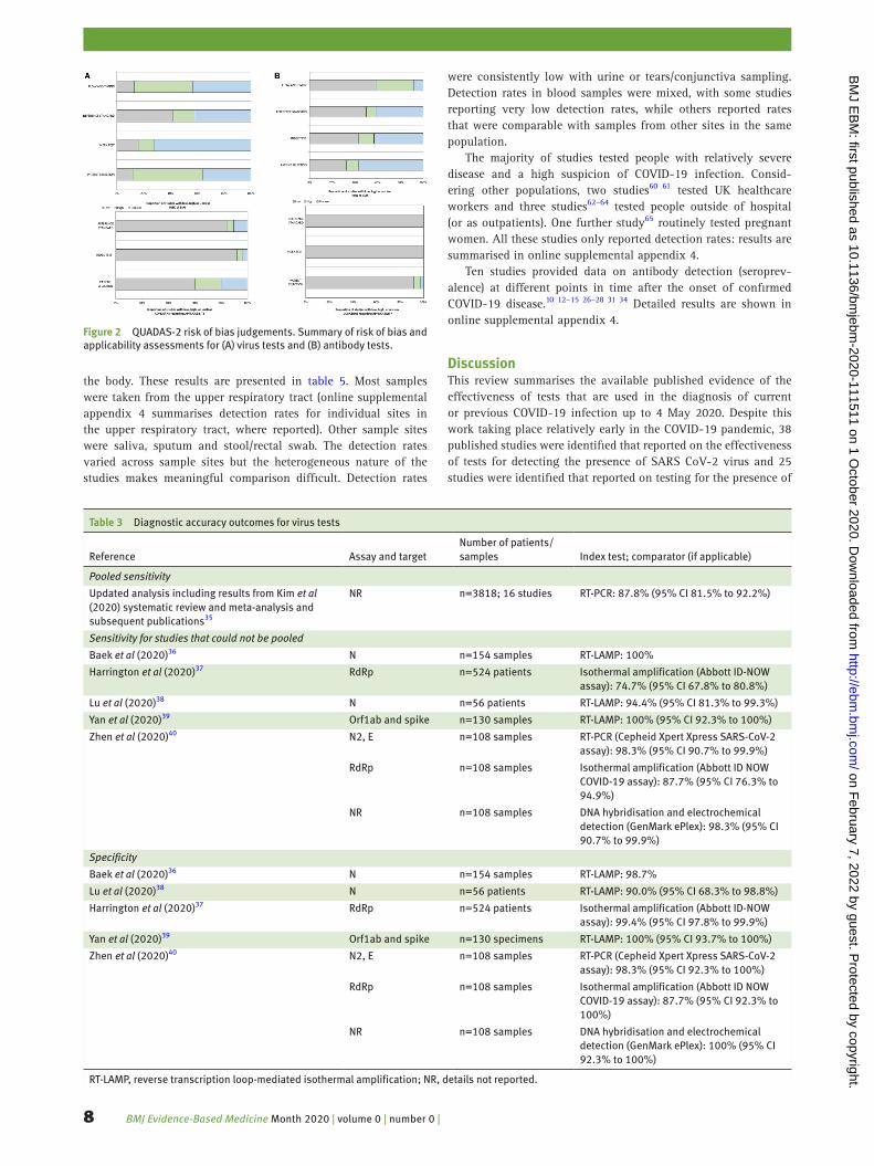
the body. These results are presented in table 5. Most samples were taken from the upper respiratory tract (online supplemental appendix 4 summarises detection rates for individual sites in the upper respiratory tract, where reported). Other sample sites were saliva, sputum and stool/rectal swab. The detection rates varied across sample sites but the heterogeneous nature of the studies makes meaningful comparison difficult. Detection rates
were consistently low with urine or tears/conjunctiva sampling. Detection rates in blood samples were mixed, with some studies reporting very low detection rates, while others reported rates that were comparable with samples from other sites in the same population.
The majority of studies tested people with relatively severe disease and a high suspicion of COVID-19 infection. Consid-ering other populations, two studies60 61 tested UK healthcare workers and three studies62–64 tested people outside of hospital (or as outpatients). One further study65 routinely tested pregnant women. All these studies only reported detection rates: results are summarised in online supplemental appendix 4.
Ten studies provided data on antibody detection (seroprev-alence) at different points in time after the onset of confirmed COVID-19 disease.10 12–15 26–28 31 34 Detailed results are shown in online supplemental appendix 4.
DiscussionThis review summarises the available published evidence of the effectiveness of tests that are used in the diagnosis of current or previous COVID-19 infection up to 4 May 2020. Despite this work taking place relatively early in the COVID-19 pandemic, 38 published studies were identified that reported on the effectiveness of tests for detecting the presence of SARS CoV-2 virus and 25 studies were identified that reported on testing for the presence of
Figure 2 QUADAS-2 risk of bias judgements. Summary of risk of bias and applicability assessments for (A) virus tests and (B) antibody tests.
Table 3 Diagnostic accuracy outcomes for virus tests
Reference Assay and targetNumber of patients/samples Index test; comparator (if applicable)
Pooled sensitivity
Updated analysis including results from Kim et al (2020) systematic review and meta- analysis and subsequent publications35
NR n=3818; 16 studies RT- PCR: 87.8% (95% CI 81.5% to 92.2%)
Sensitivity for studies that could not be pooled
Baek et al (2020)36 N n=154 samples RT- LAMP: 100%
Harrington et al (2020)37 RdRp n=524 patients Isothermal amplification (Abbott ID- NOW assay): 74.7% (95% CI 67.8% to 80.8%)
Lu et al (2020)38 N n=56 patients RT- LAMP: 94.4% (95% CI 81.3% to 99.3%)
Yan et al (2020)39 Orf1ab and spike n=130 samples RT- LAMP: 100% (95% CI 92.3% to 100%)
Zhen et al (2020)40 N2, E n=108 samples RT- PCR (Cepheid Xpert Xpress SARS- CoV-2 assay): 98.3% (95% CI 90.7% to 99.9%)
RdRp n=108 samples Isothermal amplification (Abbott ID NOW COVID-19 assay): 87.7% (95% CI 76.3% to 94.9%)
NR n=108 samples DNA hybridisation and electrochemical detection (GenMark ePlex): 98.3% (95% CI 90.7% to 99.9%)
Specificity
Baek et al (2020)36 N n=154 samples RT- LAMP: 98.7%
Lu et al (2020)38 N n=56 patients RT- LAMP: 90.0% (95% CI 68.3% to 98.8%)
Harrington et al (2020)37 RdRp n=524 patients Isothermal amplification (Abbott ID- NOW assay): 99.4% (95% CI 97.8% to 99.9%)
Yan et al (2020)39 Orf1ab and spike n=130 specimens RT- LAMP: 100% (95% CI 93.7% to 100%)
Zhen et al (2020)40 N2, E n=108 samples RT- PCR (Cepheid Xpert Xpress SARS- CoV-2 assay): 98.3% (95% CI 92.3% to 100%)
RdRp n=108 samples Isothermal amplification (Abbott ID NOW COVID-19 assay): 87.7% (95% CI 92.3% to 100%)
NR n=108 samples DNA hybridisation and electrochemical detection (GenMark ePlex): 100% (95% CI 92.3% to 100%)
RT- LAMP, reverse transcription loop- mediated isothermal amplification; NR, details not reported.
on February 7, 2022 by guest. P
rotected by copyright.http://ebm
.bmj.com
/B
MJ E
BM
: first published as 10.1136/bmjebm
-2020-111511 on 1 October 2020. D
ownloaded from

BMJ Evidence- Based Medicine Month 2020 | volume 0 | number 0 | 9
antibodies. Analysis of these studies using the QUADAS-2 frame-work revealed high or unclear risks of bias in the majority, most commonly as a result of unclear methods of patient selection and test conduct, or because of the use of a reference standard that may not definitively diagnose COVID-19. Nonetheless, the avail-able evidence provides information on which to begin to judge the possible clinical effectiveness of COVID-19 testing, although significant uncertainties remain in the evidence base regarding their clinical and public health application.
In the course of our work, the first meta- analysis of diagnostic accuracy for SARS- CoV-2 virus tests was published by Kim et al.35 This included pooled analysis of 19 studies (1502 patients) and
used the results of repeated laboratory- based RT- PCR as the refer-ence standard. This aligned closely with our own inclusion criteria for virus tests, although Kim et al included studies of any popula-tion size, we excluded studies with less than 10 patients, meaning we omitted 7 studies (46 patients). However, by including more recently published studies in our analysis the number of patients more than doubles from 1502 to 3818 patients. The analysis by Kim et al estimated that the sensitivity of an initial RT- PCR test is 89% (95% CI 81% to 94%). Our addition of data from more recent studies leads us to conclude a sensitivity of 87.8% (95% CI 81.5% to 92.2%). An analysis estimating the PPV and NPV of RT- PCR showed that the NPV is likely to be high while PPV
Table 4 Diagnostic accuracy outcomes for antibody tests
Reference Assay and target Number of patients/samples Index test; comparator (if applicable)
Sensitivity
Cassaniti et al (2020)17 LFIA, VivaChek POC n=50 patients (suspected cases only)
IgM/IgG: 18.4%
Dohla et al (2020)18 IgM/IgG POC test n=49 IgM/IgG: 36.4% (95% CI 17.2 to 59.3)
Jin et al (2020)20 CLIA (N and spike proteins) n=27 IgM: 48.1% (13/27)IgG: 88.9% (24/27)
Li et al (2020)21 Colloidal gold Population not clear IgM: 78.7%IgG: 73.0%IgM/IgG: 87.6%
Li et al (2020)22 LFIA, Jiangsu Medomics POC n=525 specimens IgM/IgG: 88.66%
Spicuzza et al (2020)24 LFIA (spike) n=37 IgG/IgM: 82.6%
Shen et al (2020)23 Colloidal gold (NR) n=150 IgM/IgG: 71.1% (95% CI 0.609 to 0.797)
Xiang et al (2020)11 ELISA (NR) n=66 IgM: 77.3% (51/66)IgG: 83.3% (55/66)
Xu et al (2020)33 Fully automated assay (NR) n=205 patients IgM: 70.24% (144/205)IgG: 96.10% (197/205)
Zhao et al (2020)12 ELISA (spike for IgM and Ab; N for IgG) n=173 samples IgM: 82.7% (143/173)IgG: 64.7% (112/173)Ab: 93.1% (161/173)RT- PCR: 67.1%* (112/?)
Specificity
Cassaniti et al (2020)17 LFIA, VivaChek POC n=50 (suspected cases only) IgM/IgG: 91.7%
Li et al (2020)21 Colloidal gold Population not clear IgM: 98.2%IgG: 99.3%IgM/IgG: 98.2%
Li et al (2020)22 LFIA, Jiangsu Medomics POC n=525 specimens IgM/IgG: 90.63%
Liu et al (2020)10 ELISA (spike) n=100 healthy controls IgM: 100% (0/100)IgG: 100% (0/100)IgM and/or IgG: 100% (0/100)
Xu et al (2020)33 Fully automated assay (NR) n=79 patients IgM: 96.20% (76/79)IgG: 92.41% (73/79)
Zhao et al (2020)12 ELISA (spike for IgM and Ab; N for IgG) Not clear Total Ab: 99.1% (211/213)IgM: 98.6% (210/213)IgA: 99.0% (195/197)
Jin et al (2020)20 CLIA (N and spike proteins) n=33 IgM: 100% (33/33)IgG: 90.9% (30/33)
Xiang et al (2020)11 ELISA (NR) n=60 IgM: 100% (60/60)IgG: 95.0% (57/60)
Dohla et al (2020)18 IgM/IgG POC test n=49 IgM/IgG: 88.9% (95% CI 70.8 to 97.7)
Spicuzza et al (2020)24 LFIA (spike) n=37 IgG/IgM 92.9%
Hoffman et al (2020)19 LFIA (NR) n=124 (controls) IgM: 100% (0/124)IgG: 99.2% (1/124)
Shen et al (2020)23 Colloidal gold (NR) n=150 IgM/IgG: 96.2% (95% CI 0.859 to 0.993)
CLIA, chemiluminescent immunoassay; LFIA, lateral flow immunoassay; NR, details not reported.
on February 7, 2022 by guest. P
rotected by copyright.http://ebm
.bmj.com
/B
MJ E
BM
: first published as 10.1136/bmjebm
-2020-111511 on 1 October 2020. D
ownloaded from

BMJ Evidence- Based Medicine Month 2020 | volume 0 | number 0 | 10
Tabl
e 5
Viru
s te
st d
etec
tion
rate
s in
stu
dies
com
pari
ng d
iffer
ent s
ampl
e si
tes
Stu
dyB
LFPh
aryn
geal
*Th
roat
was
hLi
ngua
lS
aliv
aS
putu
mPl
asm
a/bl
ood
Uri
neFa
eces
and
/or
rect
al s
wab
sTe
ars/
con
junc
tiva
l sw
abFi
brob
ronc
hosc
ope
brus
h bi
opsy
Azzi
et a
l (20
20)44
n/a
25/2
5 (1
00%
)n/
an/
a25
/25
(100
%)
n/a
n/a
n/a
n/a
n/a
n/a
Chan
et a
l (20
20),
RdR
p/H
el45
n/a
30/3
4 (8
8.2%
)n/
an/
a59
/72
(81.
9%)
13/1
4 (9
2.9%
)10
/87
(11.
5%)
0/33
(0.0
%)
7/33
(21.
2%)
n/a
n/a
Chan
et a
l (20
20),
Rd
Rp- P
245n/
a22
/34
(64.
7%)
n/a
n/a
38/7
2 (5
2.8%
)13
/14
(92.
9%)
0/87
(0.0
%)
0/33
(0.0
%)
4/33
(12.
1%)
n/a
n/a
Chen
et a
l (20
20)46
n/a
42/4
2 (1
00%
)n/
an/
an/
an/
an/
a0/
10 (0
%)
28/4
2 (6
6.7%
)n/
an/
a
Guo
et a
l (20
20)47
n/a
1/24
(4.2
%)
7/24
(29.
2%)
n/a
n/a
n/a
n/a
n/a
n/a
n/a
n/a
Fang
et a
l (20
20)43
n/a
32/3
2 (1
00%
)n/
an/
a25
/32
(78%
)n/
a23
/32
(72%
)0/
32 (0
.0%
)N
R5/
32 (1
6%)
n/a
Hua
ng e
t al (
2020
)48n/
a10
/16
n/a
n/a
n/a
16/1
6 (1
00%
)1/
161/
1611
/16
1/15
n/a
Lin
et a
l (20
20)49
n/a
23/5
2 (4
4.2%
)n/
an/
an/
a40
/52
(76.
9%)
n/a
n/a
n/a
n/a
n/a
Liu
et a
l (20
20)50
4/5
(80%
)18
43/4
818
(38.
25%
)n/
an/
an/
a28
/57
(49.
12%
)n/
an/
an/
an/
an/
a
Wan
g et
al (
2020
)5214
/15
(93%
)13
1/40
6 (3
2%)
n/a
n/a
n/a
75/1
04 (7
2%)
3/30
7 (1
%)
0/72
(0%
)44
/153
(29%
)n/
a6/
13 (4
6%)
Will
iam
s et
al (
2020
)54n/
a39
/622
(6.3
%)
n/a
n/a
33/5
22 (6
.3%
)n/
an/
an/
an/
an/
an/
a
Wu
et a
l (20
20)60
n/a
n/a
n/a
n/a
n/a
n/a
n/a
n/a
41/7
4 (5
5%)
n/a
n/a
Xia
et a
l (20
20)61
n/a
n/a
n/a
n/a
n/a
n/a
n/a
n/a
n/a
1/30
(3.3
%)
n/a
Xie
et a
l (20
20)43
n/a
9/19
n/a
n/a
n/a
n/a
0/19
0/19
8/19
n/a
n/a
Ye e
t al (
2020
)57n/
a40
/91
(44.
0%)
n/a
33/9
1 (3
6.3%
)n/
an/
an/
an/
an/
an/
an/
a
Zhan
g et
al (
2020
)58n/
an/
an/
an/
an/
an/
an/
an/
a5/
14 (3
5.7%
)n/
an/
a
Zhen
g et
al (
2020
)59n/
an/
an/
an/
a96
/96
(100
%)†
39/9
5 (4
1%)
1/67
(1%
)55
/93
(59%
)n/
an/
a
*Inc
lude
s na
soph
aryn
geal
sw
abs,
nas
opha
ryng
eal a
spir
ate,
nos
e an
d th
roat
sw
abs.
†Spu
tum
sam
ples
wer
e co
llect
ed fr
om th
e re
spir
ator
y tr
act o
f pat
ient
s w
ith
sput
um, a
nd s
aliv
a af
ter d
eep
coug
h w
as c
olle
cted
from
pat
ient
s w
itho
ut s
putu
m.
BLF
, bro
ncho
alve
olar
lava
ge fl
uid;
n/a
, not
incl
uded
in s
tudy
; NR,
sam
plin
g in
clud
ed in
stu
dy b
ut o
utco
me
not r
epor
ted.
on February 7, 2022 by guest. P
rotected by copyright.http://ebm
.bmj.com
/B
MJ E
BM
: first published as 10.1136/bmjebm
-2020-111511 on 1 October 2020. D
ownloaded from

BMJ Evidence- Based Medicine Month 2020 | volume 0 | number 0 | 11
may be low at times where the prevalence in the tested popula-tion is low. The likely prevalence in the tested population should therefore be a key consideration for decision- makers when inter-preting test results and deciding on testing strategies. Despite our finding of a high NPV for RT- PCR, uncertainty may remain with a negative test result, especially in the context of high clinical suspicion, and the possibility of a false- negative result also needs to be considered. Possible causes for false- negative tests include laboratory error, sampling error, and variability in viral shedding with the lack or negligible presence of virus nucleic acid in the tissue sampled at the time of sampling. Determining the specificity of SARS- CoV-2 nucleic acid testing is particularly challenging because of the inclusion in the published studies of patients considered to be suffering from COVID-19 as well as the lack of a reference standard that validates the absence of disease. The assessment of overall diagnostic accuracy in laboratory testing for the presence of the SARS- CoV-2 virus is hampered by the absence of a definitive reference standard and by a wide range of target primers, methods and types of sampling used in the published studies. In addition, there is very limited published information on the diagnostic accuracy of point- of- care or near- patient tests.
Of the 25 studies that assessed antibody tests, 10 reported diagnostic accuracy in terms of both sensitivity and specificity, almost all using RT- PCR (initial or repeat testing) as the reference standard.11 12 17 18 20–22 24 33 62 Accepting the limitations already discussed around the absence of a diagnostic reference standard, the overall sensitivity reported in these studies varied widely, from 18.4% to 96.1%, although the specificity was more consistent and ranged from 88.9% to 100%. The clinical implications of these data are that considerable uncertainty remains about the impli-cations of a negative antibody test with a significant possibility of false negativity, while the presence of a positive antibody test carries with it a high likelihood of previous COVID-19 infection. There is very limited information available on the accuracy of point- of- care antibody tests.
Our study has some limitations, primarily due to the nature of the evidence found by our searches. The rapid nature of this work (to help inform decision- makers at the outset of the COVID-19 pandemic in the UK) meant some steps in a full systematic review were not completed: there was minimal consultation with decision- makers on the inclusion and exclusion criteria for the review, and we did not publish our protocol in advance of commencing the review. Other limitations relate to the nature of the evidence we found, and that this work was completed during the early stages of the COVID-19 pandemic. The lack of a recognised reference standard meant we considered studies for inclusion that used any appropriate method to verify test results. While initial suspicion of COVID-19 may be based on clinical assessment combined with radiological results, WHO advice is that laboratory- based nucleic acid testing (such as RT- PCR) should be used to confirm cases with further confirmation by nucleic acid sequencing when neces-sary or feasible.66 The only suitable studies that allowed the diag-nostic accuracy of RT- PCR to be assessed compared initial test results with repeated RT- PCR testing in the same individuals: this allowed us to estimate the sensitivity of an initial RT- PCR test, using final (positive) results of the repeated test as the reference standard. Use of this reference standard, which only validates the presence of disease and not its absence, means specificity cannot be determined. We estimated the PPV and NPV of RT- PCR and at different prevalence rates; estimates from PHE were judged to provide the best current evidence for prevalence. However, it should be noted that there are limitations with this approach. Most notably, it is based on the total number of tests rather than the
number of people tested. As such, the estimates may underesti-mate prevalence as many people will have been tested more than once. Crucially, because we could not calculate specificity from the evidence found by our own systematic review, we relied on a previously published estimate of 98.0%. PPV is highly sensitive to this estimate, emphasising the need for further reliable published estimates of the sensitivity of RT- PCR to the interpretation of this test, particularly in low prevalence populations. Furthermore, the evidence included in this pooled analysis and other individual studies we identified used a range of target primers, methods and type of sampling.
We observed similar limitations with evidence on other tests. We found studies reporting diagnostic accuracy of antibody tests and of loop- mediated isothermal amplification (LAMP) as a method of virus detection. However, the reference standard used was RT- PCR (initial and repeat tests), except for one study that used either RT- PCR or clinical diagnosis to determine final disease status. As already concluded, a true assessment of the accuracy of RT- PCR test results is very challenging, and using RT- PCR for validation means the same limitations apply to the results of any antibody or LAMP tests studied in this way. These tests also varied considerably in their conduct and protocols used. These limitations led us to conclude that it was inappropriate to conduct pooled analysis of diagnostic accuracy, meaning limited conclu-sions about antibody and LAMP tests can be drawn based on the data currently available. Lastly, for all types of test, there are a wide range of different commercially available testing products and kits, as well as some that use protocols developed in- house by academic and public health testing laboratories. Where available, we have detailed the exact test used for each data source (tables 1 and 2, and online supplemental appendix 2), but our evidence synthesis does not take into account similarities or differences between specific test kits or protocols, and the results should be interpreted with this in mind.
Alternative approaches to validating COVID-19 test results could use genomic sequencing, testing for multiple primer targets, confirmatory testing for other respiratory viruses, long- term follow- up, or clinical signs and symptoms. Each would have potential advantages and disadvantages. For example, genomic sequencing would determine the exact strain of virus present and could detect cases of infection where primer target regions were not conserved, resulting in a false- negative RT- PCR result. However, sequencing could only be used to ‘rule in’ the presence of the virus and not to rule it out. Furthermore, it is time and resource intensive and so would be highly unlikely to be under-taken for all samples in routine practice. Future studies might need to use a combination of these factors as a composite refer-ence standard that could validate results from both positive and negative diagnoses, allowing sensitivity and specificity of the test to estimated with greater certainty.
In applying the results of the published studies on testing for COVID-19 to influencing the development of evidence- based testing strategies, it should be noted that the majority of published studies reported on the results of COVID-19 virus or antibody testing were done in a hospital setting and in symptomatic patients with confirmed or suspected COVID-19 infection. Data on testing in other settings are comparatively limited. Only three studies62–64 were identified that used RT- PCR to detect SARS- CoV-2 in the general population in the context of mild influenza- like symp-toms while only two studies were found60 61 that reported on the testing of UK healthcare workers. Furthermore, only one study18 was identified that reported on the results of antibody testing outside of a hospital setting. In a rapidly developing pandemic,
on February 7, 2022 by guest. P
rotected by copyright.http://ebm
.bmj.com
/B
MJ E
BM
: first published as 10.1136/bmjebm
-2020-111511 on 1 October 2020. D
ownloaded from

BMJ Evidence- Based Medicine Month 2020 | volume 0 | number 0 | 12
the widespread use of testing is an essential element in the devel-opment of effective public health strategies, but it is important to acknowledge the gaps that exist in the current evidence base and that, where possible, these should be addressed in future studies.
In regard to future research, more data are required to substantiate the effectiveness of tests to detect the presence of SARS- CoV-2 virus or antibodies to SAS- CoV-2 in different popu-lations, and more evidence is needed to compare the effectiveness of laboratory- based testing and point- of- care testing strategies. Further clarity is required about the optimal timing of tests rela-tive to symptom onset. The results of testing in people with no or minimal symptoms in a community- based setting needs further analysis and the impact of these data on public health measures needs to be fully analysed. Evidence should be prospectively collected during the implementation of public health strategies that combine testing with tracing and isolating individuals who have been in contact with COVID-19 sufferers. The UK National Institute for Health and Care Excellence have recently published an evidence standards framework which describes a three- stage approach to collecting the best possible data and evidence in the short and long term which is applicable both to established and developing COVID-19 tests.67 The framework assumes that the tests’ analytical performance is already established, that devel-opers are complying with existing quality systems for manufac-turers (ISO 13485) and laboratories (ISO 15198 or 17025), and recommends that a good diagnostic accuracy study should be followed by demonstrating the clinical significance as well as the economic impact of applying the index test.
Contributors DJ, MP, SM and PG conceived the project and prepared the review inclusion and exclusion criteria. JW prepared and ran search strategies with input from DJ. DJ and LE screened evidence, extracted data from relevant studies and carried out risk of bias assessments; data were independently checked by KC. DJ and MP analysed data to generate outcomes. DJ, LE, JW and PG wrote the manuscript with input and editing from all other authors.
Funding The authors have not declared a specific grant for this research from any funding agency in the public, commercial or not- for- profit sectors.
Competing interests None declared.
Patient consent for publication Not required.
Provenance and peer review Not commissioned; externally peer reviewed.
Data availability statement Data sharing not applicable as no datasets generated and/or analysed for this study.
This article is made freely available for use in accordance with BMJ’s website terms and conditions for the duration of the covid-19 pandemic or until otherwise determined by BMJ. You may use, download and print the article for any lawful, non- commercial purpose (including text and data mining) provided that all copyright notices and trade marks are retained.
ORCID iDDavid Jarrom http:// orcid. org/ 0000- 0002- 6008- 4758
References 1 World Health Organization. Laboratory testing strategy recommendations
for COVID-19: interim guidance, 2020. Available: https:// apps. who. int/ iris/ handle/ 10665/ 331509 [Accessed 11 May 2020].
2 World Health Organization. Surveillance strategies for COVID-19 human infection, 2020. Available: https://www. who. int/ publications/ i/ item/ surveillance- strategies- for- covid- 19- human- infection [Accessed 11 May 2020].
3 Organisation for Economic Co- operation and Development. Testing for COVID-19: a way to lift confinement restrictions, 2020. Available: http://www. oecd. org/ coronavirus/ policy- responses/ testing- for- covid- 19- a- way- to- lift- confinement- restrictions- 89756248/ [Accessed 11 May 2020].
4 EPPI- Centre. COVID-19: a living systematic map of the evidence. The NIHR policy research programme reviews facility, 2020. Available: http:// eppi. ioe. ac. uk/ cms/ Projects/ Depa rtme ntof Heal than dSoc ialCare/ Publishedreviews/ COVID- 19Li ving syst emat icma poft heev idence/ tabid/ 3765/ Default. aspx [Accessed 11 May 2020].
5 National Center for Biotechnology Information, U.S. National Library of Medicine. LitCovid - diagnosis, 2020. Available: https://www. ncbi. nlm. nih. gov/ research/ coronavirus/ [Accessed 11 May 2020].
6 World Health Organization. Global research on coronavirus disease (COVID-19), 2020. Available: https://www. who. int/ emergencies/ diseases/ novel- coronavirus- 2019/ global- research- on- novel- coronavirus- 2019- ncov [Accessed 11 May 2020].
7 Health Technology Wales. Coronavirus (COVID-19): evidence digest, 2020. Available: https://www. healthtechnology. wales/ covid- 19/ covid- 19- evidence- digest/ [Accessed 11 May 2020].
8 Whiting PF, Rutjes AWS, Westwood ME, et al. QUADAS-2: a revised tool for the quality assessment of diagnostic accuracy studies. Ann Intern Med 2011;155:529–36.
9 Freeman SC, Kerby CR, Patel A, et al. Development of an interactive web- based tool to conduct and interrogate meta- analysis of diagnostic test accuracy studies: MetaDTA. BMC Med Res Methodol 2019;19:81.
10 Liu W, Liu L, Kou G, et al. Evaluation of nucleocapsid and spike protein- based enzyme- linked immunosorbent assays for detecting antibodies against SARS- CoV-2. J Clin Microbiol 2020;58:e00461–20.
11 Xiang F, Wang X, He X, et al. Antibody detection and dynamic characteristics in patients with COVID-19. Clin Infect Dis 2020:ciaa461.
12 Zhao J, Yuan Q, Wang H, et al. Antibody responses to SARS- CoV-2 in patients of novel coronavirus disease 2019. Clin Infect Dis 2020:ciaa344.
13 Gao H- X, Li Y- N, Xu Z- G, et al. Detection of serum immunoglobulin M and immunoglobulin G antibodies in 2019 novel coronavirus infected patients from different stages. Chin Med J 2020;133:1479–80.
14 Guo L, Ren L, Yang S, et al. Profiling early humoral response to diagnose novel coronavirus disease (COVID-19). Clin Infect Dis 2020;71:778–85.
15 Sun B, Feng Y, Mo X, et al. Kinetics of SARS- CoV-2 specific IgM and IgG responses in COVID-19 patients. Emerg Microbes Infect 2020;9:940–8.
16 Zeng Z, Chen L, Pan Y, et al. Re: Profile of specific antibodies to SARS- CoV-2: the first report. J Infect 2020;81:e80–1.
17 Cassaniti I, Novazzi F, Giardina F, et al. Performance of VivaDiag COVID-19 IgM/IgG rapid test is inadequate for diagnosis of COVID-19 in acute patients referring to emergency room department. J Med Virol 2020. doi:10.1002/jmv.25800. [Epub ahead of print: 30 Mar 2020].
18 Döhla M, Boesecke C, Schulte B, et al. Rapid point- of- care testing for SARS- CoV-2 in a community screening setting shows low sensitivity. Public Health 2020;182:170–2.
19 Hoffman T, Nissen K, Krambrich J, et al. Evaluation of a COVID-19 IgM and IgG rapid test; an efficient tool for assessment of past exposure to SARS- CoV-2. Infect Ecol Epidemiol 2020;10:1754538.
20 Jin Y, Wang M, Zuo Z, et al. Diagnostic value and dynamic variance of serum antibody in coronavirus disease 2019. Int J Infect Dis 2020;94:49–52.
21 Li H, Li Y, Zhang Z, et al. Establishment and clinical performance evaluation of 2019 novel coronavirus antibody colloidal gold detection method. Chinese Journal of Infectious Diseases 2020;38:E017.
22 Li Z, Yi Y, Luo X, et al. Development and clinical application of a rapid IgM- IgG combined antibody test for SARS- CoV-2 infection diagnosis. J Med Virol 2020. doi:10.1002/jmv.25727. [Epub ahead of print: 27 Feb 2020].
23 Shen B, Zheng Y, Zhang X, et al. Clinical evaluation of a rapid colloidal gold immunochromatography assay for SARS- Cov-2 IgM/IgG. Am J Transl Res 2020;12:1348–54.
on February 7, 2022 by guest. P
rotected by copyright.http://ebm
.bmj.com
/B
MJ E
BM
: first published as 10.1136/bmjebm
-2020-111511 on 1 October 2020. D
ownloaded from
BMJ Evidence- Based Medicine Month 2020 | volume 0 | number 0 | 13
24 Spicuzza L, Montineri A, Manuele R, et al. Reliability and usefulness of a rapid IgM‐IgG antibody test for the diagnosis of SARS- CoV-2 infection: a preliminary report. J Infect 2020;81:e53–4.
25 Lee Y- L, Liao C- H, Liu P- Y, et al. Dynamics of anti- SARS- Cov-2 IgM and IgG antibodies among COVID-19 patients. J Infect 2020;81:e55–8.
26 Lippi G, Salvagno GL, Pegoraro M, et al. Assessment of immune response to SARS- CoV-2 with fully automated MAGLUMI 2019- nCoV IgG and IgM chemiluminescence immunoassays. Clin Chem Lab Med 2020;58:1156–9.
27 Long Q- X, Liu B- Z, Deng H- J, et al. Antibody responses to SARS- CoV-2 in patients with COVID-19. Nat Med 2020;26:845–8.
28 Pan Y, Li X, Yang G, et al. Serological immunochromatographic approach in diagnosis with SARS- CoV-2 infected COVID-19 patients. J Infect 2020;81:e28–32.
29 Wu X, Fu B, Chen L, et al. Serological tests facilitate identification of asymptomatic SARS- CoV-2 infection in Wuhan, China. J Med Virol 2020. doi:10.1002/jmv.25904. [Epub ahead of print: 20 Apr 2020].
30 Xie C, Jiang L, Huang G, et al. Comparison of different samples for 2019 novel coronavirus detection by nucleic acid amplification tests. Int J Infect Dis 2020;93:264–7.
31 Yong G, Yi Y, Tuantuan L, et al. Evaluation of the auxiliary diagnostic value of antibody assays for the detection of novel coronavirus (SARS- CoV-2). J Med Virol 2020. doi:10.1002/jmv.25919. [Epub ahead of print: 22 Apr 2020].
32 Yongchen Z, Shen H, Wang X, et al. Different longitudinal patterns of nucleic acid and serology testing results based on disease severity of COVID-19 patients. Emerg Microbes Infect 2020;9:833–6.
33 Xu W, Li J, He X, et al. The diagnostic value of joint detection of serum IgM and IgG antibodies to 2019- nCoV in 2019- nCoV infection. Chinese Journal of Laboratory Medicine 2020;43:E012.
34 Zhang G, Nie S, Zhang Z, et al. Longitudinal change of severe acute respiratory syndrome coronavirus 2 antibodies in patients with coronavirus disease 2019. J Infect Dis 2020;222:183–8.
35 Kim H, Hong H, Yoon SH. Diagnostic performance of CT and reverse transcriptase polymerase chain reaction for coronavirus disease 2019: a meta- analysis. Radiology 2020;296:E145–55.
36 Baek YH, Um J, Antigua KJC, et al. Development of a reverse transcription- loop- mediated isothermal amplification as a rapid early- detection method for novel SARS- CoV-2. Emerg Microbes Infect 2020;9:998–1007.
37 Harrington A, Cox B, Snowdon J, et al. Comparison of Abbott ID Now and Abbott m2000 methods for the detection of SARS- CoV-2 from nasopharyngeal and nasal swabs from symptomatic patients. J Clin Microbiol 2020;58:e00798–20.
38 Lu R, Wu X, Wan Z, et al. A novel reverse transcription loop- mediated isothermal amplification method for rapid detection of SARS- CoV-2. Int J Mol Sci 2020;21:2826.
39 Yan C, Cui J, Huang L, et al. Rapid and visual detection of 2019 novel coronavirus (SARS- CoV-2) by a reverse transcription loop- mediated isothermal amplification assay. Clin Microbiol Infect 2020. [Epub ahead of print: 8 Apr 2020].
40 Zhen W, Smith E, Manji R, et al. Clinical evaluation of three sample- to- answer platforms for detection of SARS- CoV-2. J Clin Microbiol 2020;58:e00783–20.
41 Corman VM, Landt O, Kaiser M, et al. Detection of 2019 novel coronavirus (2019- nCoV) by real- time RT- PCR. Eurosurveillance 2020;25:2000045.
42 Public Health England. COVID-19: track coronavirus cases. 2020, 2020. Available: https://www. gov. uk/ government/ publications/ covid- 19- track- coronavirus- cases [Accessed 6 Aug 2020].
43 Fang Z, Zhang Y, Hang C, et al. Comparisons of viral shedding time of SARS- CoV-2 of different samples in ICU and non- ICU patients. J Infect 2020;81:147–78.
44 Azzi L, Carcano G, Gianfagna F, et al. Saliva is a reliable tool to detect SARS- CoV-2. J Infect 2020;81:e45–50.
45 Chan JF- W, Yip CC- Y, To KK- W, et al. Improved molecular diagnosis of COVID-19 by the novel, highly sensitive and specific COVID-19- RdRp/Hel real- time reverse transcription- PCR assay validated in vitro and with clinical specimens. J Clin Microbiol 2020;58:e00310–20.
46 Chen Y, Chen L, Deng Q, et al. The presence of SARS- CoV-2 RNA in the feces of COVID-19 patients. J Med Virol 2020;92:833–40.
47 Guo W- L, Jiang Q, Ye F, et al. Effect of throat washings on detection of 2019 novel coronavirus. Clin Infect Dis;382:ciaa416.
48 Huang Y, Chen S, Yang Z, et al. SARS- CoV-2 viral load in clinical samples from critically ill patients. Am J Respir Crit Care Med 2020;201:1435–8.
49 Lin C, Xiang J, Yan M, et al. Comparison of throat swabs and sputum specimens for viral nucleic acid detection in 52 cases of novel coronavirus (SARS- Cov-2)- infected pneumonia (COVID-19). Clin Chem Lab Med 2020;58:1089–94.
50 Liu R, Han H, Liu F, et al. Positive rate of RT- PCR detection of SARS- CoV-2 infection in 4880 cases from one hospital in Wuhan, China, from Jan to Feb 2020. Clin Chim Acta 2020;505:172–5.
51 Péré H, Podglajen I, Wack M, et al. Nasal swab sampling for SARS- CoV-2: a convenient alternative in times of nasopharyngeal swab shortage. J Clin Microbiol 2020;58:e00721–20.
52 Wang W, Xu Y, Gao R, et al. Detection of SARS- CoV-2 in different types of clinical specimens. JAMA 2020;323:1843–4.
53 Wang X, Tan L, Wang X, et al. Comparison of nasopharyngeal and oropharyngeal swabs for SARS- CoV-2 detection in 353 patients received tests with both specimens simultaneously. Int J Infect Dis 2020;94:107–9.
54 Williams E, Bond K, Zhang B, et al. Saliva as a noninvasive specimen for detection of SARS- CoV-2. J Clin Microbiol 2020;58:e00776–20.
55 Wu Y, Guo C, Tang L, et al. Prolonged presence of SARS- CoV-2 viral RNA in faecal samples. Lancet Gastroenterol Hepatol 2020;5:434–5.
56 Xia J, Tong J, Liu M, et al. Evaluation of coronavirus in tears and conjunctival secretions of patients with SARS- CoV-2 infection. J Med Virol 2020;92:589–94.
57 Ye G, Li Y, Lu M, et al. Experience of different upper respiratory tract sampling strategies for detection of COVID-19. J Hosp Infect 2020;105:1–2.
58 Zhang J, Wang S, Xue Y. Fecal specimen diagnosis 2019 novel coronavirus- infected pneumonia. J Med Virol 2020;92:680–2.
59 Zheng S, Fan J, Yu F, et al. Viral load dynamics and disease severity in patients infected with SARS- CoV-2 in Zhejiang Province, China, January–March 2020: retrospective cohort study. BMJ 2020;369:m1443.
60 Hunter E, Price DA, Murphy E, et al. First experience of COVID-19 screening of health- care workers in England. Lancet 2020;395:e77–8.
61 Keeley AJ, Evans C, Colton H, et al. Roll- out of SARS- CoV-2 testing for healthcare workers at a large NHS Foundation Trust in the United Kingdom, March 2020. Eurosurveillance 2020;25:2000433.
62 Shen N, Zhu Y, Wang X, et al. Characteristics and diagnosis rate of 5630 subjects receiving SARS- CoV-2 nucleic acid tests from Wuhan, China. JCI Insight 2020;5:e137662.
63 Kong W- H, Li Y, Peng M- W, et al. SARS- CoV-2 detection in patients with influenza- like illness. Nat Microbiol 2020;5:675–8.
64 Spellberg B, Haddix M, Lee R, et al. Community prevalence of SARS- CoV-2 among patients with influenzalike illnesses presenting to a Los Angeles medical center in March 2020. JAMA 2020;323:1966–7.
65 Sutton D, Fuchs K, D'Alton M, et al. Universal screening for SARS- CoV-2 in women admitted for delivery. N Engl J Med 2020;382:2163–4.
66 World Health Organization. Global surveillance for human infection with novel coronavirus (2019- nCoV), 2020. Available: https:// apps. who. int/ iris/ bitstream/ handle/ 10665/ 330857/ WHO- 2019- nCoV- SurveillanceGuidance- 2020. 3- eng. pdf [Accessed 11 May 2020].
67 National Institute for Health and Care Excellence. Evidence standards framework for SARS- CoV-2 and anti- SARS- CoV-2 antibody diagnostic tests, 2020. Available: https://www. nice. org. uk/ Media/ Default/ About/ what- we- do/ covid- 19/ Diagnostic- tests- for- COVID- 19- evidence- standards- framework. pdf [Accessed 11 May 2020].
68 Fang Y, Zhang H, Xie J, et al. Sensitivity of chest CT for COVID-19: comparison to RT- PCR. Radiology 2020;296:E115–7.
69 He J- L, Luo L, Luo Z- D, et al. Diagnostic performance between CT and initial real- time RT- PCR for clinically suspected 2019 coronavirus disease (COVID-19) patients outside Wuhan, China. Respir Med 2020;168:105980.
70 Lee TH, Lin RJ, Lin RTP, et al. Testing for SARS- CoV-2: can we stop at two? Clin Infect Dis 2020:ciaa459.
71 Zhang J- J, Cao Y- Y, Dong X, et al. Distinct characteristics of COVID-19 patients with initial rRT- PCR- positive and rRT- PCR- negative results for SARS- CoV-2. Allergy 2020;75:1809–12.
on February 7, 2022 by guest. P
rotected by copyright.http://ebm
.bmj.com
/B
MJ E
BM
: first published as 10.1136/bmjebm
-2020-111511 on 1 October 2020. D
ownloaded from