Embed Size (px)
Citation preview
HandbookSARS-CoV-2
September 2021 (v1.1)
Key Aspects of COVID-19 & Comprehensive Overview of Related Research Reagents

antibodies-online has proudly supported over 500+ COVID-19 R&D projects in diagnostics, drug discovery and basic research across more than 25+ countries. The SARS CoV-2 Handbook is a compilation of key COVID-19 resources assembled to advance the effort against the pandemic, including the SARS-CoV-2 Lifecycle, SARS-CoV-2 Protein Interactome, and the SARS-CoV-2 Interferon Antagonism.
Part one of the Handbook touches on the basicbiology of the SARS-CoV-2 function and the involved virus and host proteins. Part two examines the factors contributing to the development of COVID-19. And Part three provides background regarding antibodies and immunoassays necessary for the study and diagnostics of SARS-CoV-2 and COVID-19.
The Handbook gives you direct access to the antibo-dies and proteins used by our community of scientists to drive the world’s leading COVID-19 work.
We hope you find this Handbook useful. We appreciate the trust you have placed with our team to deliver the most relevant and highest quality products. And, we look forward to working together with you to advance scientific discovery.
For up-to-date content and products visit our COVID-19 content hub
Contents1. SARS-CoV-2
SARS-CoV-2 Life Cycle: Stages and Inhibition Targets ........................................................................................... 4
SARS-CoV-2 Structural and Non-Structural Proteins .............................................................................................. 7
SARS-CoV-2 Mutations ........................................................................................................................................... 11
SARS-CoV-2 Protein Interactome ........................................................................................................................... 15
Global Phosphorylation Landscape of SARS-CoV-2 Infection ............................................................................. 17
2. Immune Response and COVID-19
Human Leukocyte Antigen (HLA) in Adaptive Immune Response ...................................................................... 20
B Cell Immunity ....................................................................................................................................................... 21
T Cell Immunity ....................................................................................................................................................... 23
SARS-CoV-2 Neutralizing Antibodies ..................................................................................................................... 25
Inflammasome ........................................................................................................................................................ 27
TLR Signaling ........................................................................................................................................................... 29
JAK-STAT Signaling ................................................................................................................................................. 32
Complement System............................................................................................................................................... 34
SARS-CoV-2 Interferon Antagonism ...................................................................................................................... 36
COVID-19 Cytokine Storm ....................................................................................................................................... 40
3. COVID-19 Diagnostics
SARS-CoV-2 Proteins .............................................................................................................................................. 44
SARS-CoV-2 Antibodies .......................................................................................................................................... 46
CR3022 in Assay Development .............................................................................................................................. 49
SARS-CoV-2 / COVID-19 ELISA Kits ....................................................................................................................... 51
Anti-Human IgG & IgM Antibodies for in Vitro Diagnostics .................................................................................. 53

1. SARS-CoV-24
SARS-CoV-2 Life Cycle: Stages and Inhibition Targets
7 SARS-CoV-2 Structural and Non-Structural Proteins
11 SARS-CoV-2 Mutations
15 SARS-CoV-2 Protein Interactome
17 Global Phosphorylation Landscape of SARS-CoV-2 Infection

41. SARS-CoV-2 | SARS-CoV-2 Life Cycle 2021
SARS-CoV-2 Life Cycle: Stages and Inhibition Targets
SARS-Cov-2 Replication Cycle and Inhibitors. Possible targets for inhibitors are marked in red and numbered in roman numerals.
Severe acute respiratory syndrome coronavirus 2 (SARS-CoV-2) belongs to the enveloped positive-sense RNA viruses. This virus is characterized by club-like spikes on the surface, and a unique replication strategy. Cell entry of coronaviruses depends on binding of the viral spike (S) proteins to cellular receptors and on S protein priming by host cell proteases. Unravelling which cellular factors are used by SARS-CoV-2 for entry might provide insights into viral transmission and reveal therapeutic targets.
In the following the replication cycle of SARS-CoV-2 is explained together with possible inhibitors and their respec-tive targets. This compilation is based on current literature however we make no claim to accuracy.
Virus EntrySARS-CoV-2 can hijack the cell in two ways, either via endosomes or via plasma membrane fusion. (In both ways) Spike proteins (S1, S2) of SARS-CoV-2 mediate attachment to the membrane of a host cell and engage angiotensin-con-verting enzyme 2 (ACE2) as the entry receptor. Inhibitors like Griffithsin (Inhibitor III) bind to the spike glycoprotein, thus preventing viral entry. Cell surface vimentin (VIM) acts as
a critical co-receptor and is essential for successful ACE-2 binding. Binding of heparan sulfate (HS) to the receptor binding domain (RBD) enhances binding to ACE2 as well. Viral adhesion may be inhibited by by exogenous heparin. Heparin competes with HS for binding of the SARS-CoV-2 S protein.
When virions are taken up into endosomes, cathepsin L activates the spike protein. The pH dependent cysteine protease can be blocked by lysosomotropic agents, like bafilomycin A1 or ammonium chloride (Inhibitor Classes IV,V). Alternatively, the spike protein can be cleaved between the S1 and S2 domains by the cellular serine protease TMPRSS2 in close proximity to the ACE2 receptor, which initiates fusion of the viral membrane with the plasma membrane (Inhibitor II: Camostat). 1 The plasma membrane fusion entry is less likely to trigger host cell antiviral immunity and therefore more efficient for viral replication.
Translation of Viral Replication Machinery and ReplicationAfter the viral RNA is released into the host cell, polyproteins are translated. The coronavirus genomic RNA encodes nonstructural proteins (NSPs) that have a critical role in viral

51. SARS-CoV-2 | SARS-CoV-2 Life Cycle 2021
RNA synthesis, and structural proteins which are important for virion assembly. First, polyproteins pp1a and pp1ab, are translated which are cleaved by the Papain-like protease (PLpro, Nsp3) and 3C-like protease (3CLpro, Nsp5) (Inhibitor VIII) to form functional NSPs such as Helicase or the RNA replicase–transcriptase complex (RdRp). RdRp especially can be inhibited by virostatica like Favipiravir or Penciclovir (Inhibitor VI); the replication of viral RNA in general by kinase signaling pathway inhibitors like Saracatinib (Inhibitor VII). The expression level of N protein can be decreased by resveratrol (Inhibitor X).
One of the first translated proteins is the host shutoff factor Nsp1. This viral protein interferes with translation and causes accelerated degradation of host mRNA and, thus suppressing the host’s innate immune response.
Translation of Viral Structure Proteins and Virion AssemblyRdRp (Nsp12) is responsible for replication of structural pro-tein RNA. Structural proteins S, Envelope (E), Membrane (M) are translated by ribosomes that are bound to the endoplas-mic reticulum (ER). The ER forms double membrane vesicles (DMVs) in which the viral RNA is replicated and shielded
from the host’s innate immune system. Nsp3 creates pores through which viral RNA leaves the DMVs for virion assembly. The nucleocapsid proteins (N) remain in the cytoplasm and are assembled from genomic RNA. They fuse with the virion precursor which is then transported from the ER through the Golgi Apparatus to the cell surface via small vesicles.
Release of VirusVirions are then released from the infected cell through exocytosis and search another host cell. Oseltamivir inhibits cleavage of sialic acids by neuroamidase from the cell recep-tors thus preventing release of newly formed virions from the cell surface (Inhibitor XI). One feature that sets SARS-CoV-2 apart from other coranaviruses like e.g. SARS-CoV-2 is a second cleavage site in the S protein. Proteolytic cleavage of this furin cleavage site during virion assembly is thought to prime that virus for entry into the host cells. Certain mutations within this site are also hallmarks of SARS-CoV-2 variants of concern alpha (B.1.1.7), beta (B.1.351) and delta (B.1.617.2).
Click here to see the online version of this article
References
• Henderson et al.: „Controlling the SARS-CoV-2 Spike Glycoprotein Conformation“, Nat Struct Mol Biol. (2021).
• Hoffmann et al.: „SARS-CoV-2 Cell Entry Depends on ACE2 and TMPRSS2 and Is Blocked by a Clinically Proven Protease Inhibitor“, Cell (2020).
• Lin et al.: „Effective inhibition of MERS-CoV infection by resveratrol“, BMC Infectious Diseases (2017).
• McKimm‐Breschkin: „Influenza neuraminidase inhibitors: Antiviral action and mechanisms of resistance“, Influenza and Other Respiratory Viruses
• Shin et al.: „Saracatinib Inhibits Middle East Respiratory Syndrome-Coronavirus Replication In Vitro“, Viruses (2018).
• Shirato et al.: „Wild-type human coronaviruses prefer cell-surface TMPRSS2 to endosomal cathepsins for cell entry“, Virology (2018).
• Suprewicz et al.: „Vimentin binds to SARS-CoV-2 spike protein and antibodies targeting extracellular vimentin block in vitro uptake of SARS-CoV-2 virus-like particles“, BioRxiv preprint (2021).
• Zhavoronkov et al.: „Potential COVID-2019 3C-like Protease Inhibitors Designed Using Generative Deep Learning Approaches“, ChemRxiv (2020).

61. SARS-CoV-2 | SARS-CoV-2 Life Cycle 2021
Virus EntryProduct Cat. No. Clonality Application Validations
anti-ACE2 antibody (Angiotensin I Converting Enzyme 2) ABIN1169449 Monoclonal FACS, ELISA, WB (2) (3)
anti-ACE2 antibody (Angiotensin I Converting Enzyme 2) ABIN1169446 Polyclonal ELISA, WB (1) (1)
anti-TMPRSS2 antibody (Transmembrane Protease, serine 2) (AA 254-490) ABIN1871674 Polyclonal ICC, IHC, WB (3)
Transmembrane Protease, serine 2 (TMPRSS2) (AA 1-492) protein (GST tag) ABIN4369881 AP, AA, ELISA, WB (1)
Heparan sulfate (HS) ELISA Kit ABIN6962574 ELISA (1)
anti-Vimentin antibody (VIM) (C-Term) ABIN3187471 Polyclonal ELISA, IF, IHC, WB (2)
SARS-CoV-2 Inhibitor Screening Kit ABIN6952717 ELISA, ScA (1) (3)
Translation of Viral Replication Machinery and Replication Product Cat. No. Clonality Source Validations
anti-SARS-CoV-2 Membrane Protein antibody (SARS-CoV-2 M) ABIN6952906 Polyclonal (2)
anti-SARS-CoV-2 Envelope antibody (SARS-CoV-2 E) (N-Term) ABIN1031551 Polyclonal Rabbit (8) (8)
anti-SARS-CoV-2 Nucleocapsid antibody (SARS-CoV-2 N) (AA 1-419) (Fc Tag) ABIN6952664 Chimeric HEK-293 Cells
Translation of Viral Structure Proteins and Virion AssemblyProduct Cat. No. Clonality Source Validations
SARS-CoV-2 Spike (Trimer) protein (rho-1D4 tag) ABIN6952670 HEK-293 Cells (2)
anti-SARS-CoV-2 Spike S1 antibody (RBD) ABIN6952546 Monoclonal Human (8) (5)
anti-SARS-CoV-2 Spike S1 antibody (RBD) ABIN6952547 Chimeric Rabbit (7) (3)

7 1. SARS-CoV-2 | SARS-CoV-2 Structural and Non-structural Proteins 2021
SARS-CoV-2 Structural and Non-Structural Proteins
antibodies-online provides a large selection of recombinant proteins for SARS-CoV-2 research and assay development including membrane protein, nucleocapsid protein, spike protein, S protein mutations, envelope protein, and non-struc-tural proteins.
SARS-CoV-2 Spike (S1, S2) ProteinSARS-CoV-2 uses its spike glycoprotein (S), a main target for neutralization antibody, to bind its receptor, and mediate membrane fusion and virus entry. Each monomer of trimeric, unglycoslyated S protein is about 142 kDa, and contains two subunits, S1 and S2, mediating attachment and membrane fusion, respectively. Below you can find a schematic represen-tation of SARS-CoV-2 Spike protein: NTD, N-terminal domain. FP, fusion peptide. HR1, heptad repeat 1. HR2, heptad repeat 2. TM, transmembrane domain.
SARS-CoV-2 Nucleocapsid (N) ProteinThe nucleocapsid protein is an important structural protein for the coronaviruses. It is highly abundant in the viruses. Its function involves entering the host cell, binding to the viral RNA genome, and forms the ribonucleoprotein core. N protein contains two distinct RNA-binding domains (NTD and CTD) linked by a poorly structured linkage region containing a serine/arginine-rich (SR-rich) domain.
SARS-CoV-2 Membrane (M) ProteinThe coronavirus membrane (M) protein is the key player in virion assembly. One of its functions is to mediate the incor-poration of the spikes into the viral envelope. When expressed alone, it accumulates in the Golgi complex in homomultimeric complexes.
SARS-CoV-2 Envelope (E) ProteinE protein of SARS-CoV-2 is a 75 amino acids long protein existing in both monomeric and homo-pentameric form. Approximately 20 copies of the protein have been found in the viral particle and previous mutagenesis-based studies demonstrated its pivotal role in the onset and development of the viral infection.
SARS-CoV-2 Non-structural Proteins (NSP)The SARS-CoV-2 genome encodes 16 non-structural proteins (Nsp1-16), four structural proteins, and nine putative accessory factors. NSPs include the various enzymes and transcription factors the virus uses to replicate itself, such as viral protease, RNA replicase and proteins to control the host.
The role of recombinant Proteins in SARS-CoV-2 ResearchRecombinant SARS-CoV-2 proteins are indispensable as antigens for antibody development, as capture antigens or as standards in assays. They can be used as a positive control in antigen-detecting ELISAs to accurately separate true positive results from potentially false results or as capture antigens for immunoglobin ELISAs. The trimeric, full length SARS-CoV-2 Spike protein for example is suitable for assay development and highly useful when studying neutralizing antibodies. In addition SARS-CoV-2 proteins are needed for drug discovery and drug repurposing studies. In the process of drug discovery, functional studies with active proteins are vital to verify the inhibitory effects of the tested substance. NSPs as well as the S and N proteins are in the spotlight as potential targets; their functions and interaction with the host cell are crucial for virus propagation and therefore highly relevant for inhibition strategies. SARS-CoV-2 N protein has been shown to affect the complement system whereas the SARS-CoV-2 NSPSs are responsible for virus replication.
Post-translational modifications (PTMs), like glycosylation, modify proteins as last step of maturation to promote protein folding and improve stability. The glycosylation pattern of the SARS-CoV-2 spike protein is important for the identification of immunogens for vaccine design, especially regarding steric hindrance. The spike glycoprotein exists as a homotrimeric fusion protein. Each of the trimers contains 66 glycosylation sites for host-derived N-linked glycans. In the predominant state of the trimer, one of the RBDs is in an “up” position whereas the other two are in a “down” position. Interaction of S-protein and ACE2 only takes place with one RBD in the “up” position.
SARS-CoV-2 utilizes high mannose as well as complex-type glycans structure on their spike proteins. This leads to a complex surface structures, a challenge for finding interac-tors and in the generation of neutralizing antibodies (Nabs). The Full length SARS-CoV-2 Spike protein (ABIN6952670) is, like our other active SARS-CoV-2 proteins produced in

8 1. SARS-CoV-2 | SARS-CoV-2 Structural and Non-structural Proteins 2021
HEK293-cells. The expression in cultures human cells assures the correct glycosylation pattern and the conformation with of the homotrimeric fusion protein in its active „up“ state. The Full length SARS-CoV-2 Spike protein therefore is highly useful when studying neutralizing antibodies.
In a recent Glycobiology article Shajahan et al. performed site-specific quantitative N-linked and O-linked glycan profiling on recombinant SARS-CoV-2 S protein subunit S1 and SARS-CoV-2 S protein subunit S2 through glycoproteomics using high resolution LC-MS/MS. The spike protein is comprised of two protein subunits (S1 and S2), which together possess 22 potential N-glycosylation sites. The group identified 2 unexpected O-glycosylation sites at the receptor binding domain (RBD) of subunit S1.
The N-glycans on the S protein play important roles in proper protein folding and priming by host proteases. Since glycans can shield the amino acid residues and other epitopes from cells and antibody recognition, glycosylation can enable the coronavirus to evade both the innate and adaptive immune responses. The group used recombinant SARS-CoV-2 S1 Protein and SARS-CoV-2 S2 Protein expressed in HEK293 cells and observed partial N-glycan occupancy on 17 out of 22 N-glycosylation sites. High mannose-type Man5GlcNAc2 sugar chains were implemented as predominant structure across all sites.
Thr323 and Ser325 were identified as O-glycosylation sites on the S1 subunit of SARS-CoV-2 spike protein through high resolution mass spectrometry glycoproteomic profiling. The
residues Thr323 and Ser325 are located at the RBD of the S1 subunit of SARS-CoV-2, and thus the O-glycosylation at this location could play a critical role in viral binding with hACE2 receptors.
Prefusion SARS-CoV-2 spike protein trimer with a single RBD in the „up“
position (yellow; PDB 6VSB)
Click here to see the online version of this article
References
• Andersen et al.: „The proximal origin of SARS-CoV-2“, Nature Medicine (2020).
• Bagdonaite et al.: „Global aspects of viral glycosylation“, Glycobiology (2020).
• Gordon et al.: „A SARS-CoV-2-Human Protein-Protein Interaction Map Reveals Drug Targets and Potential Drug Repurposing“, Nature (2020).
• Shang et al.: „Cell entry mechanisms of SARS-CoV-2“, Proceedings of the National Academy of Sciences (2020).
• Korber et al.: „Spike mutation pipeline reveals the emergence of a more transmissible form of SARS-CoV-2“, bioRxiv (2020).
• Lizhou Zhang et al.: „The D614G mutation in the SARS-CoV-2 spike protein reduces S1 shedding and increases infectivity“, bioRxiv (2020).
• Ou X et al.: „Characterization of spike glycoprotein of SARS-CoV-2 on virus entry and its immune cross-reactivity with SARS-CoV“, Nat Commun (2020).
• Saha et al.: „Mutations in Spike Protein of SARS-CoV-2 Modulate Receptor Binding, Membrane Fusion and Immunogenicity: An Insight into Viral Tropism and Pathogenesis of COVID-19“, chemRxiv (2020).
• Shajahan et al: „Deducing the N- and O- glycosylation profile of the spike protein of novel coronavirus SARS-CoV-2“, Glycobiology (2020).
• Tilocca et al.: „Immunoinformatic analysis of the SARS-CoV-2 envelope protein as a strategy to assess cross-protection against COVID-19“, Microbes Infect. (2020).
• Walls et al.: „Structure, Function, and Antigenicity of the SARS-CoV-2 Spike Glycoprotein“, Cell (2020).
• Watanabe et al.: „Site-specific analysis of the SARS-CoV-2 glycan shield“, BioRxiv (2020).
• Zeng et al.: „Biochemical characterization of SARS-CoV-2 nucleocapsid protein“, Biochem Biophys Res Commun. (2020).

9 1. SARS-CoV-2 | SARS-CoV-2 Structural and Non-structural Proteins 2021
Recommended Products: Trimeric SARS-CoV-2 Spike ProteinsProduct Source Cat. No. Validations
SARS-CoV-2 Spike (Trimer) protein (rho-1D4 tag) HEK-293 Cells ABIN6952670 (2)
SARS-CoV-2 Spike (Trimer) protein (His tag) HEK-293 Cells ABIN6953172
SARS-CoV-2 Spike (Super Stable Trimer) protein (His tag,AVI tag,Biotin) HEK-293 Cells ABIN6953303 (4)
SARS-CoV-2 Spike (Super Stable Trimer) protein (His tag) HEK-293 Cells ABIN6953299 (4)
SARS-CoV-2 Spike (Super Stable Trimer) protein (His tag) HEK-293 Cells ABIN6953302 (2) (5)
SARS-CoV-2 Spike (D614G), (Trimer) protein (His tag) HEK-293 Cells ABIN6953171
SARS-CoV-2 Spike (D614G), (Super Stable Trimer) protein (His tag,AVI tag,Biotin) HEK-293 Cells ABIN6953300 (4)
SARS-CoV-2 Spike (D614G), (Super Stable Trimer) protein (His tag) HEK-293 Cells ABIN6953301 (4)
SARS-CoV-2 Spike (S1, S2) ProteinsProduct Source Cat. No. Validations
SARS-CoV-2 Spike (Trimer) protein (rho-1D4 tag) HEK-293 Cells ABIN6952670 (2)
SARS-CoV-2 Spike (Super Stable Trimer) protein (His tag) HEK-293 Cells ABIN6953302 (2) (5)
SARS-CoV-2 Spike (Super Stable Trimer) protein (His tag,AVI tag,Biotin) HEK-293 Cells ABIN6953300 (4)
SARS-CoV-2 Spike S2 protein (His tag) HEK-293 Cells ABIN6952319 (1)
SARS-CoV-2 Spike S1 protein (His tag) HEK-293 Cells ABIN6952318 (1)
SARS-CoV-2 Spike S1 protein (His tag) HEK-293 Cells ABIN6952427 (4) (5)
SARS-CoV-2 Spike S1 (RBD) protein (His-SUMOstar Tag) Yeast ABIN6953166 (5)
SARS-CoV-2 Spike S1 (RBD) protein (His tag,MYC tag) Mammalian Cells ABIN6953168 (5)
SARS-CoV-2 N, M, E ProteinsProduct Source Cat. No. Validations
SARS-CoV-2 Nucleocapsid (SARS-CoV-2 N) protein (His tag) HEK-293 Cells ABIN6952454 (2) (1)
SARS-Coronavirus Membrane Protein (SARS-CoV M) Protein Escherichia coli (E. coli) ABIN1111939
SARS-CoV-2 Envelope (SARS-CoV-2 E) protein (His tag,GST tag) Escherichia coli (E. coli) ABIN6952705 (1)

10 1. SARS-CoV-2 | SARS-CoV-2 Structural and Non-structural Proteins 2021
SARS-CoV-2 Non-structural Proteins (NSP)Product Source Cat. No. Validations
SARS-CoV-2 Host Translation Inhibitor Nsp1 (NSP1) protein (His tag) Escherichia coli (E. coli) ABIN6952638 (1)
SARS-CoV-2 Host Translation Inhibitor Nsp1 (NSP1) protein (His tag) Insect Cells ABIN6952564
SARS-CoV-2 Non-Structural Protein 2 (NSP2) protein (His tag) Insect Cells ABIN6952565
SARS-CoV-2 Non-Structural Protein 4 (NSP4) protein (rho-1D4 tag) Insect Cells ABIN6952566
SARS-CoV-2 3C-Like Proteinase (NSP5) (3CL-PRO, M-Pro) (AA 1-306) protein (His-Avi Tag) Escherichia coli (E. coli) ABIN6952903
SARS-CoV-2 3C-Like Proteinase (NSP5) (3CL-PRO, M-Pro) protein (His tag) Insect Cells ABIN6952691
SARS-CoV-2 Non-Structural Protein 6 (NSP6) protein (rho-1D4 tag) Insect Cells ABIN6952568
SARS-CoV-2 Non-Structural Protein 7 (NSP7) protein (His tag) Escherichia coli (E. coli) ABIN6952707 (1)
SARS-CoV-2 Non-Structural Protein 7 (NSP7) protein (His tag) Insect Cells ABIN6952692
SARS-CoV-2 Non-Structural Protein 8 (NSP8) protein (His tag) Insect Cells ABIN6952693
SARS-CoV-2 Non-Structural Protein 10 (NSP10) protein (His tag) Insect Cells ABIN6952572
SARS-CoV-2 Helicase (NSP13) (HEL) protein (His tag) Insect Cells ABIN6952696
SARS-CoV-2 Guanine-N7 Methyltransferase (NSP14) (ExoN) protein (His tag) Insect Cells ABIN6952575
SARS-CoV-2 2‘-O-Ribose Methyltransferase (NSP16) protein (His tag) Escherichia coli (E. coli)
ABIN6953311
SARS-CoV-2 2‘-O-Ribose Methyltransferase (NSP16) protein (His tag) Insect Cells ABIN6952577

111. SARS-CoV-2 | SARS-CoV-2 Mutations 2021
SARS-CoV-2 Mutations
A rising level of immunity in the population increases the selection pressure on SARS-CoV-2 and more variants of the virus are discovered. Viral variants that can partially escape the body‘s defenses spread more rapidly. Antibodies used for vaccination also run the risk of poorer detection, thus they need to be screened for efficacy when new variants emerge.
The UK lineage B.1.1.7 with N501Y, P681H amongst other mutations is an example for higher transmissibility, resulting in rapid growth in the UK and internationally. A similar suite of deletions to B.1.1.7 shows B.1.525, however combined with E484K, Q677H and F888L mutations. The B.1.526 variant first identified in New York the E484K that has been found in variants B.1.351 and P.1 identified in South Africa and Brazil respectively. Studies by multiple laboratories have shown that the E484K change — situated in the recpetor binding motif (RBM), a protein of the S protein recognized by the host cell — weakens the potency of antibodies that can usually disable the virus. International lineage A.23.1 is characterized by F157L, V367F, Q613H and P681R mutation. Q613H is predicted to be functionally equivalent to the D614G mutation that arose early in 2020.
Independent genomic surveillance programs based in New Mexico and Louisiana simutaneously detected the rapid rise of numerous clade 20G (lineage B.1.2) infections carrying a Q677P substitution in the S Protein.The variant was first detected in the US on October 2020, within 4 month it rose to represent 27.8% and 11.3% of all SARS-CoV-2 genomes sequenced from Louisiana and New Mexico, respectively. In the same time frame B.1.617.2 was first detected in India and spread worldwide. The lineage is characterized by P681R and L452R. The proline-arginine substitution near this cleavage site at position 681 makes the sequence less acidic. This causes furin to recognize and cut more effectively, stimulating more spike proteins to enter human cells. Lineage B.1.621 was first detected in Colombia in January 2021. Until September 2021 the lineage has been reported in various countries across the globe. The lineage contains E484K, N501Y, D614G and P681H mutations among others.
Click here to see the online version of this article
References
• Barnes et al.: „SARS-CoV-2 neutralizing antibody structures inform therapeutic strategies“, Nature (2020)
• Bugembe et al.: „A SARS-CoV-2 lineage A variant (A.23.1) with altered spike has emerged and is dominating the current Uganda epidemic“, medRxiv
• Cheng et al.: „Impact of South African 501.V2 Variant on SARS-CoV-2 Spike Infectivity and Neutralization: A Structure-based Computational Assessment“
• Edara et al.: „Infection and vaccine-induced antibody binding and neutralization of the B.1.351 SARS-CoV-2 variant“, Cell Host & Microbe (2021).
• Garcia-Beltran et al.: „Multiple SARS-CoV-2 variants escape neutralization by vaccine-induced humoral immunity“, (2021).
• Hodcroft et al.: „Emergence in late 2020 of multiple lineages of SARS-CoV-2 Spike protein variants affecting amino acid position“
• Hoffmann et al.: „SARS-CoV-2 variants B.1.351 and P.1 escape from neutralizing antibodies. To appear in Cell“
• Pengfei et al.: „Increased Resistance of SARS-CoV-2 Variants B.1.351 and B.1.1.7 to Antibody Neutralization“, bioRxiv (2021).
• Reuschl et al.: „Host-directed therapies against early-lineage SARS-CoV-2 retain efficacy against B.1.1.7 variant“, bioRxiv (2021).
• Shen et al.: „SARS-CoV-2 variant B.1.1.7 is susceptible to neutralizing antibodies elicited by ancestral spike vaccines“
• Toovey et al.: „Introduction of Brazilian SARS-CoV-2 484K.V2 related variants into the UK“, J Infect. (2021).
• Yuan et al.: „A highly conserved cryptic epitope in the receptor binding domains of SARS-CoV-2 and SARS-CoV“, Science (2020).
Position 13 18 20 26 69 70 80 138
144
190
215
242
243
244
417
452
453
484
501
570
613
655
681
701
716
982
1027
1118
1176
122952 677
888
157
367
142
154
152
Wild Type
614
156
158
19 478
950
67
B.1.525 eta
B.1.427/429 epsilon
B.1.1.7 alpha
B.1.351 beta
P1 gamma
B.1.617.1 kappa
A.23.1
S1/S2 S2'
127310007505002501
S1 S2
B.1.617.2 delta (plus)
B.1.621 mu
95 145
346
RBDNTD HR1 HR2 TM CTDFP
. . .
S L T P H V D D Y F R D L A L V K L Y E N A Q H P Q A T S T D V MQ G EW DE RT T DA F
- K. . . . . . . . . . . . . . . . . . . . . . . . .R H . .. L- . ... V . . . . .. . . . . . . . . . . . . . . . . . . . . . .. ... .. R . .I . C . . .. . . . G ..
. . . . . . . . . . . . . . . . . . . . . . . .Y D H I A H- -. .. ... . . . G ..
. . . . . . . . . . . . . . . . . . . . . . . . . . .A G N VYK. .. ... . . . . ..
. . . . . . . . . . . . . . . . . . . . . .F N S Y YS T K Y I. .. .. .. . . . . F.
. . . . . . . . . . . . . . . . . . . . . . . .. ... .. K R Q R . ... .. . . . ..
. . . . . . . . . . . . . . . . . . . . . . . . . . . . .L F H R. .. ... . . . . . .
. . . . . . . . . . . . . . . . . . . . . .. ... .. R R . .. G- -.R . .K N. G
. . . . . . . . . . . . . . . . . . .. ... .. .. G.. N..
T
.
.
.
.
.
.
.
I
.
S
Y
.
.
.
.
.
.
.
.
N . ..
R
.
.
.
.
.
.
.
..
.
K K Y ..H
(N)

121. SARS-CoV-2 | SARS-CoV-2 Mutations 2021
B.1.351 Lineage
B.1.1.7 LineageProteins Product Source Cat. No.
SARS-CoV-2 Spike (B.1.1.7 lineage) protein (rho-1D4 tag) HEK-293 Cells ABIN6963742
SARS-CoV-2 Nucleocapsid (SARS-CoV-2 N) (D3L), (G204R), (R203K), (S235F) protein (His tag) HEK-293 Cells ABIN6971314
Proteins Product Source Cat. No.
SARS-CoV-2 Spike (B.1.351 lineage) protein (rho-1D4 tag) HEK-293 Cells ABIN6963740
Multiplex ELISAsProduct Cat. No.
SARS-CoV-2 and Variants IgM Antibody Multiplex ELISA ABIN6972933
SARS-CoV-2 and Variants IgG Antibody Multiplex ELISA ABIN6972931
SARS-CoV-2 and Variants IgA Antibody Multiplex ELISA ABIN6972932
Multiplex ELISAsProduct Cat. No.
SARS-CoV-2 and Variants IgM Antibody Multiplex ELISA ABIN6972933
SARS-CoV-2 and Variants IgG Antibody Multiplex ELISA ABIN6972931
SARS-CoV-2 and Variants IgA Antibody Multiplex ELISA ABIN6972932
AntibodiesProduct Clonality Clone Isotype Source Cat. No. Validations
anti-SARS-CoV-2 Spike S1 antibody (RBD) Monoclonal CR3022 IgG1 kappa Human ABIN6952546 (8)
(9)
anti-SARS-CoV-2 Spike S1 antibody (RBD) Chimeric CR3022 IgG kappa Rabbit ABIN6952547 (7)
(3)
anti-SARS-CoV-2 Nucleocap-sid antibody (SARS-CoV-2 N) Monoclonal IgG1 Mouse ABIN6953169
anti-SARS-CoV-2 Nucleocap-sid antibody (SARS-CoV-2 N) Monoclonal IgG1 Mouse ABIN6953170
anti-SARS-CoV-2 Spike S1 antibody Chimeric AM122 IgG1 Human ABIN6953206 (5)

131. SARS-CoV-2 | SARS-CoV-2 Mutations 2021
P.1 LineageProteins Product Source Cat. No.
SARS-CoV-2 Spike (P.1 lineage) protein (rho-1D4 tag) HEK-293 Cells ABIN6964443
AntibodiesProduct Clonality Clone Isotype Source Cat. No. Validations
anti-SARS-CoV-2 Spike S1 antibody (RBD) Monoclonal CR3022 IgG1 kappa Human ABIN6952546 (8)
(9)
anti-SARS-CoV-2 Spike S1 antibody (RBD) Chimeric CR3022 IgG kappa Rabbit ABIN6952547 (7)
(3)
anti-SARS-CoV-2 Nucleocap-sid antibody (SARS-CoV-2 N) Monoclonal IgG1 Mouse ABIN6953169
anti-SARS-CoV-2 Nucleocap-sid antibody (SARS-CoV-2 N) Monoclonal IgG1 Mouse ABIN6953170
B.1.617.2 LineageProteins Product Source Cat. No.
SARS-CoV-2 Spike (B.1.617.2 - delta) protein (rho-1D4 tag) HEK-293 Cells ABIN6999328
SARS-CoV-2 Spike (B.1.617.2 - delta plus), (RBD) (Active) protein (His tag) HEK-293 Cells ABIN7013114
AntibodiesProduct Clonality Clone Isotype Source Cat. No. Validations
anti-SARS-CoV-2 Spike S1 antibody (RBD) Monoclonal CR3022 IgG1 kappa Human ABIN6952546 (8)
(9)
anti-SARS-CoV-2 Nucleocap-sid antibody (SARS-CoV-2 N) Monoclonal IgG1 Mouse ABIN6953169
anti-SARS-CoV-2 Nucleocap-sid antibody (SARS-CoV-2 N) Monoclonal IgG1 Mouse ABIN6953170
B.1.617.1 LineageProteins Product Source Cat. No.
SARS-CoV-2 Spike (B.1.617.1 - kappa), (RBD) protein (His tag) HEK-293 Cells ABIN6992290
SARS-CoV-2 Spike (B.1.617.1 - kappa) protein (rho-1D4 tag) HEK-293 Cells ABIN6976302
AntibodiesProduct Clonality Clone Isotype Source Cat. No. Validations
anti-SARS-CoV-2 Spike S1 antibody (RBD) Monoclonal CR3022 IgG1 kappa Human ABIN6952546 (8)
(9)
anti-SARS-CoV-2 Nucleocap-sid antibody (SARS-CoV-2 N) Monoclonal IgG1 Mouse ABIN6953169
anti-SARS-CoV-2 Nucleocap-sid antibody (SARS-CoV-2 N) Monoclonal IgG1 Mouse ABIN6953170

141. SARS-CoV-2 | SARS-CoV-2 Mutations 2021
B.1.429 LineageProteins Product Source Cat. No.
SARS-CoV-2 Spike (B.1.429 lineage) protein (rho-1D4 tag) HEK-293 Cells ABIN6972926
AntibodiesProduct Clonality Clone Isotype Source Cat. No. Validations
anti-SARS-CoV-2 Spike S1 antibody (RBD) Monoclonal CR3022 IgG1 kappa Human ABIN6952546 (8)
(9)
anti-SARS-CoV-2 Spike S1 antibody (RBD) Chimeric CR3022 IgG kappa Rabbit ABIN6952547 (7)
(3)
anti-SARS-CoV-2 Spike S1 antibody Chimeric AM122 IgG1 Human ABIN6953206 (5)
B.1.621 Lineage Proteins Product Source Cat. No.
SARS-CoV-2 Spike (B.1.621 - mu) protein (rho-1D4 tag) HEK-293 Cells ABIN7013133
Wild Type ProteinProteins Product Source Cat. No.
SARS-CoV-2 Spike (Trimer) protein (rho-1D4 tag) HEK-293 Cells ABIN6952670
B.1.525 LineageProteins Product Source Cat. No.
SARS-CoV-2 Spike (B.1.525 lineage) protein (rho-1D4 tag) HEK-293 Cells ABIN6972924
CR3022 antibodiesProduct Clonality Clone Isotype Source Cat. No. Validations
anti-SARS-CoV-2 Spike S1 antibody (RBD) Monoclonal CR3022 IgG1 kappa Human ABIN6952546 (8)
(9)
anti-SARS-CoV-2 Spike S1 antibody (RBD) Chimeric CR3022 IgG kappa Rabbit ABIN6952547 (7)
(3)

151. SARS-CoV-2 | SARS-CoV-2 Protein Interactome 2021
SARS-CoV-2 Protein Interactome
SARS-CoV-2 Protein Interactome: 332 high-confidence interactions between 26 SARS-CoV-2 proteins (orange) and human proteins (circles; drug targets: grey;
protein complexes: yellow; proteins in the same biological process: blue).

161. SARS-CoV-2 | SARS-CoV-2 Protein Interactome 2021
The 30 kb SARS-CoV-2 genome encodes 16 non-structural proteins (Nsp1-16), four structural proteins (spike, envelope, nucleocapsid, membrane), and nine putative accessory factors. Many of these proteins and polypeptides have a number or interaction partners in particular in lung cells, the virus’ primary infection site. These interactions with the host cell determine the virus’ ability to infect the cell, reproduce its genome and trigger the production and release of new virus particles. In addition, several virus proteins appear to have interaction partners affecting innate immune pathways such as the interferon signaling pathway, NF-κB inflammatory response, type I interferon production, and IRF-3 activation.
At least some of the members of the third group of SARS-CoV-2 proteins, the nine accessory factors (Orf3a-10), have been implicated in driving progression of COVID-19. Orf3a
induces apoptosis and is thought to activate NF-kB and the NLRP3 inflammasome involved in pyroptosis, a highly inflammatory form of apoptosis. The type I interferon (IFN) antagonists Orf6 and Orf9b inhibit the IFN alpha and beta signaling, two key players of the antiviral innate immune response.
Some of these regulatory functions are shared with other pathogenic human viruses. Therefore, a deeper understan-ding of these mechanisms may lead to the identification of targets and development of novel therapeutics relevant for future virus pandemics.
Click here to see the online version of this article and antibodies, proteins as well as ELISA kits of interaction partners
References
• Báez-Santos et al.: „The SARS-coronavirus papain-like protease: Structure, function and inhibition by designed antiviral compounds“, Antiviral Res. (2015).
• Coutard et al.: „The spike glycoprotein of the new coronavirus 2019-nCoV contains a furin-like cleavage site absent in CoV of the same clade“, Antiviral Res. (2020).
• Gordon et al.: „A SARS-CoV-2-Human Protein-Protein Interaction Map Reveals Drug Targets and Potential Drug Repurposing“, Nature (2020).
• Dong, Ensheng et al.: „An interactive web-based dashboard to track COVID-19 in real time“, Lancet Infect. Dis. (2020).
• Kirchdoerfer and Ward: „Structure of the SARS-CoV nsp12 polymerase bound to nsp7 and nsp8 co-factors“, Nat. Commun. (2019).
• Li and De Clercq: „Therapeutic options for the 2019 novel coronavirus (2019-nCoV)“, Nat. Rev. Drug Discov. (2020).
• Morse et al.: „Learning from the Past: Possible Urgent Prevention and Treatment Options for Severe Acute Respiratory Infections Caused by 2019-nCoV“, Chembiochem (2020).

171. SARS-CoV-2 | Global Phosphorylation Landscape of SARS-CoV-2 Infection 2021
Global Phosphorylation Landscape of SARS-CoV-2 Infection
MARK Kinase Signaling
7SK snRNPDNA Polymerase A Ion Transport
3C-like Protease
Primase
RNA ProcessingProtein Palmitoylation
Protein Kinase A Signaling
RDRP
Phosphorylation of SARS-CoV-2 Interacting Proteins

181. SARS-CoV-2 | Global Phosphorylation Landscape of SARS-CoV-2 Infection 2021
During a COVID-19 infection both, host and viral proteins, undergo major changes in phosphorylation. The virus tries to alter activities of e.g. kinases in order to influence cellular signaling to its benefits. The map below is based on the SARS-CoV-2 virus-host protein-protein interaction map of Gordon et al. and shows 40 human proteins which are signifi-cantly differentially phosphorylated across infection at least two time points. Viral proteins are shown as green diamonds. Interacting human proteins are shown as gray or respectively dark grey circles. PHs emanate from human proteins, colored by change compared with uninfected control samples (red, increase; blue, decrease) at each time point (0, 2, 4, 8, 12, and 24 h after infection) in a clockwise fashion.
The SARS-CoV-2 N protein is known to interact with several RNA-processing proteins that are differentially phosphoryla-ted during infection, including LARP1 and RRP9. Here LARP1 phosphorylation decreases on several sites, which is known to consequently increase LARP1 affinity for 3‘ untranslated regions (UTRs) of mRNAs encoding ribosomal proteins,
driving inhibition of human protein synthesis. In addition, Nsp8 interacts with LARP7 and MEPCE which are important regulators of RNA polymerase II-mediated transcription elongation as part of the 7SK small nuclear ribonucleoprotein particle (snRNP) complex. Their phosphorylation may influence positive transcription elongation factor b (PTEFb [CDK9]) and transcriptional regulation of the virus.
10 of the 40 interacting proteins are kinases, a decrease in activity for MARK2 and PRKACA were observed while CK2 shows increased activity. The changes in kinase activity offer insights into the biology of viral infection and possible attack points to fight an infection. Therefore kinases are predestined as drug targets and further research may lead to development of novel therapeutics relevant for future virus pandemics.
Click here to see the online version of this article and antibodies, proteins as well as ELISA kits of interaction partners
References
• Bouhaddou et al.: „The Global Phosphorylation Landscape of SARS-CoV-2 Infection“, Cell (2020).
• Gordon et al.: „A SARS-CoV-2-Human Protein-Protein Interaction Map Reveals Drug Targets and Potential Drug Repurposing“, Nature (2020).
• Hong et al.: „LARP1 functions as a molecular switch for mTORC1-mediated translation of an essential class of mRNAs“, eLife (2017).
• Mbonye et al.: „Phosphorylation of HEXIM1 at Tyr271 and Tyr274 Promotes Release of PTEFb from the 7SK snRNP Complex and Enhances Proviral HIV Gene Expression“, Proteomics (2015)

2. Immune Response and COVID-19
20 Human Leukocyte Antigen (HLA) in Adaptive Immune Response
21 B Cell Immunity
23 T Cell Immunity
25 SARS-CoV-2 Neutralizing Antibodies
27 Inflammasome
29 TLR Signaling
32 JAK-STAT Signaling
34 Complement System
36 SARS-CoV-2 Interferon Antagonism
40 COVID-19 Cytokine Storm

202. Immune Response and COVID-19 | Human Leukocyte Antigen (HLA) in Adaptive Immune Response 2021
The Major Histocompatibility Complex (MHC) comprises a number of genes that occur in many species. The encoded proteins help the immune system to tell the body's own proteins apart from those of pathogens such as viruses, bacteria, and protozoans. In humans, MHC proteins are encoded by the Human Leukocyte Antigen (HLA), a group of more than 200 genes located closely together on the short arm of chromosome 6. Class I HLAs present peptides from inside the cell whereas class II HLAs present antigens from outside of the cell to T-lymphocytes. A third cluster of HLAs (class III HLAs), situated between class I and class II HLAs, encodes components of the complement system and is not involved in the adaptive immune response.
Classical class I and class II Human Leukocyte Antigen (HLA) are leading candidates for infectious disease susceptibility. Many observations point to a major role for classical HLA loci in determining susceptibility to viral infections1. One study shows that individuals with the allele HLA-B*46:01 have the fewest predicted binding peptides for SARS-CoV-2, suggesting they may be particularly vulnerable to COVID-19, as they were previously shown to be for SARS. A different allele, HLA-B*15:03, showed the greatest capacity to present highly conserved SARS-CoV-2 peptides that are shared among common human coronaviruses, suggesting it could enable cross-protective T-cell based immunity. These observations point towards a potential influence of different HLA composition - the haplotype - in the present SARS-CoV-2 pandemic. Association of various HLA haplotypes with SARS-CoV-2 infection and the course of COVID-19 could
improve assessment of viral severity in the population. Thus, it could allow strategizing prevention, treatment, vaccination, and optimizing clinical approaches.
Human Leukocyte Antigen
Click here to see the online version of this article and HLA antibodies, proteins as well as ELISA kits
References
• Blackwell et al.: „HLA and infectious diseases“, Clin Microbiol Rev. (2009).
• Nguyen et al.: „Human leukocyte antigen susceptibility map for SARS-CoV-2“, medRxiv (2020).
• Shi et al.: „COVID-19 infection: the perspectives on immune responses“, Cell Death Differ (2020).
Human Leukocyte Antigen (HLA) in Adaptive Immune Response

212. Immune Response and COVID-19 | B Cell Immunity 2021
B Cell Immunity
BCR Signaling
B cell receptor (BCR) signaling is essential for B cell survival and development and antibody production under physio-logical and pathological conditions. Antigen-driven priming signaling is important for the initiation of B cell activation and differentiation into antibody-secreting cells. On the other hand, tonic BCR signaling is required for B cell survival and development whereas chronic signaling is essential for the proliferation of B cell lymphoma cells.
Stimulation of the BCR by antigen engagement initiates receptor clustering leading to phosphorylation of CD79 and CD19 by tyrosine protein kinase Lyn (LYN). The Protein kinase Syk (SYK) binds to phospho-tyrosine residues within the CD79 ITAM domain and is activated. Adaptor proteins such as BLNK, BCAP (PIK3AP1), LAB (LAT2), and GRB2 associate with phospho-tyrosines outside the ITAM on CD79. BLNK
and BCAP are also phosphorylated by SYK. Phosphoryated BCAP and CD19 attract the regulatory subunit p85 which results in the activation of catalytic p110 PI3Kδ (PIK3CD). Conversion of phosphatidylinositol 4,5-bisphosphate (PIP2) to Phosphatidylinositol (3,4,5)-trisphosphate (PIP3) by the activated kinase then attracts PH domain containing proteins such as AKT, BTK, PLCγ2 (PLCG2), and Vav (VAV1) to the plasma membrane. Phoshporylated BLNK act as a scaffold for membrane-associated kinases BTK and PLCγ2 (PLCG2), thus facilitating their activation. This catalyzes activation of downstream NF-κB and JNK signaling through the CBM signalosome and activation of ERK signaling. In addition, Vav (VAV1) activation leads to p38 signaling and cytoskeletal rearrangement and AKT signaling leading to activation of mTORC1 and inhibition of FoxO.

222. Immune Response and COVID-19 | B Cell Immunity 2021
References
• Efremov et al.: „Mechanisms of B Cell Receptor Activation and Responses to B Cell Receptor Inhibitors in B Cell Malignancies“, Cancers (2020)
• Turvey et al.: „The CARD11-BCL10-MALT1 (CBM) signalosome complex: Stepping into the limelight of human primary immunodeficiency“, The Journal of Allergy and Clinical Immunology (2014).
• Allen et al.: “B-cell receptor translocation to lipid rafts and associated signaling“, International Journal of Hematologic Oncology (2016).
• Alsup et al.: „B-cell receptor translocation to lipid rafts and associated signaling differ between prognostically important subgroups of chronic lymphocytic leukemia“, Cancer Research (2005).
• Akimzhanov et al.: „IP3R function in cells of the immune system“, Wiley Online Library (2012).
• Wen et al.: „Immune cell profiling of COVID-19 patients in the recovery stage by single-cell sequencing“, Nature (2020).
BCR antigen engagement also leads the activation to Ca2+ dependent pathways. Activated phospholipase C-γ (PLCG2) hydrolyzes phosphatidylinositol 4,5-bisphosphate (IP2) to the second messenger 1,4,5-trisphosphate (IP3). This leads to Inositol 1,4,5-trisphosphate receptor (IP3R) mediated release of Ca2+ from the endoplasmic reticulum (ER). Upon depletion of the ER Ca2+ store additional Ca2+ enters the cell through the CRAC Channel (ORAI1), further increasing the concentration of cytoplasmic Ca2+ which is bound by Calmodulin (CaM). Ca2+ dependent signaling causes dephosphorylation nuclear factor of activated T cells (NFAT) by Calcineurin and subse-quently translocation into the nucleus and activation of NFAT promotors.
In the course of the COVID-19 pandemic and the search for therapeutic approaches, the BCR signaling pathway is increasingly being scrutinized. Epitopes of BCR as well as TCR change in the course of a COVID-19 infection and remain persistent even after the infection has subsided. Several loci are unique to COVID-19 infection indicating their SARS-CoV-2 specificity. Further understanding of B cell mechanisms has potential clinical utility in COVID-19 immunotherapies.
Click here to see the online version of this article and antibodies, proteins as well as ELISA kits of important actors

232. Immune Response and COVID-19 | T Cell Immunity 2021
The T-Cell Receptor (TCR) is a protein complex on the surface of cells responsible for the recognition of antigens on the surface of antigen presenting cells (APC). Cell surface glycoproteins CD4 and CD8 serve as coreceptors with the TCR primarily for the interaction with the major histocompa-tibility complex class II (MHC II) loaded with peptides derived from cytosolic proteins and MHC I with extracellular protein peptides respectively. Activation of the TCR induces a number of signaling cascades, ultimately leading to the transcription of several gene products essential for T cells differentiation, proliferation and secretion of a number of cytokines.
CD45 regulated activation of Src-family kinases LCK and FYN leads to phosphorylation of TCR immunoreceptor tyrosine-ba-sed activation motifs (ITAMs) in CD3, creating a docking site for ZAP-70. Phosphorylation and activation is modulated by
CD45. ZAP-70 binds to the CD3 zeta chain, which positions the protein kinase to phosphorylate the transmembrane protein linker of activated T cells (LAT). Signaling proteins like SLP-76 can now dock to LAT and are also phosphorylated by ZAP-70. SLP-76 promotes recruitment of Vav, the adaptor proteins NCK and GRAP2, and an inducible T cell kinase (Itk).
Further recruitment of other protein upon LAT and SLP-76 phosphorylation leads to calcium mobilization, Ras Activation and cytoskeletal reorganization. Phosphorylation of phospho-lipase C γ1 (PLCγ1) by the Itk results in the hydrolysis of phosphatidylinositol 4,5-bisphosphate (PIP2) to produce the second messenger inositol trisphosphate (IP3) and diacylglycerol (DAG). DAG activates PKC4 and the MAPK/Erk pathways cascade which leads to activation of transcription factor NF-κB and ATF2 activation and relocation into nucleus.
T Cell Immunity

242. Immune Response and COVID-19 | T Cell Immunity 2021
References
• Braiman and Isakov: „The Role of Crk Adaptor Proteins in T-Cell Adhesion and Migration”, Front. Immunol. (2015)
• Courtney: „A Phosphosite within the SH2 Domain of Lck Regulates Its Activation by CD45“, Mol Cell (2017)
• Liu: „Protein kinase C activation inhibits tyrosine phosphorylation of Cbl and its recruitment of Src homology 2 domain-containing proteins”, J Immunol (1999).
• Tay et al.: „The trinity of COVID-19: immunity, inflammation and intervention”, Nat Rev Immunol (2020).
• Tarke et al.: „Comprehensive analysis of T cell immunodominance and immunoprevalence of SARS-CoV-2 epitopes in COVID-19 cases”, Cell Rep Med (2021).
• van Panhuys: „CTCR Signal Strength Alters T–DC Activation and Interaction Times and Directs the Outcome of Differentiation”, Front Immunol (2016).
• Wang: „Cbl promotes ubiquitination of the T cell receptor zeta through an adaptor function of Zap-70”, J Biol Chem (2001).
• Waldman et al.: „A guide to cancer immunotherapy: from T cell basic science to clinical practice”, Nat Rev Immunol (2020).
IP3 promotes release of Ca2+ from the ER, which triggers entry of extracellular Ca2+ into cells through calcium release-activa-ted Ca2+ (CRAC) channels. Calcium-bound calmodulin (Ca2+/CaM) activates the phosphatase calcineurin. Transcription factor NFAT gets activated and promotes IL-2 gene transcrip-tion.
TCR signaling is regulated on several levels to diversify the cell response. Extracellular signals are recognized by additio-nal cell surface receptors like CD28 or LFA-1 and modulate cellular response further. Besides, tight negative regulation is essential to prevent hyperactivation of the pathway and the associated immune response.
T cell response is a crucial part of the adaptive immune response. While antibody production by B cells is the focus
to prevent infection with SARS-CoV-2, the causative agent for COVID-19, an adaptive T cell response is likely equally important, in particular considering variants of concern such as the recently emerging B.1.1.7, B.1.351, P.1 lineages. Virus-specific effector CD8+ cells resulting from an infection or vaccination can kill infected cells and form memory CD8+ cells for a later infection. Furthermore, T cells hold great promise in adoptive T cell (ATC) cancer immunotherapy using engineered chimeric antigen receptors (CARs) and are at the core of potential cancer vaccines based on tumor-derived neoantigens.
Click here to see the online version of this article along side with antibodies, proteins and ELISA kits for important actors

252. Immune Response and COVID-19 | SARS-CoV-2 Neutralizing Antibodies 2021
SARS-CoV-2 Neutralizing Antibodies
Neutralizing antibodies (nAbs) are of particular interest to scientists. They efficiently stop the infection by blocking the interaction between the SARS-CoV-2 virus and the host cells. Most neutralizing antibodies are specific for the receptor binding domain (RBD) of the spike protein, which binds directly to the cell surface receptor ACE2. antibodies-online currently offers two nAbs based on the clone CR3022.
Antibodies achieve neutralization of viral pathogens through different mechanisms: opsonization of virions or infected cells, complement and membrane attack complex (MAC) activation, antibody-dependent cytotoxicity, or binding to virion or their targets to block the initial interaction of the virus with the host cell. Most nAbs targeting SARS-CoV-2 target the virus’ S protein, in particular the RBD. For example the human antibody CR3022.
CR3022 not affected by B.1.1.7, B.1.351, P.1 and B.1.617.2 MutationsThe SARS-CoV-2 S Protein is essential for the virions’ contact to the host cell via the angiotensin-converting enzyme 2 (ACE2) receptor. ACE2 contact points are situated between amino acids K417 and Y505 of the S Protein Receptor Binding Domain (RBD). Neutralizing antibodies targeting the RBD disrupt this interaction and can thus impede SARS-CoV-2 interaction. The recombinant human neutralizing antibody CR3022 binds to epitope residues in the RBD that are not mutated in the aforementioned variants of concern and in
the variants of note B.1.621, B.1.617.1, B.1.429, B.1.525 and A.23.1. Therefore, the mutant substitutions are not expected to have a significant impact on the binding of CR3022 to its target.
Available SARS-CoV-2 Neutralizing Antibodies based on Clone CR3022A part of antibodies produced during an immune response are neutralizing antibodies. NAbs can inhibit the infectivity binding specifically to surface structures, thus preventing the interaction with its host cells. NAbs are used for passive immunization and also play a role in active immunization by vaccination.
The antibody clone CR3022 (ABIN6952546), which is the basis for both antibodies, was originally isolated from a convalescent SARS patient from Singapore. The clone was demonstrated to be effective in neutralization assays for different SARS-CoV strains in synergy with other RBD-tar-geting antibodies. Its epitope does not overlap with the ACE2 binding site, thus leaving it accessible for other neutralizing antibodies. Since the outbreak of COVID-19, CR3022 has been demonstrated to bind the SARS-CoV-2 S protein RBD in a similar fashion. Crystallization assays of CR3022 bound to its SARS-CoV-2 target have provided important insights into possible attack points for therapeutics against this virus. Moreover, CR3022 has been used as a positive control in serological assays to detect antibodies in human serum that bind SARS-CoV-2 S-protein.
One of the many advantages of recombinant Antibodies with human IgG-structures like the Spike Protein Antibody (ABIN6952546) is that they can be used as standards in the development of assays for the detection of human IgGs against SARS-CoV2.
Click here to see the online version of this article
Y369 -
A372
F374
F378 -
K386
D38
9
T430 -
G43
1
S514 -
E516
S Protein RBD
CR3022
K528
N33
1
K417
NK4
17N
K417
T
E484
KE4
84K
N50
1YN
501Y
N50
1Y
epitopes
L452
R
T478
K
B.1.1.7 alpha
B.1.351 beta
P.1 gamma
B.1.617.2delta (plus)
B.1.621mu
varia
nts o
f con
cern
A.23.1
V367
F
E484
KE4
84Q
L452
RL4
52R
B.1.617.1kappa
B.1.427/429epsilon
B.1.525eta
varia
nts o
f int
eres
t
R34
6K
E484
K
N50
1Y
(K41
7N)
Mutations in the SARS-CoV-2 S Protein RBD domain, in variants of concern (Top4) and variants of note. Yellow: Epitopes of CR3022 (ABIN6952546).
CR3022 MST Affinity Measurement:CR3022 binds Variants of Concern. View Data

262. Immune Response and COVID-19 | SARS-CoV-2 Neutralizing Antibodies 2021
Available SARS-CoV-2 Neutralizing Antibodies based on Clone CR3022Product Clonality Clone Isotype Source Cat. No. Validations
anti-SARS-CoV-2 Spike S1 antibody (RBD) Monoclonal CR3022 IgG1 kappa Human ABIN6952546 (8)
(5)
anti-SARS-CoV-2 Spike S1 antibody (RBD) Chimeric CR3022 IgG kappa Rabbit ABIN6952547 (7)
(3)
References
• Barnes et al.: „SARS-CoV-2 neutralizing antibody structures inform therapeutic strategies“, Nature (2020)
• Cheng et al.: „Impact of South African 501.V2 Variant on SARS-CoV-2 Spike Infectivity and Neutralization: A Structure-based Computational Assessment“, bioRxiv (2021).
• Garcia-Beltran et al.: „Circulating SARS-CoV-2 variants escape neutralization by vaccine-induced humoral immunity“, medRxiv (2021).
• Stadlbauer et al.: „SARS-CoV-2 Seroconversion in Humans: A Detailed Protocol for a Serological Assay, Antigen Production, and Test Setup“, Curr. Protoc. Microbiol. (2020).
• ter Meulen et al.: „Human monoclonal antibody combination against SARS coronavirus: synergy and coverage of escape mutants“, PLoS Med. (2006).
• Thao et al.: „Rapid reconstruction of SARS-CoV-2 using a synthetic genomics platform“, Nature (2020).
• Tian et al.: „Potent binding of 2019 novel coronavirus spike protein by a SARS coronavirus-specific human monoclonal antibody“, Emerg. Microbes Infect. (2020).
• Yuan et al.: „A highly conserved cryptic epitope in the receptor-binding domains of SARS-CoV-2 and SARS-CoV“, Science (2020).
• Yuan et al.: „A highly conserved cryptic epitope in the receptor binding domains of SARS-CoV-2 and SARS-CoV“, Science (2020).

272. Immune Response and COVID-19 | Inflammasome 2021
Inflammasome
Inflammasomes are multiprotein complexes located in the cytosol. Typically, they consist of a sensor protein, an adaptor protein containing an caspase recruitment domain, and a pro-inflammatory caspase. In case of the NLRP3 inflamma-some, presently the best characterized inflammasome, the respective proteins are NLRP3, ASC/PYCARD, and CASP1. The caspase promotes maturation of pro-inflammatory cytokines IL-1beta and IL-18 and Gasdermin D through proteolytic cleavage. Processing of IL-1beta, IL-18, and Gasdermin D drive pyroptosis, a highly inflammatory form of apoptosis. Subsequently to cleavage, the N-terminal part of Gasdermin D (GSDMD-N) localizes to the plasma membrane and forms pores through which IL-1beta and IL-18 are released. In addition, due to osmotic pressure the cells swells and ultimately bursts.
The formation of inflammasomes is triggered upon recogni-tion of inflammatory stimuli by cytosolic pattern recognition receptors (PRRs). NOD-like receptors (NLRs) were the first class of these sensors to be discovered. More recently, AIM2-
like receptors (ALRs) and RIG-I-like receptors (RLRs) have been added to this list. Assembly of different inflammasomes is in response to specific inflammatory ligands sensed by the respective receptors. These inflammatory ligands are molecular patterns that associated with pathogens (PAMPs) - such as bacteria, bacterial components (e.g. LPS , toxins, type III secretion systems components), and viruses - or with cellular damage (DAMPs) – such as nucleic acids, heat shock proteins, or markers for oxidative stress.
As part of the innate immune system, the primary role of the inflammasome is likely the protection against invading pathogens. It is also involved in the initiation of the adaptive immune response through stimulation of the macrophages and regulation of Th17 cell differentiation. The NLRP3 inflammasome in particular has been implicated in metabolic disorders, allergic responses to environmental stimuli, and more recently in driving the cytokine storm in COVID-19.
Click here to see the online version of this article
References
• Davis et al: „The inflammasome NLRs in immunity, inflammation, and associated diseases“, Annu. Rev. Immunol. (2011).
• de Zoete et al.: „Inflammasomes“, Cold Spring Harb. Perspect. Biol. (2014).
• Schroder and Tschopp: „The Inflammasomes“, Cell (2010).
Inflammasome - Pathway

282. Immune Response and COVID-19 | Inflammasome 2021
Selected Inflammasome Antibodies & ELISA KitsProduct Reactivity Validations Cat. No.
anti-IL1B antibody (Interleukin 1, beta) Human (2) (6)
ABIN969215
anti-NLRP3 antibody (NLR Family, Pyrin Domain Containing 3) (AA 1-93) Human, Mouse (60) (5)
ABIN1169100
IL1B ELISA Kit (Interleukin 1, beta) Rat (81) (6)
ABIN6574167
anti-HMGB1 antibody (High Mobility Group Box 1) Human, Mouse, Rat (1) ABIN1169270
anti-GSDMD antibody (Gasdermin D) Human ABIN2142907
HMGB1 ELISA Kit (High Mobility Group Box 1) Human (49) (6)
ABIN6574155
anti-IL 18 antibody (Interleukin 18) (AA 1-157) Mouse (3) (2)
ABIN964783
Lipopolysaccharides (LPS) ELISA Kit Various Species (21) (3) (1)
ABIN6574100

292. Immune Response and COVID-19 | TLR Signaling 2021
As part of the innate immune system the Toll-like receptor (TLR) signaling pathway contributes to the first line of defense against microbial pathogens. The innate immune system was historically considered nonspecific in response to different invading pathogens, targeting a wide array of pathogenic organisms, including viruses, bacteria, and fungi. This paradigm substantially shifted with the discovery of the Toll receptor in Drosophila.
To date, 10 members of the family have been identified in human and 13 in mouse. Homologs have also been discove-red in plants, illustrating the high-degree of conservation in this receptor class.
Different TLRs recognize specific pathogen-associated mole-cular patterns (PAMPs). The chemical nature of these PAMPs is highly diverse; e.g. lipopolysaccharide (LPS) of gram-ne-gative bacteria are recognized by TLR4 while TLR5 recognizes the bacterial protein flagellin. Ligands for TLR3, 7, 8, and 9 are nucleic acids, and TLR2 is specific for lipoproteins.
Binding of a TLR ligand to the N-terminal ectodomain of a TLR prompts the formation of TLR homo- or heterodimers.
Following dimerization, TLR signals are transduced via a cytoplasmic C-terminal Toll IL-1 receptor (TIR) domain to a set of adapter proteins.
Downstream, TLR signaling engages two distinct pathways in which either TRIF (TICAM2) or MyD88 are the key component. Both pathways culminate in the induction of inflammatory cytokines (TNF, IL-6, IL-12), type I interferons (IFN-alpha, IFN-beta), or apoptosis. Furthermore, TLR signaling induces dendritic cell maturation and contributes consequently to the adaptive immune response.
TLR7 and TLR8 in particular are relevant in context with SARS-CoV-2 infections because they specifically recognize viral ssRNA such as the SARS-CoV-2 genome. Therefore, some SARS-CoV-2 vaccines include TLR7/8 agonists to boost the immune response. SARS-CoV-2 effectively evades the host response by manipulating the immune answer at several points. Cytokine as well as intererfon signaling ist disturbed.
Click here to see the online version of this article
TLR Signaling

302. Immune Response and COVID-19 | TLR Signaling 2021
References
• Cook et al: „Toll-like receptors in the pathogenesis of human disease“, Nat Immunol (2004).
• Takeda and Akira: „Toll-like receptors Curr Protoc Immunol“, Chapter 14 (2007).
• Tewodros Shibabaw et al.: „Role of IFN and Complements System: Innate Immunity in SARS-CoV-2“, J Inflamm Res. (2020).
• Allison et al.: „Toll-Like Receptor 3 Signaling via TRIF Contributes to a Protective Innate Immune Response to Severe Acute Respiratory Syndrome Coronavirus Infection“, mBio (2015).
• Yamamoto et al.: „Role of adaptor TRIF in the MyD88-independent toll-like receptor signaling pathway“, Science (2004).

312. Immune Response and COVID-19 | TLR Signaling 2021
Selected TLR Signaling Antibodies & ELISA KitsProduct Reactivity Validations Cat. No.
anti-IRF3 antibody (Interferon Regulatory Factor 3) Human (2) (6)
ABIN1513098
anti-IRF5 antibody (Interferon Regulatory Factor 5) (C-Term) Human (3) ABIN184812
anti-Interleukin 6 antibody (IL6) Human (56) (3)
ABIN1383944
anti-Interleukin 6 antibody (IL6) Mouse (3) ABIN964780
anti-MAP2K3 antibody (Mitogen-Activated Protein Kinase Kinase 3) (AA 1-138) Human (9) ABIN5542528
anti-MYD88 antibody (Myeloid Differentiation Primary Response Gene (88)) (Internal Region) Human (5) ABIN185362
anti-TLR4 antibody (Toll-Like Receptor 4) Human, Mouse, Rat (3) (3)
ABIN6269449
anti-TICAM1 antibody (Toll-Like Receptor Adaptor Molecule 1) (C-Term) Human (1) (2)
ABIN2855929
anti-TICAM2 antibody (Toll-Like Receptor Adaptor Molecule 2) (AA 150-200)Cow, Human, Monkey, Mouse, Opossum, Rat
(2) ABIN960372

322. Immune Response and COVID-19 | JAK-STAT Signaling 2021
JAK-STAT Signaling
JAK-STAT signaling in vertebrates relies on a network of protein kinases and transcription factors to integrate signals from various receptor systems. The multitude of stimuli include cytokines, growth factors, and hormones, binding of which ultimately effect processes such as immune response regulation and cell growth, survival, and differentiation.
The highly conserved pathway involves essentially three levels of processing of the incoming information depending on the function of the respective components:Binding of the ligand to the receptor triggers conformational changes of the receptor molecule(s). This steric change of the receptor brings two Janus Kinases (JAK) bound to the receptor or receptor subunits into close proximity, thus enabling trans-phosphorylation. The activated JAKs phosphorylate subsequently additional targets. The major phosphorylation targets are Signal Transducer and Activator of Transcription (STAT). These transcription factors are inactive in the cytoplasm until phosphorylation by JAKs. Once the conserved tyrosine toward the C-terminus of the STAT has been phosphorylated, it can act as dimerization interface in conjunction with SH2 domains of another STAT. These activated STAT dimers are then translocated to the nucleus and bind to specific DNA motifs to activate target gene transcription.
Besides, negative regulation of these processes takes place on multiple levels: Suppressors of Cytokine Signaling (SOCS)
gene transcription is stimulated by activated STATs. SOCS inactivate signaling through binding to the phosphorylated JAKs or receptors or facilitate JAK ubiquitination. Protein Inhibitor of Activated STATs (PIAS) bind to activated STATs and prevent them from binding DNA. Protein Tyrosine Phosphatase (PTP) reverse the activity of JAKs.
The JAK-STAT signaling pathway transduces downstream of multiple cytokines critical to the pathogenesis of immune-mediated disease. It has been suggested that patients with mutations in STAT1 and STAT2 are often more likely to develop infections from bacteria and viruses. Since many cytokines function through the STAT3 transcription factor, STAT3 plays a significant role in maintaining skin immunity. In addition, because patients with JAK3 gene mutations have no functional T cells, B cells or NK cells, they would more likely to develop skin infections. Mutations of the STAT5 protein, which can signal with JAK3, has been shown to result in autoimmune disorders. Also, STAT4 mutations have been associated with rheumatoid arthritis, and STAT6 mutations are linked to asthma.
Current research on this pathway has been focusing on the inflammatory and neoplastic diseases and related drugs. One example of a JAK inhibitor drug is Ruxolitinib, which is used as a JAK2 inhibitor. JAK1 and JAK3 inhibitor Tofacitinib has been used for psoriasis and rheumatoid arthritis treatment and is under investigation for many other immune-mediated

332. Immune Response and COVID-19 | JAK-STAT Signaling 2021
diseases including systemic lupus erythematosus, inflam-matory bowel disease, and rare autoinflammatory diseases with a type 1 interferon signature. It has been reported that therapies which target STAT3 can improve the survival of patients with cancer.
In the course of the COVID-19 pandemic several JAK inhibtors have been approved for treatment of COVID-19 induced cytokine storm, an excessive inflammatory reaction in which cytokines are rapidly produced in large amount in response to an infection.
The prototypical JAK-STAT signaling pathway is rather linear. There is however considerable crosstalk with other signaling cascades like MAPK pathways, PI3K signaling and JAK independent STAT phosphorylation through receptor tyrosine kinase (RTKs).
Click here to see the online version of this article along side with antibodies, proteins and ELISA kits for important actors
References
• Kukowka et al.: „The role of janus kinases in the treatment of autoimmune skin diseases“, Farmacja Polska (2021).
• Yan et al.: „TSARS-CoV-2 drives JAK1/2-dependent local complement hyperactivation“, Science Immunology (2021).
• Demosthenous et al.: „Loss of function mutations in PTPN6 promote STAT3 deregulation via JAK3 kinase in diffuse large B-cell lymphoma“, Onco Target (2015).
• Schindler et al.: „JAK-STAT Signaling: From Interferons to Cytokines“, THE JOURNAL OF BIOLOGICAL CHEMISTRY (2007).
• Schindler et al.: „Series Introduction: JAK-STAT signaling in human disease“, J Clin Invest (2002).
• Xu et al.: „Protein tyrosine phosphatases in the JAK/STAT pathway“, Front Biosci (2008).
• Nicholson et al.: „Biology and significance of the JAK/STAT signalling pathway“, Growth factors (2012).
• Murray: „The JAK-STAT Signaling Pathway: Input and Output Integration“, Journal of Immunology (2007).
• Banerjee et al.: „JAK–STAT Signaling as a Target for Inflammatory and Autoimmune Diseases: Current and Future Prospects“, Drugs (2017).
• Villarino et al.: „Mechanisms of Jak/STAT Signaling in Immunity and Disease“, Journal of Immunology (2015).
• Luo et al.: „Targeting JAK-STAT Signaling to Control Cytokine Release Syndrome in COVID-19“, Cell Press (2020).

342. Immune Response and COVID-19 | Complement System 2021
The complement system is part of the innate immune system and plays an important role in the host defense, inflammation, tissue regeneration and other physiological processes.
Complement activation results in opsonization of pathogens and their removal by phagocytes. It also causes chemotactic attraction of phagocytes and macrophages. Furthermore, the complement system forms the terminal membrane attack complex (MAC), a membrane channel causing osmotic lysis of the respective pathogen. While complement is not adapta-ble it does complement the adaptive immune system and it is also involved in B and T cell response regulation.
Activation of complement unfolds along three different complement activation pathways depending on the nature of the pathogen: The classical pathway, the lectin pathway, and the alternative pathway. All three converge into the common
terminal pathway that leads to the formation of the MAC. In addition, anaphylatoxins C3a and C5a elicit a plethora of physiological responses that range from chemoattraction to apoptosis. The complement system consists of more than 30 proteins that are either present as soluble proteins in the blood or as membrane-associated proteins. Most exist as inactive zymogens that are then sequentially cleaved and activated. The central component in all three pathways is component C3, the most abundant complement protein found in the blood. Its activation induces the formation of the activa-tion products C3a, C3b, and C5a and ultimately the MAC.
In addition to these three established pathways, it has been shown that factors such as kallikrein, plasmin, thrombin, and factor XIIa activate the complement system independently of the C3 protein.
Complement System

352. Immune Response and COVID-19 | Complement System 2021
References
• Gao et al.: „Highly pathogenic coronavirus N protein aggravates lung injury by MASP-2-mediated complement over-activation“, medRxiv (2020).
• Risitano et al.: „Complement as a target in COVID-19?”, Nature Reviews Immunology (2020).
In COVID-19, the SARS-CoV-2 nucleocapsid protein triggers activation of the lectin pathway of the complement system through interaction with mannose binding lectin (MBL)-as-sociated serine protease (MASP). Released soluble N protein dimers interact with MASP-2, further accelerating MASP-2 activation and activation of the complement system. The positive feedback through cell lysis and release of N-protein leads to elevation of pro-inflammatory cytokines, characteri-zed as cytokine storm.
N-protein neutralization is a promising avenue for a COVID-19 therapy, as well as the targeted inhibition of MASP-2. Suppressive effects could also be observed with anti-C5a antibody treatment.
Click here to see the online version of this article along side with antibodies, proteins and ELISA kits for important actors

362. Immune Response and COVID-19 | SARS-CoV-2 Interferon Antagonism 2021
SARS-CoV-2 Interferon Antagonism
Recent studies on SARS-CoV-2 evaluating evasion of immune response reveal several SARS-CoV-2 proteins which manipu-late host response in favor of virus proliferation. Compared with other virus-related respiratory diseases, interferon I (IFN-I) and interferon III (IFN-III) signaling is more efficiently suppressed. This ultimatively leads to low interferon-stimula-ted gene (ISG) responses with impact on viral transmission and pathogenesis.
SARS-CoV-2 antagonize IFN-I Production and SignalingAn unbiased screening of SARS-CoV-2 proteins identified main antagonist to IFN-I response. Several SARS-CoV-2 proteins antagonize IFN-I production via distinct mechanisms: Similar to SARS-CoV, NSP16 of SARS-CoV-2 is able to modify the 5′ cap with its 2’-O-methyl-transferase activity, allowing the virus to efficiently evade recognition by melanoma differen-tiation-associated protein 5 (MDA5). NSP13 binds and blocks TANK binding kinase 1 (TBK1) phosphorylation, Nonstructural protein 6 (NSP6) binds TBK1 to suppress interferon regulatory factor 3 (IRF3) phosphorylation and ORF6 binds importing Karyopherin α 2 (KPNA2) to inhibit IRF3 nuclear translocation. IRF3 nuclear translocation is further inhibited by ORF3b and NSP3, the truncated ORF3b of SARS-CoV-2 suppresses IFN induction more efficiently than that of SARS-CoV, which may
contribute to the poor IFN response reported in COVID-19 patients.
Two sets of viral proteins antagonize IFN-I and IFN-III (IL28a/IL28b/IL29) signaling through blocking of signal transducer and activator of transcription 1 (STAT1)/STAT2 phosphor-ylation or nuclear translocation. NSP1, ORF3a and ORF7b antagonize STAT1 signaling, ORF7a and ORF7b antagonizes STAT2 signaling. NSP6, and NSP13 are key actors in inter-feron antagonism. They suppress both signaling pathways alongside with IRF3 nuclear translocation. Taken together, SARS-CoV-2 inhibits IFN-I signaling through suppression and inhibition of STAT1 and STAT2 phosphorylation and through blockign of STAT1 nuclear translocation. This ultimatively leads to low expression of ISGs and a limited antiviral response.
Interferon Antagonism as a driver of severe COVID-19Besides viral proteins suppressing IFN-1 signaling, autoan-tibodies have been found to hamper the type I interferon antiviral response. Approximately one in ten patients that develop severe COVID-19, produce antibodies that block type I interferons. The proportion of patients possessing these

372. Immune Response and COVID-19 | SARS-CoV-2 Interferon Antagonism 2021
autoantibodies appears to be positively correlate with age, from slightly less than 10% in those younger than 40 years up to slightly more than 20% in COVID-19 patients older than 80 years. Type I interferons are involved in the innate immunity against viral infections. The innate immune response is thought to be an important factor of SARS-CoV-2. This is consistent with the observation that genetic defect of the IFN I immune response can underlie severe COVID-19. It is also consistent with the observation that children, that rely more on the innate immune response, are relatively spared from a severe course of the disease.
Potential therapeutic SARS-CoV-2 TargetsPairo-Castineira used Mendelian randomisation to find potential targets for repurposing of licensed medications. A low expression of Interferon-alpha/beta receptor beta chain (IFNAR2), and high expression of TYK2 and C-C chemokine
receptor type 2 (CCR2) is associated with critical illness. TYK2 is a target for JAK inhibitors already in medical use. The receptor CCR2 is able to bind monocyte chemotactic protein 1 (MCP-1). MCP-1 concentrations are associated with more severe disease. Anti-CCR2 monoclonal antibody therapy in treatment of rheumatoid arthritis is safe.
Dipeptidyl peptidase 9 (DPP9) encodes a serine protease with diverse intracellular functions, including cleavage of the key antiviral signalling mediator CXCL10 and key roles in antigen presentation, and inflammasome activation.
Another potential target is therapeutic target is PDE-12. Upon contact with viral dsDNA, OAS1 produces 2‘-5‘A which activates an effector enzyme, RNAse L. RNAse L degrades double-stranded RNA which consequently activates MDA5 leading to interferon production. Endogenous or exogenus phosphodiesterase 12 (PDE-12) activity degrades the host antiviral mediator 2-5A.
Click here to see the online version of this article
References
• Blanco-Melo et al.: „Imbalanced Host Response to SARS-CoV-2 Drives Development of COVID-19“, Cell (2020).
• Conno et al.: „SARS-CoV-2 SARS-CoV-2 ORF3b Is a Potent Interferon Antagonist Whose Activity Is Increased by a Naturally Occurring Elongation Variant“, Cell (2020).
• Lokugamage et al.: „SARS-CoV-2 is sensitive to type I interferon pretreatment“, Journal of Virology (2020).
• Pairo-Castineira et al.: „Genetic mechanisms of critical illness in Covid-19“, Naturel (2020).
• Park et al.: „Type I and Type III Interferons – Induction, Signaling, Evasion, and Application to Combat COVID-19“, Cell Host Microbe (2020).
• Wood et al.: „The Role of Phosphodiesterase 12 (PDE12) as a Negative Regulator of the Innate Immune Response and the Discovery of Antiviral Inhibitors“, J Biol Chem. (2015).
• Xia et al.: „SARS-CoV-2 Disrupts Splicing, Translation,and Protein Trafficking to Suppress Host Defenses“, Cell (2020)
• Bastard, P. et al.: “Autoantibodies against type I IFNs in patients with life-threatening COVID-19.” Science (2020).
• Bastard, P. et al.: “Autoantibodies neutralizing type I IFNs are present in ~4% of uninfected individuals over 70 years old and account for ~20% of COVID-19 deaths.” Sci. Immunology (2021).
• Irfan, O. et al.: “Risk of infection and transmission of SARS-CoV-2 among children and adolescents in households, communities and educational settings: A systematic review and meta-analysis.” J Glob Health (2021).
• Zhang, Z. et al.: “Inborn errors of type I IFN immunity in patients with life-threatening COVID-19.” Science (2020).

382. Immune Response and COVID-19 | SARS-CoV-2 Interferon Antagonism 2021
Interferon Antagonism related AntibodiesProduct Clonality Application Cat. No. Validations
anti-TBK1 antibody (TANK-Binding Kinase 1) (AA 514-527) Polyclonal ELISA, IHC, WB ABIN1590112 (2)
anti-STAT2 antibody (Signal Transducer and Activator of Transcription 2, 113kDa) (Tyr1221) Polyclonal ELISA, IHC (p), WB ABIN3187617 (5)
anti-STAT1 antibody (Signal Transducer and Activator of Transcription 1, 91kDa) (pSer727) Polyclonal ELISA, ICC, IF, IHC,
WB ABIN6256410 (1) (8)
anti-MAVS antibody (Mitochondrial Antiviral Signaling Protein) (AA 160-450) Monoclonal ICC, IP, IHC, WB ABIN1169132 (3)
(1)
anti-IL29 antibody (Interleukin 29) (Internal Region) Polyclonal ELISA, WB ABIN3181015 (1)
anti-IL28A antibody (Interleukin 28A (Interferon, lambda 2)) (AA 1-200) Monoclonal ELISA, FACS, ICC,
IHC, WB ABIN5542627 (6)
anti-Interleukin 28 (IL28) (C-Term) antibody Polyclonal ELISA, IHC (p), WB ABIN3181017 (6)
anti-IFNB1 antibody (Interferon, beta 1, Fibroblast) Polyclonal IHC, WB ABIN3022168 (4)
anti-IFNA1 antibody (Interferon, alpha 1) Monoclonal FACS, IF, IHC (fro), WB ABIN6939689 (1)
anti-IFNA1 antibody (Interferon, alpha 1) (C-Term) Polyclonal ELISA, WB ABIN3180993 (1)
anti-IRF9 antibody (Interferon Regulatory Factor 9) (AA 171-393) Polyclonal ICC, IF, IHC, IHC (p), WB ABIN1498905 (3)
anti-IRF3 antibody (Interferon Regulatory Factor 3) Polyclonal ChIP, IHC, WB ABIN6142528 (1) 4)
anti-IRF3 antibody (Interferon Regulatory Factor 3) Polyclonal IF, IHC, WB ABIN1513098 (2) (6)
anti-IFNAR2 antibody (Interferon alpha/beta Receptor 2) (Center) Polyclonal ICC, IF, WB ABIN2856183 (1) (2)
anti-IFNAR1 antibody (Interferon (Alpha, beta and Omega) Receptor 1) (AA 1-436) Polyclonal ELISA, IHC (p), WB ABIN2692327 (3)
anti-DDX58 antibody (DEAD (Asp-Glu-Ala-Asp) Box Polypeptide 58) Polyclonal IHC, WB ABIN3023649 (2)
(4)

392. Immune Response and COVID-19 | SARS-CoV-2 Interferon Antagonism 2021
Interferon Antagonism related AntibodiesProduct Clonality Application Cat. No. Validations
anti-TYK2 antibody (Tyrosine Kinase 2) Monoclonal ELISA, WB ABIN969448 (2) (1)
anti-TICAM1 antibody (Toll-Like Receptor Adaptor Molecule 1) (C-Term) Polyclonal ICC, IF, WB ABIN2855929 (1)
(2)
anti-TLR8 antibody (Toll-Like Receptor 8) Polyclonal IHC, WB ABIN3022015 (2)
anti-TLR7 antibody (Toll-Like Receptor 7) (AA 900-950) Polyclonal FACS, IHC, IHC (p), WB ABIN604913 (3)
anti-TLR4 antibody (Toll-Like Receptor 4) (AA 420-435) Polyclonal ICC, FACS, IF, IHC, WB ABIN361724 (3)
(2)
anti-TLR3 antibody (Toll-Like Receptor 3) (C-Term) Polyclonal ELISA, ICC, IHC, IHC (p), WB ABIN461896 (4)
anti-MYD88 antibody (Myeloid Differentiation Primary Response Gene (88)) (Internal Region) Polyclonal ELISA, EIA, IHC, WB ABIN185362 (5)
anti-Interleukin 28 Receptor, alpha (Interferon, lambda Receptor) (IL28RA) (Internal Region) antibody Polyclonal ELISA, WB ABIN3180976 (1)
anti-IFIH1 antibody (Interferon Induced with Helicase C Domain 1) (N-Term) Polyclonal ELISA, IHC, WB ABIN185002 (3)
(1)
anti-IFNG antibody (Interferon gamma) (FITC) MonoclonalFunc, ICC, FACS, IP, IHC (p), RIA, ELISA, WB
ABIN1981884 (4) (1)
anti-CCR2 antibody (Chemokine (C-C Motif) Receptor 2) (Internal Region) Polyclonal ELISA, WB ABIN3181473 (2)
anti-CCL2 antibody (Chemokine (C-C Motif) Ligand 2) Monoclonal ICC, FACS, IHC, ELISA, WB ABIN969505 (2)
(7)
anti-OAS2 antibody (2‘-5‘-Oligoadenylate Synthetase 2, 69/71kDa) (Internal Region) Polyclonal ELISA, IHC (p), WB ABIN3181072 (4)
anti-OAS3 antibody (2‘-5‘ Oligoadenylate Synthetase 3) (Internal Region) Polyclonal ELISA, WB ABIN1782149
anti-OAS1 antibody (2‘,5‘-Oligoadenylate Synthetase 1, 40/46kDa) (C-Term) Polyclonal IHC, IHC (p), WB ABIN6748615 (2)
Interferon Antagonism related SARS-CoV-2 ProteinsProduct Source Cat. No. Validations
SARS-CoV-2 ORF3a (AA 126-275) protein (His tag) Escherichia coli (E. coli) ABIN6952944
SARS-CoV-2 Non-Structural Protein 6 (NSP6) protein (rho-1D4 tag) Insect Cells ABIN6952568
SARS-CoV-2 Host Translation Inhibitor Nsp1 (NSP1) protein (His tag)collections(1)
Escherichia coli (E. coli) ABIN6952638 (1)
SARS-CoV-2 Host Translation Inhibitor Nsp1 (NSP1) protein (His tag) Insect Cells ABIN6952564
SARS-CoV-2 Helicase (NSP13) (HEL) protein (His tag) Insect Cells ABIN6952696

402. Immune Response and COVID-19 | COVID-19 Cytokine Storm 2021
In most patients developing severe COVID-19, inflammatory processes do not subside. Instead, a cytokine storm emerges, a hyper-inflammatory response involving the dysregulated activation of a large number of immune and inflammatory cells.
IL-6 levels continue to increase and the levels of IL- 2, IL-7, IL-10, TNF-α, G-CSF, CXCL10, CCL2, and CCL3 are also sub-stantially higher in COVID-19 patients. CD4+ and CD8+ T cell numbers are anti-proportional to the levels of TNF-alpha, IL-6, and IL-10 in COVID-19 patients. Expression of the exhaustion markers PD-1 and HAVCR2 are also increased in these cells.On the other hand, in severe cases of COVID-19 numbers of CD14+ CD16+ inflammatory monocytes are increased in peripheral blood. CD14+ CD16+ monocytes have also been linked to Kawasaki Disease, a rare acute inflammatory disea-se of the arteries in young children that has been observed in conjunction with COVID-19 recently. These CD14+CD16+ monocytes are also CD11b+, CD14+, CD16+, CD68+, CD80+, CD163+, CD206+ and secrete IL-6, IL-10 and TNF-alpha, thus further contributing to inflammation.
All these factors contribute to the development of a cytokine release syndrome or cytokine storm, an excessive inflam-matory reaction in which cytokines are rapidly produced in large amount in response to an infection. Cytokine storm is considered an important contributor to acute respiratorydistress syndrome (ARDS) and multiple organ dysfunctionsyndrome (MODS).
Evidence suggests an increased susceptibility to a severe course of COVID-19 due to life style related low-grade inflammation or genetics. These factors can lead to enhanced
COVID-19 Cytokine Storm
exposure to DAMPs and further NLRP3 inflammasome activation. One of the DAMPs downstream of NLRP3 inflam-masome activation is HMGB1. It is typically found in elevated serum concentrations during inflammatory events and acts as a central mediator of an excessive inflammatory response in case of viral infections. HMGB1 has been proposed to be one of the main contributors to the cytokine storm through a positive feedback loop involving induction of IL-17 production by Th17 cells and the subsequent neutrophil infiltration, leading to further NLRP3 inflammasome activation.
The SARS-CoV-2 N protein triggers activation of the lectin pathway of the complement system through interaction with mannose binding lectin (MBL)-associated serine protease (MASP). Released soluble N protein dimers interact with MASP-2, further accelerating MASP-2 activation and activation of the complements system. The positive feedback through cell lysis and release of N-protein leads to further increase of pro-inflammatory cytokines and aggravation of the cytokine storm.
In addition to the damaging effect on the alveolar structure, inflammatory cytokines IL-1 and TNF induce increases expression of HA-synthase-2 (HAS2) in CD31+ endothelium, EpCAM+ lung alveolar epithelial cells, and fibroblasts. HAS2 catalyzes polymerization of hyaluronan, a component of the extracellular matrix that can absorb water up to a 1000 times its weight. Accumulation of this liquid jelly in the damaged lungs further limits the gas exchange in the lung, leading to low oxygen saturation of the blood.
Click here to see the online version of this article

412. Immune Response and COVID-19 | COVID-19 Cytokine Storm 2021
References
• Blanco-Melo et al.: „Imbalanced Host Response to SARS-CoV-2 Drives Development of COVID-19“, Cell (2020).
• Gao et al.: „Highly pathogenic coronavirus N protein aggravates lung injury by MASP-2- mediated complement over-activation“, medRxiv (2020).
• Lin et al.: „ORF8 contributes to cytokine storm during SARS-CoV-2 infection by activating IL-17 pathway“, iScience (2021).
• Tay et al.: „The trinity of COVID-19: immunity, inflammation and intervention“, Nat. Rev. Immunol. (2020).
• van den Berg et al.: „COVID-19: NLRP3 Inflammasome Dysregulated“, Frontiers in Immunology (2020).
• Zhang et al.: „COVID-19 infection induces readily detectable morphological and inflammation-related phenotypic changes in peripheral blood monocytes, the severity of which correlate with patient outcome“, medRxiv (2020).
• Ziegler-Heitbrock: „The CD14+ CD16+ blood monocytes: their role in infection and inflammation“, J. Leukoc. Biol. (2007).

422. Immune Response and COVID-19 | COVID-19 Cytokine Storm 2021
Cytokine Storm related AntibodiesProduct Reactivity Clonality Application Validations Cat. No.
anti-Caspase 1 antibody (CASP1) Human, Mouse, Rat Polyclonal IHC, WB (2) (1)
ABIN3021171
anti-CD46 antibody (CD46 Molecule, Complement Regulatory Protein) Cow, Human Monoclonal FACS, IP, WB (8)
(1)ABIN94149
anti-CCL2 antibody (Chemokine (C-C Motif) Ligand 2)
Human, Monkey, Mouse Monoclonal ICC, FACS, IHC,
ELISA, WB (2) (7)
ABIN969505
anti-CCL3 antibody (Chemokine (C-C Motif) Ligand 3) (C-Term) Human Polyclonal ELISA ABIN185352
anti-CCL5 antibody (Chemokine (C-C Motif) Ligand 5) Human Monoclonal ELISA, WB (1) ABIN1574139
anti-CXCL10 antibody (Chemokine (C-X-C Motif) Ligand 10) Human Monoclonal IA, IHC (fro), FACS,
WB (3) ABIN2191895
anti-GSDMD antibody (Gasdermin D) (AA 1-485) Human Monoclonal ELISA, IF, IHC, IHC
(p), WB (2) ABIN574462
anti-HMGB1 antibody (High Mobility Group Box 1) Human, Mouse, Rat Monoclonal ELISA, WB (1) ABIN1169270
anti-IFNA antibody (Interferon alpha) Human, Mouse, Rat Polyclonal WB (2) ABIN3020881
anti-IRF3 antibody (Interferon Regulatory Factor 3) Human Polyclonal IF, IHC, WB (2)
(6)ABIN1513098
Cytokine Storm related ELISA KitsProduct Method Type Sample Type Detection Method Validations Cat. No.
IL2 ELISA Kit (Interleukin 2) Sandwich ELISA
Cell Culture Supernatant, Cell Lysate, Plasma, Serum, Tissue Homogenate
Colorimetric(Pre-coated)
(4) (5)
ABIN6730879
IL17 ELISA Kit (Interleukin 17) Sandwich ELISA
Cell Culture Supernatant, Cell Lysate, Plasma, Serum, Tissue Homogenate
Colorimetric(Pre-coated)
(6) (5)
ABIN6730906
IL-10 ELISA Kit (Interleukin 10) Sandwich ELISA
Cell Culture Supernatant, Cell Lysate, Plasma, Serum, Tissue Homogenate
Colorimetric(Pre-coated)
(16) (5)
ABIN6574129
IL1B ELISA Kit (Interleukin 1, beta) Sandwich ELISA
Cell Culture Supernatant, Cell Lysate, Plasma, Serum, Tissue Homogenate
Colorimetric(Pre-coated)
(31) (5)
ABIN6574165
IL1A ELISA Kit (Interleukin 1 alpha) Sandwich ELISA
Cell Culture Supernatant, Cell Lysate, Plasma, Serum, Tissue Homogenate
Colorimetric(Pre-coated)
(4) (4)
ABIN6730878
Human TNF-a ELISpot Kit Sterile plate ABIN1447108
Human Interferon gamma ELISpot Kit Sterile plate ABIN1446656
HMGB1 ELISA Kit (High Mobility Group Box 1) Sandwich ELISA Plasma, Serum Colorimetric
(Pre-coated) (49) (6)
ABIN6574155
HMGB1 ELISA Kit (High Mobility Group Box 1) Sandwich ELISA Plasma, Serum Colorimetric
(Pre-coated) (25) (6)
ABIN6574156
Caspase 1 ELISA Kit (CASP1) Sandwich ELISA
Cell Culture Supernatant, Cell Lysate, Tissue Homogenate
Colorimetric(Pre-coated)
(3) (5)
ABIN6574284

3. COVID-19 Diagnostics
44 SARS-CoV-2 Proteins
46 SARS-CoV-2 Antibodies
49 CR3022 in Assay Development
51 SARS-CoV-2 / COVID-19 ELISA Kits
53 Anti-Human IgG & IgM Antibodies for in Vitro Diagnostics

443. Diagnostics | SARS-CoV-2 Proteine 2021
SARS-CoV-2 Proteins
Recombinant SARS-CoV-2 proteins are being used as antigens for antibody development, as capture antigens or as standards in assays. They can be used as a positive control in antigen-detecting ELISAs to accurately separate true positive results from potentially false results or as capture antigens for immunoglobin ELISAs. Our trimeric full-length SARS-CoV-2 S proteins for example aresuitable for assay development and are highly useful forstudying neutralizing antibodies.
SARS-CoV-2 Spike (B.1.1.7) Protein (rho-1D4 tag)
SARS-CoV-2 Spike B.1.351 Protein (rho-1D4 tag)
SARS-CoV-2 Spike Trimer Protein (rho-1D4 tag)
• Incl. N501Y Mutation.• Source: HEK-293 Cells.• For ELISA, Ligand
Binding Assay.• In Stock, Fast Delivery.
• Incl. N501Y, E484K.• Source: HEK-293 Cells.• For ELISA, Ligand
Binding Assay.• In Stock, Fast Delivery.
Recommended SARS-CoV-2 Spike Protein Wildytpe
Recommended SARS-CoV-2 Spike Protein Variants
• Wild type, full length Cov-2 spike protein.
• For assay development.• In Stock, Fast Delivery.
Click here to see this product online
Click here to see this product onlineClick here to see this product online
SARS-CoV-2 Spike (P.1 lineage) Protein (rho-1D4 tag)
• Incl. E484K, N501Y Mutation.
• Source: HEK-293 Cells.• For ELISA, Ligand
Binding Assay.• In Stock, Fast Delivery.
Click here to see this product online
SARS-CoV-2 Spike (B.1.617.2 lineage) Protein
• Incl. D614G, P681R Mutation.
• Source: HEK-293 Cells.• For ELISA, Ligand
Binding Assay.• In Stock, Fast Delivery.
Click here to see this product online
T1027I, V1176F
K417T, E484KN501Y
L18F, T20N, P26S, D138Y, R190S, H655Y
N501YK417N, E484K
del 144, A570D, D614G, P681H
T716I, S982A, D1118H
T716I, S982A, D1118H
del 69-70
N501Y
del 144, A570D, D614G, P681H
D950N
T478KL452R
D614G, P681R
del 156, del 157, R158G

453. Diagnostics | SARS-CoV-2 Proteine 2021
SARS-CoV-2 Spike B.1.429 Protein (rho-1D4 tag)
SARS-CoV-2 Spike (B.1.525 lineage) Protein (rho-1D4 tag)
• Incl. W152C, L452R, D614G Mutation.
• Source: HEK-293 Cells.• For ELISA, Ligand
Binding Assay.• In Stock, Fast Delivery.
Recommended SARS-CoV-2 Spike Protein Variants
• Incl. E484K, F888L, Q677H Mutation.
• Source: HEK-293 Cells.• For ELISA, Ligand
Binding Assay.• In Stock, Fast Delivery.
SARS-CoV-2 Spike (B.1.621 lineage) Protein (rho-1D4 tag)
• Incl. E484K, N501Y , D614G, P681H Mutation.
• Source: HEK-293 Cells.• For ELISA, Ligand
Binding Assay.• In Stock, Fast Delivery.
Click here to see this product online
SARS-CoV-2 Spike (B.1.617.1 lineage) Protein (rho-1D4 tag)
• Incl. E484Q, P681R Mutation.
• Source: HEK-293 Cells.• For ELISA, Ligand
Binding Assay.• In Stock, Fast Delivery.
Click here to see this product online Click here to see this product online
Click here to see this product online
E484K
F888L
Q677H
L454RD614G
W152C
D614G, P681H
Y144S, Y145N
R346K
E484KN501Y
D950N
E484QL452R
P681R
E154K

463. Diagnostics | SARS-CoV-2 Antibodies 2021
antibodies-online offers a broad collection against SARS-CoV targets. Antibodies against spike protein, membrane protein, envelope protein, nucleocapsid protein and non structural proteins are available. These antibodies can be used for detection of the virus in a variety of applications including IHC, ELISA, WB, IF staining. We also offer a set of neutralizing antibodies based on clone CR3022.
SARS-CoV-2 Spike Antibodies (S1, S2, RBD)The SARS-CoV-2 spike protein protrudes from the envelopeof the virion and plays a pivotal role in the receptor hostselectivity and cellular attachment. SARS-CoV-2 spike pro-teins interact with angiotensin-converting enzyme 2 (ACE2). The S1 subunit contains a receptor-binding domain (RBD) mediating this interaction. The region is an important target of neutralizing antibodies (nAbs) as it is crucial for receptor-binding and virion entry. nAbs targeting the RBD disrupt the functionality and can thus impede SARS-CoV-2 interaction. The recombinant human neutralizing antibody ABIN6952546 and ABIN6952547 (clone CR3022) bind to epitope residues in the RBD.These antibodies can be utilized as a positive control in serological assays to detect antibodies in human serum that bind SARS-CoV-2 S-protein
SARS-CoV-2 Nucleocapsid AntibodiesThe nucleocapsid (N) protein is an important antigen for coronavirus, which participate in RNA package and virus particle release. After infection, the N protein enters the host cell together with the viral RNA to facilitate its replication and process the virus particle assembly and release. SARS-CoV-2 N protein contains two distinct RNA-binding domains (NTD and CTD) linked by a poorly structured linkage region contai-ning a serine/arginine-rich (SR-rich) domain.
Rabbit monoclonals offer an improved immune response to small epitopes and a better response compared to mouse
antibodies. They give a better reaction to antigens than those from rodents such as mice. Because of their advantages rabbit monoclonal antibodies are becoming more preferred in research and clinical applications.
SARS-CoV-2 Envelope Protein AntibodiesThe E protein is the smallest of the major structural proteins of SARS-CoV-2 and participates in viral assembly and budding. During the replication cycle, E is abundantly expressed inside the infected cell, but only a small portion is incorporated into the virion envelope. The majority of the protein is localised at the site of intracellular trafficking. We currently offer two E-protein antibodies SARS-CoV-2 E-protein antibody ABIN1031551 and SARS-CoV-2 E-protein antibody ABIN6952904
SARS-CoV-2 Membrane Protein AntibodiesThe coronavirus membrane (M) protein is the key player in virion assembly. One of its functions is to mediate the incorporation of the spikes into the viral envelope. When expressed alone, it accumulates in the Golgi complex in homomultimeric complexes. However, in combination with the E protein, virus-like particles (VLPs) similar to authentic virions in size and shape are assembled, demonstrating that the M and E proteins are the minimal requirements for envelope formation.
Click here to see the online version of this article
SARS-CoV-2 Antibodies
References
• Ortega et al.: „Role of changes in SARS-CoV-2 spike protein in the interaction with the human ACE2 receptor: An in silico analysis“, EXCLI J. (2020).
• Roujian Lu et al.: „Genomic characterisation and epidemiology of 2019 novel coronavirus: implications for virus origins and receptor binding“ (2020).
• Thao et al.: „Rapid reconstruction of SARS-CoV-2 using a synthetic genomics platform“, Nature (2020).
• Weihong Zeng et al.: „Biochemical characterization of SARS-CoV-2 nucleocapsid protein“, Biochem Biophys Res Commun. (2020)

473. Diagnostics | CR3020 in Assay Development 2021
Rabbit Monoclonal SARS-CoV-2 N-Protein AntibodiesProduct Clonality Clone Protein Type Application Cat. No. Validations
anti-SARS-CoV-2 Spike antibody Monoclonal DM42 Recombinant Antibody ELISA ABIN6964042 (1)
anti-SARS-CoV-2 Spike antibody (AA 319-541) Monoclonal DM54 Recombinant
Antibody ELISA, FACS ABIN6964062 (2)
anti-SARS-CoV-2 Nucleocapsid antibody (SARS-CoV-2 N) Monoclonal DM38 ELISA ABIN6961070 (1)
anti-ACE2 antibody (Angiotensin I Converting Enzyme 2) (AA 18-740) Monoclonal DM48 Recombinant
Antibody ELISA, FACS ABIN6964048 (3)
SARS-CoV-2 Spike Antibodies (S1, S2, RBD)Product Clonality Application Cat. No. Validations
anti-SARS-CoV-2 Spike S2 antibody (C-Term) Polyclonal ELISA, IF, IHC, WB ABIN1030641 (7) (11)
anti-SARS-CoV-2 Spike S1 antibody (Biotin) Monoclonal ELISA ABIN6953155 (1)
anti-SARS-CoV-2 Spike S1 antibody (RBD) Monoclonal GICA, ELISA, Neut ABIN6953152 (4)
anti-SARS-CoV-2 Spike S1 antibody (RBD) Monoclonal ELISA, Neut ABIN6952616 (3) (10)
anti-SARS-CoV-2 Spike S1 antibody (RBD) Monoclonal Crys, ELISA, Neut, SPR ABIN6952546 (8)
(5)
anti-SARS-CoV-2 Spike S1 antibody (RBD) Chimeric Crys, ELISA, Neut, SPR ABIN6952547 (7)
(3)
SARS-CoV-2 N-Protein AntibodiesProduct Clonality Application Cat. No. Validations
anti-SARS-CoV-2 Nucleocapsid antibody (SARS-CoV-2 N) Chimeric ELISA, IF ABIN6953059 (1) (1)
anti-SARS-CoV-2 Nucleocapsid antibody (SARS-CoV-2 N) Monoclonal ELISA, IF, WB ABIN6952432 (2) (2)
anti-SARS-CoV-2 Nucleocapsid antibody (SARS-CoV-2 N) Polyclonal ELISA ABIN6952440 (1)
anti-SARS-CoV-2 Nucleocapsid antibody (SARS-CoV-2 N) (AA 1-419) (Fc Tag) Chimeric GICA, ELISA, WB ABIN6952664
anti-SARS-Coronavirus Nucleocapsid Protein (SARS-CoV N) antibody Polyclonal ELISA, FACS, IF,
IHC, WB ABIN6952544 (21) (10)
SARS-CoV-2 Envelope Protein AntibodiesProduct Clonality Application Cat. No. Validations
anti-SARS-CoV-2 Envelope antibody (SARS-CoV-2 E) Polyclonal ELISA, WB ABIN6952904
anti-SARS-CoV-2 Envelope antibody (SARS-CoV-2 E) (N-Term) Polyclonal ELISA, IF, IHC ABIN1031551 (8) (8)
SARS-CoV-2 Membrane Protein AntibodiesProduct Clonality Application Cat. No. Validations
anti-SARS-CoV-2 Membrane Protein antibody (SARS-CoV-2 M) Polyclonal ELISA, WB ABIN6952906 (1)
anti-SARS-Coronavirus Membrane Protein (SARS-CoV M) (N-Term) antibody Polyclonal ELISA ABIN1031552 (1)
(1)

483. Diagnostics | CR3020 in Assay Development 2021
SARS-CoV-2 ORF AntibodiesProduct Clonality Application Cat. No. Validations
anti-SARS-CoV-2 ORF10 antibody Polyclonal ELISA, WB ABIN6952939
anti-SARS-CoV-2 ORF3a antibody Polyclonal ELISA, WB ABIN6952940
anti-SARS-CoV-2 ORF6 antibody Polyclonal ELISA, WB ABIN6952945
anti-SARS-CoV-2 ORF7a antibody Polyclonal ELISA, WB ABIN6952946
anti-SARS-CoV-2 ORF8 antibody Polyclonal ELISA, WB ABIN6952948
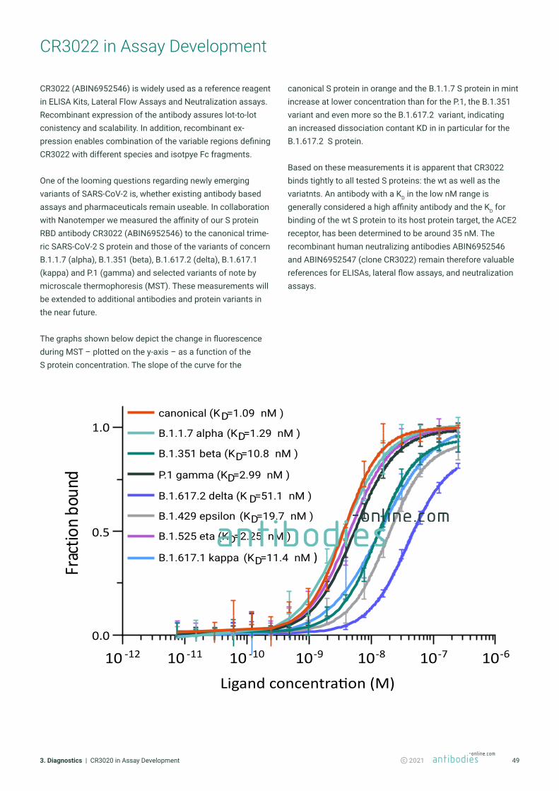
493. Diagnostics | CR3020 in Assay Development 2021
CR3022 in Assay Development
CR3022 (ABIN6952546) is widely used as a reference reagent in ELISA Kits, Lateral Flow Assays and Neutralization assays. Recombinant expression of the antibody assures lot-to-lot conistency and scalability. In addition, recombinant ex-pression enables combination of the variable regions defining CR3022 with different species and isotpye Fc fragments.
One of the looming questions regarding newly emerging variants of SARS-CoV-2 is, whether existing antibody based assays and pharmaceuticals remain useable. In collaboration with Nanotemper we measured the affinity of our S protein RBD antibody CR3022 (ABIN6952546) to the canonical trime-ric SARS-CoV-2 S protein and those of the variants of concern B.1.1.7 (alpha), B.1.351 (beta), B.1.617.2 (delta), B.1.617.1 (kappa) and P.1 (gamma) and selected variants of note by microscale thermophoresis (MST). These measurements will be extended to additional antibodies and protein variants in the near future.
The graphs shown below depict the change in fluorescence during MST – plotted on the y-axis – as a function of the S protein concentration. The slope of the curve for the
B.1.429 epsilon (KD=19.7 nM )
B.1.617.2 delta (K D=51.1 nM )
canonical (KD=1.09 nM )
B.1.1.7 alpha (KD=1.29 nM )
B.1.351 beta (KD=10.8 nM )
P.1 gamma (KD=2.99 nM )
B.1.525 eta (KD=2.25 nM )
B.1.617.1 kappa (KD=11.4 nM )
10 -12 10 -11 10 -10 10-9 10-8 10-7 10-6
0.0
0.5
1.0
Ligand concentra�on (M)
Frac
tionb
ound
canonical S protein in orange and the B.1.1.7 S protein in mint increase at lower concentration than for the P.1, the B.1.351 variant and even more so the B.1.617.2 variant, indicating an increased dissociation contant KD in in particular for the B.1.617.2 S protein.
Based on these measurements it is apparent that CR3022 binds tightly to all tested S proteins: the wt as well as the variatnts. An antibody with a KD in the low nM range is generally considered a high affinity antibody and the KD for binding of the wt S protein to its host protein target, the ACE2 receptor, has been determined to be around 35 nM. The recombinant human neutralizing antibodies ABIN6952546 and ABIN6952547 (clone CR3022) remain therefore valuable references for ELISAs, lateral flow assays, and neutralization assays.

503. Diagnostics | CR3020 in Assay Development 2021
SARS-CoV-2 Spike S1 antibody (RBD)
SARS-CoV-2 Spike S1 antibody (RBD)
• Recombinant SARS-CoV-2 Antibody. • Synergizes with other hNAbs: binds a highly conserved epitope,
not interefering with the S-protein‘s ACE2 RBD.• Reference in S-protein ELISAs and neutralization assays.• In Stock, Fast Delivery.
• Chimeric Rabbit CR3022 Antibody. • Epitope does not overlap with ACE2-Binding Site.• Reference in S-protein ELISAs and neutralization assays.• In Stock, Fast Delivery.
Recommended
Click here to see this product online
Click here to see this product online

513. Diagnostics | SARS-CoV-2 ELISA Kits 2021
SARS-CoV-2 / COVID-19 ELISA Kits
We offer you a handpicked selection of proven quantitative SARS-CoV-2 ELISA Kits for the detection of COVID-19 immu-noglobulins such as IgG and IgM in human serum or plasma. Quantitative measurement of immunoglobulins helps to understand kinectics of antibody response and the longevity of N, RBD and S1 antibodies. Furthermore it enables to draw conclusions about antibody level required for protection from reinfection and its duration. All kit advantages at one glance:
• Low Sample Volume (5µl)• Short Incubation Time (100 min)• Tested with NIBSC Panel Sera 20/B770
Our SARS-CoV-2 sandwich ELISA immunoglobulin kits have been extensively tested against anti-SARS-CoV-2 verification panel for serology assays 20/B770 from the National Institute for Biological Standards and Control (NIBSC). The panel contains 23 Anti-SARS-CoV-2 positive samples of varying reactivity and antibody composition & 14 Anti-SARS-CoV-2
negative samples provided by the WHO (example above). For more information please contact our customer service.
Click here to see our full portfolio of SARS-CoV-2 / COVID-19 ELISA Kits
References
• Zhao et al: „Antibody responses to SARS-CoV-2 in patients of novel coronavirus disease.“ Pre-print medRxiv (2020)
• Ahn et al: „Use of Convalescent Plasma Therapy in Two COVID-19 Patients with Acute Respiratory Distress Syndrome in Korea.“ in: Journal of Korean medical science, Vol. 35, Issue 14, pp. e149, (2020)
• Krüttgen et al „Comparison of four new commercial serologic assays for determination of SARS-CoV-2 IgG.“ in: Journal of clinical virology : the official publication of the Pan American Society for Clinical Virology, Vol. 128, pp. 104394, (2020)
• Bundschuh et al: „Evaluation of the EDI enzyme linked immunosorbent assays for the detection of SARS-CoV-2 IgM and IgG antibodies in human plasma.“ in: Clinica chimica acta; international journal of clinical chemistry, Vol. 509, pp. 79-82, (2020)
• Egger et al: „Comparison of the Elecsys® Anti-SARS-CoV-2 immunoassay with the EDI™ enzyme linked immunosorbent assays for the detection of SARS-CoV-2 antibodies in human plasma.“ in: Clinica chimica acta; international journal of clinical chemistry, Vol. 509, pp. 18-21, (2020)
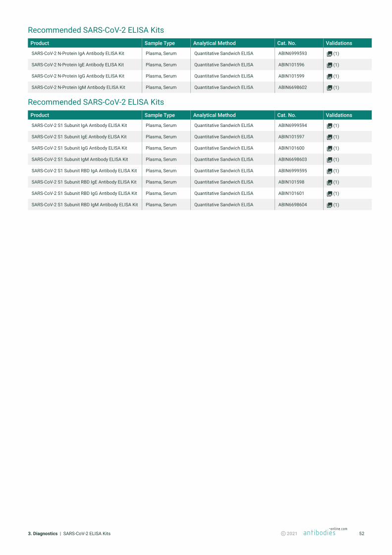
523. Diagnostics | SARS-CoV-2 ELISA Kits 2021
Recommended SARS-CoV-2 ELISA KitsProduct Sample Type Analytical Method Cat. No. Validations
SARS-CoV-2 N-Protein IgA Antibody ELISA Kit Plasma, Serum Quantitative Sandwich ELISA ABIN6999593 (1)
SARS-CoV-2 N-Protein IgE Antibody ELISA Kit Plasma, Serum Quantitative Sandwich ELISA ABIN101596 (1)
SARS-CoV-2 N-Protein IgG Antibody ELISA Kit Plasma, Serum Quantitative Sandwich ELISA ABIN101599 (1)
SARS-CoV-2 N-Protein IgM Antibody ELISA Kit Plasma, Serum Quantitative Sandwich ELISA ABIN6698602 (1)
Recommended SARS-CoV-2 ELISA KitsProduct Sample Type Analytical Method Cat. No. Validations
SARS-CoV-2 S1 Subunit IgA Antibody ELISA Kit Plasma, Serum Quantitative Sandwich ELISA ABIN6999594 (1)
SARS-CoV-2 S1 Subunit IgE Antibody ELISA Kit Plasma, Serum Quantitative Sandwich ELISA ABIN101597 (1)
SARS-CoV-2 S1 Subunit IgG Antibody ELISA Kit Plasma, Serum Quantitative Sandwich ELISA ABIN101600 (1)
SARS-CoV-2 S1 Subunit IgM Antibody ELISA Kit Plasma, Serum Quantitative Sandwich ELISA ABIN6698603 (1)
SARS-CoV-2 S1 Subunit RBD IgA Antibody ELISA Kit Plasma, Serum Quantitative Sandwich ELISA ABIN6999595 (1)
SARS-CoV-2 S1 Subunit RBD IgE Antibody ELISA Kit Plasma, Serum Quantitative Sandwich ELISA ABIN101598 (1)
SARS-CoV-2 S1 Subunit RBD IgG Antibody ELISA Kit Plasma, Serum Quantitative Sandwich ELISA ABIN101601 (1)
SARS-CoV-2 S1 Subunit RBD IgM Antibody ELISA Kit Plasma, Serum Quantitative Sandwich ELISA ABIN6698604 (1)

533. Diagnostics | Anti-Human IgG & IgM Antibodies for in Vitro Diagnostics 2021
Anti-Human IgG & IgM Antibodies for in Vitro Diagnostics
The SARS‐CoV‐2 ongoing development of serological tests is massive. Various techniques are available, enzyme‐linked immunosorbent assay (ELISA), chemiluminescence enzyme immunoassays, fluorescence immunoassays, lateral flow immunoassays to detect immunoglobulin G (IgG), immuno-globulin A (IgA), immunoglobulin M (IgM) (separately or in combination) as well as different antibody targets. Validation of anti-Human Ig Antibodies used in the assays is crucial before use in clinical routine practice.
IgM is the first immunoglobulin to be produced in response to an antigen and is primarily detected during the early onset of disease. Broad patient studies showed positive rates of SARS‐CoV‐2 IgM peaking within 15–21 days following by a slow decline. The positive rates of IgG were increased with the disease course and reached the peak between 22 and 39 days. IgG tests perform better compared with IgA or IgM ones and show better sensitivity when the samples were taken minimum 2 weeks after the RT‐qPCR positive detection.
Separately, IgG detection assays perform better compared to IgA and IgM assays and show better sensitivity when the samples were taken minimum 2 weeks after the RT‐qPCR positive detection. A combined IgG/IgA/IgM test seems to be a better choice in terms of sensitivity than measuring either antibody alone, additionally the period of reliable detection is extended. Another approach to increase sensitivity of the test is a combination of different antigens improves diagnostic performances.
antibodies-online offers anti-human IgG and IgM antibodies for the reliable detection in in vitro diagnostic. We can ensure supply chain stability for the selection of antibodies below.
Click here to see our full portfolio of secondary antibodies
References
• Krüttgen et al: „Comparison of four new commercial serologic assays for determination of SARS-CoV-2 IgG“, Journal of clinical virology: the official publication of the Pan American Society for Clinical Virology (2020).
• Lee et al: „Dynamics of anti-SARS-Cov-2 IgM and IgG antibodies among COVID-19 patients“, J Infect (2020).
• Li et al: „Dynamic changes in anti-SARS-CoV-2 antibodies during SARS-CoV-2 infection and recovery from COVID-19“, Nat Commun (2020).
• Tré-Hardy et al.: „Analytical and clinical validation of an ELISA for specific SARS‐CoV‐2 IgG, IgA, and IgM antibodies“, J Med Virol (2020).
• Wu et al.: „Clinical significance of the serum IgM and IgG to SARS‐CoV‐2 in coronavirus disease‐2019“, J Clin Lab Anal (2021).
• Zhao et al.: „Antibody responses to SARS-CoV-2 in patients of novel coronavirus disease“ (2020).

543. Diagnostics | Anti-Human IgG & IgM Antibodies for in Vitro Diagnostics 2021
Recommended Anti-Human IgG & IgM AntibodiesProduct Conjugate Application Cat. No. Validations
Goat anti-Human IgA, IgG, IgM (Heavy & Light Chain) Antibody (Alkaline Phosphatase (AP)) - Preadsorbed AP ELISA, IHC, WB ABIN100789 (1)
Goat anti-Human IgG (Heavy Chain) Antibody (Alkaline Phosphatase (AP)) - Preadsorbed AP ELISA, IHC, WB ABIN101591 (1)
Goat anti-Human IgG (Fc Region) Antibody (Biotin) - Preadsorbed Biotin ELISA, IHC, WB ABIN101660 (1)
Goat anti-Human IgG Antibody (DyLight 549) - Preadsorbed DyLight 549 FLISA, FM, WB ABIN6698923 (1)
Goat anti-Human IgG (Heavy & Light Chain) Antibody (HRP) HRP IA ABIN101662
Goat anti-Human IgG (Heavy & Light Chain) Antibody (HRP) HRP DB, IEM, IHC, ELISA, WB ABIN1043965 (1)
(1)
Goat anti-Human IgM (Chain mu) Antibody (HRP) HRP ELISA, WB ABIN102628 (3)
Goat anti-Human IgG (F(ab‘)2 Region) Antibody - Preadsorbed ELISA, IHC, WB ABIN101607
Goat anti-Human IgG (Heavy & Light Chain) Antibody - Preadsorbed ELISA, IHC, WB ABIN965345 (1)
Rabbit anti-Human IgG (Fc Region) Antibody - Preadsorbed ELISA, IHC, WB ABIN101666 (1)

antibodies-online Inc.PO Box 5201Limerick, PA 19468USA
WebsiteEmailPhoneFax
[email protected]+1 877 302 8632+1 888 205 9894 (Toll-free)
[email protected]+49 (0)241 95 163 153+49 (0)241 95 163 155
antibodies-online GmbHSchloss-Rahe-Str. 1552072 AachenDeutschland
WebsiteEmailPhone Fax
Stay up-to-date
Visit our SARS-CoV-2 Mutations TrackerFind the latest product updates for lineages of concern
Contact our customer support Talk to our PhD experts and discuss how we can support your project