Embed Size (px)
Citation preview

9 – ORIGINAL ARTICLEWound Healing
Effect of a combination of medium chain triglycerides, linoleic acid, soy lecithin andvitamins A and E on wound healing in rats1
Efeito da combinação de triglicerídeos de cadeia média, ácido linoléico, lecitina de soja evitaminas A e E na cicatrização de ferida em ratos
Maria Sonia Felício MagalhãesI, Francisco Vagnaldo FechineII, Rafael Nogueira de MacedoIII, Diego Levi Silveira MonteiroIII,Cecília Carvalho OliveiraIV, Gerly Anne de Castro BritoV, Maria Elisabate Amaral de MoraesVI, Manoel Odorico de MoraesVI
I Fellow PhD degree, Department of Surgery, Federal University of Ceará (UFC), Brazil.II PhD, Department of Physiology and Pharmacology, UFC, Brazil.III Graduate students, School of Medicine, UFC, Brazil.IV Fellow Master degree, Pharmacology, UFC, Brazil.V PhD, Associate Professor, Department of Morfology and Histology, UFC, Brazil.VI PhD, Associate Professor, Department of Physiology and Pharmacology, UFC, Brazil.
ABSTRACTPurpose: The aim of the study was to determine the effect of a combination of medium chain triglycerides (caprylic, capric,caproic and lauric acids), linoleic acid (essential fatty acid), vitamins A and E and soy lecithin, through a morphometric study, onthe wound healing kinetics of experimental cutaneous ulcers. Methods: A total of 45 male Wistar rats were used, in which a skinflap of total thickness with an area of 4 cm2 was removed. The animals were divided randomly into 3 groups of 15 rats each,Control, Reference and Test groups, which were treated topically with 0.9% NaCl, a preparation of clostebol combined withneomycin sulfate and the test formulation, respectively. The wound areas were measured by digital planimetry at days zero, 3, 7and 12 postoperative. Based on the wound area, we determined the degree of tissue repair and mean rate of repair at different timeintervals. Results: At day 3, an expansion of the wound area was observed in the Reference group and slight contraction in theControl and Test groups. On the subsequent days, the healing process, according to the degree of repair, proceeded in a linearmanner, such that, at day 12, the healed area reached 77.95% of the initial ulcerated region in the Control group, 78.40% in theReference group and 83.49% in the Test group, showing no significant differences. The overall mean rate of repair was equallysimilar at 12 days of treatment: 25.79 mm2/dia in the Control group, 25.42 mm2/dia in the Reference group and 27.38 mm2/dia inthe Test group. Conclusion: The test preparation, applied topically on the experimentally induced cutaneous ulcers in rats, did notaccelerate the process of tissue repair by secondary union.Key words: Triglycerides. Linoleic Acid. Wound Healing. Rats.
RESUMOObjetivo: Avaliar o efeito da associação de triglicerídeos de cadeia média (ácidos caprílico, cáprico, capróico e láurico), ácidolinoléico (ácido graxo essencial), vitaminas A e E e lecitina de soja, através de estudo morfométrico, na cinética de reparação deúlceras cutâneas experimentais. Métodos: Utilizaram-se 45 ratos, machos, da linhagem Wistar, nos quais foi removido um retalhocutâneo de espessura total com 4 cm2 de área. Os animais foram divididos aleatoriamente em 3 grupos constituídos de 15 ratos,Controle, Referência e Teste, que foram tratados por via tópica respectivamente, com solução salina 0,9%, composto de clostebolassociado a sulfato de neomicina e a formulação em teste. As áreas das feridas foram mensuradas por planimetria digital nos diaszero, 3, 7 e 12 de pós-operatório. A partir da área da ferida, calcularam-se ainda o grau de reparação e a taxa média de reparaçãoem intervalo de tempo. Resultados: No 3o dia observou-se uma expansão da área da ferida no grupo referência e uma levecontração nos grupos controle e teste. Nos dias subseqüentes o processo de reparação, medido pela variável grau de reparação,evoluiu de forma linear, de modo que, no 12o dia, a área reparada alcançou 77,95% da região ulcerada inicial no grupo Controle,78,40% no grupo Referência e 83,49% no grupo Teste, não sendo constatadas diferenças estatisticamente significante. Igualmentesemelhantes foram os valores da taxa média de reparação referente aos 12 dias de tratamento: 25,79 mm2/dia no grupo Controle,25,42 mm2/dia no grupo Referência e 27,38 mm2/dia no grupo Teste Conclusão: O composto em Teste, aplicado por via tópicaem úlceras cutâneas experimentalmente induzidas em ratos, não acelerou o processo de reparação recidual por segunda intenção.Descritores: Triglicerídeos. Ácido Linoléico. Cicatrização de Feridas. Ratos.1. Research perfomed at Experimental Surgical Research Laboratory (LABCEX), Department of Surgery, Post-Graduation Program, FederalUniversity of Ceará (UFC), Brazil.
262 - Acta Cirúrgica Brasileira - Vol. 23 (3) 2008

Effect of a combination of medium chain triglycerides, linoleic acid, soy lecithin and vitamins A and E on wound healing in rats
Introduction
The phenomenon of wound healing permits surgicalinterventions to be performed in animals. In antiquity, humansobserved that wounds sustained from injuries closed themselvesafter a period of time. Hippocrates conducted studies on woundsand noted that tissues have the power of natural wound heal-ing. Some studies were conducted on wound healing, but basedon empirical observations in battle injuries. In the beginning ofthe XXth century, studies on laboratory animals were begun,comparing wounds in humans. From then on, experimentationsintensified and permitted a greater understanding of this sub-ject1.
The wound healing process consists of the perfect andcoordinated cascade of cellular and molecular events that inter-act to bring about the repair and reconstitution of the tissue2.This is a dynamic process that involves biochemical and physi-ological phenomena occurring in a harmonious manner to guar-antee tissue restoration.
The wound can be defined as any alteration in the ana-tomic integrity of the skin, resulting from any type of trauma,where it can even be classified as intentional (surgical incisions)or accidental3. Wound healing by secondary union are submit-ted to the influence of various factors that can contribute to thedelay of wound healing, resulting in the majority of cases ininflammation, edema and hypertrophic and unesthetic scars. Allwounds, regardless of their etiology, are a disruption of conti-nuity in the tissue, which results in the interruption of bloodflow, in the perturbation of sensitivity, in the accumulation ofdead cell debris and in a variable degree of contamination (withor without infection). With the aim of restoring the integrity ofthe skin, the organism utilizes a complex mechanism calledwound healing4.
The process of wound healing begins immediately af-ter the lesion and comprises the inflammatory, proliferative andmaturation phases. The inflammatory phase is characterized bythe recruitment of leukocytes, such as neutrophils and macroph-ages, to the lesion site. In the proliferative phase, the migrationand proliferation of keratinocytes, fibroblasts and endothelialcells result in re-epithelization and formation of granulation tis-sue. Finally, in the maturation phase, the excess of collagen isdegraded various proteolytic enzymes promoting complete tis-sue repair5.
The substance tested here is a oily formulation mar-keted as a hydrating oil and dermoprotector for topical use, withbasic composition consisting of medium chain triglycerides (ca-prylic, capric, caproic and lauric acids) and linoleic acid (es-sential fatty acid), vitamins A and E and soy lecithin.
MCT (medium chain triglycerides) contain in theirstructure predominantly fatty acids with eight carbons (caprylic),ten carbons (capric), six carbons (caproic) and twelve carbons(lauric). The triacylglicerols of capric and caprylic acids de-serve special attention as esters. In being classified as MCT,they are useful as a nutritional source, solvent, and vehicle andstabilizer of products to be administered orally, topically orparenterally. They can have uses in the treatment and preven-tion of ammoniacal dermatitis and bedsores, by forming a pro-tective barrier for the skin, impeding maceration, besides being
of importance in processes of cellular inflammation, providingalleviaton after first application, and local cell nutrition, in ad-dition to having a great capacity of tissue regeneration. Me-dium chain fatty acids, such as monoacylglycerols, have beenshown to have antimicrobial activity, reducing the formation ofdental caries in a study conducted in laboratory animals6.
Fatty acids belong to a class of compounds formed bya long hydrocarbon chain and a terminal carboxylic group. Theyhave three main functions: they are structural components ofbiological membranes, they act as precursors of intracellularmessengers and they are oxidized producing ATP (adenosinetriphosphate)7.
From the beginning of the 1970s, there have been stud-ies on the effects of fatty acids on the immune response. Suchcompounds interfere with various events of the inflammatoryprocess, such as vascular contraction, chemotaxis, adhesion,diapedesis, activation and cell death, where the majority of theseoccur via arachidonic acids such as prostaglandins, leukotrienes,tromboxanes and lipoxins7.
PUFAs (polyunsaturated fatty acids) should be pointedout among the various fatty acids present in the plasma and inleukocytes. Besides their structural function, they can modu-late cell-cell interactions and intracellular signaling. Thus, thealteration of the composition of fatty acids of phospholipids inthe cell membrana can modify fluidity and change the bindingof cytokines to their receptors8.
Vitamin A interferes with wound healing causing lysisof lysosomal membranes stimulation fibroblasts and collagendeposition9. The biological activities of vitamin A have notbeen completely elucidated, but it appears to act as an anti-inflammatory agent and its antioxidant properties suggest a pro-tective function for growing cells against oxygen radicals re-leased by leukocytes10.
Studies have shown that besides essential fatty acids,soy lecithin and vitamins A and E also contribute to the processof tissue repair. Soy lecithin, besides being a protective agent,provides maintenance of tissue hydration and helps in the pro-cess of wound healing of the skin9.
Linoleic acid is an essential fatty acid of 18 carbons.Through a desaturation process, it gives rise to arachidonic acid(20 carbons), a precursor of prostaglandins, leukotrienes, throm-boxanes and lipoxins, which in turn act as mediators of plateletfunction and of inflammatory, vascular, motor and sensory pro-cesses, among others2,7.
It has been observed that linoleic acid is capable ofinhibiting the growth of Staphylococcus aureus by altering itsprotein synthesis, cell wall, nucleic acids and cell membraneduring division11.
Linoleic acid has also been shown to participate in cellproliferation and inflammatory process, where in the latter itplays a role as a mediator of leukocyte function having chemo-tactic and stimulatory effects on neutrophils12.
Since antiquity, humans developed the capacity to per-form relatively reliable measurements. Primitive human popu-lations performed astronomical measurements for religious andagricultural purposes, for the construction of religious temples,palaces and houses. During the Industrial Revolution, there wasa standarization of weights and measures with the introduction
Acta Cirúrgica Brasileira - Vol. 23 (3) 2008 - 263
of the decimal metric system in France.The word morphometry is formed from the Greek pre-
fix morphe which means shape, combined with the Greek suf-fix metrikos, or in Latin metricu, which means the act of mea-suring or the process of establishing dimensions13.
This term has a wide application in science, but in bio-medicine its meaning is measurement of anatomic structures. Itis a method for the purpose of giving more objectivity and pre-cision to the collection, presentation and analysis of results inresearch and routine laboratory work, thereby permitting ana-tomical structures to be compared like functions. Some mor-phometric measurements are areas (surfaces), weights, volumes,lengths, angles, diameters and perimenters. These are measure-ments that can be determined in microscopic, mesoscopic andmacroscopic structures13,14.
The measurement of areas can be performed with ac-curacy utilizing different planimetric methods, among which isplanimetry by linear integration and by counting points. Suchtechniques are applicable only with flat images14.
The advent of computational systems for the process-ing of digital images made it possible to carry out planimetricmeasurements that are more objective, rapid and accurate13.
The aim of this work was to determine the effect ofthe combination of medium chain triglycerides (caprylic, ca-pric, caproic and lauric acids), linoleic acid (essential fatty acid),vitamins A and E and soy lecithin, on the tissue repair kineticsof experimental cutaneous ulcers, by means of a morphometricstudy.
Methods
The study was conducted in the Laboratory of Ex-perimental Surgery in the Department of Surgery of the FederalUniversity of Ceara (UFC) following the protocol No 31/05,approved by the Committee on Ethics in Animal Research(CEPA) of the same instituition, which was in accordance withthe Ethical Principles in Animal Experimentation adopted bythe Brazilian College of Animal Experimentation (COBEA).
A total of 45 male Wistar rats (Rattus norvegicus) wereused, with a mean body weight of 149.69 g varying from 106 to171g, obtained from the Central Animal Facility of UFC. Theanimals were previously examined with respect to general healthconditions, and received an appropriate balanced ration for thespecies and water ad libitum. The animals were housed in indi-vidual cages without sawdust, so that it would not stick to thewounds; a paper bottom was provided which was changed daily.They were kept at room temperature with controled humidityand a 12-h photoperiod.
The surgical wound model utilized involved the re-moval of a skin flap of full thickness on the back of the animal.The rats were initially anesthesized with an intraperitoneal in-jection of ketamine hydrochoride (90 mg/kg) and xylazine hy-drochloride (10 mg/kg), and the dorsal was then shaved. After-ward, a 2x2 cm (4 cm2) area was outlined with a template madeof cellulose film, on the dorsal medial line, caudally correspond-ing to the forelimbs. Incisions were made on the skin and thesquare skin flap was removed.
The rats were divided randomly into 3 groups consist-ing of 15 animals each: Control, Reference and Test. From thefirst day postoperative up to the 12th day of treatment, the woundswere cleaned daily with isotonic saline (0.9% NaCl) in the threegroups. In the Reference group, the wound was treated with adaily topical application of a fine layer (0.5232 g) of a creamcomposed of clostebol acetate and neomycin sulfate. In the Testgroup, the wound was treated once a day with a topical applica-tion of 0.1435 g of test solution containing medium chain trig-lycerides, linoleic acid, vitamins A and E and soy lecithin.
Digital images of the ulcers were taken, in vivo, in astandardized manner, immediately after the surgical procedure(day zero) and on days 3, 7 and 12 postoperative. An analogvideo camera (Hitachi VCC-151, Japan) was used coupled to asurgical microscope (D.F. Vasconcellos M90, D.F. VasconcellosS.A., São Paulo, Brazil), whose signal was transferred to a videocapture plate (PixelView PV-TV304P, Prolink MicrosystemsCorp., Taiwan) installed in a microcomputer. The axis of themicroscope was fixed perpendicular to the horizontal plane,keeping the objective at a distance of 33 cm from this plane.Such distance provided optimal focusing of the ulcer, with amagnification of 6X, when the animal was in the ventral decu-bitus position, on the horizontal plane, with the center of theulcer coinciding with the center of the microscope field. Thedigitized images were stored in Windows Bitmap (BMP) for-mat, with the dimensions of 320x240 pixels, each pixel corre-sponding to 24 bits, based on the red, green, blue (RGB) colormodel.
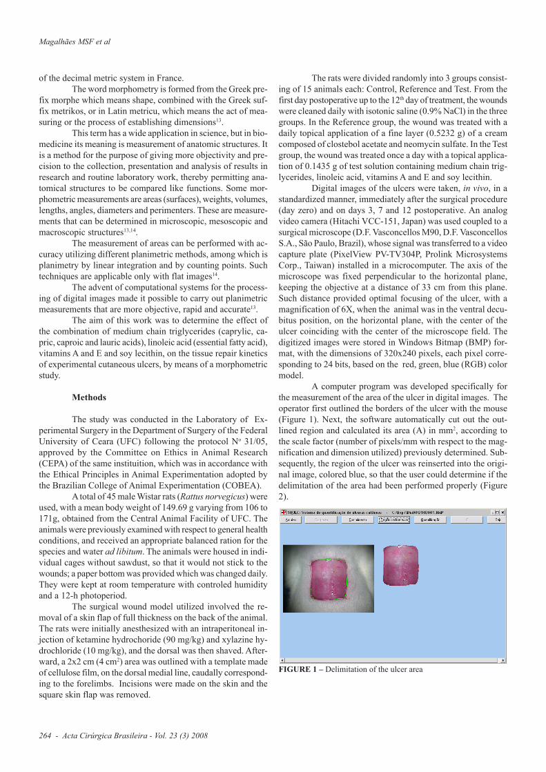
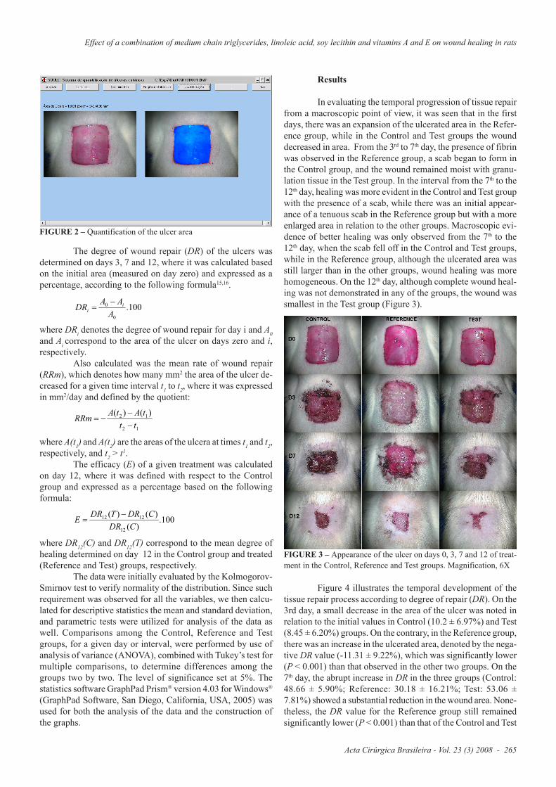
A computer program was developed specifically forthe measurement of the area of the ulcer in digital images. Theoperator first outlined the borders of the ulcer with the mouse(Figure 1). Next, the software automatically cut out the out-lined region and calculated its area (A) in mm2, according tothe scale factor (number of pixels/mm with respect to the mag-nification and dimension utilized) previously determined. Sub-sequently, the region of the ulcer was reinserted into the origi-nal image, colored blue, so that the user could determine if thedelimitation of the area had been performed properly (Figure2).
FIGURE 1 – Delimitation of the ulcer area
264 - Acta Cirúrgica Brasileira - Vol. 23 (3) 2008
Magalhães MSF et al
Effect of a combination of medium chain triglycerides, linoleic acid, soy lecithin and vitamins A and E on wound healing in rats
FIGURE 2 – Quantification of the ulcer area
Results
In evaluating the temporal progression of tissue repairfrom a macroscopic point of view, it was seen that in the firstdays, there was an expansion of the ulcerated area in the Refer-ence group, while in the Control and Test groups the wounddecreased in area. From the 3rd to 7th day, the presence of fibrinwas observed in the Reference group, a scab began to form inthe Control group, and the wound remained moist with granu-lation tissue in the Test group. In the interval from the 7th to the12th day, healing was more evident in the Control and Test groupwith the presence of a scab, while there was an initial appear-ance of a tenuous scab in the Reference group but with a moreenlarged area in relation to the other groups. Macroscopic evi-dence of better healing was only observed from the 7th to the12th day, when the scab fell off in the Control and Test groups,while in the Reference group, although the ulcerated area wasstill larger than in the other groups, wound healing was morehomogeneous. On the 12th day, although complete wound heal-ing was not demonstrated in any of the groups, the wound wassmallest in the Test group (Figure 3).
The degree of wound repair (DR) of the ulcers wasdetermined on days 3, 7 and 12, where it was calculated basedon the initial area (measured on day zero) and expressed as apercentage, according to the following formula15,16.
where DRi denotes the degree of wound repair for day i and A0and Ai correspond to the area of the ulcer on days zero and i,respectively.
Also calculated was the mean rate of wound repair(RRm), which denotes how many mm2 the area of the ulcer de-creased for a given time interval t1 to t2, where it was expressedin mm2/day and defined by the quotient:
where A(t1) and A(t2) are the areas of the ulcera at times t1 and t2,respectively, and t2 > t1.
The efficacy (E) of a given treatment was calculatedon day 12, where it was defined with respect to the Controlgroup and expressed as a percentage based on the followingformula:
where DR12(C) and DR12(T) correspond to the mean degree ofhealing determined on day 12 in the Control group and treated(Reference and Test) groups, respectively.
The data were initially evaluated by the Kolmogorov-Smirnov test to verify normality of the distribution. Since suchrequirement was observed for all the variables, we then calcu-lated for descriptive statistics the mean and standard deviation,and parametric tests were utilized for analysis of the data aswell. Comparisons among the Control, Reference and Testgroups, for a given day or interval, were performed by use ofanalysis of variance (ANOVA), combined with Tukey’s test formultiple comparisons, to determine differences among thegroups two by two. The level of significance set at 5%. Thestatistics software GraphPad Prism® version 4.03 for Windows®
(GraphPad Software, San Diego, California, USA, 2005) wasused for both the analysis of the data and the construction ofthe graphs.
100.0
0
A
AADR i
i
12
12 )()(
tt
tAtARRm
100.)(
)()(
12
1212
CDR
CDRTDRE
FIGURE 3 – Appearance of the ulcer on days 0, 3, 7 and 12 of treat-ment in the Control, Reference and Test groups. Magnification, 6X
Figure 4 illustrates the temporal development of thetissue repair process according to degree of repair (DR). On the3rd day, a small decrease in the area of the ulcer was noted inrelation to the initial values in Control (10.2 ± 6.97%) and Test(8.45 ± 6.20%) groups. On the contrary, in the Reference group,there was an increase in the ulcerated area, denoted by the nega-tive DR value (-11.31 ± 9.22%), which was significantly lower(P < 0.001) than that observed in the other two groups. On the7th day, the abrupt increase in DR in the three groups (Control:48.66 ± 5.90%; Reference: 30.18 ± 16.21%; Test: 53.06 ±7.81%) showed a substantial reduction in the wound area. None-theless, the DR value for the Reference group still remainedsignificantly lower (P < 0.001) than that of the Control and Test
Acta Cirúrgica Brasileira - Vol. 23 (3) 2008 - 265

groups. On the 12th day, however, there were no statisticallysignificant differences among the groups. In fact, a decrease inthe slope of the DR curve was seen in Control (77.95 ± 10.71%)and Test (83.49 ± 3.50%) groups, indicating that in relation today 7, the area of the ulcer regressed at a slower rate. On theother hand, in the Reference group, the tissue repair processproceded at the same intensity (78.40 ± 11.57%), as seen by thelinear behavior of DR in the period between the 3rd and 12th day.
FIGURE 5 – Mean rate of wound repair (RRm) for the intervals be-tween days 0 and 3, 3 and 7, and 7 and 12. Data expressed as mean ofmeasurements for 15 animals of each group. Analysis of variance wasused to compare the three groups, followed by Tukey’s test to deter-mine differences between all pairs of groups. *** P < 0.001: Refer-ence lower than Control and Test; +++ P < 0.001. Reference higherthan Control and Test (Tukey’s test)
FIGURE 4 – Temporal development of the tissue repair process in theControl, Reference and Test groups, based on the degree of tissue re-pair (DR). Data expressed as means and standard deviation of mea-surements for 15 animals of each group. Analysis of variance was usedto compare the three groups, followed by Tukey’s test to determinedifferences between all pairs of groups. No statistically significantdifferences were found. *** P < 0.001. Reference lower than Controland Test (Tukey’s test)
0 1 2 3 4 5 6 7 8 9 10 11 12-30
-15
0
15
30
45
60
75
90Control
Test
Reference
***
***
Time (days)
Deg
ree o
f re
pair
(%
)
The kinetics of tissue repair can be better understoodby the analysis of the variable mean rate of wound repair (RRm)which represents the mean velocity of wound closing (Figure5). In the period between days 0 and 3, as a result of the expan-sion of the ulcerated area, RRm for the Reference group wasnegative (-14.77 ± 12.59 mm2/day) and significantly lower (P <0.001) than that of Control (13.72 ± 9.57 mm2/day) and Test(11.33 ± 8.29 mm2/day) groups. In the interval between days 3and 7, however, RRm of the Reference group increased mark-edly (40.61 ± 17.52 mm2/day), reaching values similar to thatobserved in the Control (38.10 ± 8.68 mm2/day) and Test (43.86± 6.89 mm2/day), such that no statistically significant differ-ences were found. In this interval, in the three groups, RRmreached their maximal values, characterizing the phase of great-est intensity of tissue repair. In the period between day 7 and12, on the other hand, there was a deceleration in the repair ofthe ulcer in the Control (23.18 ± 8.04 mm2/day) and Test (23.83± 4.38 mm2/day) groups, denoted by the diminution in the RRmvalues. On the contrary, in the Reference group, RRm remainedpractically unchanged (37.37 ± 6.15 mm2/day), such that it wassignificantly greater than the of the other groups (P < 0.001).
Although RRm by intervals revealed differences amongthe groups, RRm overall, that is, that refering to the period be-tween day 0 and 12 (Figure 6), was similar in the three groups:Control (25.79 ± 3.78 mm2/day), Reference (25.42 ± 4.76 mm2/day), Test (27.38 ± 1.95 mm2/day). Therefore, there were nostatistically significant differences found (Figure 6).
FIGURE 6 – Mean rate of wound repair (RRm) for the interval be-tween days 0 and 12. Data expressed as mean and standard deviationof the measurements from 15 animals of each group. Analysis of vari-ance was used to compare the three groups, followed by Tukey’s testto determine differences between all pairs of groups. No statisticallysignificant differences were found
0 1 2 3 4 5 6 7 8 9 10 11 12-20
-10
0
10
20
30
40
50
Control TestReference
***
+++
Time (days)
Mean
rate
of
rep
air
(m
m2/d
ay)
Control Reference Test0
10
20
30
40
Mean
rate
of
rep
air
(mm
2/d
ay)
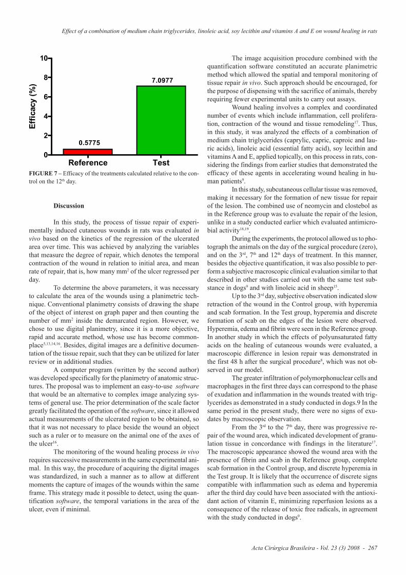
The efficacy of the treatments was determined rela-tive to the control, based on the mean DR for day 12 (Figure 7).In the Reference group, efficacy was 0.5775%, while in theTest group it was 7.0977%. Such percentages indicated that thetreatments had effects similar to that of the control, albeit aslightly better effect with test treatment.
266 - Acta Cirúrgica Brasileira - Vol. 23 (3) 2008
Magalhães MSF et al
Effect of a combination of medium chain triglycerides, linoleic acid, soy lecithin and vitamins A and E on wound healing in rats
FIGURE 7 – Efficacy of the treatments calculated relative to the con-trol on the 12th day.
Discussion
In this study, the process of tissue repair of experi-mentally induced cutaneous wounds in rats was evaluated invivo based on the kinetics of the regression of the ulceratedarea over time. This was achieved by analyzing the variablesthat measure the degree of repair, which denotes the temporalcontraction of the wound in relation to initial area, and meanrate of repair, that is, how many mm2 of the ulcer regressed perday.
To determine the above parameters, it was necessaryto calculate the area of the wounds using a planimetric tech-nique. Conventional planimetry consists of drawing the shapeof the object of interest on graph paper and then counting thenumber of mm2 inside the demarcated region. However, wechose to use digital planimetry, since it is a more objective,rapid and accurate method, whose use has become common-place5,13,14,16. Besides, digital images are a definitive documen-tation of the tissue repair, such that they can be utilized for laterreview or in additional studies.
A computer program (written by the second author)was developed specifically for the planimetry of anatomic struc-tures. The proposal was to implement an easy-to-use softwarethat would be an alternative to complex image analyzing sys-tems of general use. The prior determination of the scale factorgreatly facilitated the operation of the software, since it allowedactual measurements of the ulcerated region to be obtained, sothat it was not necessary to place beside the wound an objectsuch as a ruler or to measure on the animal one of the axes ofthe ulcer16.
The monitoring of the wound healing process in vivorequires successive measurements in the same experimental ani-mal. In this way, the procedure of acquiring the digital imageswas standardized, in such a manner as to allow at differentmoments the capture of images of the wounds within the sameframe. This strategy made it possible to detect, using the quan-tification software, the temporal variations in the area of theulcer, even if minimal.
The image acquisition procedure combined with thequantification software constituted an accurate planimetricmethod which allowed the spatial and temporal monitoring oftissue repair in vivo. Such approach should be encouraged, forthe purpose of dispensing with the sacrifice of animals, therebyrequiring fewer experimental units to carry out assays.
Wound healing involves a complex and coordinatednumber of events which include inflammation, cell prolifera-tion, contraction of the wound and tissue remodeling17. Thus,in this study, it was analyzed the effects of a combination ofmedium chain triglycerides (caprylic, capric, caproic and lau-ric acids), linoleic acid (essential fatty acid), soy lecithin andvitamins A and E, applied topically, on this process in rats, con-sidering the findings from earlier studies that demonstrated theefficacy of these agents in accelerating wound healing in hu-man patients9.
In this study, subcutaneous cellular tissue was removed,making it necessary for the formation of new tissue for repairof the lesion. The combined use of neomycin and clostebol asin the Reference group was to evaluate the repair of the lesion,unlike in a study conducted earlier which evaluated antimicro-bial activity18,19.
During the experiments, the protocol allowed us to pho-tograph the animals on the day of the surgical procedure (zero),and on the 3rd, 7th and 12th days of treatment. In this manner,besides the objective quantification, it was also possible to per-form a subjective macroscopic clinical evaluation similar to thatdescribed in other studies carried out with the same test sub-stance in dogs9 and with linoleic acid in sheep15.
Up to the 3rd day, subjective observation indicated slowretraction of the wound in the Control group, with hyperemiaand scab formation. In the Test group, hyperemia and discreteformation of scab on the edges of the lesion were observed.Hyperemia, edema and fibrin were seen in the Reference group.In another study in which the effects of polyunsaturated fattyacids on the healing of cutaneous wounds were evaluated, amacroscopic difference in lesion repair was demonstrated inthe first 48 h after the surgical procedure8, which was not ob-served in our model.
The greater infiltration of polymorphonuclear cells andmacrophages in the first three days can correspond to the phaseof exudation and inflammation in the wounds treated with trig-lycerides as demonstrated in a study conducted in dogs.9 In thesame period in the present study, there were no signs of exu-dates by macroscopic observation.
From the 3rd to the 7th day, there was progressive re-pair of the wound area, which indicated development of granu-lation tissue in concordance with findings in the literature17.The macroscopic appearance showed the wound area with thepresence of fibrin and scab in the Reference group, completescab formation in the Control group, and discrete hyperemia inthe Test group. It is likely that the occurrence of discrete signscompatible with inflammation such as edema and hyperemiaafter the third day could have been associated with the antioxi-dant action of vitamin E, minimizing reperfusion lesions as aconsequence of the release of toxic free radicals, in agreementwith the study conducted in dogs9.
Reference Test0
2
4
6
8
10
0.5775
7.0977
Eff
icacy (
%)
Acta Cirúrgica Brasileira - Vol. 23 (3) 2008 - 267
The action of soy lecithin provides hydration of thetissues, constituting a favorable factor in the repair of the le-sion. Keeping the wound hydrated promotes autolytic debride-ment and contributes as a stimulus of epithelization, formationof granulation tissue and angiogenesis9. Thus, hydration pro-moted by soy lecithin, complemented by normal saline solu-tion, made surgical debridement of the wounds unnecessary inthis study. On the other hand, previous studies demonstratedthat open and dried wounds undergo re-epitelization moreslowly17.
From the 7th to 12th day, a greater rate of repair oc-curred in the Reference group. The test preparation showedevident granulation tissue and greater tissue contraction aroundthe edges of the wound, which had become irregular. In theControl group, gaps in the scab were seen on the 7th day, alongwith irregular edges due to contraction of the wound, besidesthe presence of granulation tissue. The Reference group re-mained with the largest wound area and with irregular edges,which was also found in a study using linoleic acid15. The treat-ments in the three groups contributed to the almost completeclosing of the lesions, suggesting that growth factors were prob-ably responsible for the hyperplasia of the epithelium as re-ported in the literature17. In this period, there was agreementwith a study that considered cytokines as being important me-diators of neoangiogenesis, fibroplasia and maturation, whichare released by cells such as platelets, neutrophils, macroph-ages, lymphocytes, mast cells and fibroblasts, making it easy tounderstand the importance of chemotactic properties of the testpreparation in the repair of the lesions9.
A study that examined wound healing in sheep showedthat linoleic acid constituted a powerful pro-inflammatory me-diator, being essential for the regulation of the biochemicalevents that precede fibroplasia in addition to stimulating growthfactors and neovascularization15. It is possible that in this studythe presence of linoleic acid in the test preparation contributedto a similar event.
A study carried out with polyunsaturated fatty acidsshowed a tendency for the wound area to diminish in the firstten days of treatment, and demonstrated overall that PUFAsmay play an important role in wound healing8.
On the 12th day, clinical observation showed a smallerwound area characterized by the proximity of the edges (con-traction) with irregular outlines and better presence of granula-tion tissue, like that seen in the study with linoleic acid in sheep15.
Proliferation (fibroplasia and matrix formation) wasdemonstrated by scholars in the past as being extremely impor-tant in the formation granulation tissue. This depends on fibro-blasts which produce elastin, fibronectin, glucosaminoglycansand proteases17. In this phase, the presence of vitamin A in thetest preparation was important, because authors evaluating thesame preparation found that it stimulates fibroblasts, the depo-sition of collagen and formation of connective tissue9.
The repair of wounds by secondary union showed thatcontraction could have been responsible for the reduction inwound area in the three groups, in concordance with a study inwhich contraction was shown to be able reduce the surface ofthe cutaneous defect by 62%17.
In the model presented here, triglycerides favored agreater repair from a macroscopic clinical viewpoint, where itcontributed to the wound area being less on the 12th day of treat-ment. However, compared to Control group, the efficacy of thetest preparation was only 7.0977%, while the efficacy of thereference treatment was lower at only 0.5775%.
Conclusion
The test preparation applied topically to the surgicalwound produced in this model did not accelerate significantlythe process of tissue healing by secondary union.
References
1. Carrel A. The treatment of wounds. JAMA. 1910;55:2148-50.2. Ortonne JP, Clévy JP. Physiology of cutaneous cicatrization.Rev Prat. 1994; 44(13):1733-7.3. Araújo CFR, Souza Fo ZA, Greca FH, Guerreiro MHCPM,Leite AL, Mansur AE C, Kantor DC, Nassif AE. Effects ofAgarol® and Trigliceril® on cutaneous wound healing: experi-mental study in rats. Acta Cir Bras. 1998;13(4):232-7.4. Banda MJ, Knighton DR, Hunt TK, Werb Z. Isolation of anonmitogenic angiogenesis factor from wound fluid. Proc NatAcad Sci. 1982;79:7773-7.5. Mori, R.; Kondo, T.; Nishiie, T.; Ohshima, T.; Asano, M.Impairment of skin wound healing in -1,4-galactosyltransferase-deficient mice with reduced leukocyterecruitment. Am J Pathol. 2004;164(4):1303-14.6. D’Agostini D. Obtenção de lipídíos estruturados porintereterificação de triacilglicerois de cadeia média e longa [Tesede Doutorado]. Universidade de São Paulo, Faculdade deCiências Farmacêuticas; 2001.7. Hatanaka E, Curi R. Fatty acids and wound healing: a re-view. RevRv Bras Farmacol. 2007;88(2):53-8.8. Cardoso CRB, Souza MA, Ferro EAV, Favoreto S, Pena JDO.Influence of topical administration of n-3 and n-6 essential andn-9 nonessential fatty acids on the healing of cutaneous wounds.Wound Rep Reg. 2004;12:235-43.9. De Nardi AB, Rodaski S, Sousa RS, Baudi DLK, Castro JHT.Secondary cicatrization in dermoepidermal wounds treated withessential fatty acids, vitamins A and E, soy lecithin andpolynylpyrrolidone-iodine in dogs. Arc Vet Sci. 2004;9(1):1-1610. Ehrlich HP , Hunt TK. Effects of cortisone and vitamin Aon wound healing. Ann Surg. 1968;167:324-8.11. Greenway DLA & Dyke KGH. Mechanism of the inhibi-tory action of linoleic acid on the growth of Staphylococcusaureus. J Gen Microbiol. 1979;115:233-45.12. Moch D, Sheva T, Heihn H, Schimidt D, Buntroc P. Thelinoleic acid metabolite 2 Ds-hidroxi-10, 12 (E-Z): octadecadi-enoic acid is a strong, proinflammatory mediator in a experi-mental would healing model of the rat. Biom Biochem Acta.1990;49:201-7.
268 - Acta Cirúrgica Brasileira - Vol. 23 (3) 2008
Magalhães MSF et al

Effect of a combination of medium chain triglycerides, linoleic acid, soy lecithin and vitamins A and E on wound healing in rats
13. Teixeira VPA, Pereira SAL, Rodrigues DBR, Lino RSJ.Princípios básicos e aplicações da morfometria. Disponível em:h t t p : / / w w w. u f t m . e d u . b r / i n s t p u b / f m t m / p a t g e /morfometria01.htm. Acesso em: 17 jul.2007.14. Mandarin-de-Lacerda, CA. Stereological tools in biomedi-cal reserarch. An Acad Bras Cienc. 2003;75(4):469-86.15. Marques SR, Peixoto CA, Messias JB, Albuqurque AR, SilvaJr VA. The effects of topical application of sunflower-seed oilon open wound healing in lambs. Acta Cir Bras. 2004;19(3):196-209.16.Martins NLP, Malafaia O, Ribas-Filho JM, Heibel M, BaldezRN, Vasconcelos PRL, Moreira H, Mazza M, Nassif PAN,Wallbach TZ. Healing process in cutaneous surgical wounds in
rats under the influence of Orbignya phalerata aqueous extract.Acta Cir Bras. 2006;21(supl 3):66-75.17. Mandelbaum SH, Di Santis EP, Mandelbaum MHSA. Cica-trization: current and auxiliary resources-Part 1. An BrasDermatol. 2003;78(4):393-410.18. Rodrigues KL, Cardoso CC, Caputo LR, Carvalho JCT,Fiorini JE, Schneedorf JM. Cicatrizing and antimicrobial prop-erties of an ozonised oil from sunflower seeds.Inflammopharmacology. 2004;12(3):261-70.19. Sakuma CH, Matera JM, Valente NS. Clinical study of skinflap application during oncologic surgery in dog. Bras J VetRes Anim Sci. 2003;40 Suppl:32-7.
Conflict of interest: noneFinancial source: none
Correspondence:Manoel Odorico de MoraesRua Cel. Nunes de Melo, 112760430-270 Fortaleza – Ceará BrazilPhone: (55 85)[email protected]
Received: November 19, 2007Review: January 23, 2008
Accepted: February 19, 2008
How to cite this articleMagalhães MSF, Fechine FV, Macedo RN, Monteiro DLS, Oliveira CC, Brito GAC, Moraes MEA, Moraes MO. Effect of acombination of medium chain triglycerides, linoleic acid, soy lecithin and vitamins A and E on wound healing in rats. Acta CirBras. [serial on the Internet] 2008 May-June;23(3). Available from URL: http://www.scielo.br/acb
*Color figures available from www.scielo.br/acb
Acta Cirúrgica Brasileira - Vol. 23 (3) 2008 - 269


![i5 Literature [081610DP] · Chicory), Beta Glucans, Oat Fiber) Sunflower oil, Proprietary Immune Blend, Natural Flavors, Medium Chain Triglycerides, Arabinogalactan, Broccoli Seed](https://img.dokumen.tips/doc/110x75/6013a3b270ad005e46206382/i5-literature-081610dp-chicory-beta-glucans-oat-fiber-sunflower-oil-proprietary.jpg)