Embed Size (px)
Citation preview

S. Nishida, M. D. Fortes and N. Miyazaki, eds.Coastal Marine Science in Southeast Asia —Synthesis Report of the Core University Program of the JapanSociety for the Promotion of Science: Coastal Marine Science (2001–2010), pp. 23–48.© by TERRAPUB 2011.
cal characters of the organisms. Each sec-tion was prepared by leader(s) of 4–10 sci-entists who worked on each topic frommember countries, i .e. Pyrodiniumbahamense by Furio and Cayme,Alexandrium and Gymnodinium catenatumby Po Teen, benthic dinoflagellates byOmura and Fukuyo, taxonomy and distri-bution of Pseudo-nitzschia by Po Teen,domoic acid production of Pseudo-nitzschia by Dao, Nitzschia by Kotaki,Cochlodinium by Matsuoka, Heterocapsaby Iwataki, and Noctiluca by Sriwoon,Lirdwitayaprasit, and Furuya. Fukuyo andKodama made an editorial arrangement.
Chapter 3
Ecology and oceanography of harmful marine microalgae(Project-2)
Yasuwo Fukuyo1, Masaaki Kodama1, Takuo Omura1, Ken Furuya2, Elsa F. Furio3,Mirriam Cayme3, Lim Po Teen4, Dao Viet Ha5, Yuichi Kotaki6, Kazumi Matsuoka7,Mitsunori Iwataki8, Rujinard Sriwoon9 and Thaithaworn Lirdwitayaprasit10
1Asian Natural Environmental Science Center, The University of Tokyo, 1-1-1 Yayoi, Bunkyo-ku, Tokyo 113-8657, Japan2Graduate School of Agricultural and Life Sciences, The University of Tokyo, 1-1-1 Yayoi, Bunkyo-ku, Tokyo 113-8657, Japan3National Fisheries Research and Development Institute, Bureau of Fisheries, PCA Bldg. Diliman, Quezon City, The Philippines4Faculty of Resource Science and Technology, Universiti Malaysia Sarawak, 94300 Kota Samarahan, Sarawak, Malaysia5Department of Biochemistry, Institute of Oceanography, 01 Cau Da, NhaTrang, Vietnam6School of Marine Biosciences, Kitasato University, Okirai, Sanriku-machi, Ofunato 022-0101, Japan7Institute for East China Sea Research, Nagasaki University, 1-14 Bunkyo-machi, Nagasaki 852-8521, Japan8Faculty of Science,Yamagata University, 1-4-12 Kojirakawa-machi, Yamagata 990-8560, Japan9Faculty of Science, Prince of Songkla University, Hatyai, Songkhla 90112, Thailand10Department of Marine Science, Faculty of Science, Chulalongkorn University, Phayathai Road, Pathumwan, Bangkok 10330, Thailand
Introduction
In the last decade many HAB scientistsnoticed that HAB phenomena in SoutheastAsian region were changing its nature;occurrence of new HAB species, their in-creased frequency, widen affected geo-graphical area, and prolonged duration ofthe occurrences. Moreover variety of phe-nomena, i.e. new types of toxins and mor-tality of marine organisms, also increased.In this chapter features of the problemscaused by several endemic and newly in-vaded HAB species are described in de-tails, together with biological and chemi-

24 Y. FUKUYO et al.
Toxin Producing Microalgae
PSP toxin producing microalgae
Pyrodinium bahamenseThe toxic dinoflagellate Pyrodinium
bahamense var. compressum (Fig. 1) hascaused adverse socio-economic problemsin the Southeast Asian region for more than3 decades now. This organism producessaxitoxin and other toxin derivatives thatcause Paralytic Shellfish Poisoning (PSP),resulting from human ingestion of shell-fish, commonly the filter-feeding bivalvesthat accumulate toxins as they fed on thisorganism. The organism is therefore re-sponsible for human illnesses and deathsdue to PSP, and repeated closures of har-vesting of both wild and farmed shellfishand small pelagic fish from affected areasand marketing/trading seafood ban to con-suming public.
The occurrence of P. bahamense var.compressum blooms and PSP episodesseemed to have limited geographical dis-tribution in the Southeast Asian regionmainly confined in Brunei Darussalam,west coast of Sabah, Malaysia, Indonesiaand the Philippines (Fig. 2). Among them,the Philippines has the greatest number ofbloom outbreaks and affected areas withhighest number of PSP cases recorded(Azanza and Taylor 2001, Relox andBajarias 2006).
Indonesia: PSP phenomenon in KaoBay, Halmahera, East Indonesia was firstreported in 1977, but only in 1993 PSPproblem became obvious when the causa-tive organism was first identified asPyrodinium bahamense var. compressum(Praseno and Wiadnyana 1996). In 1994,harmful algal bloom occurred in AmbonBay and Wiadnyana and Sidabutar (1997)also identified the same causative organ-ism, with a cell count of 1.6 × 10⁶ cells/l.Since then, P. bahamense blooms havebeen sporadically reported in 13 coastalareas in Indonesia (Fig. 2) such as thosedescribed in various coastal waters in
Lampung Bay, Jakarta Bay, UdjungPandang, East Flores waters, off SeabitikIsland, and Hurun Bay Ambon Bay (Thohaand Pangabean, pers. comm.). There havebeen a total of 427 PSP cases with 17deaths reported in the country from 1983to 1987 (Azanza and Taylor 2001). Arecord on PSP cases caused by P.bahamense blooms in the country has notbeen updated.
Malaysia: Blooms of P. bahamense as-sociated with PSP events have been re-ported in the entire west coast of Sabah,Malaysia since 1976 when it first bloomedin Brunei Bay (Roy 1977). During thisoutbreak which lasted for 4 months, 202cases of PSP with 7 fatalities were recorded(Roy 1977). Since then it has been reportedthat a frequently recurring event confinedonly in the west coast of Sabah (Fig. 2),particularly in Kimanis Bay and KotaKinabalu Bay (Usup et al. 2002). The re-mainder of 407 PSP cases with 37 deaths(Fig. 3) has been reported countrywide in-cluding those accounted for other PSP-causing organisms such as Alexandriumspecies (Azanza and Taylor 2001). Addi-tional PSP incidents in Sebatu, Melaka andTumpat Kelantan have been documentedin 1997 and September 2001, respectively,but the toxicity in shellfish were attributedto the occurrence of various species ofAlexandrium (A. tamiyavanichii , A.minutum, A. lusitanicum and A. tropicale)
Fig. 1. Motile cells of toxic dinoflagellatePyrodinium bahamense Böhm.

Ecology and oceanography of harmful marine microalgae 25
(Usup et al. 2002). No occurrence of P.bahamense bloom has been ever docu-mented in Sarawak.
Philippines: The first PSP outbreak as-sociated with P. bahamense bloom in West-ern Samar Bays, Central Philippines in1983 has been inferred as gradual HABdispersal event within the region. Evidenceof the increasing frequency and intensityof P. bahamense bloom in the country hasbeen observed since the late eighties andearly nineties. Blooms of P. bahamenseand PSP have been widely affected thePhilippine coastline causing extensivelosses to the shellfish industry and humanhealth problems (Estudillo and Gonzales1984, Corrales and Gomez 1990, Azanzaand Taylor 2001, Furio and Gonzales 2002,Relox and Bajarias 2006). The country hasexperienced more than 40 outbreaks of P.bahamense blooms in 27 coastal areassince 1983. There have been 2,465 re-ported PSP cases with 146 deaths from1983 to date (Relox et al. pers. comm.).Figure 2 shows the various coastal areasin the Philippines where P. bahamense
blooms occurred. Outbreaks in 1983, 1987and 1988 resulted in direct losses to themussel industry of $5 million each year,with equivalent indirect losses due to lackof consumer confidence in seafood prod-ucts (Gonzales 1989a, b).
Dinoflagellate cysts play an importantrole in the initiation, recurrence and geo-graphical expansion of HABs. The geo-graphical distribution and abundance ofcysts in marine sediment has become veryessential information in giving early warn-ings of the presence of toxic species andthe continuing recurrence of HABs in agiven area.
Cysts of P. bahamense var. compressumare widespread in relatively high concen-trations in the sediments of most coastalwaters in the region where the species isendemic. Cysts of other toxicdinoflagellates are present at significantlylow counts as follows: A. cf. minutum inmariculture areas in Pangasinan (Baula etal. 2008), and in Subic Bay (Furio et al.unpublished) all in NW Philippines; A. cf.tamiyavanichii and Protoceratium
Fig. 2. Occurrence of Pyrodinium bahamense var. compressum, Alexandrium spp. andGymnodinium catenatum in Southeast Asia.

26 Y. FUKUYO et al.
reticulatum in Masinloc Bay, NW Philip-pines and A. cf. minutum and P. reticulatumin Western Samar Bays, Central Philip-pines (Furio et al . unpublished).Protoceratium reticulatum cyst is alsopresent in the surface sediments of coastalwaters of Sabah, Malaysia (Furio et al.2006) and in other several basins in thePhilippines (Reotita et al. 2008). The pres-ence of cysts as enumerated above couldwell be a useful tool for explaining thepopulation dynamics of these toxic specieswithin the region as they could also posepotential risks for future bloom events.
AlexandriumThe genus Alexandrium Halim com-
prised of more than 30 known species. Onethird of the species have been reported glo-bally to cause shellfish poisonings by theirproduction of saxitoxin (STX) and its ana-logues (Anderson et al. 1990). The humanintoxication incidence was due to the con-sumption of contaminated shellfishmollusks by the toxins which is commonlyknown as paralytic shellfish poisoning(PSP). In Southeast Asia, Pyrodiniumbahamense var. compressum remained themain causative organism in the region,with more than two thousand cases of poi-soning reported up to date (Furio andGonzales 2002). In recent years, PSP
events become more and more complicatedwith the presence of other species of toxinproducers, Alexandrium species andGymnodinium catenatum.
There are two species of Alexandrium,A. tamiyavanichii and A. minutum are thetwo progeners of PSP in Southeast Asianregions. A. tamiyavanichii was first de-scribed as Protogonyaulax cohorticula(Kodama et al. 1988) from the Gulf ofThailand and subsequently designated asA. cohorticula (Ogata et al. 1990). How-ever, Balech (1994, 1995) re-examined theThai specimens and based on some mor-phological differences described it as anew species, A. tamiyavanichii.
The thecal morphology of A.tamiyavanichii differs slightly from itsmorphological very closely related species,A. cohorticula. The two species can be dis-tinguished by their anterior sulcal plate(s.a.), in conjunction to the posterior mar-gin of the first apical plate (1′) (Balech1995). Detailed thecal morphology of theMalaysian strains was investigated anddocumented (Lim et al. 2007a) (Fig. 4A).A. tamiyavanichii generally contains tox-ins GTX1-5, C1-2, neoSTX and STX.However the toxin composition of South-east Asia strains differed slightly comparedto the temperate strains from Japan (Ogata
Fig. 3. Paralytic Shellfish Poisoning (PSP) cases include deaths by Pyrodinium bahamenseblooms in Southeast Asian region (1987–2010).

Ecology and oceanography of harmful marine microalgae 27
et al. 1990, Nagai et al. 2005). The toxincomposition of Malaysian and Thai strainsis dominated by GTX4+1 with more than80% mole toxin (Lim et al. 2007a). How-ever, it is interesting to note that theMalaysian strain and Japanese strains areclosely related genetically with no geneticdivergence observed in the LSU ribosomalgene sequences (Leaw et al . 2005).Phylogenetic analysis revealed that thespecies is closely related to the A.tamarense/fundyense/catenella speciescomplex (Leaw et al. 2005).
A. minutum is among the smallest cellof Alexandrium; the species was first de-scribed by Halim in 1960 from Alexandriaharbor, Egypt. The species shares the fea-ture of short posterior sulcal plates withits closely related taxa with some distin-guishable characters (e.g. Balech 1995,MacKenzie and Todd 2002, Hansen et al.2003, Montresor et al. 2004, Lim et al.2007a) (Fig. 4B) . This small rounddinoflagellate showed unique growthphysiology to other Alexandrium. A.minutum in the Southeast Asia region is aeuryhaline species which is commonlyfound in low salinity waters (Lim et al.2004, 2007b). This is consistent with theexperimental studies of cultures from Ma-laysia and Vietnam (Lim et al. 2005,
2007b). The high salinity tolerance of A.minutum was also reported in other regions(Hwang and Lu 2000). The species canalso adapt to low light intensity with opti-mal growth (Lim et al . 2006).Ultrastructures examination of thyllakoidperipheral arrangement in the species pro-vided further evidence for the shade adap-tation strategy of the species. In contrast,several other Alexandrium species havebeen shown to be light-adapted (Lim un-published).
Toxin profiles of A. minutum fromSoutheast Asia regions possess uniquetoxin composition compared to its temper-ate counterparts (Hansen et al. 2003, Limet al. 2007a). GTX4 and GTX1 remainedthe predominant toxin congeners regard-less of environmental changes (Lim et al.2005, 2007b). Recently, a new toxin con-gener was discovered from A. minutumfrom northern Vietnam (Lim et al. 2007b).Toxin profile cluster analysis as well asgenetic analysis of ribosomal genes of theSoutheast Asia strains indicated low diver-gence among the strains found in the re-gions (Lim et al. 2007a).
Alexandrium tamiyavanichii have beenreported to co-exist with Pyrodiniumblooms in Manila Bay, Philippine (Furioand Gonzales, 2002) and Kota Kinabalu
Fig. 4. Motile cells of Alexandrium species. A: Alexandrium tamiyavanichii in chain fromSebatu, Malacca, Malaysia. B: A. minutum from Tumpat, Kelantan, Malaysia.
28 Y. FUKUYO et al.
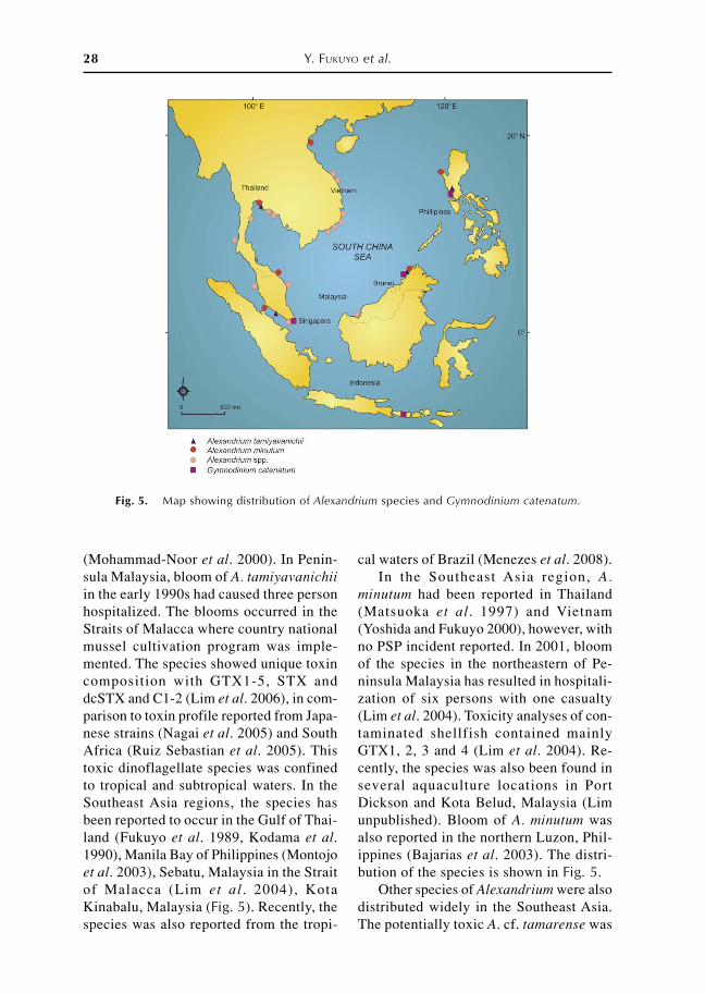
(Mohammad-Noor et al. 2000). In Penin-sula Malaysia, bloom of A. tamiyavanichiiin the early 1990s had caused three personhospitalized. The blooms occurred in theStraits of Malacca where country nationalmussel cultivation program was imple-mented. The species showed unique toxincomposition with GTX1-5, STX anddcSTX and C1-2 (Lim et al. 2006), in com-parison to toxin profile reported from Japa-nese strains (Nagai et al. 2005) and SouthAfrica (Ruiz Sebastian et al. 2005). Thistoxic dinoflagellate species was confinedto tropical and subtropical waters. In theSoutheast Asia regions, the species hasbeen reported to occur in the Gulf of Thai-land (Fukuyo et al. 1989, Kodama et al.1990), Manila Bay of Philippines (Montojoet al. 2003), Sebatu, Malaysia in the Straitof Malacca (Lim et al . 2004), KotaKinabalu, Malaysia (Fig. 5). Recently, thespecies was also reported from the tropi-
cal waters of Brazil (Menezes et al. 2008).In the Southeast Asia region, A.
minutum had been reported in Thailand(Matsuoka et al. 1997) and Vietnam(Yoshida and Fukuyo 2000), however, withno PSP incident reported. In 2001, bloomof the species in the northeastern of Pe-ninsula Malaysia has resulted in hospitali-zation of six persons with one casualty(Lim et al. 2004). Toxicity analyses of con-taminated shellfish contained mainlyGTX1, 2, 3 and 4 (Lim et al. 2004). Re-cently, the species was also been found inseveral aquaculture locations in PortDickson and Kota Belud, Malaysia (Limunpublished). Bloom of A. minutum wasalso reported in the northern Luzon, Phil-ippines (Bajarias et al. 2003). The distri-bution of the species is shown in Fig. 5.
Other species of Alexandrium were alsodistributed widely in the Southeast Asia.The potentially toxic A. cf. tamarense was
Fig. 5. Map showing distribution of Alexandrium species and Gymnodinium catenatum.

Ecology and oceanography of harmful marine microalgae 29
found to occur in the waters of Thailandand Malaysia (Piumsomboon et al. 2000,Usup et al. 2002, Lim et al. 2003), whileA. taylori and A. peruvianum were founddistributed in the Kuching waters of Ma-laysia (Lim and Ogata 2005). In Vietnam-ese waters, more than 16 species ofAlexandrium have been reported (Nguyen-Ngoc 2004).
Gymnodinium catenatumGymnodinium catenatum is the only
naked dinoflagellate that is associated withparalytic shellfish poisoning (PSP). Theorganism forms chain in the natural envi-ronment, with moderate cell size (Fig. 6).Blooms of this species have been resultedin shellfish contamination worldwide. Inthe temperate Pacific region, G. catenatumhas been reported from Australia(Hallegraeff et al. 1988), Japan (Matsuokaand Fukuyo 1994), and Korea (Park et al.2004). In the Southeast Asia regions, thespecies is only found in certain locationswith very few reports of occurrences, suchas in the Manila Bay of Philippines(Fukuyo et al. 1993) and Kota Kinabalu,Malaysia (Mohammad-Noor et al. 2002)which co-existed with the Pyrodiniumblooms. The species was also found to bepresent in the phytoplankton assemblagein Singaporean waters (Holmes et al.
2002), Lombok, Indonesia (Sidharta andAhyadi 2007) and most recently in Thai-land (Lirdwitayaprasit et al. 2008). TheThai strain was reported with n-sulfocarbomoyl C1-2 as the major conge-ners and GTX1-4 as minor components inthe toxin composition (Lirdwitayaprasit etal. 2008). The Singapore strains of G.catenatum on the other hand were reportedwith unique toxin profile which dominatedby GTX1+4, with small amount ofGTX2+3, neoSTX and STX (Holmes et al.2002).
Benthic dinoflagellates potentially caus-ing ciguatera fish poisoning
Ciguatera fish poisoning (CFP) is acommon food-borne disease related to theconsumption of subtropical and tropicalmarine finfish. CFP is the most commonand widespread seafood poisoning afflict-ing approximately 50,000 victims/year inthe world (Fleming et al. 2000). The con-cerned ciguatoxic-fish are either feedingon small algae species known asdinoflagellates or feeding on toxic herbiv-ore fish. The main toxic dinoflagellate isGambierdiscus toxicus (Adachi andFukuyo 1979) which is found primarily insub- and tropical areas where it lives inassociation with macroalgae, sand androck. Ostreopsis and Prorocentrum areknown as species occurring withGambierdiscus toxicus (Fukuyo 1981), andmay have implication in some symptomsof CFP.
In Japan CFP has been known for a longtime only in Okinawa, the most southernislands having subtropical climate. How-ever, several food poisoning cases sus-pected as CFP occurred in recent years af-ter eating spotted knifejaw (Oplegnathuspunctatus) caught by surf fishing at sev-eral cities facing the Pacific Ocean in thetemperate area of Japan. CFP causativespecies Gambierdiscus toxicus seems tohave expanded the distribution area to thetemperate region (Omura and Fukuyo un-
Fig. 6. Motile cells of Gymnodiniumcatenatum.

30 Y. FUKUYO et al.
published). Recent reports of potentiallyCFP causative species, together with someassociating ones, in Southeast Asia andJapan were summarized as follows.
GambierdiscusTen species of Gambierdiscus has been
known in coastal waters as benthicdinoflagellates in the world: G. australesFaust et Chinain 1999, G. belizeanus Faust1995, G. caribaeus Litaker et al. 2009, G.carolinianus Litaker et al. 2009, G. car-penter Litaker et al. 2009, G. pacificusChinain et Faust 1999, G. polynesiensisChinain et Faust 1999, G. ruetzleri Litakeret al. 2009, G. toxicus Adachi et Fukuyo1979, G. yasumotoi Holmes 1998. Amongthem several species of Gambierdiscus areknown to occur in Southeast Asian waters.
G. pacificus was reported from KotaKinabalu and Sipadan Island, Malaysia(Mohammad-Noor et al. 2006). G. toxicuswas reported from Long Vi Island and NhaTrang Bay, Vietnam (Larsen and Nguyen2004), Okinawa Islands to main land, Ja-pan (Hara and Horiguchi 1982, Koike etal. 1991, Fukuyo et al. 2002, Ishikawa andKurashima 2010, Omura et al. 2010) (Fig.7). G. yasumotoi was reported from Sin-gapore (Holmes 1998). In addition to themKuno et al. (2010) and Leaw et al. (2001)also reported Gambierdiscus from Japanand Malaysia, respectively, but speciesfound by them were not described.
Ostreopsis and ProrocentrumNine species of Ostreopsis has been
known in the world (Ostreopsis belizeanus,
Fig. 7. Gambierdiscus toxicus. A: live specimen from Wakayama Pref. Japan. B and C: fixedspecimen from Hachijo Island, Japan. C: cell in apical view with the fish-hook shaped apicalpore.
Fig. 8. Ostreopsis ovata from Hachijo Island, Japan (live specimen). A: cells attaching onsurface of macroalga (Gelidium sp.). B and C: enlarged photomicrograph of A.

Ecology and oceanography of harmful marine microalgae 31
O. caribbenaus, O. heptagona, O. labens,O. lenticularis , O. marinus , O.mascarenensis, O. ovata, O. siamensis).They usually occur associating withGambierdiscus and other benthicmicroalgae. Among them, Ostreopsislabens, O. lenticularis and O. ovata werereported from Malaysia (Leaw et al. 2001,Mohammad-Noor et al . 2006), O.lenticularis, O. marinus and O. ovata fromVietnam (Larsen and Nguyen 2004), O.ovata, O. siamensis from the Ryukyu Is-land, Japan (Fukuyo 1981), O. cf. marinusand O. cf. ovata from main land, Japan(Omura unpublished) (Fig. 8).
The genus Prorocentrum has species ofplanktonic and benthic habitat. As for thebenthic Prorocentrum species,Prorocentrum arenarium, P. concavum, P.cf. faustiae and P. lima were reported fromMalaysia (Mohammad-Noor et al. 2006),P. concavum, P. emarginatum, P. lima and
P. rhathymum from Vietnam (Larsen andNguyen 2004), P. concavum , P.emarginatum, P. hoffmannianum, P. lima,P. mexicanum and P. ruetzlerianum fromPhilippines (Marasigan et al. 2001), P.concavum, P. emarginatum and P. limafrom Japan (Fukuyo 1981, Koike et al.1991).
ASP toxin producing microalgae
Pseudo-nitzschiaPseudo-nitzschia is a genus of marine
pennate diatom comprised of thirty fourspecies (Table 1) known up to date, withalmost half of the known species are re-ported to produce naturally neurotoxins,domoic acid (DA) and its derivatives(Bates 2000). Contamination of DA inshellfish mollusks are the cause of humanintoxication in Canada in the late 1980s,as well as death of marine birds and mam-mals in the coasts of the United States
Country
Species Malaysia Thailand Indonesia Vietnam Philippines
Pseudo-nitzschia americana + b Pseudo-nitzschia brasiliana +b,d,h +e +e +b +c
Pseudo-nitzschia caciantha +c
Pseudo-nitzschia calliantha** +b +f +b Pseudo-nitzschia cuspidata +b Pseudo-nitzschia delicatissima** +b +f +b Pseudo-nitzschia dolorosa +d Pseudo-nitzschia fraudulenta* * +b Pseudo-nitzschia cf. granii +b Pseudo-nitzschia heimii +f Pseudo-nitzschia inflatula +f +b Pseudo-nitzschia micropora +b +f +b +c
Pseudo-nitzschia multistriata * * +b +b Pseudo-nitzschia pungens +d,h +g +b +a,c
Pseudo-nitzschia pseudodelicatissima * * +c
Pseudo-nitzschia cf. sinica +f +b Pseudo-nitzschia subpacifica +f
Table 1. Occurrence of Pseudo-nitzschia species in the Southeast Asia.
Sources: aBajarias et al. 2006, bLarsen and Nguyen 2004, cYap-Dejeto et al. 2008, dLim 2010, eLundholmet al. 2002, fPrissholm et al. 2002, gSidabutar et al. 2000, hSu 2010.**Toxic species.

32 Y. FUKUYO et al.
(Scholin et al. 2000).Under light microscope, Pseudo-
nitzschia can be differentiated from othergenera of diatoms based on the character-istic of colonies or chains formed withoverlapping cells. Pseudo-nitzschia is di-vided into two subgroups, seriata-groupwith valve width larger than 3 µm anddelicatissima-group with valve widthsmaller than 3 µm (Hasle and Syvertsen1997). The ultrastructural observation offrustules of the cell is important in speciesidentification. Identification to species isonly possible with the aid of advancedelectron microscopy.
In Southeast Asia, a total of seventeenspecies of Pseudo-nitzschia have beenrecord thus far, with five species knownto be toxic (Lundholm et al . 2002,Prissholm et al. 2002, Larsen and Nguyen2004, Bajarias et al. 2006, Yap-Dejeto2010, Su 2010). No serious ASP case wasreported from the region, however, con-tamination of DA in shellfish was reportedin Philippines (Bajarias et al. 2006), Viet-nam (Dao et al. 2009) and some tropicalAsian countries (Takata et al. 2009) withno records of human poisoning.
In Malaysian waters, Skov noted thepresence of five species in Sabah waters,namely, P. brasiliana, P. calliantha, P.delicatissima , P. micropora and P.multistriata (Skov pers. comm. in Larsenand Nguyen 2004). In the recent studies,four species of Pseudo-nitzschia weredocumented and identified based on cul-tures and natural samples. They are P.pungens, P. brasiliana, P. dorolosa and P.calliantha (Lim unpublished data). Inter-estingly, P. pungens and P. brasiliana arewidely distributed in Malaysian waters andwere observed in samples collected fromboth the Straits of Malacca and SouthChina Sea; occurrence of P. dorolosa andP. calliantha on the other hand is only con-fined to limited locations (Lim unpub-lished data).
Very few studies have been carried out
to understand the bloom mechanism ofPseudo-nitzschia especially in the tropicalwaters. In Malaysian waters, a study wasconducted to investigate the seasonal oc-currence of Pseudo-nitzschia and its re-lated environmental conditions at two es-tuarine waters of Sarawak, Malaysia. Celldensity of Pseudo-nitzschia peaked duringthe months of April–May over the sam-pling period of 2007–2010 (Su 2010). In-creases of Pseudo-nitzschia cell densitywas likely corresponding to low precipi-tation, high salinity and pH, with no cleartrend with changes of macronutrient con-dition of the waters (Su 2010). ThePseudo-nitzschia cells were postulated tobe brought into the estuaries by the semi-diurnal tidal cycle (Su 2010). Higher celldensity of Pseudo-nitzschia was also ob-served during the dry season from Decem-ber to May in Philippines (Yap-Dejeto etal. 2008) and Thailand (Udomratana et al.2008). Presence of potentially toxic spe-cies might pose potential threats of ASPin the areas with shellfish farming if nomonitoring effort was implemented.
Domoic acid (DA) is well known as acausative toxin of amnesic shellfish poi-soning (ASP) which was first found inCanada (Wright et al. 1989). Since a pen-nate diatom Pseudo-nitzschia multiserieswas identified as the causative planktonspecies in Canadian case, several speciesof Pseudo-nitzschia have been reported toproduce a significant level of domoic acid(Bates et al. 1989). However, these stud-ies have been limited in temperate and coldwaters (Bates 2000, Trainer et al. 2008).Little is known about the DA-producingdiatom in tropical waters. No informationon the accumulation of DA has been ob-tained for tropical bivalves. Under thesecircumstances, Takata et al . (2009)screened the occurrence of DA in SouthEast Asian countries and reported thatbivalves belonging to the genus Spondylusaccumulate a significant level of DA whileother bivalve species do not. These find-

Ecology and oceanography of harmful marine microalgae 33
ings indicate the occurrence of DA-pro-ducing plankton such as Pseudo-nitzschiaspp. in these areas. As a considerable levelof DA was also detected in S. versicolorin Nha Phu Bay, Vietnam, studies on DAin Nha Phu Bay have been conducted incollaboration with Japanese members un-der the current project (Dao et al. 2009a,2009b).
In the monitoring study on DA level ofS. versicolor in association with that ofplankton net samples, clear correlation wasobserved between them, indicating thepresence of DA-producing plankton in thebay. Light microscopic observation of theplankton net samples showed the occur-rence of Pseudo-nitzschia spp. but theircell number was not dominant. In a trial inwhich plankton cells in the samples werefractionated by successive filtrationthrough sieves with different pore sizes,most of DA in the plankton sample wasfound to be concentrated in the fractionwith small size particles (0.6–10 µm frac-tion). Light microscopic observationshowed that Pseudo-nitzschia spp. andNitzschia spp. were dominant in the frac-tion (Dao et al. 2009b). These results sug-gest that Pseudo-nitzschia species in thefraction is the causative species of DA.Thus, unicellular cultures were made fromPseudo-nitzschia species observed in 0.6–10 µm fraction. DA was detected by LC-MS/MS analysis in more than half thestrains established, though the level wassignificantly low (unpublished data). In-terestingly, all the DA-producing strainswere identified as Pseudo-nitzschiacaciantha, indicating that this species isat least one of the causative organisms forDA in S. versicolor in Nha Phu Bay, Viet-nam.
Although DA production is notanalyzed, more than 10 species of Pseudo-nitzschia including P. caciantha are listedin Vietnamese (Larsen and Nguyen 2004)and Japanese water (Yap-Dejeto et al.2010). These include potentially toxic spe-
cies such as P. calliantha, P. delicatissima,P. multistriata, and P. pungens (IOC, http://www.marinespecies.org/hab/index.php).DA production of Pseudo-nitzschia is re-ported to be influenced by various factors.Further study on DA production of P.caciantha is under progress.
Nitzschia navis-varingicaIn the field survey on harmful algal
species in Vietnam, plankton net samplescollected from a resting shrimp culturepond in Do Son, Vietnam were broughtback to Japan. Cells of algal species sus-pected to produce DA were isolated forunialgal cultures, and tested for DA pro-duction by HPLC with fluorescence detec-tion (Pocklington et al. 1990, Kotaki et al.1999). Significant level of DA productionwas detected in some of the culture strains(maximum 1.7 pg/cell) (Kotaki et al.2000). Cells of all the strains positive forDA production were morphologically thesame as follows.
LM observation revealed that the cellspossess two chloroplasts at each end of thecell and are lanceolate in valve view. Cellsare 38–110 µm long and 9–11 µm wide. Ingirdle view, the cells are rectangular andslightly indented at the middle. Most cellsmake ribbon-shaped colonies while grow-ing (Fig. 9). In TEM, characteristic silicaridges are observed in the wall of the ra-
Fig. 9. Nitzschia navis-varingica. Scalebar: 50 µm.
34 Y. FUKUYO et al.
found to be distributed widely in the brack-ish waters of South East Asian countries.
In the screening of N. navis-varingicain the Philippines, some strains of the spe-cies isolated from Bulacan Estuary, Ma-nila Bay, were found to produce onlyisodomoic acids A (IA) and B (IB). No DAwas detected in these strains (Kotaki et al.2005). Isodomoic acid C was reported to-gether with DA in shellfish and in thecausative diatom P. australis (Rhodes etal. 2003, Holland et al. 2005). Traceamount of isodomoic acids A, D, E, F and5′-epi-DA were detected together with DAin the tropical bivalves Spondylus spp.(Takata et al. 2009). Although it is uncer-tain whether these isomers are the artifactsof DA (Wright et al. 1989, Quilliam 2003)or bio-synthesized one by diatom, traceamount of isomers were detected togetherwith high level of DA in Pseudo-nitzschiamultiseries (Takata et al. 2009). However,the level of isomers in P. multiseries isusually vanishingly low. The finding of the
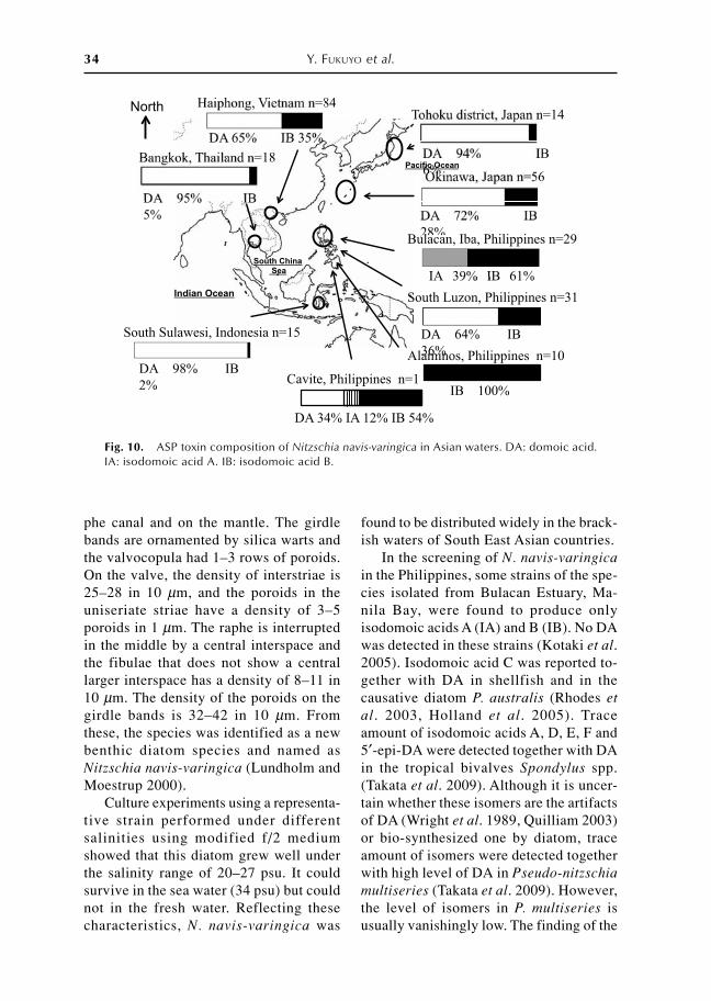
Fig. 10. ASP toxin composition of Nitzschia navis-varingica in Asian waters. DA: domoic acid.IA: isodomoic acid A. IB: isodomoic acid B.
phe canal and on the mantle. The girdlebands are ornamented by silica warts andthe valvocopula had 1–3 rows of poroids.On the valve, the density of interstriae is25–28 in 10 µm, and the poroids in theuniseriate striae have a density of 3–5poroids in 1 µm. The raphe is interruptedin the middle by a central interspace andthe fibulae that does not show a centrallarger interspace has a density of 8–11 in10 µm. The density of the poroids on thegirdle bands is 32–42 in 10 µm. Fromthese, the species was identified as a newbenthic diatom species and named asNitzschia navis-varingica (Lundholm andMoestrup 2000).
Culture experiments using a representa-tive strain performed under differentsalinities using modified f/2 mediumshowed that this diatom grew well underthe salinity range of 20–27 psu. It couldsurvive in the sea water (34 psu) but couldnot in the fresh water. Reflecting thesecharacteristics, N. navis-varingica was

Ecology and oceanography of harmful marine microalgae 35
Fig. 11. Harmful Cochlodinium speciesoccurring from the Southeast Asia. Allscale bars 10 µm. 1, 2: Cochlodiniumfulvescens collected from Harun Bay ofSumatra, Indonesia. 3, 5, 6: Cochlodiniumpolykrikoides collected from Sabah, Ma-laysia (3: by courtesy of Prof. A. Anton).4: Cochlodinium convolutum collectedfrom Sebatu in south of Malacca Strait,Malaysia (by courtesy of Dr. P.-T. Lim).
N. navis-varingica strains which produceonly isomers of DA seems to be interest-ing from the standpoint of metabolism in-cluding biosynthesis of DA.
The toxicity of IA, IB and IC was re-ported to be significantly lower than thatof DA (Munday et al. 2008), suggestingthat these toxins pose a lower risk to hu-mans. However, the possible risk due tobioconversion of isomers to DA in theshellfish could not be ruled out. Metabolictransformation of isomers to DA is cur-rently under investigation.
After finding N. navis-varingica strainsthat produce IA and IB instead of DA,screening of toxin producing N. navis-varingica was performed not only for DAbut also for IA and IB. As a result, 5 typesof toxin composition namely DA, DA-IB,IA-IB, IB, DA-IA-IB were confirmedamong the N. navis-varingica strains tested(Fig. 10). Comparison of the toxin com-position between the sub-strains and theparental strain showed that the toxin com-position is stable in a strain. DA and DA-IB types were the major toxin compositiontypes, because these types were seen in allisolates obtained from Vietnam (nearHaiphong), southern Philippines (southernpart of Manila Bay and some areas nearTacloban), Japan (Tohoku and Okinawadistricts) (Kotaki et al. 2005, 2008), Thai-land (near Bangkok) (Bajarias et al. 2006)and Indonesia (south Sulawesi) (unpub-lished data). Only the isolates obtainedfrom limited areas in Luzon Island, thePhilippines showed the rest toxin compo-sition types. The isolates obtained fromBulacan Estuary, Manila Bay and Iba Es-tuary, Zambales showed an IA-IB toxincomposition (Kotaki et al. 2005, Bajariaset al. 2006). The isolates from Alaminos,Pangasinan showed the toxin compositionof only IB. One isolate from Cavite Estu-ary, Manila Bay showed the DA-IA-IBtoxin composition (unpublished data). Allof the diatoms were morphologically thesame species N. navis-varingica.
When axenic cultures were made fromsome uni-algal culture of DA-IB type, thetoxin composition changed to IA-IB type,suggesting the bacterial effect on control-ling the toxin composition (Kotaki et al.2008). Possible factors affecting the toxincomposition such as bacteria, salinity, pHand/or genetic difference among strains areimportant to solve the toxin productionmechanism of the diatom. Efforts to solvethese factors are currently under way.
Red Tide Causative Species
Cochlodinium: Red tide causative specieswith fish kills
Taxonomy of Cochlodinium polykrikoidesand morphologically similar species
In Asian waters, five species tentativelyattributable to the genus Cochlodinium

36 Y. FUKUYO et al.
Tab
le 2
. M
orp
holo
gica
l fea
ture
s o
f fo
ur
HA
B s
pec
ies
attr
ibu
tab
le to
the
gen
us
Co
chlo
din
ium
.
C.
con
volu
tum
C.
cf.
gem
inat
um
C.
po
lykr
iko
ides
C.
fulv
e sce
ns
Cel
l si
zeca
. 60
−70
µ mca
. 40
−70
µmca
. 30
−40
µ mca
. 45
−50
µmEy
e sp
ot
no
nn
on
Do
rsal
ep
ico
neD
ors
al e
pic
one
Cin
gu
lum
ca.
1.5
ca.
1.5
ca.
2ca
. 2
Sulc
us
deep
deep
Shal
low
, ju
st b
elo
w t
he c
ing u
l um
Shal
l ow
, be
twee
n th
e ci
ngu
lum
Nuc
leus
elo
ngat
eSp
h eri
cal,
cen
ter
Sph e
rica
l, ep
ico
ne
Sph e
rica
l, ep
ico
ne
Ch
l oro
pl a
stR
etic
ulat
eR
etic
ulat
eR
od
- l ike
, lo
ngi
tud
inal
l yg r
anul
ate
Oth
erTw
o ce
lls c
hai
nTw
o ce
l l s c
ha i
n16
cel
ls c
hain
Four
cel
l s c
ha i
n
have been observed up to date (Fig. 11).These are all photosynthetic with morpho-logically variable chloroplasts, and havemore or less made blooms; Cochlodiniumcatenatum Okamura, Cochlodiniumconvolutum Kofoid and Swezy,Cochlodinium fulvescens Iwataki, Kawamiand Matsuoka, Cochlodinium cf.geminatum (Schütt), and Cochlodiniumpolykrikoides Margalef (Table 2). C.catenatum described from Tokyo Bay in1916 by Okamura was morphologicallysimilar to C. polykrikoides (Matsuoka etal. 2008), however, the taxonomical rela-tionship of these species is still unclear.
Cochlodinium polykrikoides Margalefhas been well known to form large-scaleblooms (e.g. Matsuoka et al. 2008, 2010).In Asian waters, this species was first ob-served in the Yatsushiro Sea, Japan in 1976accompanied with huge economic damage(Kagoshima Prefectural Fisheries Station1995). Thereafter, this species has occurredalmost every summer in Japan and Koreaduring the last thirty years. In SoutheastAsia, C. polykrikoides was first detectedin the Philippines in 2002 (Relox Jr. andBajarias 2003) and then in coastal watersof Hong Kong, Philippines and MalaysianSabah (Iwataki et al. 2008, Matsuoka etal. 2008). Recent progress on a molecularphylogenetic study on C. polykrikoides, atleast four ribo-types (American-Malaysiatype, Philippines type, East Asian type in-cluding the so-called Cochlodinium sp.Kasasa type) were recognized (Iwataki etal. 2008, Matsuoka et al. 2010).
Cochlodinium fulvescens, morphologi-cally similar to C. polykrikoides, was firstdescribed in the Asian coastal waters aswell as the east coast of the North Americaby Iwataki et al. (2007). Cochlodinium cf.geminatum was initially found in the coastof Hong Kong accompanied with fish-kill.This species can produce a resting cyst pre-servative in sediments. According to thisnature, the cyst of Cochlodinium cf.geminatum has been recorded from tropi-

Ecology and oceanography of harmful marine microalgae 37
Fig. 12. Geographical distribution of harmful Cochlodinium in East and Southeast Asia.
cal to temperate regions of the East andSoutheast Asia as a cyst of C. polykrikoidesfollowed by Matsuoka and Fukuyo (2000).Cochlodinium convolutum characterizedby having a roundly rectangular nucleuslocated in the center appeared frequentlyin west Japan, but rather harmless.
Distribution (Fig. 12)C. polykrikoides has been reported
from Sabah, Malaysia (Anton et al. 2008),Palawan, the Philippines (Azanza et al.2008), Balayan Bay of Luzon Island, thePhilippines (Relox Jr. and Bajarias 2003)up to date. According to Iwataki et al.(2008), C. polykrikoides occurring fromSabah was assignable to the American-Malaysian ribo-type and C. polykrikoidesfrom Luzon Island to the Philippines ribo-type. Around the East China Sea, most ofC. polykrikoides were designated into theEast Asian ribo-type (Iwataki et al. 2008,Matsuoka et al. 2010).
C. fulvescens occurred in Hurun Bayof Sumatra, Indonesia (Matsuoka et al.2008). C. convolutum has been recordedfrom Sebatu in south of Malacca Strait,Malaysia (Lim pers. comm.). These occur-rences of C. fulvescens and C. convolutumnever caused any serious damages foraquaculture. Other occurrences ofCochlodinium were recorded in the innerGulf of Thailand in 1991 and 1992 withdiscolorations (Lirdwitayaprasit 2003), butunfortunately species of theseCochlodinuim were unknown.
Bloom with fish kill in Southeast Asia (Fig.13)
In Southeast Asia, only few cases offish-kill events caused by C. polykrikoideshave been reported from Luzon and offPalawan of the Philippines and Saba ofMalaysia. As previously mentioned, otherphotosynthetic species of Cochlodinium,C. fulvescens and C. cf. geminatum are
38 Y. FUKUYO et al.
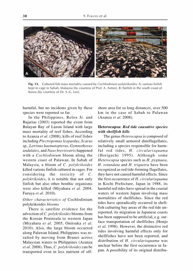
Fig. 13. Cultured fish mass mortality caused by Cochlodinium polykrikoides. A: various finfishkept in cage in Sabah, Malaysia (by courtesy of Prof. A. Anton). B: flatfish in the south coast ofKorea (by courtesy of Dr. S.-G. Lee).
harmful, but no incidents given by thesespecies were reported so far.
In the Philippines, Relox Jr. andBajarias (2003) reported the event fromBalayan Bay of Luzon Island with largemass mortality of reef fishes. Accordingto Azanza et al. (2008), kills of reef fishesincluding Plectropomus leopardus, Scarussp., Letrinus haematopterus, Gymnothoraxundulates, and Naso brevirostris happenedwith a Cochlodinium bloom along thewestern coast of Palawan. In Sabah ofMalaysia, a bloom of C. polykrikoideskilled various finfish cultured in cages. Forconsidering the toxicity of C.polykrikoides, it is notable that not onlyfinfish but also other benthic organismswere also killed (Miyahara et al. 2004,Furuya et al. 2010).
Other characteristics of Cochlodiniumpolykrikoides blooms
There is satellite evidence for theadvection of C. polykrikoides blooms fromthe Korean Peninsula to western Japan(Miyahara et al. 2005, Onitsuka et al.2010). Also, the large bloom occurredalong Palawan Island, Philippines was re-sulted by moving from Brunei acrossMalaysian waters to Philippines (Azanzaet al. 2008). Thus, C. polykrikoides can betransported even in less nutrient of off-
shore area for so long distances, over 500km in the case of Sabah to Palawan(Azanza et al. 2008).
Heterocapsa: Red tide causative specieswith shellfish kills
The genus Heterocapsa is composed ofrelatively small armored dinoflagellates,including a species responsible for harm-ful red tides, H. circularisquama(Horiguchi 1995). Although someHeterocapsa species such as H. pygmaea,H. rotundata and H. triquetra have beenrecognized as red tide-forming flagellates,they have not caused harmful effects. Sincethe first occurrence of H. circularisquamain Kochi Prefecture, Japan in 1988, itsharmful red tides have spread in the coastalwaters of western Japan involving massmortalities of shellfishes. Since the redtides have sporadically occurred in shell-fish culturing bay areas of the red tide un-reported, its migration in Japanese coastshas been supposed to be artificial, e.g. sur-face transportation of shellfishes (Honjoet al. 1998). However, the distinctive redtides involving harmful effects only forshellfishes have not been reported, anddistribution of H. circularisquama wasunclear before the first occurrence in Ja-pan. A possibility of its original distribu-

Ecology and oceanography of harmful marine microalgae 39
tion was assumed to the tropical or sub-tropical areas because the optimum tem-perature and salinity for growth were rela-tively high compared to other red tide flag-ellates occurred in Japan (Yamaguchi et al.1997). This presumption was supported bythe evidence for H. circularisquama redtides in Hong Kong during 1986–1987,immediately before the first occurrence inJapan (Iwataki et al. 2002b). It also sug-gested that H. circularisquama might dis-tribute in Southeast Asian coast with simi-lar environmental condition to Hong Kong.For understanding of the H.circularisquama red tides we carried outtaxonomic study on the genus Heterocapsato distinguish the harmful species, and dis-tribution survey including otherHeterocapsa species.
TaxonomyAfter the species description of H.
circularisquama in 1995, eightHeterocapsa species (H. arctica , H.horiguchii, H. huensis, H. lanceolata, H.orientalis, H. ovata, H. psammophila andH. pseudotriquetra) were described mainlyfor the purpose of discrimination from theharmful species H. circularisquama(Horiguchi 1997, Iwataki et al. 2002a,2003, 2004, 2009, Tamura et al. 2005).Most recently a subspecies, H. arcticasubsp. frigida was reported from the Bal-tic Sea (Rintala et al. 2010), and conse-quently 16 species have so far been as-signed to the genus. Since Heterocapsaspecies share similar morphological char-acters, light microscopic identification isdifficult for some species. Thecal platearrangements of these species have beenrecognized to be identical, Po, cp, 5′, 3a,7″, 6c, 5s, 5�, 2″″, even though the varia-tion are often found in culture condition.Using light microscopy, the position of thenucleus and pyrenoid, i.e. located in theepitheca or hypotheca, and the cell shapesuch as the larger epitheca or presence ofantapical horn, are also available for pro-visional species identification, however,
these combination are inadequate for un-ambiguous identification for manyHeterocapsa species (Iwataki 2008). Onthe other hand, ultrastructure of body scaleis a reliable morphological character forspecies identification, and species recentlydescribed have been established basedmainly on this diagnostic character. Thebody scale is tiny organic cell coveringsituated on the cell surface, composed ofa reticulated basal plate ca. 200–500 nmin diameter and a three-dimensional frame-work on the plate. Since the size, shape ofbasal plate, numbers of vertical and hori-zontal bars are congruous in each speciesand apparently different from other spe-cies, the scale structure is available forspecies discrimination. For example, scaleof H. circularisquama consists of a circu-lar basal plate with a central and six mar-ginal uprights connected by horizontal barsone another, by which species can be dis-tinguished from others. Ultrastructure ofHeterocapsa body scales was summarizedin Iwataki et al. (2004) and almost all spe-cies can be identified based on the struc-ture. Moreover, the body scale structure iskept in preserved phytoplankton specimenand therefore presence of H.circularisquama in Hong Kong was dem-onstrated after 15 years from the bloom-ing (Iwataki et al. 2002b).
Distribution of H. circularisquamaSince it was strongly suggested that the
harmful species H. circularisquama is dis-tributed not only in western Japanesecoasts but also in the coasts of SoutheastAsia, we surveyed presence of this speciesin Vietnamese coasts, Hai Phong, Hue, NhaTrang and Phu Quoc Island during in 2006and 2007. In the samples collected fromthese coasts, a Heterocapsa was found attwo locations in Hue. The cell is relativelysmall and resembling H. pygmaea due tohaving plural pyrenoids located above thenucleus in the hypotheca, while the bodyscale is similar to that of H. illdefina witha difference in number of marginal up-

40 Y. FUKUYO et al.
rights on basal plate. This species was re-vealed to be an undescribed Heterocapsaspecies on the basis of body scale struc-ture and the sequencing of ITS region as amolecular marker. Consequently it was de-scribed as a new species H. huensis undercollaboration with Vietnamese scientists(Iwataki et al. 2009). The harmful species,H. circularisquama, has not been detectedfrom Vietnamese coasts. Therefore the oc-currence has so far been reported only fromHong Kong in Asia, except for the con-tinuous reports from Japanese coasts in-volving economic losses of shellfish cul-tures. Recently, the presence of H.circularisquama was unexpectedly re-ported from Cuba, as a preliminary iden-tification without body scale structure(Moreira González 2010). This implies thathabitation expansion of H.circularisquama due to artificial transferor its cryptic flora is now unveiling, andsupports the importance to monitor theoccurrence of H. circularisquama inSoutheast Asia to understand its distribu-tion and mechanism of globalization.
Noctiluca: Red tide species associatedwith eutrophication
Noctiluca scintillans is a common redtide species which has world wide distrib-uted in the coastal area. Noctiluca red tidesappear as pinkish red in various temperateand subtropical waters but causes green-ish discoloration in tropical waters of thewestern Pacific and the Indian Ocean(Elbrächter and Qi 1998, Harrison et al.pers. comm.). This difference in color isdue to the presence of a tiny green flagel-late endosymbiont, Pedinomonasnoctilucae, in N. scintillans cells (Sweeney1971). Therefore, N. scintillans harboringthe symbiotic green algae is referred to asgreen Noctiluca and red Noctiluca meansthose lack of the symbionts. The outbreakof green Noctiluca red tide is also the com-mon phenomena in the eutrophicated wa-ters such as Jakarta Bay (Adnan 1984),
Manila Bay (Jacinto et al. 2006) and theupper Gulf of Thailand (Suvapeepun1989). In Manila Bay, since 1998 up to thepresent, green Noctiluca has dominatedother phytoplankton species during theperiod that Pyrodinium used to bloom(Jacinto et al. 2006) and the bloom of greenNoctiluca formed occasionally almostwhole area of the bay since 2001(Furuyaet al. 2006a). In the Gulf of Thailand, greenNoctiluca is a main causative red tide spe-cies. The bloom of green Noctiluca wasfirst reported in 1957 (Charernphol 1958),since then the blooms occurred more fre-quently in the eastern part of the upperGulf and the study on seawater discolora-tion has been focused on its impact on fish-eries. The dense blooms of green Noctilucaoccasionally cause the mass mortality ofboth coastal fishes and shrimp culture(Adnan 1989, Suvapepun 1989,Lirdwitayaprasit et al. 1995, PollutionControl Department 2003). GreenNoctiluca is selected as one of targetedspecies for cooperative international re-search in GEOHAB Asia (Furuya et al.2010).
Red Noctiluca is characterized as avoracious predator with a diverse dietranging from phytoplankton to copepodsand fish eggs (Hattori 1962, Schaumannet al. 1988, Nakamura 1998). A linkage ofincreasing blooming events of redNoctiluca with progressive eutrophicationof coastal waters is suggested from in-creasing prey availabili ty due toeutrophication (Porumb 1992). In contrast,the presence of photosyntheticendosymbiont in green Noctiluca impliesdifferent dependence on environmentalconditions from that of red Noctiluca. Thesymbiont ensures the survival of greenNoctiluca during the shortage of food par-ticles (Saito et al. 2006). Thus, it is highlyprobable that nutrient and light intensitiesaffect growth of green Noctiluca directlyand indirectly through the growth of bothphytoplankton as prey and the symbiont.

Ecology and oceanography of harmful marine microalgae 41
Therefore, the research group of Noctilucain the JSPS Program on Coastal MarineScience (the research group hereafter) aimsto understand the mechanism of the appar-ent expansion of bloom of green Noctilucain the Southeast Asian waters byecophysiological research of this species.
Life history and physiologyThe life cycle of red Noctiluca com-
prises both asexual binary fission andsexual reproduction (Zigmark 1970). Thecomplete sexual process in red Noctilucawas reported by Fukuda and Endoh, 2006while green Noctiluca performs sexual re-production in the same manner as the redone (Lirdwitayaprasit 2002). Gametocytesof green Noctiluca are frequently observedin the upper Gulf of Thailand during theSW monsoon (Sriwoon et al. 2008). Incultures of green Noctiluca, gametocytesare constantly observed during the expo-nential growth phase (T. Lirdwitayaprasitand K. Furuya unpublished data). There-fore, the marked occurrence ofgametocytes in the upper gulf was indica-tive of active growth of green Noctiluca.
Since green Noctiluca harbors P.noctilucae, photosynthesis of P. noctilucaeis of a particular concern of the researchgroup. Saito et al. (2006) showed that cul-tures of non-feeding strains, isolated fromthe upper Gulf of Thailand, growphotoautotrophically for generations, butthey also feed on D. tertiolecta, indicat-ing phagotrophy is facultative. Net photo-synthesis was significantly higher in thenon-feeding strains than the feeding ones.The difference is due to high respirationactivity in the feeding strains. This is con-sistent with an observation in a naturalpopulation of Manila Bay, where net pho-tosynthesis was significantly higher incells lacking food vacuoles than those withfood vacuoles (Saito et al. unpublisheddata). The relationship of photosynthesiswith irradiance is characterized by satura-tion at low light intensity and absence orweak photoinhibition, showing efficient
util ization of a wide range of l ightintensities.
The vegetative growth of greenNoctiluca was also investigated using cul-tures and in natural populations. The re-search group found that under optimalgrowth conditions there is no significantdifference in growth rate between greenNoctiluca (0.33 day–1) and red Noctiluca(0.28 day–1) isolated from Gulf of Thai-land and the Seto Inland Sea, respectively(Furuya et al . 2006b). The role ofphagotrophy on population growth ofgreen Noctiluca in the upper Gulf of Thai-land clearly showed that the higher abun-dance of N. scintillans in the SW monsoonthan in the NE monsoon was consequenceof active growth supported by phagotrophy(Sriwoon et al. 2008). This was consistentwith laboratory evidence that feedingstrains grow faster than non-feeding strains(Furuya et al. 2006b).
A cell cycle analysis conducted in anatural population in Manila Bay revealeda diurnal rhythm in cell division, peakingduring early morning. The in situ specificgrowth rate of 0.16 d–1 as determined froma diurnal rhythm of nuclear DNA contentis within a range reported for the hetero-trophic N. scintillans in temperate waters(Furuya et al. 2006b).
EcologyPopulation dynamics of green
Noctiluca associated with the monsooncycle in the upper Gulf of Thailand wasinvestigated and found that there is a dis-tinct association between the abundance ofN. scintillans and the monsoon cycle, withits blooms occurring during the southwest(SW) monsoon from May to September,and low abundance during the northeast(NE) monsoon from November to Febru-ary. The higher nutrient in SW monsoonrather than NE monsoon is the favor con-dition for algal growth and the higherabundance of N. scintillans in the SWmonsoon is manifested primarily by highergrowth of Noctiluca through both sexual

42 Y. FUKUYO et al.
and asexual reproduction supported byphagotrophy (Sriwoon et al. 2008). Hansenet al. (2004) revealed that phagotrophy onPyrodinium bahamense var. compressumcontributed significantly (30%) to the di-rect growth of green Noctiluca, suggest-ing that this species may play an impor-tant role as the grazer to control otherphytoplankton population. Sriwoon et al.(2008) concluded that growth ofphytoplankton as prey of green Noctilucaduring the SW monsoon is a key factor ofthe bottom-up control of the populationdynamics of N. scintillans, and that theseasonal shift in the circulation patternassociated with the monsoon cycle plays acrucial role in blooming of N. scintillansby producing favorable food conditions.
Bloom and eutrophicationAs consequences of progressive
eutrophication in the Southeast Asiacoastal water, in particular near megacities,increased phytoplankton standing stockand zooplankton production persist formonths. Thus, the elevated food availabil-ity likely favors phagotrophy of greenNoctiluca and enhances its active growth.The endosymbiosis apparently providesadvantage to green Noctiluca overphytoplankton by increased chance to sur-vive or maintain its population underunflavored conditions such as during theNE monsoon when nutrient concentrationsdecrease. Although green Noctiluca isharmless, negative impacts of its densebloom on fisheries are well recognized par-ticularly during a decay process of bloomsdue to massive slime production which canclog the gills, oxygen depletion, high am-
monia concentration released from greenNoctiluca cells (Subrahmanian 1985,Schaumann et al. 1988, Xie et al. 1993).Therefore, the prevention of its densebloom is of a public concern. Our under-standing of ecophysiology of greenNoctiluca has much advanced during thelast decade. However, our knowledge onfield phenomena is still much limited inthe upper Gulf of Thailand and ManilaBay. Therefore, we are not sure how muchapplicable the existing knowledge to a cer-tain area. For example, Jakarata Bay isknown for its highly eutrophic conditions,and occurrence of green Noctiluca blooms,but little is known for bloom formationmechanisms. This is the case in variousSoutheast Asian waters. After the JSPSProgram on Coastal Marine Science, theestablishment of a new framework for in-ternational cooperative studies oneutrophication and expansion of greenblooming is requisite not only to advanceour knowledge, but also to obtain bettergovernance of coastal waters.
Acknowledgments
This study was supported by the Multilateral CoreUniversity Program “Coastal Marine Science” ofJapan Society for the Promotion of Science (JSPS).Thanks are also due to all members for their col-laboration during laboratory and field study through-out the project, and also generous assistance to ob-tain permit of field observation and sample collec-tion. On behalf of the HAB study members, we dedi-cate this report to the memory of our good friendand colleague Ms. Fe Farida Bajarias of Bureau ofFisheries and Aquatic Resources, Philippines, whodied in 2005, and Ms. Suchana Wissesang ofChulalongkorn University, Thailand, who passedaway in 2007.
References
Adachi R, Fukuyo Y (1979) The thecal structure of the marine toxic dinoflagellate Gambierdiscus toxicusgen sp. Nov. Collected in a ciguatera-endemic area. Bull. Japan. Soc. Scient. Fish. 45: 67–71.
Adnan Q (1989) Red tides due to Noctiluca scintillans (Macartney) Ehrenberg and mass mortality offish in Jakarta Bay. p. 53–55. In Red Tides. Biology, Environmental Science and Toxicology (eds.Okaichi T, Anderson DM, Nemoto T). Elsevier, New York.

Ecology and oceanography of harmful marine microalgae 43
Anderson DM, Kulis D, Sullivan J, Hall S, Lee C (1990) Dynamics and physiology of saxitoxin produc-tion by the dinoflagellates Alexandrium spp. Mar. Biol. 104: 511–524.
Anton A, Teoh PL, Mohd-Shaleh SR, Mohammad-Nor N (2008) First occurrence of Cochlodinium bloomsin Sabah, Malaysia. Harmful Algae 7: 331–336.
Azanza RV, Taylor FJ (2001) Are Pyrodinium blooms in the Southeast Asian Region recurring and spread-ing? A view at the end of the millennium. AMBIO: A Journal of the Human Environment 30: 356–364.
Azanza RV, David L, Borja RT, Baula IU, Fukuyo Y (2008) An extensive Cochlodinium bloom along thewestern coast of Palawan, Philippines. Harmful Algae 7: 324–330.
Bajarias FA, Montojo UM, Juan Relox J, Sato S, Kodama M, Yoshida M, Fukuyo Y (2003) Paralyticshellfish poisoning due to Alexandrium minutum Halim in Northwestern Philippines. p. 18. In Proc.NRCT-JSPS Joint Sem. Mar. Sci., December 2–3, 1993 (eds. Snidvongs A, Utoomprukporn W,Hungspreugs M). Songkhla, Thailand.
Bajarias FFA, Kotaki Y, Juan Relox J, Romero MLJ, Furio EF, Lundholm N, Koike K, Fukuyo Y, Kodama M(2006) Screening of diatoms producing domoic acid and its derivatives in the Philippines. Coast.Mar. Sci. 30: 121–129.
Balech E (1994) Three new species of the genus Alexandrium (Dinoflagellata). Trans. Amer. Micros.Soc. 113: 216–220.
Balech E (1995) The Genus Alexandrium Halim (Dinoflagellata). Sherkin Island Marine Station, Cork,Ireland, 151 pp.
Bates SS (2000) Domoic-acid-producing diatoms: another genus added! J. Phycol. 36: 978–983.Bates SS, Bird CJ, de Freitas ASW, Foxall R, Gilgan M, Hanic LA, Johnson GR, McCulloch AW, Odense
P, Pocklington R, Quilliam MA, Sim PG, Smith JC, Subba Rao DV, Todd ECD, Walter JA, Wright JLC(1989) Pennate diatom Nitzschia pungens as the primary source of domoic acid, a toxin in shellfishfrom eastern Prince Edward Island, Canada. Can. J. Fish. Aquat. Sci. 46: 1203–1215.
Charernphol S (1958) Preliminary study on discoloration of seawater in the Gulf of Thailand. p. 131–134. In Proceedings of Ninth Pacific Science Congress.
Chinain M, Faust MA, Pauillac S (1999) Morphology and molecular analysis of three toxic species ofGambierdiscus (Dinophyceae): G. pacificus, sp. nov., G. australes, sp. nov., and G. polynesiensis, sp.nov. J. Phycol. 35: 1282–1296.
Corrales RA, Gomez ED (1990) Red tide outbreaks and their management in the Philippines. p. 453–458. In Toxic Marine Phytoplankton (eds. Graneli E, Sundstrom B, Edler L, Anderson DM). Elsevier,New York.
Dao VH, Takata Y, Omura T, Sato S, Fukuyo Y, Kodama M (2009a) Seasonal variation of domoic acid ina bivalve Spondylus versicolor in association with that in plankton samples in Nha Phu Bay, KhanhHoa, Vietnam. Fish. Sci. 75: 507–512.
Dao VH, Takata Y, Omura T, Nguyen TD, Nguyen TH, Sato S, Fukuyo Y, Kodama M (2009b) Domoicacid in small-sized plankton in Nha Phu Bay, Khanh Hoa Province, Vietnam. La mer, 46: 117–120.
Elbrächter M, Qi Y (1998) Aspects of Noctiluca (Dinophyceae) population dynamics. p. 315–336. InPhysiological Ecology of Harmful Algal Blooms (eds. Anderson DM, Cembella AD, Hallegraeff GM).Springer, Berlin.
Estudillo R, Gonzales C (1984) Red tides and paralytic shellfish poisoning in the Philippines. p. 52–79.In Toxic Red Tides and Shellfish Toxicity in Southeast Asia (eds. White AW, Anraku M, Hooi KK).Southeast Asian Fisheries Development Center.
Faust MA (1995) Observation of sand-dwelling toxic dinoflagellates (Dinophyceae) from widely differ-ing sites, including two new species. J. Phycol. 31: 996–1003.
Fleming L, Dewailly E, Banden DG (2000) The epidemiologic of marine harmful algal blooms. Epide-miology 11: 143.
Fukuda Y, Endoh H (2006) New details from the complete life cycle of the red tide dinoflagellateNoctiluca scintillans (Ehrenberg) McCartney. Eur. J. Protistol. 42: 209–219.
Fukuyo Y (1981) Taxonomical study on benthic dinoflagellates collected in coral reef. Bull. Japan. Soc.Sci. Fish. 47: 967–978.
Fukuyo Y, Yoshida K, Ogata T, Ishimaru T, Kodama M, Pholpunthin P, Wisessang S, Phanichyakarn V,Piyakarnchana T (1989). Suspected causative dinoflagellates of paralytic shellfish poisoning in theGulf of Thailand. p. 403–406. In Red Tides: Biology, Environmental Science, and Toxicology (eds.Okaichi T, Anderson DM, Nemoto T). Elsevier, New York.
Fukuyo Y, Kodama M, Ogata T, Ishimaru T, Matsuoka K, Okaichi T, Maala AM, Ordones JA (1993)Occurrence of Gymnodinium catenatum in Manila Bay, the Philippines. p. 875–880. In Toxic

44 Y. FUKUYO et al.
Phytoplankton Blooms in the Sea (eds. Smayda TJ, Shimizu Y). Elsevier, New York.Furio E, Gonzales C (2002) Toxic red tide and paralytic shellfish poisoning profiles in the Philippines.
p. 168. In Practical Guide on Paralytic Shellfish Poisoning Monitoring in the Philippines (eds. GonzalesC, Sakamoto S, Furio EF, Ogata T, Kodama M, Fukuyo Y). Bureau of Fisheries and Aquatic Re-sources, Manila.
Furuya K, Saito H, Sriwoon R, Vijayan AK, Omura T, Furio EF, Borja VM, Boonyapiwat S, LirdwitayaprasitT (2006a) Persistent whole-bay red tide of Noctiluca scintillans in Manila Bay, Philippines. Coast.Mar. Sci. 30: 74–79.
Furuya K, Saito H, Sriwoon R, Omura T, Furio EF, Borja VM, Lirdwitayaprasit T (2006b) Vegetativegrowth of Noctiluca scintillans with green flagellate endosymbiont Pedinomonas noctilucae. Af. J.Mar. Sci. 28: 305–308.
Furuya K, Gilbert PM, Zhou M-J, Raine R (eds.) (2010) Global Ecology and Oceanography of HarmfulAlgal Blooms in Asia; A Regional Comparative Programme. IOC and SCOR, Paris and Newark, Dela-ware, 68 pp.
Gonzales CL (1989a) Pyrodinium blooms and paralytic shellfish poisoning in the Philippines. p. 39–47.In Biology, Epidemiology and Management of Pyrodinium Red Tides (eds. Hallegraeff GM, MacLeanJL). ICLARM Conference Proc. 21. Fisheries Department, Ministry of Development, BruneiDarussalam, and International Center for Living Aquatic Resources Management, Manila, Philip-pines.
Gonzales CL (1989b) Management of toxic red tides in the Philippines. p. 141–147. In Biology, Epide-miology and Management of Pyrodinium Red Tides (eds. Hallegraeff GM, MacLean JL). ICLARMConference Proceedings 21. Fisheries Department, Ministry of Development, Brunei Darussalam,and International Center for Living Aquatic Resources Management, Manila, Philippines.
Hallegraeff GM, Steffensen DA, Wetherbee R (1988) Three estuarine Australian dinoflagellates thatcan produce paralytic shellfish toxins. J. Plankton Res. 10: 533–541.
Hansen G, Daugbjerg N, Franco JM (2003) Morphology, toxin composition and LSU rDNA phylogenyof Alexandrium minutum (Dinophyceae) from Denmark, with some morphological observations onother European strains. Harmful Algae 2: 317–335.
Hansen PJ, Miranda L, Azanza R (2004) Green Noctiluca scintillans: a dinoflagellate with its own green-house. Mar. Ecol. Prog. Ser. 275: 79–87.
Hara Y, Horiguchi T (1982) A floristic study of the marine microalgae along the coast of the Izu Penin-sula. Mem. Natl. Sci. Mus. 15: 99–108.
Hasle GR, Syvertsen, EE (1997) Marine diatoms. p. 5–385. In Identifying Marine Phytoplankton (ed.Tomas CR). Academic Press, San Diego.
Hattori S (1962) Predatory activity of Noctiluca on anchovy eggs. Bull. Tokai. Reg. Fish. Res. Lab. 9:211–220.
Holland PT, Selwood AI, Mountfort DO, Wilkins AL, McNabb P, Rhodes LL, Doucette GJ, Mikulski CM,King KL (2005) Isodomoic acid C, an unusual amnesic shellfish poisoning toxin from Pseudo-nitzschiaaustralis. Chem. Res. Toxicol. 18: 814–816.
Holmes MJ (1998) Gambierdiscus yasumotoi sp. nov. (Dinophyceae), a toxic benthic dinoflagellatefrom southeastern Asia. J. Phycol. 34: 661–668.
Holmes MJ, Bolch CJS, Green DH, Cembella AD, Teo SLM (2002) Singapore isolates of the dinoflagellateGymnodinium catenatum (Dinophyceae) produce a unique profile of paralytic shellfish poisoningtoxins. J. Phycol. 38: 96–106.
Honjo T, Imada N, Maema Y, Nagai K, Matsuyama Y, Uchida T (1998) Potential transfer of Heterocapsacircularisquama with pearl oyster consignments. p. 224–226. In The 8th International Conferenceon Harmful Algae (eds. Reguera B, Blanco J, Fernádez MaL, Wyatt T). Vigo, UNESCO.
Horiguchi T (1995) Heterocapsa circularisquama sp. nov. (Peridiniales, Dinophyceae): A new marinedinoflagellate causing mass mortality of bivalves in Japan. Phycol. Res. 43: 129–136.
Horiguchi T (1997) Heterocapsa arctica sp. nov. (Peridiniales, Dinophyceae), a new marine dinoflagellatefrom the arctic. Phycologia 36: 488–491.
Hwang DF, Lu YH (2000) Influence of environmental and nutritional factors on growth, toxicity, andtoxin profile of dinoflagellate Alexandrium minutum. Toxicon 38: 1491–1503.
Ishikawa A, Kurashima A (2010) Occurrence of the toxic benthic dinoflagellate Gambierdiscus toxicusin Ago Bay, central part of Japan. Bull. Japan. Soc. Fish. Oceanogr. 74: 13–19.
Iwataki M (2008) Taxonomy and identification of the armored dinoflagellate genus Heterocapsa(Peridiniales, Dinophyceae). Plankton Benthos Res. 3: 135–142.
Iwataki M, Takayama H, Matsuoka K, Fukuyo Y (2002a) Heterocapsa lanceolata sp. nov. and Heterocapsa

Ecology and oceanography of harmful marine microalgae 45
horiguchii sp. nov. (Peridiniales, Dinophyceae), two new marine dinoflagellates from coastal Japan.Phycologia 41: 470–479.
Iwataki M, Wong MW, Fukuyo Y (2002b) New record of Heterocapsa circularisquama (Dinophyceae)from Hong Kong. Fish. Sci. 68: 1159–1161.
Iwataki M, Botes L, Sawaguchi T, Sekiguchi T, Fukuyo Y (2003) Cellular and body scale structure ofHeterocapsa ovata sp. nov. and Heterocapsa orientalis sp. nov. (Peridiniales, Dinophyceae).Phycologia 42: 629–637.
Iwataki M, Hansen G, Sawaguchi T, Hiroishi S, Fukuyo Y (2004) Investigations of body scales in twelveHeterocapsa species (Peridiniales, Dinophyceae), including a new species H. pseudotriquetra sp.nov. Phycologia 43: 394–403.
Iwataki M, Kawami H, Matsuoka K (2007) Cochlodinium fulvescens sp. nov. (Gymnodiniales,Dinophyceae), a new chain-forming unarmored dinoflagellate from Asian coasts. Phycol. Res. 55:231–239.
Iwataki M, Kawami H, Mizushima K, Mikulski CM, Doucette GJ, Relox JRJr, Anton A, Fukuyo Y, MatsuokaK (2008) Phylogenetic relationships in the harmful dinoflagellate Cochlodinium polykrikoides(Gymnodiniales, Dinophyceae) inferred from LSU rDNA sequences. Harmful Algae 7: 271–277.
Iwataki M, Kawami H, Nguyen NV, Doc LQ, Phap TT, Fukuyo Y, Matsuoka K (2009) Cellular and bodyscale morphology of Heterocapsa huensis sp. nov. (Peridiniales, Dinophyceae) found in Hue, Viet-nam. Phycol. Res. 57: 87–93.
Jacinto GS, Azanza RV, Velasquez IB, Sirigan FP (2006) Manila Bay: Environmental challenges andopportunities. p. 309–328. In The Environment in Asia Pacific Harbours (ed. Wolanski E). Springer,The Netherlands.
Kagoshima Prefectural Fisheries Experimental Station (1995) Red-Tide Causative Organisms in KagoshimaPrefecture (Supplemental Edition), A-7-1. Kagoshima Prefecture (in Japanese).
Kodama M, Ogata T, Fukuyo Y, Ishimaru T, Wisessang S, Saitanu K, Panichyakarn V, Piyakarnchana T(1988) Protogonyaulax cohorticula, a toxic dinoflagellate found in the Gulf of Thailand. Toxicon 26:707–712.
Koike K, Ishimaru T, Murano M (1991) Distribution of benthic dinoflagellates in Akajima Island, Okinawa,Japan. Bull. Japan. Soc. Sci. Fish. 57: 2261–2264.
Kotaki Y, Koike K, Sato S, Ogata T, Fukuyo Y, Kodama M (1999) Confirmation of domoic acid produc-tion of Pseudo-nitzschia multiseries isolated from Ofunato Bay, Japan. Toxicon 37: 677–682.
Kotaki Y, Koike K, Yoshida M, Thuoc CV, Huyen NTM, Hoi NC, Fukuyo Y, Kodama M (2000) Domoicacid production in Nitzschia sp. (Bacillariophyceae) isolated from a shrimp-culture pond in Do Son,Vietnam. J. Phycol. 36: 1057–1060.
Kotaki Y, Furio EF, Satake M, Lundholm N, Katayama T, Koike K, Fulgueras VP, Bajarias FA, Takata Y,Kobayashi K, Sato S, Fukuyo Y, Kodama M (2005) Production of isodomoic acids A and B as majortoxin components of a pennate diatom Nitzschia navis-varingica. Toxicon 46: 946–953.
Kotaki Y, Lundholm N, Katayama T, Furio EF, Romero ML, Relox JR, Yasumoto T, Naoki H, Hirose MY,Thann TD, Thuoc CV, Huyen NTM, Thu PT, Takata Y, Kodama M, Fukuyo Y (2008) ASP toxins ofpennate diatoms and bacterial effects on the variation in toxin composition. p. 300–302. In Harm-ful Algae, The Proceeding of 12th International Conference on Harmful Algae (eds. Moestrup O etal.). Intergovermental Oceanographic Commission, UNESCO, Copenhagen, Denmark.
Kuno S, Kamikawa R, Yoshimatsu S, Sagara T, Nihio S, Sako Y (2010) Genetic diversity of Gambierdiscusspp. (Gonyaulacales, Dinophyceae) in Japan coastal areas. Phycol. Res. 58: 44–52.
Larsen J, Nguyen NL (2004) Potentially Toxic Microalgae of Vietnamese Waters. Opera Botanica 140.Council for Nordic Publications in Botany, Copenhagen, 216 pp.
Leaw CP, Lim PT, Ahmad A, Usup G (2001) Genetic diversity of Ostreopsis ovata (Dinophyceae) fromMalaysia. Mar. Biotechnol. 3: 246–255.
Leaw CP, Lim PT, Ng BK, Cheah MY, Ahmad A, Usup G (2005) Phylogenetic analysis of Alexandriumspecies and Pyrodinium bahamense (Dinophyceae) based on theca morphology and nuclear ribos-omal gene sequence. Phycologia 44: 550–565.
Lim PT, Ogata T (2005) Salinity effect on growth and toxin production of four tropical Alexandriumspecies (Dinophyceae). Toxicon 45: 699–710.
Lim PT, Leaw CP, Usup G (2003) Identification of Alexandrium Halim (Dinophyceae) using EPI-fluores-cence microscopy. Annals of Microscopy 3: 102–107.
Lim PT, Leaw CP, Usup G (2004) First incidence of paralytic shellfish poisoning on the east coast ofPeninsular Malaysia. p. 661–667. In Marine Science into the New Millennium: New Perspectivesand Challenges (eds. Phang SM, Chong VC, Ho SS, Mokhtar N, Ooi JLS). University of Malaya

46 Y. FUKUYO et al.
Maritime Research Centre, Kuala Lumpur, Malaysia.Lim PT, Leaw CP, Usup G, Kobiyama A, Koike K, Ogata T (2006) Effects of light and temperature on
growth, nitrate uptake, and toxin production of two tropical dinoflagellates: Alexandriumtamiyavanichii and Alexandrium minutum (Dinophyceae). J. Phycol. 42: 786–799.
Lim PT, Leaw CP, Ogata T (2007a) Morphological variation of two Alexandrium species responsible forparalytic shellfish poisoning in Southeast Asia. Botanica Marina 50: 14–21.
Lim PT, Sato S, Thuoc CV, Tu PT, Huyen NTM, Takata Y, Yoshida M, Kobiyama A, Koike K, Ogata T(2007b) Toxic Alexandrium minutum (Dinophyceae) from Vietnam with new gonyautoxin analogue.Harmful Algae 6: 321–331.
Lirdwitayaprasit T (2003) Red tide in the inner Gulf of Thailand. p. 53–56. In Extended Abstracts ofWorkshop on Red Tide Monitoring in Asian Coastal Waters. March 10–12, 2003 (eds. Furuya K,Fukuyo Y). The University of Tokyo, Tokyo.
Lirdwitayaprasit T, Vicharangsan T, Sawetwong N (1995) Occurrence of red tide phenomena in theinner Gulf of Thailand during 1991–1993. p. 106–110. In Proc. NRCT-JSPS Joint Sem. Mar. Sci.,December 2–3, 1993 (eds. Snidvongs A, Utoomprukporn W, Hungspreugs M). Songkhla, Thailand.
Lirdwitayaprasit T, Meksumpun S, Rungsupa S, Furuya K (2006) Seasonal variations in cell abundanceof Noctiluca scintillans in the coastal waters off Chonbuti Province, the upper Gulf of Thailand.Coast. Mar. Sci. 30: 80–84.
Lirdwitayaprasit T, Panuksubkasul D, Takata Y, Sato S, Kodama M, Fukuyo Y (2008) Occurrence ofGymnodinium catenatum in the Gulf of Thailand. Mar. Res. Indonesia 33: 87–89.
Litaker RW, Vandersea MW, Faust MA, Kibler SR, Chinain M, Holmes MJ, Holland WC, Tester PA(2009) Taxonomy of Gambierdiscus including four new species, Gambierdiscus caribaeus,Gambierdiscus carolinianus, Gambierdiscus carpenteri and Gambierdiscus ruetzleri (Gonyaulacales,Dinophyceae). Phycologia 48: 344–390.
Lundholm N, Moestrup O (2000) Morphology of the marine diatom Nitzschia navis-varingica, sp. nov.(Bacillariophyceae), another producer of the neurotoxin domoic acid. J. Phycol. 36: 1162–1174.
MacKenzie L, Todd K (2002) Alexandrium camurascutulum sp. nov. (Dinophyceae): a new dinoflagellatespecies from New Zealand. Harmful Algae 1: 295–300.
Marasigan AN, Tamse AF, Fukuyo Y (2001) Prorocentrum (Prorocentrales: Dinophyceae) populationson seagrass-blade surface in Taklong Island, Guimaras Province, Philippines. Plankton Biol. Ecol.48: 79–84.
Matsuoka K, Fukuyo Y (1994) Geographical distribution of the toxic dinoflagellate Gymnodiniumcatenatum Graham in Japanese coastal waters. Botanica Marina 37: 495–503.
Matsuoka K, Fukuyo Y (2000) Technical Guide for Modern Dinoflagellate Cyst Study. i + 29 pp., 17figures, 7 tables, 22 plates, WESTPAC-HAB/WESTPAC/IOC, Japan Society for the Promotion ofScience.
Matsuoka K, Fukuyo Y, Yoshida M (1997) Alexandrium minutum Halim collected from aquacultureponds in tropical and subtropical coastal waters. p. 85–94. In Marine Conservation and ResourceRehabilitation (ed. Sudara S). Chulalongkorn University, Chiangrai, Thailand.
Matsuoka K, Iwataki M, Kawami H (2008) Morphology and taxonomy of chain-forming species of thegenus Cochlodinium (Dinophyceae). Harmful Algae 7: 261–270.
Matsuoka K, Mizuno A, Iwataki M, Takano Y, Yamatogi T, Yoon YH, Lee JB (2010) Seed populations ofa harmful unarmored dinoflagellate Cochlodinium polykrikoides Margalef in the East China Sea.Harmful Algae 9: 548–556.
Menezes M, Varela D, de Oliveira Proenca LA, Paredes J (2008) Characterization of Alexandirumtamiyavanichii (Dinophyceae) in tropical Brazilian coast. The 13th International Conference onHarmful Algae, 3–7 Nov 2008, Hong Kong. Book of abstract, p. 87.
Miyahara K, Uji R, Yamada H, Matsui Y, Nishikawa T, Onitsuka G (2005) A harmful bloom ofCochlodinium polykrikoides Margalef (Dinophyceae) in the coastal area of San-in, western part ofthe Japan Sea, in September 2003. Bull. Plankton Soc. Jpn. 52: 11–18.
Mohammad-Noor N, Anton A, Alexander J (2002) HAB Species in Sabah, Malaysia: New records ofAlexandrium tamiyavanichii and Gymnodinium sp. The 10th International Conference on HarmfulAlgae, 21–25 October 2002, Florida, USA. Book of abstract.
Mohammad-Noor N, Daugbjerg N, Moestrup O, Anton A (2006) Marine epibenthic dinoflagellatesfrom Malaysia—a study of five cultures and preserved samples based on light and scanning electronmicroscopy. Nord. J. Bot. 24: 629–690.
Montojo UM, Sakamoto S, Furio EF, Gatdula NC, Borja VM, Formeloza Ma, Yoshida M, Fukuyo Y,Ogata T, Kodama M (2003) Occurrence of three species of toxic dinoflagellates in Manila Bay,

Ecology and oceanography of harmful marine microalgae 47
Philippines. p. 19. In Proc. NRCT-JSPS Joint Sem. Mar. Sci., December 2–3, 1993 (eds. Snidvongs A,Utoomprukporn W, Hungspreugs M). Songkhla, Thailand.
Montresor M, John U, Beran A, Medlin LK (2004) Alexandrium tamutum sp. nov. (Dinophyceae): a newnontoxic species in the genus Alexandrium. J. Phycol. 40: 398–411.
Moreira González AR (2010) Dinoflagellate blooms in eutrophic zones of Bahía de Cienfuegos, Cuba.Harmful Algae News 41: 10–11.
Munday R, Holland PT, McNabb P, Selwood AI, Rhodes LL (2008) Comparative toxicity to mice ofdomoic acid and isodomoic acids A, B and C. Toxicon 52: 954–956.
Nagai S, Suzuki M, Matsuyama Y, Hakura S, Go J (2005) Molecular and toxicity analysis of toxicdinoflagellate A. tamiyavanichii isolated from Seto Inland Sea. p. 259. In Annual Meeting of theJapanese Society of Fisheries Science, Conference Abstract. Tokyo, Japan.
Nakamura Y (1998) Growth and grazing of a large heterotrophic dinoflagellate, Noctiluca scintillans inlaboratory cultures. J. Plankton Res. 20: 1711–1720.
Nguyen-Ngoc L (2004) An autecological study of the potentially toxic dinoflagellate Alexandrium affineisolated from Vietnamese waters. Harmful Algae 3: 117–129.
Ogata T, Pholpunthin P, Fukuyo Y, Kodama M (1990) Occurrence of Alexandrium cohorticula in Japa-nese coastal water. J. Appl. Phycol. 2: 351–356.
Omura T, Nagahama Y, Fukuyo Y (2010) Ciguatera causative species found in main land of Japan.GEOHAB Open Science Meeting on the Core Research Project: HABs in Benthic Systems, 21–23June 2010, Hawaii, USA. Book of abstract, p. 31.
Park TG, Kim CH, Oshima Y (2004) Paralytic shellfish toxin profiles of different geographic populationsof Gymnodinium catenatum (Dinophyceae) in Korean coastal waters. Phycological Res. 52: 300–305.
Pocklington R, Milley JE, Bates SS, Bird CJ, deFreitas ASW, Quilliam MA (1990) Trace determination ofdomoic acid in seawater and phytoplankton by high-performance liquid chromatography of thefluorenylmethoxycarbonyl (FMOC) derivative. Intern. J. Environ. Anal. Chem. 38: 351–368.
Porumb F (1992) On the development of Noctiluca scintillans under eutrophication of Romanian BlackSea waters. Science of the Total Environment, Supplement: 907–920.
Praseno DP, Wiadnyana NN (1996) Proc. 5th Canadian Workshop on Harmful Algae, p. 69–75.Quilliam MA (2003) Chemical methods for domoic acid, the amnesic shellfish poisoning (ASP) toxin.
p. 247–265. In Manual on Harmful Marine Microalgae (Hallegrave GM, Anderson DM, CembellaAD). UNESCO, Paris.
Relox RJJr, Bajarias FA (2003) Harmful algal booms (HABs) in the Philippines. p. 65–68. In ExtendedAbstracts of Workshop on Red Tide Monitoring in Asian Coastal Waters (eds. Furuya K, Fukuyo Y).March 10–12, 2003, The University of Tokyo, Tokyo.
Relox RJJr, Bajarias FA (2006) PSP in the Philippines: three decades of monitoring a disaster. Coast.Mar. Sci. 30: 104–106.
Rhodes LL, Holland PT, Adamson JE, McNabb P, Selwood AI (2003) Production of a new isomer ofdomoic acid by New Zealand isolates of the diatom Pseudo-nitzschia ausutralis. p. 43–48. InMolluscan Shellfish Safety (eds. Villalba BR, Romalde J, Beiras). Xanta de Galicia & IOC of UNESCO,Santiago de Compostela.
Rintala JM, Hällfors H, Hällfors S, Hällfors G, Majaneva M, Blomster J (2010) Heterocapsa arctica subsp.frigida subsp. nov. (Peridiniales, Dinophyceae)—description of a new dinoflagellate and its occur-rence in the Baltic Sea. J. Phycol. 46: 751–762.
Roy RN (1997) Red tide and outbreak of paralytic shellfish poisoning in Sabah. Med. J. Malaysia 31:247–251.
Ruiz Sebastian C, Etheridge SM, Cook PA, O’Ryan C, Pitcher GC (2005) Phylogenetic analysis of toxicAlexandrium (Dinophyceae) isolates from South Africa: implications for the global phylogeographyof the Alexandrium tamarense species complex. Phycologia 44: 49–60.
Saito H, Furuya K, Lirdwitayaprasit T (2006) Photoautotrophic growth of Noctiluca scintillans with theendosymbiont Pedinomonas noctilucae. Plankton Benthos Res. 1: 97–101.
Schaumann K, Gerdes D, Hesse K (1988) Hydrographic and biological characteristics of a Noctilucascintillans red tide in the German Bight, 1984. Meeresforsch 32: 77–91.
Scholin CA, Gulland F, Doucette GJ, Benson S, Busman M, Chavez FP, Cordaro J, DeLong R, De VogelaereA, Harvey J, Haulena M, Lefebvre K, Lipscomb T, Loscutoff S, Lowenstine L, Marin R III, Miller PE,Mclellan WA, Moeller PDR, Powell CL, Rowles T, Silvagni P, Silver M, Spraker T, Trainer V, VanDolah FM (2000) Mortality of sea lions along the central California coast linked to a toxic diatomBloom. Nature 403: 80–84.

48 Y. FUKUYO et al.
Sidharta BR, Ahyadi H (2007) Possible occurrence of toxic and harmful phytoplankton in Lembar Bay,Lombok, Indonesia. Mar. Res. Indonesia 32: 197–202.
Skov J, Ton TP, Do TBL (2004) Bacillariophyceae. In Potentially Toxic Microalgae of Vietnamese Waters(eds. Larsen J, Nguyen NL). Opera Botanica 140: 23–52.
Sriwoon R, Pholpunthin P, Lirdwitayaprasit T, Kishino M, Furuya K (2008) Population dynamics of greenNoctiluca scintillans (Dinophyceae) associated with the monsoon cycle in the Upper Gulf of Thai-land. J. Phycol. 44: 605–615.
Su SNP (2010) Ecology and taxonomy of potential amnesic shellfish poisoning toxin producing diatomPseudo-nitzschia from Malaysian waters. Msc Thesis, Universiti Malaysia Sarawak, Malaysia.
Subramanian A (1985) Noxious dinoflagellates in Indian waters. p. 525–528. In Toxic dinoflagellates(eds. Anderson DM, White AW, Baden DG). Elsevier, New York.
Suvapepun S (1989) Occurrences of red tide in the Gulf of Thailand. p. 41–44. In Red Tides. Biology,Environmental Science and Toxicology. Proceedings of the First International Symposium Red Tides,November 10–14, Takamatsu, Japan (eds. Okaichi T, Anderson DM, Nemoto T). Elsevier, New York.
Sweeney BM (1971) Laboratory studies of green Noctiluca from New Guinea. J. Phycol. 7: 53–58.Takata Y, Sato S, Dao VH, Montojo UM, Lirdwitayaprasit T, Kamolsiripichaiporn S, Kotaki Y, Fukuyo Y,
Kodama M (2009) Occurrence of domoic acid in tropical bivalves. Fish. Sci. 75: 473–480.Tamura M, Iwataki M, Horiguchi T (2005) Heterocapsa psammophila sp. nov. (Peridiniales, Dinophyceae),
a new sand-dewling marine dinoflagelate. Phycol. Res. 53: 303–311.Trainer V, Hickey B, Bates S (2008) Toxic diatoms. p. 219–237. In Oceans and Human Health: Risks and
Remedies from the Sea (eds. Walsh P, Smith S, Fleming L, Solo-Gabriele H, Gerwick W). Elsevier,New York.
Udomratana P, Songroop C, Sivaipram I, Piumsomboon A (2008) Temporal variation of phytoplanktonbloom in the Upper Gulf of Thailand. The 13th International Conference on Harmful Algae, 3–7Nov 2008, Hong Kong. Book of abstract, p. 109.
Usup G, Leaw CP, Asmat A (2002) Increasing importance of harmful algal blooms in Malaysia. p. 144–153. In Proceedings of the Regional Symposium on Environment and Natural Resources 10–11thApril 2002, Hotel Renaissance, Kuala Lumpur, Malaysia, Vol. 1P (eds. Omar R, Ali Rahman Z, LatifMT, Lihan T, Adam JH).
Wiadnyana NN, Sidabutar T (1997) Monitoring of harmful Dinoflagellates in the east Indonesian wa-ters. p. 1119–1129. In Proc. ASEAN-Canada Technical Conference on Marine Science: Quality Crite-ria and Monitoring for Aquatic Life and Human Health Protection (eds. Vigers G, Ong KS, McPhersonC, Millson N, Watson I, Tang A). EVS Environment Consultants, North Vancouver & Department ofFisheries Malaysia.
Wright JLC, Boyd RK, de Freitas ASW, Falk M, Foxall RA, Jamieson WD, Laycock MV, McCulloch AW,McInnes AG, Odense P, Pathak VP, Quilliam MA, Ragan MA, Sim PG, Thibault P, Walter JA, GilganM, Richard DJA, Dewar D (1989) Identification of domoic acid, a neuroexcitatory amino acid, intoxic mussel from eastern Prince Edward Island. Can. J. Chem. 67: 481–490.
Xie J, Li J, Lu S, Chen Q, Yang L (1993) Features of red tide caused by Noctiluca scintillans off Yantiancoast within Dapeng Bay. Mar. Sci. Bull. 12: 1–6 (in Chinese with English abstract).
Yamaguchi M, Itakura S, Nagasaki K, Matsuyama Y, Uchida T, Imai I (1997) Effects of temperature andsalinity on the growth of the red tide flagellates Heterocapsa circularisquama (Dinophyceae) andChattonella verruculosa (Raphidophyceae). J. Plankton Res. 19: 1167–1174.
Yap-Dejeto L, Omura T, Nagahama Y, Fukuyo Y (2010a) Observations of eleven Pseudo-nitzschia spe-cies in Tokyo Bay, Japan. La mer 48: 1–16.
Yap-Dejeto L, Omura T, Cinco G, Cobacha M, Fukuyo Y (2010b) Evaluation of sampling protocols todetect and quantify the diatom Pseudo-nitzschia in San Pedro Bay, Philippines. J. Nature Studies 8:123–128.
Yoshida M, Fukuyo Y (2000) Taxonomy of armored dinoflagellate Alexandrium Halim based on mor-phology. Bull. Plankton Soc. Japan 47: 34–43.
Zigmark RG (1970) Sexual reproduction in the dinoflagellate Noctiluca miliaris Souriray. J. Phycol. 6:122–126.


![Industrial application of microalgae in the circular ... · Industrial application of microalgae in the circular bioeconomy Dorinde Kleinegris [Applied Biotechnology / Microalgae]](https://img.dokumen.tips/doc/110x75/5ead3c152d0239422909016e/industrial-application-of-microalgae-in-the-circular-industrial-application.jpg)