Embed Size (px)
Citation preview

09/11/2011
1
‘Dry’ Age-related Macular Degeneration (AMD)
Adnan TufailMoorfields Eye Hospital
Declaration of Interest
Consultant for the following companiesAllergan, Novartis, Bayer, GSK, Neuronsystems,
Thrombogenics

09/11/2011
2
Aim
•Overview of the disease•Disease Definitions•Epidemiology•Natural History
• Diagnosis and Management•Clinical & Investigations•Current standard of care
• Potential parameters that predict visual outcome
• Methods of quantifying lesion growth
Background- Normal Vision
•Normal vision occurs when light is focused on the retina •The macula is the central part of the retina •The macula has the highest density of photoreceptors which facilitate central vision and permit high resolution vision

09/11/2011
3
AMD (Dry) Pathogenesis
Neural retina (photoreceptor) produces waste throughout life
With aging, the ability of RPE cells to digest these molecules decreases
Excessive accumulation of intra and extracellular waste (drusen) results in
inflammation
Bruch membrane and the RPE cells degenerate and atrophy sets in leading
slowly to severe visual lossTufail
Overview-Disease Definitions
Age-related maculopathy• Progressive disorder of the macula
• Characteristic features– Drusen deposits >63 microns– Pigmentary changes (hypo- or hyper-) of the RPE– Atrophic macular degeneration (=geographic atrophy)– Neovascular macular degeneration

09/11/2011
4
Neovascular AMD
Early age-related maculopathy
Normal Vision Geographic atrophy
Late age-related maculopathy =AMD
The ‘natural’ endpoint for the macula is Geographic Atrophy
Early ‘dry’ AMD
Why are we interested in atrophic AMD?A Major Public Health Issue
Medical Need• AMD is the most common cause of legal
blindness in the developed world • Atrophic AMD is more prevalent with age, and
proportion is probably increasing– Under diagnosed– Treatment of wet AMD with anti-VEGF may result in
increased number of patients with atrophy– Iceland 50% late AMD population have GA – high fish oil
intake
• NO EFFECTIVE TREATMENT AVAILABLE

09/11/2011
5
Classification of Age-related maculopathy in epidemiological studies
Age-related maculopathy (ARM)• Different shapes/sizes of drusen used in definitions of various studies –
as well as functional changesMost Epidemiological studies use International Classification
or similar (WARMGS) [Bird et al Surv Ophthal 1995]Detection Grading of colour fundus transparencies using grid 6000 micron
diameter
Overall term Age-related maculopathy
Exclusion Other diseases mimicking features of ARM e.g. myopia
Early ARM Drusen>63microns, pigmentary changes
Late ARM=age-relatedmacular degeneration (AMD)
Atrophic or neovascular
-atrophic AMD=geographic atrophy
Sharply delineated lesion >175 microns diam with apparentabsence of RPE in which enhance choroidal vessel visibility
- exudative AMD RPE detachment, neovascular membrane, subretinal heam, scar
Atrophic AMD will become more common
• The increase in population aged over 80 is expected to be more than five fold by 2050
• One major implication of this demographic change is the emergence of conditions that are directly related to aging

09/11/2011
6
Risk Factors for development of Late ‘dry’ AMD (GA)
Only consistent risk factors for incident GASystemic1- Smoking 2- total serum cholesterol
3-Age (RR, 2.81 [95% CI, 1.33–5.94] for �79 years vs. 50–59 years)
Ocular1. greater retinal area covered by drusen and pigment change(RR, 5.10 [95% CI, 2.57–10.1] for >25% vs. <10%), RPE depigmentation (RR, 2.64 [95% CI, 1.26–5.53), RPE hyperpigmentation (RR, 10.4 [95% CI, 4.51–24.0] for >250microns vs. none)
(CAPT Res Group Ophthalmol 2008)
(
(Risk factors for Incident AMD: Pooled findings from 3 continents, Tomany et al. Ophthalmol 2004)
`
How many people are affected with the Atrophic form of AMD?
GA exponential increase with age
Prevalence over 90 years 22%20% of legal blindness from AMD
Risk factors for Incident AMD: pooled findings from 3 continents Ophthalmology 2004
09/11/2011
7
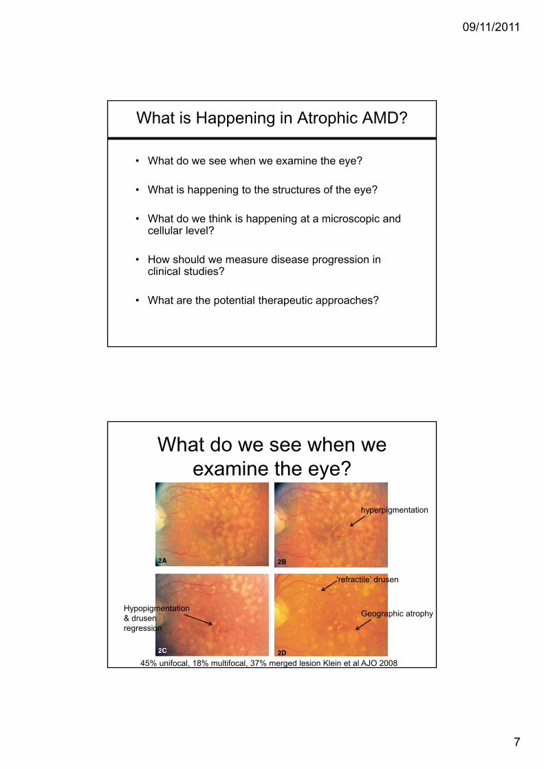
What is Happening in Atrophic AMD?
• What do we see when we examine the eye?
• What is happening to the structures of the eye?
• What do we think is happening at a microscopic and cellular level?
• How should we measure disease progression in clinical studies?
• What are the potential therapeutic approaches?
What do we see when we examine the eye?
hyperpigmentation
Hypopigmentation & drusenregression
‘refractile’ drusen
Geographic atrophy
45% unifocal, 18% multifocal, 37% merged lesion Klein et al AJO 2008

09/11/2011
8
Typical expansion rate 1.5 to 2 mm sq/year
Adapted from Holz FG et al. Invest Ophthalmol Vis Sci. 2001;42:1051-1056.
Patient#2
Patient#1
P2
FundusPhotographs Fundus Autofluorescence (AF) Photographs
P1
Initial Baseline AF Year 1 AF Year 2 AF Year 3 AF
Lipofuscin Autofluorescence Precedes Death of Photoreceptor and RPE Cells in Patients With Age-
related Macular Degeneration
What is happening to the structures of the eye?
Neural retina (photoreceptor) produces waste throughout life
With aging, the ability of RPE cells to digest these molecules
decreases
Excessive accumulation of intra(residual bodies) and
extracellular waste (drusen) results in inflammation
Bruch membrane and the RPE cells degenerate and atrophy
sets in, associated with choroicapillaris atrophy and photoreceptor loss leading slowly to severe visual loss

09/11/2011
9
What is happening to the structures of the eye?
• Rods cells lost with age (30% by age 90)
• Cone cells relatively well preserved with age
• In patients with GA, rod cells lost before cones, but cones seem structurally abnormal
What does the patient experience?Extrafoveal GAPoor vision in dim lightDifficulty readingImpaired ContrastReasonable central VA50% loose 3+ lines vision within 2 years
Subfoveal GASevere central vision lossEccentric fixation

09/11/2011
10
Diagnosis and Management
Clinical & Investigations• Diagnosis usually made clinically• Atrophy in the presence of drusen/rpe change with the
exclusion of other mimicking disorders
Current standard of care• No proven intervention halts progression of GA• Smoking cessation?• AREDS vitamins may prevent nAMD but theoretically may
worsen atophy• Removing drusen (laser etc) increased risk of nAMD
More than on cause of Atrophic AMD different therapeutic approaches
cf age-related choroidal atrophy Spaide AJO 2008

09/11/2011
11
Natural History of Geographic Atrophy (Sunness et al. Ophthalmol 2007)Atrophy expands at median 2.1mmsq/yr)
Long-term Natural History of Geographic Atrophy from Age-Related Macular Degeneration:Sunness et al Ophthalmol 2007
• Digitised colour photos• Median time to develop central GA
(after GA diagnosis) 2.5yrs {95% CI 2-3}
• Av VA decrease after ‘central’ GA development 3.7 letters initially then 22 letters at 5 years

09/11/2011
12
Potential biomarkers affecting progression of GA
• Genetics- none proven for CFH, C2, C3, APOE, or TLR3 genes. There was a nominally significant association with the LOC387715/ARMS2/HTRA1
(Progression of geographic atrophy and genotype in AMD Klein et al Ophthalmol 2010)• Systemic disease – none to date eg. BMI, CHD, diabetes > cholesterol
(Dreyhaupt et al Methods Inf Med 4/2007)• Rate of progression- 1.52 mm(2)/year (IQR, 0.81 to 2.33)
(Holz et al AJO 2007)• Autofluoresence Pattern – yes -banded (median 1.81 mm(2)/year) diffuse FAF
pattern (1.77 mm(2)/year) were significantly higher compared to eyes without FAF abnormalities (0.38 mm(2)/year) and focal FAF patterns (0.81 mm(2)/year, P < .0001).
How should we measure disease progression in clinical studies?
• Structural• Colour• AF• OCT• IR reflectance
• Functional • microperimetery• VA – standard or low luminance• Reading• Dark adaptation• Contrast sensitivity
Colour Topcon AF HRA-AFSchmitz-Valckenberg et al IOVS 2009

09/11/2011
13
Why is the standard endpoint of high contrast visual acuity problematic?
Sunness et al. Ophthalmol 2007
Alternatives•Low-luminance visual acuitySunness JS et al.. Low luminance visual dysfunction as a predictor of subsequent visual acuity loss from geographic atrophy in age-related macular degeneration. Ophthalmology. 2008 Sep;115(9):1480-8
•Reading SpeedPatel PJ, Chen FK, Da Cruz L, Rubin GS, Tufail A. Test-retest variability of reading performance metrics using MNREAD in patients with age-related macular degeneration. Invest Ophthalmol Vis Sci. 2011 Jun 1;52(6):3854-9
Reading Speed
Reading is a complex task – required2 degrees horizontal 1 above below and about 5 degrees for ‘perceptual span’
10 5
What is the best parameter to take?•Maximal reading speed•Critical print size
Lesion position may affect reading speed – language dependent
Repeatability in AMD for MNread The 95% coefficient of repeatability (CR) was 0.30 logMAR for reading acuity. The CR for critical print size and maximum reading speed varied depending on the analysis method applied
Patel PJ, Chen FK, Da Cruz L, Rubin GS, Tufail A. Test-retest variability of reading performance metrics using MNREAD in patients with age-related macular degeneration. Invest Ophthalmol Vis Sci. 2011 Jun 1;52(6):3854-9

09/11/2011
14
• Coeff of repeatability ofPointwise sensitivity 5.56dbAv sensitivity of centralMacula 2.13db for MP-1
•Still has potential to detectDecline in function withStable VA. Chen FK, Patel PJ, Webster AR, Coffey PJ, Tufail A, Da Cruz L. Nidek MP1 is able to detect subtle decline in function in inherited and age-related atrophic macular disease with stable visual acuity. Retina. 2011 Feb;31(2):371-9
•How do the different micro perimeters compare and which is best suited to measuring change?
IOVS July 2009
Other Functional tests
• Dark AdaptationDelayed cone & rod adaptation (Owsley Ophthalmol 2001, Brown 1986, Dimitrov IOVS 2008)- What is the best method of measurement what is the repeatability?
• Contrast SensitivityImpaired in AMD. Intersession 95% coef of repeatability 7 letters (Patel PJ, Chen FK, Rubin GS, Tufail A. Intersession repeatability of contrast sensitivity scores in age-related macular degeneration. Invest Ophthalmol Vis Sci. 2009 Jun;50(6):2621-5)
• Quality of Life QuestionnairesStandard questionnaires inadequate to assess tasks in low luminance. (NEI-VFQ, VF-14 only have 1-3 items). 32 Item LLQ developed) Owsley C, McGwin G Jr, Scilley K, KalliesK. Development of a questionnaire to assess vision problems under low luminance in age-related maculopathy. Invest Ophthalmol Vis Sci. 2006 Feb;47(2):528-35

09/11/2011
15
Colour-AF-OCT fundus
Yehoshua et al Ophthalmology April 2011
95% limits of agreement for 2 readers of colour images -1.54 to 1.25 mmsqAREDS report 26
GA progression over time
Median progression: ca. 1.52 mm2/year (IQR, 0.81 to 2.33) (range 0 to 13.8)Holz et al. 2007 FAM Study Group
Fovea not involved until the late course of the diseaseMaguire et Vine 1986; Schatz and McDonald 1989; Sunness et al. 1999

09/11/2011
16
Typical expansion rate 1.5 to 2 mm sq/year
Adapted from Holz FG et al. Invest Ophthalmol Vis Sci. 2001;42:1051-1056.
Patient#2
Patient#1
P2
FundusPhotographs Fundus Autofluorescence (AF) Photographs
P1
Initial Baseline AF Year 1 AF Year 2 AF Year 3 AF
Lipofuscin Autofluorescence Precedes Death of Photoreceptor and RPE Cells in Patients With Age-
related Macular Degeneration
Findings from the FAM study group‐implications in clinical trials
• Holz F et al. Progression of geographic atrophy and impact of fundus autofluorescence patterns in age‐related macular degeneration. Am J Ophthalmol. 2007 Mar;143(3):463‐72
• Fleckenstein et al.; FAM Study Group. Concordance of disease progression in bilateral geographic atrophy due to AMD. Invest Ophthalmol Vis Sci. 2010
concordance correlation coefficient between the eyes was 0.310 (95% CI, 0.097–0.495) for visual acuity, 0.706 (95% CI, 0.575–0.801) for GA size, and 0.756 (95% CI, 0.644–0.837) for GA progression rate

09/11/2011
17
Influence of nuclear lens opacities
AREDS Report No. 4Am J Ophthalmol. 2001
1
2
3
4
1Lens clear Nuclear sclerosis
Qua
lity
cSLO
Fundus camera
Influence of nuclear lens opacities
Insufficient
Good
Schmitz-Valckenberg S, Fleckenstein M, Göbel AP, Sehmi K, Fitzke FW, Holz FG, Tufail A. Evaluation of autofluorescence imaging with the scanning laser ophthalmoscope and the fundus camera in age-related geographic atrophy. Am J Ophthalmol. 2008 Aug;146(2):183-92

09/11/2011
18
Optical Coherence Tomography (OCT)
Fleckenstein et al. Tracking progression with spectral-domain optical coherence tomography in geographic atrophy caused by age-related macular degeneration. Invest Ophthalmol Vis Sci. 2010 Aug;51(8):3846-52
09/11/2011
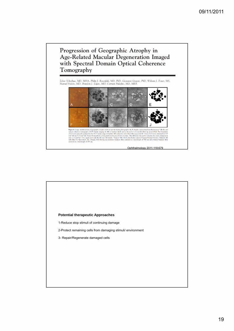
19
Ophthalmology 2011:118:679
Potential therapeutic Approaches
1-Reduce stop stimuli of continuing damage
2-Protect remaining cells from damaging stimuli/ environment
3- Repair/Regenerate damaged cells

09/11/2011
20
Can we avoid the development of Atrophic AMD?
‘Prevention is better than cure’
Genetics
Environment
Pharmaceutical
10-15%
5+%
Summary
• The geographic atrophy form of AMD will become an increasingly common cause of severe vision loss
• No proven treatments yet halt progression
• Current outcomes measures in AMD trials (high contrast VA) may not be optimal for GA trial
• Functional testing in low luminance or test that measure parafoveal function may be most suitable, but noisey
• Structural changes can reproducibly measured and may represent a good endpoint for clinical trials