Embed Size (px)
Citation preview

© 2017 Al-Zamil and Yassin. This work is published and licensed by Dove Medical Press Limited. The full terms of this license are available at https://www.dovepress.com/terms.php and incorporate the Creative Commons Attribution – Non Commercial (unported, v3.0) License (http://creativecommons.org/licenses/by-nc/3.0/). By accessing the work you
hereby accept the Terms. Non-commercial uses of the work are permitted without any further permission from Dove Medical Press Limited, provided the work is properly attributed. For permission for commercial use of this work, please see paragraphs 4.2 and 5 of our Terms (https://www.dovepress.com/terms.php).
Clinical Interventions in Aging 2017:12 1313–1330
Clinical Interventions in Aging Dovepress
submit your manuscript | www.dovepress.com
Dovepress 1313
R e v I e w
open access to scientific and medical research
Open Access Full Text Article
http://dx.doi.org/10.2147/CIA.S143508
Recent developments in age-related macular degeneration: a review
waseem M Al-ZamilSanaa A YassinDepartment of Ophthalmology, Imam Abdulrahman Bin Faisal University, Al-Khobar, Saudi Arabia
Background: Visual impairment in elderly people is a considerable health problem that
significantly affects quality of life of millions worldwide. The magnitude of this issue is becoming
more evident with an aging population and an increasing number of older individuals.
Objective: The objective of this article was to review the clinical and pathological aspects of
age-related macular degeneration (AMD), diagnostic tools, and therapeutic modalities presently
available or underway for both atrophic and wet forms of the disease.
Methods: An online review of the PubMed database was performed, searching for the key
words. The search was limited to articles published since 1980 to date.
Results: Several risk factors have been linked to AMD, such as age (.60 years), lifestyle
(smoking and diet), and family history. Although the pathogenesis of AMD remains unclear,
genetic factors have been implicated in the condition. Treatment for atrophic AMD is mainly
close observation, coupled with nutritional supplements such as zinc and antioxidants, whereas
treatment of wet AMD is based on targeting choroidal neovascular membranes.
Conclusion: Identification of modifiable risk factors would improve the possibilities of prevent-
ing the progression of AMD. The role of anti-vascular endothelial growth factor (anti-VEGF)
agents has transformed the therapeutic approach of the potentially blinding disease “wet AMD”
into a more favorable outcome.
Keywords: age-related macular degeneration, anti-VEGF, risk factors, treatment
IntroductionAge-related macular degeneration (AMD) is an acquired disease of the macula
characterized by progressive visual impairment because of late-onset neurodegenera-
tion of the photoreceptor–retinal pigment epithelial complex. AMD is a major cause of
central visual loss in the developed world affecting 10% of people older than 65 years
and more than 25% of people older than 75 years.1 In the US alone, ~2 million individuals
have advanced AMD and .8 million individuals have an intermediate form of the
disease. These numbers are expected to rise by 50% in 2020.2 In Saudi Arabia, AMD
represents 3.3% of the major causes of blindness in individuals older than 50 years.3
Human retinal pigment epithelium (RPE) is a non-dividing cell, which has several
functions essential for the maintenance of photoreceptor cells. RPE undergoes various
changes during aging, leading to the emergence of a clinically detectable focal yellow
accumulation of extracellular, polymorphous material, called drusen, at the interface
between the RPE and the inner collagenous zone of Bruch’s membrane. The pres-
ence of drusen within the macula is the hallmark sign of AMD. However, individuals
with small drusen in the absence of other ocular abnormalities are at a decreased
risk to progress to the more severe forms of the disease. A widely variable clinical
presentation related to drusen characteristics and pigmentary disturbances, such as
Correspondence: Sanaa A YassinDepartment of Ophthalmology, Imam Abdulrahman Bin Faisal University, PO Box 40097, Al-Khobar 31952, Saudi ArabiaTel +966 5 0480 5108Fax +966 3 896 6776email [email protected]
Journal name: Clinical Interventions in AgingArticle Designation: ReviewYear: 2017Volume: 12Running head verso: Al-Zamil and YassinRunning head recto: Recent developments in AMDDOI: http://dx.doi.org/10.2147/CIA.S143508

Clinical Interventions in Aging 2017:12submit your manuscript | www.dovepress.com
Dovepress
Dovepress
1314
Al-Zamil and Yassin
hypopigmentation and hyperpigmentation, can be found in
individuals with AMD.4 Various pathologies, including focal
detachment of the RPE, outer retinal atrophy, and new blood
vessel growth between Bruch’s membrane and the retina, can
progress into either geographic atrophy (GA) or choroidal
neovascularization (CNV) AMD, which are also known as
dry or wet AMD, respectively.
The aim of this review was to recapitulate the extensive
work conducted over the last 3 decades in the clinical and
pathological aspects of AMD, diagnostic tools, and thera-
peutic modalities presently available or underway for both
atrophic and wet forms of the disease. An online review of
the database PubMed was performed, searching for the key
words: age-related macular degeneration, risk factors, VEGF,
prevention, genetics, management, and phrases in combina-
tion. Some literature was derived from the reference lists of
identified publications. Additionally literature search of the
Clinical Trials (https://ClinicalTrials.gov) was performed
using “age-related macular degeneration” as keyword. The
search was limited to articles published since 1980 to date.
PathophysiologyThe exact pathophysiology of AMD is not fully understood;
however, findings from ongoing studies are expanding our
knowledge of the disease and the underlying mechanism.
It is believed that the pathogenesis of AMD is the result
of a complex multifactorial interaction between metabolic,
functional, genetic, and environmental factors.5 With aging,
intracellular residual bodies containing lipofuscin accumulate
in RPE cells.6 RPE cells express such materials that would
normally be eliminated by the choriocapillaris; however,
as RPE dysfunction progresses, it results in alterations in
the permeability of Bruch’s membrane, which leads to an
accumulation of extruded material (drusen) between the
two layers.7 The appearance of drusen may either be found
with or is proceeded by a thickening of the collagenous
layers of Bruch’s membrane, a degeneration of elastin
and collagen within the membrane, and its calcification.7
Furthermore, it has been observed that the choriocapillaris
thins in patients with AMD, which may contribute to the
decline in the removal of extracellular material that results
in drusen formation.8
Twin studies have demonstrated that family history is an
established risk factor for AMD.9 One study recently reported
that drusen formation may stimulate an inflammatory cascade
that has a role in AMD progression.10 Another study found evi-
dence that polymorphisms in genes that encode complement
factors, which have an important role in the immune system,
can affect AMD risk either by increasing the risk for or by
protecting against development of the disease.11 Evidence of
associations between increased risk of AMD and complement
factor H (CFH) on chromosome 112 and pleckstrin homology
domain containing A1 (PLEKHA1) and the hypothetical gene
LOC387715 on chromosome 10 has been documented.13
In contrast, another study found protective effects of genetic
polymorphisms in complement factor B (CFB), complement
component 2 (C2) on chromosome 6, and specific haplotypes
in CFH that demonstrated decreased risk of AMD.11
CFH suppresses complement pathway; hence, in the
presence of abnormal CFH activity, the complement cascade
is activated with a consequent downstream inflammatory
response to subretinal tissues.14 Inflammatory components
from the cascade pathway have been identified within
drusen,10 and additionally, environmental factors such as
smoking can decrease CFH levels, which may account for
smokers having a significantly increased risk of developing
AMD compared to nonsmokers.15,16 Furthermore, comple-
ment pathway activation could be inhibited because of the
protective effects conferred by polymorphisms in CFB and
C2, thereby limiting the degree of chronic inflammation.11
Additional evidence suggests that choroidal C-reactive
protein-specific deposition contributes to AMD pathogenesis
in individuals with homozygous CFH.17
Drusen formation signals RPE dysfunction, which pro-
motes RPE loss with further progression that results in photo-
receptor death.18 As previously described, RPE degeneration
consequently leads to dysfunction of Bruch’s membrane.
Progressive damage to Bruch’s membrane with the upregula-
tion of vascular endothelial growth factor (VEGF) promotes
the outgrowth of abnormal choroidal vessels underneath
the RPE and, subsequently, under the retina. Initially, these
abnormal vessels present with subretinal extravasations that
may hemorrhage before they regress and form a disciform
scar. Thus, the visual outcome of end-stage wet AMD is the
permanent loss of central vision.8
Several studies have investigated the molecular pathway
that underlies GA and vision loss.19,20 This pathway implies
that RPE death consequently leads to photoreceptor loss,
which progressively results in visual loss over time. Low
levels of the ribonucleic acid (RNA)-cleaving enzyme,
DICER1, in RPE cells were observed in patients with dry
AMD.21 It was reported that decreased levels of DICER1 lead
to a decreased rate in the breakdown of RNA-Alu molecules,
which are noncoding sequences of RNA. The overabundance
of cytoplasmic RNA-Alu activates inflammatory proteins
such as NLRP3 in the inflammasome, which results in the

Clinical Interventions in Aging 2017:12 submit your manuscript | www.dovepress.com
Dovepress
Dovepress
1315
Recent developments in AMD
activation of a cascade of molecular reactions that result in
RPE cell loss.22,23 Furthermore, mitochondrial dysfunction has
been associated with the development of dry AMD. The mito-
chondrial dysmorphology observed in the RPE in individuals
with AMD was consistent with severe dysfunction.24
Risk factorsAgeThe development of advanced AMD has multifactorial risk
factors, such as increasing age, ethnicity, and genetics. Age
is the strongest predictor of AMD.25 Although infrequent in
people younger than 50 years, the risk of acquiring AMD
increases more than threefold in patients older than 75 years
compared to patients between 65 and 74 years of age.26,27
In the US, AMD is found in 30% of individuals older than
85 years.26
Family history and geneticsAMD is most frequently found in Caucasians, followed
by Hispanics and Asians with the lowest rate reported in
African Americans.2 There is an increased risk in individuals
with a positive family history of AMD, which is typically
multifactorial in nature. Siblings of an affected individual
have a threefold to sixfold higher risk than those of the
general population.28
To date, 34 genetic loci including 52 gene variants have
been identified that have been linked to AMD.29 Using
genome-wide screening approaches, several sets of gene
variants have been identified in different chromosomes,
including chromosomes 1, 6, and 10.30,31 These culprit genes
play role in controlling immune response, inflammatory
processes, and retina homeostasis, and the extent of dysfunc-
tion of these reactions in individuals with AMD is attributed
to variations found in these loci. The most investigated
candidate genes are CFH on chromosome 1 at 1q31.3, high-
temperature requirement serine peptidase 1 (HTRA1; also
known as age-related maculopathy [ARM] susceptibility 2
[ARMS2]) on chromosome 10 at 10q26, and CFB/C2 on
chromosome 6 at 6p21.3.32,33 Other genetic variants putatively
related to AMD include a polymorphism in lipase C hepatic
type (LIPC), which is involved in high-density lipoprotein
cholesterol metabolism;34 the single-nucleotide polymor-
phism (SNP) rs3775291 in toll-like receptor 3 (TLR3);19 and
the tissue inhibitor of metalloproteinase 3 (TIMP3).35
The contribution and role of genetic predisposition extend
beyond increased susceptibility of an individual developing
AMD and may also affect treatment response. Smailhodzic
et al36 demonstrated a cumulative effect of high-risk alleles
in CFH, ARMS2, and VEGFA that were associated with a
younger age of onset and inadequate response to intravitreal
anti-vascular endothelial growth factor (anti-VEGF) agents
in individuals with AMD. Medina et al37 also found that in
individuals with a homozygous CC group, variants of the
CFH gene polymorphism T1277C were associated with
delayed functional and limited morphological response to
the initial intravitreal injection of Avastin (bevacizumab) in
wet AMD. Therefore, further pharmacogenomic studies may
aid in developing a rational guide to treatment regimens and
to optimize treatment response tailored to an individual’s
genetic background.
Lifestyle, diet, and nutritionSmoking is the main influential modifiable risk factor, and
patients should be encouraged at each visit to refrain from
smoking to prevent further visual loss. Smokers for .40 years
are two to four times more likely to develop AMD than
nonsmokers of the same age.38 The ALIENOR study from
France showed that high pulse pressure was associated with
an increased risk of late-stage AMD, whereas systolic or
diastolic blood pressure or the use of antihypertensive medi-
cations was not significantly associated with an increased
risk of either early- or late-stage AMD.39
A high intake of certain fats, such as saturated fats, trans
fats, and omega-6 fatty acids, has been associated with
a twofold increase in the prevalence of AMD, whereas
monounsaturated fats were potentially protective.40 There is
conflicting evidence concerning the role of sunlight exposure
in AMD occurrence. In a study that compared sun exposure
of individuals with end-stage AMD to unaffected spouses,
researchers found no evidence of an association;41 however,
other studies have shown that high-energy visible light may
contribute to AMD.42,43 Furthermore, data from several large
population-based studies have indicated that there may be a
gender effect with women at a higher risk to develop AMD
compared to men.26,44
It has been investigated whether dietary antioxidants, such
as vitamins C and E, carotenoids (eg, lutein and zeaxanthin),
and zinc, are additional risk factors for AMD; however, data
from observational studies revealed insufficient evidence
supporting a role.45,46
Aspirin useThere is inconsistent evidence linking aspirin use and AMD.
The Beaver Dam Eye Study demonstrated that the incidence
of late-stage AMD in patients who used aspirin at least twice
weekly for 10 years was higher compared to in those who

Clinical Interventions in Aging 2017:12submit your manuscript | www.dovepress.com
Dovepress
Dovepress
1316
Al-Zamil and Yassin
did not use aspirin,47 whereas a different study reported
that aspirin conferred a potentially protective effect against
developing the disease.48 A meta-analysis of 10 studies,
including .171,000 patients, concluded that aspirin use
was not a risk factor for AMD.49 Based on the available
information on aspirin use in patients with AMD, the current
preferred practice is to continue aspirin therapy prescribed
by physicians.
Other risk factorsCombined analysis of longitudinal data from two large
population-based cohorts suggests an increased risk of
developing late-stage AMD, particularly neovascular AMD
in older individuals who undergo cataract surgery.50
Other proposed risk factors for AMD include abdominal
obesity, especially among men;51 hyperlipidemia;52 hyperopia;53
light iris color;41 cardiovascular disease;25 hormonal status;54
alcohol use;55 vitamin B and D status;56,57 and elevated
C-reactive protein.58
A systematic review including 18 prospective and cross-
sectional studies and six case–control studies involving
113,780 individuals identified age (.60 years), smoking,
previous cataract surgery, and a family history of AMD as
strong risk factors for AMD, whereas increased body mass
index, previous cardiovascular disease, hypertension, and
higher plasma fibrinogen were found to be moderate risk
factors.25 Table 1 summarizes the risk factors associated
with AMD.
Classification and severity gradingVarious classification systems are used to delineate and
characterize AMD for both clinical and research purposes.
Currently, there is no universally accepted consensus on
specific definitions, although conventionally, AMD has two
main types of macular degeneration known as dry and wet
AMD. Dry AMD, which is also known as the nonexudative
form, is the most common type, comprising ~90% of all
diagnosed cases. GA is the advanced stage of dry AMD.
Wet AMD is also known as the exudative form, and although
less common than dry AMD, it is associated with a more rapid
progression to advanced vision loss. The main manifestations
of wet AMD are CNV and pigment epithelial detachment
(PED). Approximately 10%–20% of patients with nonexuda-
tive AMD may develop the wet form, which is estimated to
affect 1.75 million people in the US.2,59
The development of standardized photographic retinal
grading methods such as the Wisconsin ARM grading
system60 was followed by the International ARM Epidemio-
logical Study for classification and grading,61 which redefined
the diagnostic system and led to the adoption of a stricter
criterion for diagnosing AMD. The ARM grading system
was characterized by minimal or moderate nonexudative
age-related changes in the macula. By definition, the presence
of advanced RPE atrophy (ie, GA) or CNV was essential to
establish a diagnosis of nonexudative AMD or wet AMD,
respectively. After applying the International Classifica-
tion criteria, ARM accounted for ~85%–90% of cases with
AMD representing 10%–15% of affected individuals with
age-related macular changes.
The Age-Related Eye Disease Study (AREDS) defined
the categories of AMD based on the presenting features of
drusen, atrophy, and neovascularization (Table 2). Drusen
were classified as small (,63 μm in diameter), intermediate
(63–124 μm), or large (.125 μm). ARED categories of
AMD were defined as: 1) no AMD if there were fewer than
five small drusen; 2) mild AMD based on the identification
of multiple small drusen or at least one intermediate-sized
drusen; 3) intermediate AMD classified by extensive
intermediate-sized drusen, more than one large drusen, or
non-central GA; and 4) advanced AMD determined by central
GA or CNV causing vision loss (defined as visual acuity
[VA] worse than 20/32) in one eye.62
Klein et al developed a risk assessment model using data
from AREDS; the model incorporated demographic, envi-
ronmental, phenotypic, and genetic covariates. The model
gave satisfactory results, with very good discrimination,
Table 1 Summary of risk factors for AMD
Risk factors Strength and consistency of association
Older age Strong and consistentCigarette smokingPrevious cataract surgeryFamily history of AMDHigher body mass index Moderate and consistentHistory of cardiovascular diseaseHypertensionHigher plasma fibrinogenGender weak and inconsistentethnicityDiabetes Iris colorHistory of cerebrovascular diseaseSerum total cholesterolHDL cholesterolTriglyceride levels
Abbreviations: AMD, age-related macular degeneration; HDL, high-density lipoprotein.

Clinical Interventions in Aging 2017:12 submit your manuscript | www.dovepress.com
Dovepress
Dovepress
1317
Recent developments in AMD
calibration, and overall performance. This risk assessment
tool was made available for online use.63
Clinical features and diagnosisAMD is characterized by a variable presentation of clinically
observable changes at the posterior pole.
Variable types, sizes, and distribution of drusen within the
posterior pole with a high degree of symmetry between the
two eyes of an individual are characteristics of dry AMD.64
Pseudodrusen are a different entity from conventional drusen.
They are drusenoid deposits located in the subretinal
space above the RPE.65 Pseudodrusen are associated with
advanced AMD.66 They have been classified into three
groups, dot, reticular, and confluent pseudodrusen, depending
on different morphological appearances.67 It was found that
the prevalence of GA was associated with the presence of
ribbon-dominant (confluent)-type pseudodrusen, large soft
drusen, and female gender.68 Pigmentary disturbances are a
frequent finding in AMD that can be in the form of either
focal hyperpigmentation or hypopigmentation. Progressive
well-demarcated atrophic patches of the RPE may appear in
the perifoveal area. Over time, these atrophic patches enlarge
and may coalesce, resulting in GA.7 Wet AMD may present
with drusen, PED, intraretinal hemorrhages, and CNV, which
appears as a well-demarcated graying area of the retina.59
There are different methods for diagnosis, including
fundus imaging and optical coherence tomography (OCT).
Digital fundus photography is an effective approach to
Table 2 Demonstration of the classification of AMD, according to AREDS
Classification Category Clinical signs
No AMD 1 0–5 small drusen (,63 μm in diameter)
early AMD 2 Multiple small drusen or a few intermediate-sized (63–124 μm in diameter) drusen, or macular pigmentary changes
Intermediate AMD 3 extensive intermediate drusen or at least one large ($125 μm) drusen, or GA not involving the foveal center
Advanced AMD 4 GA involving the foveal center or any evidence of choroidal neovascularizationa
Note: aSubretinal hemorrhage, serous retinal or RPE detachments, lipid exudates, or fibrovascular scar.Abbreviations: AMD, age-related macular degeneration; AReDS, Age-Related eye Disease Study; GA, geographic atrophy; RPe, retinal pigment epithelium.

Clinical Interventions in Aging 2017:12submit your manuscript | www.dovepress.com
Dovepress
Dovepress
1318
Al-Zamil and Yassin
document clinical findings.60 However, there are a few
difficulties that hinder precision of color photograph measure-
ments, such as image quality, resolution, noise, and dynamic,
as well as the sensitivity of the camera, media clarity, and
fundus pigmentation. Foveal involvement is also difficult
to determine. Fundus autofluorescence (FAF) imaging
depends on the stimulated emission of light from molecules
in the RPE, mainly lipofuscin.69 FAF imaging is a helpful
tool for evaluating and monitoring the topographic structure
and health of the RPE because the localized deposition of
lipofuscin in RPE cells results in increased autofluorescence,
whereas in contrast, a decrease or absence of RPE lipofuscin
will result in a decreased FAF signal.70,71
In case of GA, a well-circumscribed dark area typically
appears in FAF images that clearly delineates atrophy of
the RPE.72 Increased autofluorescence surrounding the GA
area has been considered a risk factor for the progression
of AMD.71,73 The multicenter Fundus Autofluorescence in
Age-related Macular Degeneration (FAM) study using a
confocal scanning laser ophthalmoscopy classified patterns
of abnormal FAF in the junctional zone of GA in patients
with AMD into five different patterns as follows: none, focal
increased, banded, patchy, and diffuse.70 The progression rate
of GA was highest in eyes with the diffuse trickling pattern
followed by the banded pattern in a study on 54 eyes.74
A hyperpigmented area in the fundus can either show as
decreased FAF intensities because of absorption by melanin
granules or as increased FAF signal, which is believed to
arise from melanolipofuscin. Similarly, variable increased
or decreased intensities of FAF can be observed in non-RPE-
related changes, such as subpigment epithelial or subneuro-
sensory detachments, areas with extracellular fluid exudation
or hemorrhage with subsequent biosynthesis of fluorophores
that possess autofluorescent properties.69,75
Spectral-domain optical coherence tomography (SD-OCT)
has provided an improved understanding of the microstruc-
tural changes related to AMD.76 For example, drusen may
appear as elevations at the level of the RPE. In GA, OCT
demonstrated loss of the RPE layer and photoreceptor bands
within the atrophic lesion.76 In addition, OCT is used to deter-
mine subretinal or intraretinal blood or fluid.77 Fluid appears
as black gaps or elevations and is indicative of wet AMD.
OCT is often used when fluorescein angiography (FA) is
equivocal and is used to monitor treatment responses.78 The
technology of OCT has progressed rapidly since its first intro-
duction to ophthalmology in 1995.79 Improved axial resolution
and increased scanning speed have facilitated the introduc-
tion of new OCT techniques, which can more effectively
visualize the posterior segment.80 En-face OCT is one such
visualization approach that has significantly benefited from
technical advancements. En-face OCT in current systems is
based on software reconstruction of OCT images.
Nunes et al81 used en-face OCT to study the progression
of GA at the inner/outer segment junction of 30 patients with
AMD. Interestingly, they found outer retinal disruption that
extended beyond the borders of GA, which are visualized
as focal hyporeflective areas on en-face OCT, predicted GA
progression for 1 year in 43.3% of eyes in their patient cohort.
Another novel technique OCT angiography (OCT-A) is an
in vivo noninvasive imaging of blood vessels with a high
resolution in three dimensions, thus enabling a qualitative
and quantitative analysis of characteristics of normal and
pathological blood vessels at different layers of the retina and
choroid. OCT-A may have applications in both neovascular
and non-neovascular AMD. In neovascular AMD, OCT-A
can be used to monitor treatment response over time,82 while
in non-neovascular AMD, it is useful to study the choriocapil-
laris and other vascular structures believed to be important
in the pathogenesis of the disease.8,77
FA reveals drusen that bind fluorescein and hyperfluores-
cence in the late stage of angiography; however, fluorescein
uptake depends on the components found in drusen that
can vary from being hydrophobic to hydrophilic (Figure 1).
Consequently, only 50% of clinically identifiable drusen will
stain with fluorescein.83 Large drusen tend to stain with fluo-
rescein more frequently than smaller drusen. During the transit
phase of FA, areas of GA appear as well-demarcated patches
of hyperfluorescence surrounded by a rim of blocked fluo-
rescence with visualization of the choroidal vessels passing
through the region. In the late phase of FA, some staining
may persist.84 The gold standard for confirming diagnosis
whenever CNV is suspected is FA. In addition, FA may
assist in determining the pattern (classic or occult), borders,
composition, and location of the neovascular complex as well
as guide treatment with laser or intravitreal injections.84
In the early phases of FA, CNV presents as a hyperfluo-
rescent lesion that enlarges in size and intensity in late phases
as the fluorescein leaks from the neovascular membranes. The
two major angiographic patterns are classic and occult forms
of CNV. Differences between the two patterns are thought
to arise from the location of the neovascular membrane.
In classic lesions, CNV penetrates the RPE and grows in front
of the RPE, whereas occult lesions are sub-RPE.85 As shown in
Figure 2, classic CNV is characterized by well-circumscribed
lesions with early hyperfluorescence and progressive leakage
in later frames. A concomitant pooling of fluorescein in the

Clinical Interventions in Aging 2017:12 submit your manuscript | www.dovepress.com
Dovepress
Dovepress
1319
Recent developments in AMD
subretinal space can also be observed. Occult CNV is catego-
rized as an ill-defined leakage from an undetermined source.
Occult CNV is also presumed when a fibrovascular PED is
suspected because these detachments are accompanied with
an irregular RPE elevation and a stippled hyperfluorescent
pattern with or without leakage or pooling.84
The value of indocyanine green (ICG) angiography in
assessing and treating AMD has been debated. Nevertheless,
ICG is useful in assessing specific forms of AMD, such as
occult CNV, which is poorly defined, and lesions like retinal
angiomatous proliferation or idiopathic polypoidal choroidal
vasculopathy.86
Although their specific role in clinical practice is not
distinctly determined, other tests such as microperimetry
can be used to quantify retinal sensitivity and fixation in
AMD.87,88
Figure 1 Fundus photograph and late FA phase demonstrating drusen.Note: FA shows fewer drusen than clinically identifiable.Abbreviation: FA, fluorescein angiography.
Figure 2 Classic choroidal neovascular membrane in AMD is depicted on FA, showing early hyperfluorescence with progressively increasing hyperfluorescence on successive images, surrounded by hypofluorescence due to blockage from subretinal hemorrhage.Abbreviations: AMD, age-related macular degeneration; FA, fluorescein angiography.

Clinical Interventions in Aging 2017:12submit your manuscript | www.dovepress.com
Dovepress
Dovepress
1320
Al-Zamil and Yassin
Prophylaxis and treatment of dry AMDCurrent management of GA in AMD basically depends on
documentation, observation, and surveillance for early rec-
ognition of any changes in visual function and detection of
CNV at a treatable stage.89 Home monitoring for changes in
the central visual field is usually advised using an Amsler
grid to detect the presence of metamorphopsia or scotoma in
individuals with dry AMD, although the sensitivity of Amsler
charts in revealing macular disease can be suboptimal.90
The identification of modifiable risk factors and the devel-
opment of future preventive treatments are crucial because
there is currently no available treatment of dry AMD and
population aging will result in a major upsurge in AMD bur-
den on both families and socioeconomically.91 Thus, intense
efforts have been made to identify possible therapeutic targets
to reduce disease progression. Tobacco smoking is consis-
tently associated with AMD, and because it is a modifiable
risk factor, a primary therapeutic recommendation should
advocate the significance of smoking cessation.25 In addi-
tion, the preventive role of antioxidant supplements was
evaluated in several studies.62,92,93 Oxidative damage from
various sources, such as light exposure, inflammation, and
oxidative stress, to the retina has been strongly linked with
AMD. Although results from initial epidemiological studies
indicated an association, later studies were inconclusive.94–97
The AREDS study was designed to determine whether anti-
oxidant supplementation could have protective effects against
AMD.62,92 AREDS was a multicenter, randomized, double-
masked, clinical trial that enrolled 3,640 participants and was
stratified into four categories of severity. The study demon-
strated that a daily dose of vitamin C (500 mg), vitamin E
(400 IU), β-carotene (15 mg), zinc oxide (80 mg), and cupric
oxide (2 mg) significantly reduced the odds of developing
advanced AMD in individuals with high-risk characteristics.
The greatest risk reduction (34% odds reduction) was seen
in participants from categories 3 and 4, who received both
antioxidants and zinc (odds ratio [OR] =0.66; P=0.001),
compared to other categories in the treatment arm.
A second AREDS study (AREDS2) was started in 2006 to
evaluate the role of oral supplementation with high doses of
lutein, zeaxanthin, and omega-3 fatty acids (docosahexaenoic
acid [DHA] and its precursor eicosapentaenoic acid [EPA]).98
A second objective of the study was to assess the effects of
eliminating beta-carotene and reducing the zinc dose from the
original AREDS formula. After a median 5-year follow-up,
there was no convincing support toward including DHA/EPA
or lutein/zeaxanthin to the AREDS formula, although
subgroup analysis indicated that substitution of beta-carotene
with lutein and zeaxanthin showed a slightly decreased risk of
progression (hazard ratio =0.82; P=0.02). However, partici-
pants who were former smokers and took the revised formula
with beta-carotene were more prone to lung cancer compared
to those whose formula did not include beta-carotene (2.0%
vs 0.9%, respectively; P=0.04). The final recommendation
from AREDS2 was to substitute beta-carotene with lutein/
zeaxanthin in the updated formula.98
Dry AMD has no approved treatment, which is driving
efforts toward understanding the pathophysiology of the
disease for the scientific development of rationale treatments.
Several innovative treatments for dry AMD are in progress.
Because inflammation and polymorphisms in genes involved
in the complement pathway have been consistently linked to
AMD pathogenesis, several trials were performed to evaluate
the safety, tolerability, and response of different comple-
ment inhibitors to treat dry AMD.99 The 18-month Phase II
MAHALO study assessed the efficacy of monthly intravit-
real injections of lampalizumab in halting the progression
of GA in patients with bilateral disease.100 Lampalizumab
(anti-factor D Fab) is a humanized monoclonal antibody
(Fab fragment), which is a selective inhibitor of complement
factor D (CFD). Using autofluorescence and color fundus
photographs, it was found that patients had a 20% reduction
in mean change from baseline in the GA area at month 18.
Remarkably, in a subset of complement factor I-positive
patients, intravitreal administration of lampalizumab was
associated with a significant 44% reduction in the GA
area progression at month 18 compared to sham treatment
(P,0.005). Lampalizumab is under further evaluation in two
large, prospective, Phase III, randomized, double-masked
clinical trials, CHROMA and SPECTRI.101,102 Currently,
lampalizumab is the most promising potential therapeutic
agent for dry AMD that may be commercially available as
early as 2019.
There is evidence that tetracyclines can target the low-
grade inflammation caused by aberrant complement pathway
activation, which is believed to underlie the pathogenesis of
dry AMD, and may also slow the progression of GA. Doxy-
cycline is a broad-spectrum antibiotic at a dose of 100 mg
a day; however, at a lower dosage (20–40 mg/day), it acts
as an anti-inflammatory.103 Low-dose oral doxycycline is
currently approved for treating the inflammatory lesions of
rosacea and is under investigation in a randomized, double-
masked, placebo-controlled study (TOGA) to determine
its efficacy and safety in slowing the progression of GA in
patients with AMD.104

Clinical Interventions in Aging 2017:12 submit your manuscript | www.dovepress.com
Dovepress
Dovepress
1321
Recent developments in AMD
Statins are lipid-lowering anti-inflammatory agents with
pleiotropic actions. There is emerging genetic and pathologi-
cal evidence from a cross-sectional study that included 5,604
participants in the National Health and Nutrition Examina-
tion Survey from 2005 to 2008, $40 years of age, who
were confirmed to diagnosis of AMD, the use of statins, and
comorbidities and health-related behaviors such as smoking.
The study suggests a possible role for statins in delaying the
progression of AMD and its prevention in individuals at
the age of 68 years or older;105 however, additional studies
are needed to fully evaluate their therapeutic importance.
A novel mitochondrial protective compound, MTP-131
(Ocuvia), is a topical ophthalmological investigational drug
under development to treat dry AMD. Preliminary testing
of MTP-131 in cell culture and in a mouse model of AMD
was performed, and the results indicated that the investi-
gational drug was highly effective in several experimental
models.106 Currently, a Phase I/II open-label dose-escalation
clinical study on topical MTP-131 is carried on to determine
its safety and tolerability in patients with diabetic macular
edema and dry AMD.107
Rapamycin (Sirolimus) is an inhibitor of the mammalian
target of rapamycin complex (mTOR), which is a pleiotropic
protein kinase involved in regulating organismal growth
and homeostasis. Sirolimus is clinically administered as an
immunosuppressive agent following organ transplantation for
its ability to suppress T-cell and B-cell proliferation and anti-
body production.108 However, intravitreal or subconjunctival
(440 μg/every 3 months) administration of sirolimus in six
eyes or 11 eyes, respectively, showed no effect on VA, GA
size, or GA progression in patients with dry AMD.108,109
Stem-cell therapy is under investigation as a potential
cell-replacement approach for damaged and lost photorecep-
tors and RPE cells in GA lesions. A prospective study of nine
patients with atrophic AMD (median age of 77 years) showed
that subretinal transplantation of human embryonic stem cell-
derived retinal epithelial cells improved VA at 12 months in the
treated eye compared to that in the untreated eye.110 Although
the procedure was well tolerated and patients were followed
for up to 3 years, these findings only support proof of concept
and are too preliminary to suggest its use in treatment. Table 3
lists the emerging treatments targeting advanced AMD.
Other promising concepts of therapeutic approaches in
the pipeline are to target inflammasomes,111 visual cycle
modulators,112,113 glatiramer acetate (suppressor of T cells
and downregulates inflammatory cytokines),114 and neuro-
protection.113,115 As part of regular patient care and to ensure
continued quality of life, individuals with poor vision should
be supported by low vision aids as needed.
Treatment of wet AMDManagement of wet AMD has undergone significant
advances in recent years. So far, none of the used treatments
could certainly cure the disease or reverse its course. Macular
photocoagulation studies (1970s) showed the effectiveness
and efficacy of laser photocoagulation in the treatment of
wet AMD. Remarkable visual outcome was described in
extrafoveal lesions,116,117 while parafoveal lesions showed
less favorable results.118,119
Laser therapy was not considered as an ideal procedure
for treating subfoveal lesions despite reported benefits.120,121
Currently, the adoption of laser photocoagulation in the
treatment of wet AMD is hampered by several issues, includ-
ing a high recurrence rate; a risk of producing vision loss,
especially with subfoveal membranes; and a limited visual
improvement potential. In the era of anti-VEGF therapies,
the role of direct photocoagulation as a major treatment
approach for wet AMD is waning. However, if used, this
treatment should be limited to treat very small lesions outside
the central macula.
Table 3 emerging treatments targeting advanced AMD are being tested actively in clinical trials
Agent Targeted pathology
Route of administration
Mechanism of action
Lampalizumab99 GA Intravitreal Anti-factor D FabOracea104 GA Oral Antibiotic–anti-inflammatoryMTP-131 (Ocuvia)107 GA Topical Mitochondrial protective compoundMA09-hRPe110 GA Subretinal injection Human umbilical tissue-derived cellsBrimonidine tartrate implant113 GA Intravitreal implant Alpha-2-antagonisteculizumab188 GA Intravitreal mAb against complement factor C5e10030172,173 Neovascular AMD Intravitreal Anti-PDGF PeGylated aptamerProton radiation189 Neovascular AMD external radiation Radiation: proton radiationAbicipar pegol190 Neovascular AMD Intravitreal injection Anti-veGFRTH258191 Neovascular AMD Intravitreal injection Anti-veGF
Abbreviations: AMD, age-related macular degeneration; GA, geographic atrophy; PDGF, platelet-derived growth factor; veGF, vascular endothelial growth factor.

Clinical Interventions in Aging 2017:12submit your manuscript | www.dovepress.com
Dovepress
Dovepress
1322
Al-Zamil and Yassin
Photodynamic therapy (PDT) was introduced in the late
1990s.122 The photosensitizer verteporfin is administered
intravenously at a dose of 6 mg/m2 of body surface area and
is activated by photons with a low-energy visible red laser
(689 nm) using a specific type of contact lens. The activated
dye forms reactive-free radicals that damage the vascular
endothelium and induce occlusion of new vessels. Verteporfin
is an approved photosensitizer by the US Food and Drug
Administration (FDA) for ophthalmic use. The selectivity of
the photosensitizer to the CNV, which retains the dye more
avidly than normal vessels, allows directed targeting at the
lesion without damaging surrounding tissues. In a Phase III
trial, 609 participants with subfoveal neovascular AMD were
randomized to PDT or placebo; it demonstrated that more
eyes treated with PDT were spared from moderate vision
loss than those treated with placebo (loss of ,15 letters of
VA at 12 months; 61% vs 46%, respectively; P,0.001).122
In subgroup analyses, the VA benefit (,15 letters lost) found
in the PDT arm was robustly demonstrated in eyes with pre-
dominantly classic lesions on FA compared to the placebo
arm (67% vs 39%, respectively; P,0.001). The effects were
maintained at 2123 and 3 years124 of follow-up. The use of
PDT became more prevalent worldwide for a brief period.
However, because of inadequate and unpredictable effects
of PDT on CNV, which have led to a large number of recur-
rences and the need for retreatments,125,126 and with the rise
of other treatment options, the use of PDT in treating wet
AMD has declined. In the current anti-VEGF treatment era,
PDT alone is infrequently used in practice. However, its use is
usually considered in combination with an anti-VEGF agent
and/or steroid administration as a second line of therapy in
eyes that are not responding to monotherapy.126,127
The first anti-VEGF agent that received FDA approval
for the treatment of wet AMD was in 2004. Macugen
(pegaptanib) was considered a unique therapy in its objec-
tive and mechanism of action.128 It is a RNA aptamer that
specifically binds to the VEGF-165 isoform. In the VISION
studies, participants with different types of subfoveal CNV
were randomized to receive 0.3, 1.0, or 3.0 mg of intravitreal
pegaptanib injections or sham treatment every 6 weeks over
a period of 48 weeks.128 At 48 weeks, patients receiving
0.3 mg pegaptanib were more likely to have maintained their
VA or to have gained acuity compared to those who received
sham injection (33% vs 23%, respectively; P=0.003). How-
ever, since this study, newer and more effective anti-VEGF
agents have widely substituted pegaptanib.
Lucentis (ranibizumab) is a recombinant humanized
IgG1 monoclonal Fab fragment created from the same parent
antibody as bevacizumab. It inhibits all biologically active
VEGF-A isoforms. In 2006, ranibizumab was approved by
the FDA for the treatment of wet AMD after its effectiveness
was determined in the ANCHOR129 and MARINA130 trials.
In the ANCHOR study, 423 patients with predominantly
classic CNV were randomized 1:1:1 to verteporfin PDT plus
monthly sham intraocular injection or to sham verteporfin
PDT plus monthly intravitreal ranibizumab (0.3 or 0.5 mg)
injection. The role of ranibizumab in the treatment of predom-
inantly classic CNV was highlighted and showed improved
VA (35.7%–40.3%) in patients at 1 year with a low rate of
severe ocular adverse events.129 The MARINA trial investi-
gated the role of intravitreal administration of ranibizumab
in the management of 716 patients with minimally classic or
occult CNV for 2 years and demonstrated that ranibizumab
prevented loss ,15 letters in 94.5% of patients and improved
mean VA in 24.8%–33.8% of patients.130
A recombinant humanized monoclonal IgG1, bevaci-
zumab, is an anti-VEGF-A antibody originally developed
for systemic administration as a chemotherapeutic agent,
receiving FDA approval for the treatment of colorectal
cancer, non-small cell lung cancer, glioblastoma multi-
forme, renal cell carcinoma, cervical cancer, ovarian cancer,
fallopian tube cancer, and peritoneal cancer.131 However,
because of its low cost, its off-label use became prevalent
as an alternative intravitreal anti-VEGF for the treatment
of several retinal diseases.131 The National Eye Institute
funded the randomized Comparison of AMD Treatments
Trials (CATT) to compare the effectiveness of bevacizumab
versus ranibizumab for neovascular AMD.132,133 A total of
1,208 participants with neovascular AMD were random-
ized to ranibizumab or bevacizumab on either a monthly or
as-needed schedule. Both bevacizumab and ranibizumab had
similar efficacies on VA over 24 months, and there were no
differences between the two drugs in the frequency of death
or arteriothrombotic events. The remaining issue was the
unresolved debate of higher rates of serious adverse events
with bevacizumab because of a lack of specificity to condi-
tions associated with inhibition of VEGF. The Inhibition of
VEGF in Age-related Choroidal Neovascularization (IVAN)
study was a second head-to-head trial with an analogous
protocol, enrolling 610 patients, and its primary outcome
of best VA at 2 years again supported the fact that bevaci-
zumab was not inferior to ranibizumab.134 A meta-analysis
evaluating the results of the data from both the CATT and
the IVAN trials convincingly demonstrated that bevacizumab
was not inferior to ranibizumab in best-corrected VA in
patients with wet AMD.135 Since these studies, two additional

Clinical Interventions in Aging 2017:12 submit your manuscript | www.dovepress.com
Dovepress
Dovepress
1323
Recent developments in AMD
randomized trials evaluated both drugs and demonstrated
non-inferiority136 or equivalent efficacy137 of bevacizumab
compared to ranibizumab.
Eylea (aflibercept) is the latest approved anti-VEGF
agent, which is a VEGF-A receptor decoy with a high affinity
to all VEGF-A and VEGF-B isoforms and to placental growth
factor.138,139 It is a recombinant fusion protein consisting of
the ligand-binding elements of human VEGF receptor 1 and
receptor 2 fused to the human immunoglobulin G1 fragment
crystallizable (Fc) region. Aflibercept received FDA approval
in 2011 and promptly gained popularity competing with
other anti-VEGF agents on the market. The VIEW trials
included two parallel trials that randomly assigned 2,419
participants to 0.5 mg aflibercept monthly, 2.0 mg afliber-
cept monthly, 2.0 mg aflibercept every 2 months after three
initial monthly doses, or 0.5 mg ranibizumab monthly.140
All the aflibercept groups, including the bimonthly group,
were shown to be non-inferior to monthly ranibizumab.140
The sustained effects of aflibercept can be accounted for its
higher binding affinity.141
Basically, four dosing regimens of anti-VEGF treatment
in neovascular AMD are in clinical use, a fixed regimen
using monthly or bimonthly injections, a pro re nata (PRN)
strategy, a treat-and-extend regimen, and an observe-and-
plan regimen.142 Generally, initial treatment with intravitreal
anti-VEGF agents is given at a fixed monthly interval.130,132,140
However, once disease stability is achieved, the follow-up
and treatment plan are tailored according to clinical status
and the judgment of the treating physician, in an attempt to
reduce the treatment frequency and inconvenience on patients’
life.142 In the PRN strategy, the retreatment is an individualized
regimen based on monthly evaluation visits, to detect early
disease recurrence.143–146 This regimen allows for a reduced
number of injections; however, the tremendous burden of
monthly visit on patients and the health care system is unre-
solved. The treat-and-extend regimen is based on progres-
sive lengthening of the intervals between the visit-injection
dates.147,148 Each visit is combined with an injection, and the
visit result determines the subsequent interval to the next
visit-injection date. This approach is becoming increasingly
popular. It allowed for reducing the number of injections and
simultaneously the number of visits with lower costs com-
pared with fixed, monthly retreatment, along with maintaining
an overall good VA outcome.149–151 Treat-and-extend regimen
showed several advantages over the PRN regimen, reducing
the number of visits along with the number of injections, rang-
ing from 7.6 to 8.4 in the first year.149–151 The observe-and-plan
regimen is based on an initial three loading doses, followed
by a monthly observation; once signs of recurrence appeared
on SD-OCT, the ideal treatment interval is considered to be
2 weeks shorter than the elapsed interval.142 Subsequently, this
interval is then applied for several fixed injections without
intermittent evaluation. Monitoring visits following each
series of injections aim to tune the interval in the subsequent
injection series. This emerging regimen achieved a favorable
functional outcome with fewer clinic visits.152,153
Several cost-effective analyses studies compared the
less frequent injections of aflibercept with ranibizumab and
bevacizumab.154–156 All studies agreed that irrespective of
the treatment protocol used, bevacizumab is cost-efficient
in a comparison with aflibercept, which in turn is more cost-
efficient than ranibizumab.154–156
Currently, the three widely used intravitreal anti-VEGF
agents, ranibizumab, bevacizumab, and aflibercept, have
proven to be highly effective treatments that can effectively
prevent legal blindness in patients with wet AMD.157–159
However, there is a compelling need for a long-lasting
therapy solution for patients with AMD because of the cost
of the drugs, patient access and adherence to treatment, and
the increasing burden of frequent office visits to receive
injections, especially as the population ages. Considerable
research efforts continue toward developing extended-release
therapeutics and intraocular drug-delivery devices.131,160–165
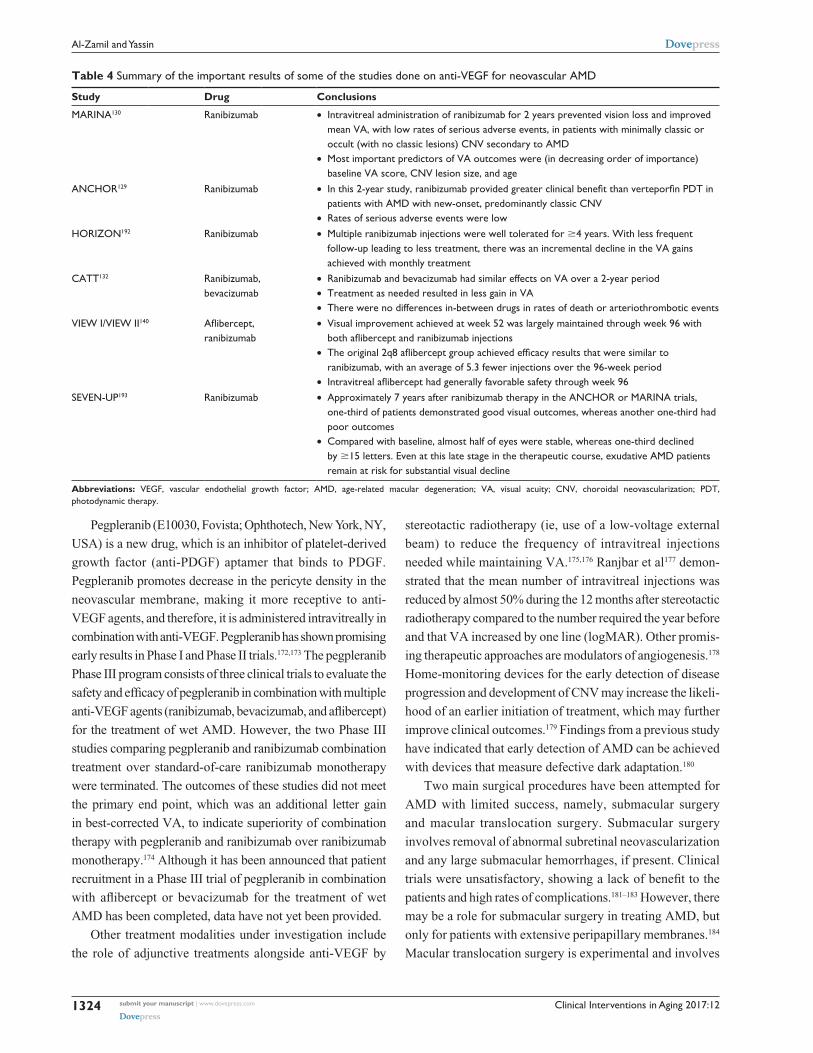
Table 4 summarizes the important results of some of the
studies done on anti-VEGF for neovascular AMD.
An adjuvant treatment used in combination with VEGF
inhibition is transpupillary thermotherapy (TTT). TTT
delivers a waveform near the infrared spectrum through the
pupil to the target tissue. At low doses (136 mW/mm), the
surrounding neurosensory retina is not damaged. In a trial
using sham procedure as a control, 100 patients with wet
AMD were randomized to receive low-dose TTT or sham
TTT every 3 months for 2 years. Patients in the TTT group
required fewer treatments with ranibizumab compared to
those in the sham group (mean 8.0 vs 6.3 over 2 years,
respectively). There were no differences in the corrected VA
or the lesion area between the two groups.166
PDT in combination with anti-VEGF and steroid admin-
istration is currently reserved as a second-line therapy for
patients not responding to monotherapy with an anti-VEGF
agent. Combined therapy using ranibizumab and verteporfin
PDT was more effective than PDT alone,167 and the combi-
nation of PDT with bevacizumab has also been effective in
many cases.168,169 Reports on whether intravitreal adminis-
tration of triamcinolone in combination with PDT provides
added benefits are conflicting.170,171
Clinical Interventions in Aging 2017:12submit your manuscript | www.dovepress.com
Dovepress
Dovepress
1324
Al-Zamil and Yassin
Pegpleranib (E10030, Fovista; Ophthotech, New York, NY,
USA) is a new drug, which is an inhibitor of platelet-derived
growth factor (anti-PDGF) aptamer that binds to PDGF.
Pegpleranib promotes decrease in the pericyte density in the
neovascular membrane, making it more receptive to anti-
VEGF agents, and therefore, it is administered intravitreally in
combination with anti-VEGF. Pegpleranib has shown promising
early results in Phase I and Phase II trials.172,173 The pegpleranib
Phase III program consists of three clinical trials to evaluate the
safety and efficacy of pegpleranib in combination with multiple
anti-VEGF agents (ranibizumab, bevacizumab, and aflibercept)
for the treatment of wet AMD. However, the two Phase III
studies comparing pegpleranib and ranibizumab combination
treatment over standard-of-care ranibizumab monotherapy
were terminated. The outcomes of these studies did not meet
the primary end point, which was an additional letter gain
in best-corrected VA, to indicate superiority of combination
therapy with pegpleranib and ranibizumab over ranibizumab
monotherapy.174 Although it has been announced that patient
recruitment in a Phase III trial of pegpleranib in combination
with aflibercept or bevacizumab for the treatment of wet
AMD has been completed, data have not yet been provided.
Other treatment modalities under investigation include
the role of adjunctive treatments alongside anti-VEGF by
stereotactic radiotherapy (ie, use of a low-voltage external
beam) to reduce the frequency of intravitreal injections
needed while maintaining VA.175,176 Ranjbar et al177 demon-
strated that the mean number of intravitreal injections was
reduced by almost 50% during the 12 months after stereotactic
radiotherapy compared to the number required the year before
and that VA increased by one line (logMAR). Other promis-
ing therapeutic approaches are modulators of angiogenesis.178
Home-monitoring devices for the early detection of disease
progression and development of CNV may increase the likeli-
hood of an earlier initiation of treatment, which may further
improve clinical outcomes.179 Findings from a previous study
have indicated that early detection of AMD can be achieved
with devices that measure defective dark adaptation.180
Two main surgical procedures have been attempted for
AMD with limited success, namely, submacular surgery
and macular translocation surgery. Submacular surgery
involves removal of abnormal subretinal neovascularization
and any large submacular hemorrhages, if present. Clinical
trials were unsatisfactory, showing a lack of benefit to the
patients and high rates of complications.181–183 However, there
may be a role for submacular surgery in treating AMD, but
only for patients with extensive peripapillary membranes.184
Macular translocation surgery is experimental and involves
Table 4 Summary of the important results of some of the studies done on anti-veGF for neovascular AMD
Study Drug Conclusions
MARINA130 Ranibizumab • Intravitreal administration of ranibizumab for 2 years prevented vision loss and improved mean vA, with low rates of serious adverse events, in patients with minimally classic or occult (with no classic lesions) CNv secondary to AMD
• Most important predictors of vA outcomes were (in decreasing order of importance) baseline vA score, CNv lesion size, and age
ANCHOR129 Ranibizumab • In this 2-year study, ranibizumab provided greater clinical benefit than verteporfin PDT in patients with AMD with new-onset, predominantly classic CNv
• Rates of serious adverse events were lowHORIZON192 Ranibizumab • Multiple ranibizumab injections were well tolerated for $4 years. with less frequent
follow-up leading to less treatment, there was an incremental decline in the vA gains achieved with monthly treatment
CATT132 Ranibizumab, bevacizumab
• Ranibizumab and bevacizumab had similar effects on vA over a 2-year period• Treatment as needed resulted in less gain in vA• There were no differences in-between drugs in rates of death or arteriothrombotic events
vIew I/vIew II140 Aflibercept, ranibizumab
• visual improvement achieved at week 52 was largely maintained through week 96 with both aflibercept and ranibizumab injections
• The original 2q8 aflibercept group achieved efficacy results that were similar to ranibizumab, with an average of 5.3 fewer injections over the 96-week period
• Intravitreal aflibercept had generally favorable safety through week 96SeveN-UP193 Ranibizumab • Approximately 7 years after ranibizumab therapy in the ANCHOR or MARINA trials,
one-third of patients demonstrated good visual outcomes, whereas another one-third had poor outcomes
• Compared with baseline, almost half of eyes were stable, whereas one-third declined by $15 letters. even at this late stage in the therapeutic course, exudative AMD patients remain at risk for substantial visual decline
Abbreviations: veGF, vascular endothelial growth factor; AMD, age-related macular degeneration; vA, visual acuity; CNv, choroidal neovascularization; PDT, photodynamic therapy.

Clinical Interventions in Aging 2017:12 submit your manuscript | www.dovepress.com
Dovepress
Dovepress
1325
Recent developments in AMD
relocating the macula to a less affected area of the retina in
patients with subfoveal CNV.185,186 The advent of effective
pharmacological therapy has limited the use of this surgical
modality to either patients with large submacular hemor-
rhages or patients unresponsive to anti-VEGF treatment.187
The surgical risks are serious, including retinal detachment,
proliferative vitreoretinopathy, and diplopia.183
ConclusionAvailable evidence indicates that risk profiling of AMD
is largely based on advanced age of the patient, lifestyle
decisions (smoking and diet), family history of AMD, and
signs of soft drusen and pigmentary abnormalities. The
identification of major susceptibility genes over the last
decade has opened new insights toward understanding the
role of complement-mediated inflammation and oxidative
stress in disease pathogenesis and, consequently, identified
underlying mechanistic pathways for the development of
novel therapeutic approaches. Treatment of atrophic AMD
is largely close observation coupled with nutritional supple-
ments, such as zinc and antioxidants. However, treatment of
wet AMD is based on targeting choroidal neovascular mem-
branes. Advances in anti-VEGF agents have transformed the
approach in treating patients with wet AMD. These advances
have resulted in more favorable outcomes for a previously
blinding disease. Substantial research efforts continue in the
identification and evaluation of new therapeutic modalities
for both forms of this disease.
DisclosureThe authors report no conflicts of interest in this work.
References1. Smith W, Assink J, Klein R, et al. Risk factors for age-related macular
degeneration: pooled findings from three continents. Ophthalmology. 2001;108(4):697–704.
2. Chou R, Dana T, Bougatsos C, Grusing S, Blazina I. Screening for impaired visual acuity in older adults: updated evidence report and systematic review for the US Preventive Services Task Force. JAMA. 2016;315(9):915–933.
3. Hajar S, Al Hazmi A, Wasli M, Mousa A, Rabiu M. Prevalence and causes of blindness and diabetic retinopathy in Southern Saudi Arabia. Saudi Med J. 2015;36(4):449–455.
4. Ferris FL, Davis MD, Clemons TE, et al; Age-Related Eye Disease Study (AREDS) Research Group. A simplified severity scale for age-related macular degeneration: AREDS Report No. 18. Arch Ophthalmol. 2005; 123(11):1570–1574.
5. Kijlstra A, Berendschot TT. Age-related macular degeneration: a comple-mentopathy? Ophthalmic Res. 2015;54(2):64–73.
6. Okubo A, Rosa RH Jr, Bunce CV, et al. The relationships of age changes in retinal pigment epithelium and Bruch’s membrane. Invest Ophthalmol Vis Sci. 1999;40(2):443–449.
7. Green WR, McDonnell PJ, Yeo JH. Pathologic features of senile macular degeneration. Ophthalmology. 1985;92(5):615–627.
8. Biesemeier A, Taubitz T, Julien S, Yoeruek E, Schraermeyer U. Cho-riocapillaris breakdown precedes retinal degeneration in age-related macular degeneration. Neurobiol Aging. 2014;35(11):2562–2573.
9. Haddad S, Chen CA, Santangelo SL, Seddon JM. The genetics of age-related macular degeneration: a review of progress to date. Surv Ophthalmol. 2006;51(4):316–363.
10. Johnson LV, Leitner WP, Staples MK, Anderson DH. Complement activation and inflammatory processes in Drusen formation and age related macular degeneration. Exp Eye Res. 2001;73(6):887–896.
11. Gold B, Merriam JE, Zernant J, et al. Variation in factor B (BF) and complement component 2 (C2) genes is associated with age-related macular degeneration. Nat Genet. 2006;38(4):458–462.
12. Edwards AO, Ritter R 3rd, Abel KJ, Manning A, Panhuysen C, Farrer LA. Complement factor H polymorphism and age-related macular degeneration. Science. 2005;308(5720):421–424.
13. Jakobsdottir J, Conley YP, Weeks DE, Mah TS, Ferrell RE, Gorin MB. Susceptibility genes for age-related maculopathy on chromosome 10q26. Am J Hum Genet. 2005;77(3):389–407.
14. Hageman GS, Anderson DH, Johnson LV, et al. A common haplotype in the complement regulatory gene factor H (HF1/CFH) predisposes individuals to age-related macular degeneration. Proc Natl Acad Sci U S A. 2005;102(20):7227–7232.
15. Esparza-Gordillo J, Soria JM, Buil A, et al. Genetic and environmental factors influencing the human factor H plasma levels. Immunogenetics. 2004;56(2):77–82.
16. Sepp T, Khan JC, Thurlby DA, et al. Complement factor H variant Y402H is a major risk determinant for geographic atrophy and choroidal neovascularization in smokers and nonsmokers. Invest Ophthalmol Vis Sci. 2006;47(2):536–540.
17. Johnson PT, Betts KE, Radeke MJ, Hageman GS, Anderson DH, Johnson LV. Individuals homozygous for the age-related macular degeneration risk-conferring variant of complement factor H have elevated levels of CRP in the choroid. Proc Natl Acad Sci U S A. 2006; 103(46):17456–17461.
18. Bressler NM, Silva JC, Bressler SB, Fine SL, Green WR. Clinico-pathologic correlation of drusen and retinal pigment epithelial abnor-malities in age-related macular degeneration. Retina. 1994;14(2): 130–142.
19. Yang Z, Stratton C, Francis PJ, et al. Toll-like receptor 3 and geographic atrophy in age-related macular degeneration. N Engl J Med. 2008; 359(14):1456–1463.
20. Shaw PX, Stiles T, Douglas C, et al. Oxidative stress, innate immu-nity, and age-related macular degeneration. AIMS Mol Sci. 2016;3(2): 196–221.
21. Ambati J, Ambati BK, Yoo SH, Ianchulev S, Adamis AP. Age-related macular degeneration: etiology, pathogenesis, and therapeutic strategies. Surv Ophthalmol. 2003;48(3):257–293.
22. Tarallo V, Hirano Y, Gelfand BD, et al. DICER1 loss and Alu RNA induce age-related macular degeneration via the NLRP3 inflammasome and MyD88. Cell. 2012;149(4):847–859.
23. Kaneko H, Dridi S, Tarallo V, et al. DICER1 deficit induces Alu RNA toxicity in age-related macular degeneration. Nature. 2011; 471(7338):325–330.
24. Nordgaard CL, Karunadharma PP, Feng X, Olsen TW, Ferrington DA. Mitochondrial proteomics of the retinal pigment epithelium at pro-gressive stages of age-related macular degeneration. Invest Ophthalmol Vis Sci. 2008;49(7):2848–2855.
25. Chakravarthy U, Wong TY, Fletcher A, et al. Clinical risk factors for age-related macular degeneration: a systematic review and meta-analysis. BMC Ophthalmol. 2010;10:31.
26. Klein R, Klein BE, Linton KL. Prevalence of age-related maculopathy. The Beaver Dam Eye Study. Ophthalmology. 1992;99(6):933–943.
27. Leibowitz HM, Krueger DE, Maunder LR, et al. The Framingham Eye Study monograph: an ophthalmological and epidemiological study of cataract, glaucoma, diabetic retinopathy, macular degeneration, and visual acuity in a general population of 2631 adults, 1973–1975. Surv Ophthalmol. 1980;24(suppl):335–610.

Clinical Interventions in Aging 2017:12submit your manuscript | www.dovepress.com
Dovepress
Dovepress
1326
Al-Zamil and Yassin
28. Maller J, George S, Purcell S, et al. Common variation in three genes, including a noncoding variant in CFH, strongly influences risk of age-related macular degeneration. Nat Genet. 2006;38(9):1055–1059.
29. Fritsche LG, Igl W, Bailey JN, et al. A large genome-wide association study of age-related macular degeneration highlights contributions of rare and common variants. Nat Genet. 2016;48(2):134–143.
30. Klein ML, Schultz DW, Edwards A, et al. Age-related macular degen-eration. Clinical features in a large family and linkage to chromosome 1q. Arch Ophthalmol. 1998;116(8):1082–1088.
31. Majewski J, Schultz DW, Weleber RG, et al. Age-related macular degeneration – a genome scan in extended families. Am J Hum Genet. 2003;73(3):540–550.
32. Hughes AE, Orr N, Esfandiary H, Diaz-Torres M, Goodship T, Chakravarthy U. A common CFH haplotype, with deletion of CFHR1 and CFHR3, is associated with lower risk of age-related macular degeneration. Nat Genet. 2006;38(10):1173–1177.
33. Cipriani V, Leung HT, Plagnol V, et al. Genome-wide association study of age-related macular degeneration identifies associated variants in the TNXB-FKBPL-NOTCH4 region of chromosome 6p21.3. Hum Mol Genet. 2012;21(18):4138–4150.
34. Neale BM, Fagerness J, Reynolds R, et al. Genome-wide association study of advanced age-related macular degeneration identifies a role of the hepatic lipase gene (LIPC). Proc Natl Acad Sci U S A. 2010; 107(16):7395–7400.
35. Weber BH, Vogt G, Pruett RC, Stohr H, Felbor U. Mutations in the tissue inhibitor of metalloproteinases-3 (TIMP3) in patients with Sorsby’s fundus dystrophy. Nat Genet. 1994;8(4):352–356.
36. Smailhodzic D, Muether PS, Chen J, et al. Cumulative effect of risk alleles in CFH, ARMS2, and VEGFA on the response to ranibizumab treatment in age-related macular degeneration. Ophthalmology. 2012; 119(11):2304–2311.
37. Medina FM, Alves Lopes da Motta A, Takahashi WY, et al. Pharmaco-genetic effect of complement factor H gene polymorphism in response to the initial intravitreal injection of bevacizumab for wet age-related macular degeneration. Ophthalmic Res. 2015;54(4):169–174.
38. McCarty CA, Mukesh BN, Fu CL, Mitchell P, Wang JJ, Taylor HR. Risk factors for age-related maculopathy: the Visual Impairment Project. Arch Ophthalmol. 2001;119(10):1455–1462.
39. Cougnard-Gregoire A, Delyfer MN, Korobelnik JF, et al. Long-term blood pressure and age-related macular degeneration: the ALIENOR study. Invest Ophthalmol Vis Sci. 2013;54(3):1905–1912.
40. Parekh N, Voland RP, Moeller SM, et al; CAREDS Research Study Group. Association between dietary fat intake and age-related macular degeneration in the Carotenoids in Age-Related Eye Disease Study (CAREDS): an ancillary study of the women’s health initiative. Arch Ophthalmol. 2009;127(11):1483–1493.
41. Khan JC, Shahid H, Thurlby DA, et al; Genetic Factors in AMD Study. Age related macular degeneration and sun exposure, iris colour, and skin sensitivity to sunlight. Br J Ophthalmol. 2006;90(1):29–32.
42. Glazer-Hockstein C, Dunaief JL. Could blue light-blocking lenses decrease the risk of age-related macular degeneration? Retina. 2006; 26(1):1–4.
43. Margrain TH, Boulton M, Marshall J, Sliney DH. Do blue light filters confer protection against age-related macular degeneration? Prog Retinal Eye Res. 2004;23(5):523–531.
44. Klein R, Rowland ML, Harris MI. Racial/ethnic differences in age-related maculopathy. Third National Health and Nutrition Examination Survey. Ophthalmology. 1995;102(3):371–381.
45. Chong EW, Wong TY, Kreis AJ, Simpson JA, Guymer RH. Dietary anti-oxidants and primary prevention of age related macular degeneration: systematic review and meta-analysis. BMJ. 2007;335(7623):755.
46. Moriarty-Craige SE, Adkison J, Lynn M, et al. Antioxidant supple-ments prevent oxidation of cysteine/cystine redox in patients with age-related macular degeneration. Am J Ophthalmol. 2005;140(6): 1020–1026.
47. Klein BE, Howard KP, Gangnon RE, Dreyer JO, Lee KE, Klein R. Long-term use of aspirin and age-related macular degeneration. JAMA. 2012;308(23):2469–2478.
48. Christen WG, Glynn RJ, Ajani UA, et al. Age-related maculopathy in a randomized trial of low-dose aspirin among US physicians. Arch Ophthalmol. 2001;119(8):1143–1149.
49. Zhu W, Wu Y, Xu D, et al. Aspirin use and risk of age-related macular degeneration: a meta-analysis. PLoS One. 2013;8(3):e58821.
50. Wang JJ, Klein R, Smith W, Klein BE, Tomany S, Mitchell P. Cataract surgery and the 5-year incidence of late-stage age-related maculopathy: pooled findings from the Beaver Dam and Blue Mountains eye studies. Ophthalmology. 2003;110(10):1960–1967.
51. Adams MK, Simpson JA, Aung KZ, et al. Abdominal obesity and age-related macular degeneration. Am J Epidemiol. 2011;173(11): 1246–1255.
52. Dasari B, Prasanthi JR, Marwarha G, Singh BB, Ghribi O. Cholesterol-enriched diet causes age-related macular degeneration-like pathology in rabbit retina. BMC Ophthalmol. 2011;11:22.
53. Sandberg MA, Tolentino MJ, Miller S, Berson EL, Gaudio AR. Hyperopia and neovascularization in age-related macular degeneration. Ophthalmology. 1993;100(7):1009–1013.
54. Feskanich D, Cho E, Schaumberg DA, Colditz GA, Hankinson SE. Menopausal and reproductive factors and risk of age-related macular degeneration. Arch Ophthalmol. 2008;126(4):519–524.
55. Chong EW, Kreis AJ, Wong TY, Simpson JA, Guymer RH. Alcohol consumption and the risk of age-related macular degeneration: a sys-tematic review and meta-analysis. Am J Ophthalmol. 2008;145(4): 707–715.
56. Gopinath B, Flood VM, Rochtchina E, Wang JJ, Mitchell P. Homo-cysteine, folate, vitamin B-12, and 10-y incidence of age-related macular degeneration. Am J Clin Nutr. 2013;98(1):129–135.
57. Millen AE, Voland R, Sondel SA, et al; CAREDS Study Group. Vitamin D status and early age-related macular degeneration in postmenopausal women. Arch Ophthalmol. 2011;129(4):481–489.
58. Schaumberg DA, Christen WG, Buring JE, Glynn RJ, Rifai N, Ridker PM. High-sensitivity C-reactive protein, other markers of inflammation, and the incidence of macular degeneration in women. Arch Ophthalmol. 2007;125(3):300–305.
59. Tielsch JM, Javitt JC, Coleman A, Katz J, Sommer A. The prevalence of blindness and visual impairment among nursing home residents in Baltimore. N Engl J Med. 1995;332(18):1205–1209.
60. Klein R, Davis MD, Magli YL, Segal P, Klein BE, Hubbard L. The Wisconsin age-related maculopathy grading system. Ophthalmology. 1991;98(7):1128–1134.
61. Bird AC, Bressler NM, Bressler SB, et al. An international classifica-tion and grading system for age-related maculopathy and age-related macular degeneration. The International ARM Epidemiological Study Group. Surv Ophthalmol. 1995;39(5):367–374.
62. AREDS-Study-Group. A randomized, placebo-controlled, clinical trial of high-dose supplementation with vitamins C and E, beta carotene, and zinc for age-related macular degeneration and vision loss: AREDS report no. 8. Arch Ophthalmol. 2001;119(10):1417–1436.
63. Klein ML, Francis PJ, Ferris FL 3rd, Hamon SC, Clemons TE. Risk assessment model for development of advanced age-related macular degeneration. Arch Ophthalmol. 2011;129(12):1543–1550.
64. Barondes M, Pauleikhoff D, Chisholm IC, Minassian D, Bird AC. Bilaterality of drusen. Br J Ophthalmol. 1990;74(3):180–182.
65. Zweifel SA, Imamura Y, Spaide TC, Fujiwara T, Spaide RF. Prevalence and significance of subretinal drusenoid deposits (reticular pseudod-rusen) in age-related macular degeneration. Ophthalmology. 2010; 117(9):1775–1781.
66. Klein R, Meuer SM, Knudtson MD, Iyengar SK, Klein BE. The epidemiol-ogy of retinal reticular drusen. Am J Ophthalmol. 2008;145(2):317–326.
67. Zhou Q, Daniel E, Maguire MG, et al; Comparison of Age-Related Macular Degeneration Treatments Trials Research Group. Pseudod-rusen and incidence of late age-related macular degeneration in fellow eyes in the comparison of age-related macular degeneration treatments trials. Ophthalmology. 2016;123(7):1530–1540.
68. Shijo T, Sakurada Y, Yoneyama S, et al. Prevalence and characteristics of pseudodrusen subtypes in advanced age-related macular degenera-tion. Graefes Arch Clin Exp Ophthalmol. 2017;255(6):1125–1131.

Clinical Interventions in Aging 2017:12 submit your manuscript | www.dovepress.com
Dovepress
Dovepress
1327
Recent developments in AMD
69. von Ruckmann A, Fitzke FW, Bird AC. Fundus autofluorescence in age-related macular disease imaged with a laser scanning ophthalmoscope. Invest Ophthalmol Vis Sci. 1997;38(2):478–486.
70. Bindewald A, Schmitz-Valckenberg S, Jorzik JJ, et al. Classification of abnormal fundus autofluorescence patterns in the junctional zone of geographic atrophy in patients with age related macular degeneration. Br J Ophthalmol. 2005;89(7):874–878.
71. Schmitz-Valckenberg S, Bindewald-Wittich A, Dolar-Szczasny J, et al; Fundus Autofluorescence in Age-Related Macular Degeneration Study Group. Correlation between the area of increased autofluorescence sur-rounding geographic atrophy and disease progression in patients with AMD. Invest Ophthalmol Vis Sci. 2006;47(6):2648–2654.
72. von Ruckmann A, Fitzke FW, Bird AC. Distribution of fundus auto-fluorescence with a scanning laser ophthalmoscope. Br J Ophthalmol. 1995;79(5):407–412.
73. Bearelly S, Khanifar AA, Lederer DE, et al. Use of fundus autofluo-rescence images to predict geographic atrophy progression. Retina. 2011;31(1):81–86.
74. Batioglu F, Gedik Oguz Y, Demirel S, Ozmert E. Geographic atrophy progression in eyes with age-related macular degeneration: role of fundus autofluorescence patterns, fellow eye and baseline atrophy area. Ophthalmic Res. 2014;52(2):53–59.
75. Sawa M, Ober MD, Spaide RF. Autofluorescence and retinal pigment epithelial atrophy after subretinal hemorrhage. Retina. 2006;26(1): 119–120.
76. Wu Z, Luu CD, Ayton LN, et al. Optical coherence tomography-defined changes preceding the development of drusen-associated atrophy in age-related macular degeneration. Ophthalmology. 2014; 121(12):2415–2422.
77. Jia Y, Bailey ST, Wilson DJ, et al. Quantitative optical coherence tomography angiography of choroidal neovascularization in age-related macular degeneration. Ophthalmology. 2014;121(7):1435–1444.
78. Kaiser PK. The Massachusetts Eye and Ear Infirmary Illustrated Manual of Ophthalmology. London: Elsevier Health Sciences; 2014.
79. Puliafito CA, Hee MR, Lin CP, et al. Imaging of macular diseases with optical coherence tomography. Ophthalmology. 1995;102(2): 217–229.
80. Gabriele ML, Wollstein G, Ishikawa H, et al. Optical coherence tomog-raphy: history, current status, and laboratory work. Invest Ophthalmol Vis Sci. 2011;52(5):2425–2436.
81. Nunes RP, Gregori G, Yehoshua Z, et al. Predicting the progression of geographic atrophy in age-related macular degeneration with SD-OCT en face imaging of the outer retina. Ophthalmic Surg Lasers Imaging Retina. 2013;44(4):344–359.
82. Coscas G, Lupidi M, Coscas F, Francais C, Cagini C, Souied EH. Optical coherence tomography angiography during follow-up: quali-tative and quantitative analysis of mixed type I and II choroidal neo-vascularization after vascular endothelial growth factor trap therapy. Ophthalmic Res. 2015;54(2):57–63.
83. Friedman D, Parker JS, Kimble JA, Delori FC, McGwin G Jr, Curcio CA. Quantification of fluorescein-stained drusen associated with age-related macular degeneration. Retina. 2012;32(1):19–24.
84. Bressler BA. Neovascular (exudative or “wet”) age-related macular degeneration. Ryan’s Retinal Imaging and Diagnostics. 5th ed. Elsevier Health Sciences; 2013:1183–1212.
85. Lim LS, Mitchell P, Seddon JM, Holz FG, Wong TY. Age-related macular degeneration. Lancet. 2012;379(9827):1728–1738.
86. Spaide RF, Yannuzzi LA, Slakter JS, Sorenson J, Orlach DA. Indo-cyanine green videoangiography of idiopathic polypoidal choroidal vasculopathy. Retina. 1995;15(2):100–110.
87. Midena E, Vujosevic S, Convento E, Manfre A, Cavarzeran F, Pilotto E. Microperimetry and fundus autofluorescence in patients with early age-related macular degeneration. Br J Ophthalmol. 2007;91(11): 1499–1503.
88. Sunness JS, Margalit E, Srikumaran D, et al. The long-term natural history of geographic atrophy from age-related macular degeneration: enlargement of atrophy and implications for interventional clinical trials. Ophthalmology. 2007;114(2):271–277.
89. Jager RD, Mieler WF, Miller JW. Age-related macular degeneration. N Engl J Med. 2008;358(24):2606–2617.
90. Crossland M, Rubin G. The Amsler chart: absence of evidence is not evidence of absence. Br J Ophthalmol. 2007;91(3):391–393.
91. Krishnadev N, Meleth AD, Chew EY. Nutritional supplements for age-related macular degeneration. Curr Opin Ophthalmol. 2010;21(3): 184–189.
92. AREDS-Study-Group. Risk factors associated with age-related macu-lar degeneration. A case-control study in the age-related eye disease study: age-related eye disease study report number 3. Ophthalmology. 2000;107(12):2224–2232.
93. Chew EY, Clemons TE, Agron E, et al; Age-Related Eye Disease Study. Ten-year follow-up of age-related macular degeneration in the age-related eye disease study: AREDS report no. 36. JAMA Ophthalmol. 2014;132(3):272–277.
94. Taylor HR, Tikellis G, Robman LD, McCarty CA, McNeil JJ. Vitamin E supplementation and macular degeneration: randomised controlled trial. BMJ. 2002;325(7354):11.
95. Parisi V, Tedeschi M, Gallinaro G, et al; CARMIS Study Group. Carotenoids and antioxidants in age-related maculopathy Italian study: multifocal electroretinogram modifications after 1 year. Ophthalmology. 2008;115(2):324–333.e2.
96. Christen WG, Gaziano JM, Hennekens CH. Design of Physicians’ Health Study II – a randomized trial of beta-carotene, vitamins E and C, and multivitamins, in prevention of cancer, cardiovascular disease, and eye disease, and review of results of completed trials. Ann Epidemiol. 2000;10(2):125–134.
97. Newsome DA, Swartz M, Leone NC, Elston RC, Miller E. Oral zinc in macular degeneration. Arch Ophthalmol. 1988;106(2):192–198.
98. AREDS2-Study-Group. Lutein + zeaxanthin and omega-3 fatty acids for age-related macular degeneration: the Age-Related Eye Disease Study 2 (AREDS2) randomized clinical trial. JAMA. 2013; 309(19):2005–2015.
99. Geerlings MJ, de Jong EK, den Hollander AI. The complement system in age-related macular degeneration: a review of rare genetic variants and implications for personalized treatment. Mol Immunol. 2017;84:65–76.
100. Rhoades W, Dickson D, Do DV. Potential role of lampalizumab for treat-ment of geographic atrophy. Clin Ophthalmol. 2015;9:1049–1056.
101. ClinicalTrials.gov. A study investigating the efficacy and safety of lampalizumab intravitreal injections in participants with geographic atrophy secondary to age-related macular degeneration (CHROMA). Available from: https://clinicaltrials.gov/ct2/show/NCT02247479. NLM identifier: NCT02247479. Accessed November 17, 2016.
102. ClinicalTrials.gov. A study investigating the efficacy and safety of lampalizumab intravitreal injections in participants with geographic atrophy secondary to age-related macular degeneration (SPECTRI). Available from: https://clinicaltrials.gov/ct2/show/NCT02247531. NLM identifier: NCT02247531. Accessed July 28, 2017.
103. Di Caprio R, Lembo S, Di Costanzo L, Balato A, Monfrecola G. Anti-inflammatory properties of low and high doxycycline doses: an in vitro study. Mediators Inflamm. 2015;2015:329418.
104. ClinicalTrials.gov. Clinical study to evaluate treatment with Oracea® for geographic atrophy (TOGA) (TOGA). Available from: https://clinicaltrials.gov/ct2/show/NCT01782989. NLM identifier: NCT01782989. Accessed July 28, 2017.
105. Barbosa DT, Mendes TS, Cintron-Colon HR, et al. Age-related macular degeneration and protective effect of HMG Co-A reductase inhibitors (statins): results from the National Health and Nutrition Examination Survey 2005–2008. Eye (Lond). 2014;28(4):472–480.
106. Szeto HH, Birk AV. Serendipity and the discovery of novel compounds that restore mitochondrial plasticity. Clin Pharmacol Ther. 2014; 96(6):672–683.
107. ClinicalTrials.gov. A study of MTP-131 topical ophthalmic solution in subjects with diabetic macular edema and non-exudative intermedi-ate age-related macular degeneration (SPIOC-101). Available from: https://clinicaltrials.gov/ct2/show/NCT02314299?term=MTP-131& rank=4. NLM identifier: NCT02314299. Accessed April 17, 2015.

Clinical Interventions in Aging 2017:12submit your manuscript | www.dovepress.com
Dovepress
Dovepress
1328
Al-Zamil and Yassin
108. Wong WT, Dresner S, Forooghian F, et al. Treatment of geographic atrophy with subconjunctival sirolimus: results of a phase I/II clinical trial. Invest Ophthalmol Vis Sci. 2013;54(4):2941–2950.
109. Petrou PA, Cunningham D, Shimel K, et al. Intravitreal sirolimus for the treatment of geographic atrophy: results of a phase I/II clinical trial. Invest Ophthalmol Vis Sci. 2014;56(1):330–338.
110. Schwartz SD, Regillo CD, Lam BL, et al. Human embryonic stem cell-derived retinal pigment epithelium in patients with age-related macular degeneration and Stargardt’s macular dystrophy: follow-up of two open-label phase 1/2 studies. Lancet. 2015;385(9967):509–516.
111. Gao J, Liu RT, Cao S, et al. NLRP3 inflammasome: activation and regulation in age-related macular degeneration. Mediators Inflamm. 2015;2015:690243.
112. Kubota R, Boman NL, David R, Mallikaarjun S, Patil S, Birch D. Safety and effect on rod function of ACU-4429, a novel small-molecule visual cycle modulator. Retina. 2012;32(1):183–188.
113. Tsai JC. Canadian Journal of Ophthalmology Lecture: translational research advances in glaucoma neuroprotection. Can J Ophthalmol. 2013;48(3):141–145.
114. Landa G, Butovsky O, Shoshani J, Schwartz M, Pollack A. Weekly vaccination with Copaxone (glatiramer acetate) as a potential therapy for dry age-related macular degeneration. Curr Eye Res. 2008;33(11): 1011–1013.
115. Wong WT, Kam W, Cunningham D, et al. Treatment of geographic atrophy by the topical administration of OT-551: results of a phase II clinical trial. Invest Ophthalmol Vis Sci. 2010;51(12):6131–6139.
116. [No authors listed] Argon laser photocoagulation for senile macular degeneration. Results of a randomized clinical trial. Arch Ophthalmol. 1982;100(6):912–918.
117. Macular Photocoagulation Study Group. Argon laser photocoagulation for neovascular maculopathy. Three-year results from randomized clinical trials. Arch Ophthalmol. 1986;104(5):694–701.
118. Macular Photocoagulation Study Group. Laser photocoagulation for juxtafoveal choroidal neovascularization. Five-year results from randomized clinical trials. Arch Ophthalmol. 1994;112(4):500–509.
119. Macular Photocoagulation Study Group. The influence of treatment extent on the visual acuity of eyes treated with Krypton laser for jux-tafoveal choroidal neovascularization. Arch Ophthalmol. 1995;113(2): 190–194.
120. Group MPS. Persistent and recurrent neovascularization after laser photocoagulation for subfoveal choroidal neovascularization of age-related macular degeneration. Arch Ophthalmol. 1994;112(4):489–499.
121. Group MPS. Laser photocoagulation of subfoveal neovascular lesions of age-related macular degeneration. Updated findings from two clinical trials. Arch Ophthalmol. 1993;111(9):1200–1209.
122. Group TS. Photodynamic therapy of subfoveal choroidal neovascular-ization in age-related macular degeneration with verteporfin: one-year results of 2 randomized clinical trials – TAP report. Treatment of age-related macular degeneration with photodynamic therapy (TAP) Study Group. Arch Ophthalmol. 1999;117(10):1329–1345.
123. Bressler NM; Treatment of Age-Related Macular Degeneration with Photodynamic Therapy (TAP) Study Group. Photodynamic therapy of subfoveal choroidal neovascularization in age-related macular degen-eration with verteporfin: two-year results of 2 randomized clinical trials-tap report 2. Arch Ophthalmol. 2001;119(2):198–207.
124. Blumenkranz MS, Bressler NM, Bressler SB, et al; Treatment of Age-Related Macular Degeneration with Photodynamic Therapy (TAP) Study Group. Verteporfin therapy for subfoveal choroidal neovascu-larization in age-related macular degeneration: three-year results of an open-label extension of 2 randomized clinical trials – TAP Report no. 5. Arch Ophthalmol. 2002;120(10):1307–1314.
125. Gaynes BI, Fiscella RG. Safety of verteporfin for treatment of sub-foveal choroidal neovascular membranes associated with age-related macular degeneration. Expert Opin Drug Saf. 2004;3(4):345–361.
126. Michels S, Hansmann F, Geitzenauer W, Schmidt-Erfurth U. Influence of treatment parameters on selectivity of verteporfin therapy. Invest Ophthalmol Vis Sci. 2006;47(1):371–376.
127. Schmidt-Erfurth UM, Richard G, Augustin A, et al; European Society for Retina Specialists’ Guidelines Committee (EURETINA). Guidance for the treatment of neovascular age-related macular degeneration. Acta Ophthalmol Scand. 2007;85(5):486–494.
128. Gragoudas ES, Adamis AP, Cunningham ET Jr, et al; VEGF Inhibition Study in Ocular Neovascularization Clinical Trial Group. Pegaptanib for neovascular age-related macular degeneration. N Engl J Med. 2004;351(27):2805–2816.
129. Brown DM, Michels M, Kaiser PK, et al; ANCHOR Study Group. Ranibizumab versus verteporfin photodynamic therapy for neovascular age-related macular degeneration: two-year results of the ANCHOR study. Ophthalmology. 2009;116(1):57–65.e5.
130. Rosenfeld PJ, Brown DM, Heier JS, et al; MARINA Study Group. Ranibizumab for neovascular age-related macular degeneration. N Engl J Med. 2006;355(14):1419–1431.
131. Yonekawa Y, Miller JW, Kim IK. Age-related macular degenera-tion: advances in management and diagnosis. J Clin Med. 2015;4(2): 343–359.
132. Comparison of Age-related Macular Degeneration Treatments Trials (CATT) Research Group; Martin DF, Maguire MG, et al. Ranibi-zumab and bevacizumab for treatment of neovascular age-related macular degeneration: two-year results. Ophthalmology. 2012;119(7): 1388–1398.
133. Kim BJ, Ying GS, Huang J, Levy NE, Maguire MG; CATT Research Group. Sporadic visual acuity loss in the comparison of age-related macular degeneration treatments trials (CATT). Am J Ophthalmol. 2014;158(1):128–135.e10.
134. Chakravarthy U, Harding SP, Rogers CA, et al; IVAN Study Investiga-tors. Alternative treatments to inhibit VEGF in age-related choroidal neovascularisation: 2-year findings of the IVAN randomised controlled trial. Lancet. 2013;382(9900):1258–1267.
135. Chakravarthy U, Harding SP, Rogers CA, et al. A randomised con-trolled trial to assess the clinical effectiveness and cost-effectiveness of alternative treatments to Inhibit VEGF in Age-related choroidal Neovascularisation (IVAN). Health Technol Assess. 2015;19(78): 1–298.
136. Kodjikian L, Souied EH, Mimoun G, et al; GEFAL Study Group. Ranibizumab versus bevacizumab for neovascular age-related macular degeneration: results from the GEFAL noninferiority randomized trial. Ophthalmology. 2013;120(11):2300–2309.
137. Berg K, Hadzalic E, Gjertsen I, et al. Ranibizumab or bevacizumab for neovascular age-related macular degeneration according to the lucentis compared to avastin study treat-and-extend protocol: two-year results. Ophthalmology. 2016;123(1):51–59.
138. Browning DJ, Kaiser PK, Rosenfeld PJ, Stewart MW. Aflibercept for age-related macular degeneration: a game-changer or quiet addition? Am J Ophthalmol. 2012;154(2):222–226.
139. Stewart MW. Aflibercept (VEGF Trap-eye): the newest anti-VEGF drug. Br J Ophthalmol. 2012;96(9):1157–1158.
140. Heier JS, Brown DM, Chong V, et al; VIEW 1 and VIEW 2 Study Groups. Intravitreal aflibercept (VEGF trap-eye) in wet age-related macular degeneration. Ophthalmology. 2012;119(12):2537–2548.
141. Papadopoulos N, Martin J, Ruan Q, et al. Binding and neutralization of vascular endothelial growth factor (VEGF) and related ligands by VEGF Trap, ranibizumab and bevacizumab. Angiogenesis. 2012; 15(2):171–185.
142. Mantel I. Optimizing the anti-VEGF treatment strategy for neovascular age-related macular degeneration: from clinical trials to real-life requirements. Transl Vis Sci Technol. 2015;4(3):6.
143. Busbee BG, Ho AC, Brown DM, et al; HARBOR Study Group. Twelve-month efficacy and safety of 0.5 mg or 2.0 mg ranibizumab in patients with subfoveal neovascular age-related macular degeneration. Ophthalmology. 2013;120(5):1046–1056.
144. Fung AE, Lalwani GA, Rosenfeld PJ, et al. An optical coherence tomography-guided, variable dosing regimen with intravitreal ranibi-zumab (Lucentis) for neovascular age-related macular degeneration. Am J Ophthalmol. 2007;143(4):566–583.

Clinical Interventions in Aging 2017:12 submit your manuscript | www.dovepress.com
Dovepress
Dovepress
1329
Recent developments in AMD
145. IVAN Study Investigators, Chakravarthy U, Harding SP, et al. Ranibi-zumab versus bevacizumab to treat neovascular age-related macular degeneration: one-year findings from the IVAN randomized trial. Ophthalmology. 2012;119(7):1399–1411.
146. CATT Research Group, Martin DF, Maguire MG, et al. Ranibizumab and bevacizumab for neovascular age-related macular degeneration. N Engl J Med. 2011;364(20):1897–1908.
147. Lalwani GA, Rosenfeld PJ, Fung AE, et al. A variable-dosing regimen with intravitreal ranibizumab for neovascular age-related macular degeneration: year 2 of the PrONTO Study. Am J Ophthalmol. 2009; 148(1):43–58.e1.
148. Engelbert M, Zweifel SA, Freund KB. Long-term follow-up for type 1 (subretinal pigment epithelium) neovascularization using a modified “treat and extend” dosing regimen of intravitreal antivascular endothe-lial growth factor therapy. Retina. 2010;30(9):1368–1375.
149. Gupta OP, Shienbaum G, Patel AH, Fecarotta C, Kaiser RS, Regillo CD. A treat and extend regimen using ranibizumab for neovascular age-related macular degeneration clinical and economic impact. Ophthalmology. 2010;117(11):2134–2140.
150. Rayess N, Houston SK 3rd, Gupta OP, Ho AC, Regillo CD. Treatment outcomes after 3 years in neovascular age-related macular degen-eration using a treat-and-extend regimen. Am J Ophthalmol. 2015; 159(1):3.e1–8.e1.
151. Shienbaum G, Gupta OP, Fecarotta C, Patel AH, Kaiser RS, Regillo CD. Bevacizumab for neovascular age-related macular degeneration using a treat-and-extend regimen: clinical and economic impact. Am J Ophthalmol. 2012;153(3):468.e1–473.e1.
152. Mantel I, Niderprim SA, Gianniou C, Deli A, Ambresin A. Reducing the clinical burden of ranibizumab treatment for neovascular age-related macular degeneration using an individually planned regimen. Br J Ophthalmol. 2014;98(9):1192–1196.
153. Gianniou C, Dirani A, Ferrini W, et al. Two-year outcome of an observe-and-plan regimen for neovascular age-related macular degen-eration: how to alleviate the clinical burden with maintained functional results. Eye (Lond). 2015;29(3):342–349.
154. Vottonen P, Kankaanpaa E. Cost-effectiveness of treating wet age-related macular degeneration at the Kuopio University Hospital in Finland based on a two-eye Markov transition model. Acta Ophthalmol. 2016;94(7):652–656.
155. Elshout M, van der Reis MI, Webers CA, Schouten JS. The cost-utility of aflibercept for the treatment of age-related macular degeneration compared to bevacizumab and ranibizumab and the influence of model parameters. Graefes Arch Clin Exp Ophthalmol. 2014;252(12): 1911–1920.
156. Shaikh AH, Toussaint BW, Miller DM, et al. Cost comparison of intravitreal aflibercept with bevacizumab and ranibizumab for the treatment of wet age-related macular degeneration. Ophthalmic Surg Lasers Imaging Retina. 2015;46(1):62–66.
157. Park DH, Sun HJ, Lee SJ. A comparison of responses to intravitreal bevacizumab, ranibizumab, or aflibercept injections for neovascular age-related macular degeneration. Int Ophthalmol. Epub 2016 Nov 8.
158. Au A, Parikh VS, Singh RP, et al. Comparison of anti-VEGF therapies on fibrovascular pigment epithelial detachments in age-related macular degeneration. Br J Ophthalmol. 2017;101(7):970–975.
159. Ba J, Peng RS, Xu D, et al. Intravitreal anti-VEGF injections for treating wet age-related macular degeneration: a systematic review and meta-analysis. Drug Des Devel Ther. 2015;9:5397–5405.
160. Campochiaro PA, Channa R, Berger BB, et al. Treatment of diabetic macular edema with a designed ankyrin repeat protein that binds vas-cular endothelial growth factor: a phase I/II study. Am J Ophthalmol. 2013;155(4):697–704, 704.e1–e2.
161. Souied EH, Devin F, Mauget-Faysse M, et al; MP0112 Study Group. Treatment of exudative age-related macular degeneration with a designed ankyrin repeat protein that binds vascular endothelial growth factor: a phase I/II study. Am J Ophthalmol. 2014;158(4): 724.e2–732.e2.
162. Dugel PU, Warburton J, Weichselberger A, Sallstig P. A 2-year study comparing the efficacy and safety of brolucizumab vs aflibercept in subjects with neovascular age-related macular degeneration: testing an alternative treatment regimen. Invest Ophthalmol Vis Sci. 2016; 57(12):5018–5018.
163. Guerrero-Naranjo JL, Quiroz-Mercado H, Sanchez-Bermudez G, et al. Safety of implantation of the NT-503 device in patients with Choroidal Neovascularization secondary to age-related macular degeneration. Invest Ophthalmol Vis Sci. 2013;54(15):3298–3298.
164. ClinicalTrials.gov. Study of the efficacy and safety of the ranibi-zumab port delivery system for sustained delivery of ranibizumab in participants with subfoveal neovascular age-related macular degeneration (LADDER). Available from: https://clinicaltrials.gov/ct2/show/NCT02510794. NLM identifier: NCT02510794. Accessed July 28, 2017.
165. Replenish [webpage on the Internet]. Our Technology: Ophthalmic Micro-pump System. Available from: www.replenishinc.com/our-technology/ophthalmic-micropump-system. Accessed July 28, 2017.
166. Soderberg AC, Algvere PV, Hengstler JC, Soderberg P, Seregard S, Kvanta A. Combination therapy with low-dose transpupillary ther-motherapy and intravitreal ranibizumab for neovascular age-related macular degeneration: a 24-month prospective randomised clinical study. Br J Ophthalmol. 2012;96(5):714–718.
167. Antoszyk AN, Tuomi L, Chung CY, Singh A; FOCUS Study Group. Ranibizumab combined with verteporfin photodynamic therapy in neovascular age-related macular degeneration (FOCUS): year 2 results. Am J Ophthalmol. 2008;145(5):862–874.
168. Costa RA, Jorge R, Calucci D, Melo LA Jr, Cardillo JA, Scott IU. Intravitreal bevacizumab (Avastin) in combination with verteporfin photodynamic therapy for choroidal neovascularization associated with age-related macular degeneration (IBeVe Study). Graefes Arch Clin Exp Ophthalmol. 2007;245(9):1273–1280.
169. Kaiser PK, Boyer DS, Garcia R, et al. Verteporfin photodynamic therapy combined with intravitreal bevacizumab for neovascular age-related macular degeneration. Ophthalmology. 2009;116(4):747–755, 755.e741.
170. Piermarocchi S, Sartore M, Lo Giudice G, Maritan V, Midena E, Segato T. Combination of photodynamic therapy and intraocular triamcinolone for exudative age-related macular degeneration and long-term chorioreti-nal macular atrophy. Arch Ophthalmol. 2008;126(10):1367–1374.
171. Kovacs KD, Quirk MT, Kinoshita T, et al. A retrospective analysis of triple combination therapy with intravitreal bevacizumab, posterior sub-tenon’s triamcinolone acetonide, and low-fluence verteporfin photodynamic therapy in patients with neovascular age-related macular degeneration. Retina. 2011;31(3):446–452.
172. ClinicalTrials.gov. A phase 1, safety, tolerability and pharmacokinetic profile of intravitreous injections of E10030 (anti-PDGF pegylated aptamer) in subjects with neovascular age-related macular degenera-tion. Available from: https://clinicaltrials.gov/show/NCT00569140. NLM identifier: NCT00569140. Accessed July 28, 2017.
173. ClinicalTrials.gov. A safety and efficacy study of E10030 (anti-PDGF pegylated aptamer) plus lucentis for neovascular age-related macular degeneration. Available from: https://clinicaltrials.gov/ct2/show/results/NCT01089517?term=E10030&rank=3. NLM identifier: NCT01089517. Accessed July 28, 2017.
174. ClinicalTrials.gov. A phase 3 safety and efficacy study of Fovista® (E10030) intravitreous administration in combination with Lucentis® compared to Lucentis® monotherapy. Available from: https://clinical-trials.gov/ct2/show/NCT01940900. NLM identifier: NCT01940900. Accessed July 28, 2017.
175. Jackson TL, Chakravarthy U, Kaiser PK, et al; INTREPID Study Group. Stereotactic radiotherapy for neovascular age-related macular degeneration: 52-week safety and efficacy results of the INTREPID study. Ophthalmology. 2013;120(9):1893–1900.
176. Neffendorf JE, Desai R, Wang Y, et al. StereoTactic radiotherapy for wet age-related macular degeneration (STAR): study protocol for a randomised controlled clinical trial. Trials. 2016;17(1):560.

Clinical Interventions in Aging
Publish your work in this journal
Submit your manuscript here: http://www.dovepress.com/clinical-interventions-in-aging-journal
Clinical Interventions in Aging is an international, peer-reviewed journal focusing on evidence-based reports on the value or lack thereof of treatments intended to prevent or delay the onset of maladaptive correlates of aging in human beings. This journal is indexed on PubMed Central, MedLine,
CAS, Scopus and the Elsevier Bibliographic databases. The manuscript management system is completely online and includes a very quick and fair peer-review system, which is all easy to use. Visit http://www.dovepress.com/testimonials.php to read real quotes from published authors.
Clinical Interventions in Aging 2017:12submit your manuscript | www.dovepress.com
Dovepress
Dovepress
Dovepress
1330
Al-Zamil and Yassin
177. Ranjbar M, Kurz M, Holzhey A, Melchert C, Rades D, Grisanti S. Stereotactic radiotherapy in neovascular age-related macular degen-eration: real-life efficacy and morphological evaluation of the outer retina-choroid complex. Medicine. 2016;95(52):e5729.
178. Yanai R, Mulki L, Hasegawa E, et al. Cytochrome P450-generated metabolites derived from omega-3 fatty acids attenuate neovascular-ization. Proc Natl Acad Sci U S A. 2014;111(26):9603–9608.
179. AREDS2-HOME Study Research Group, Chew EY, Clemons TE, et al. Randomized trial of a home monitoring system for early detection of choroidal neovascularization home monitoring of the Eye (HOME) study. Ophthalmology. 2014;121(2):535–544.
180. Jackson GR, Scott IU, Kim IK, Quillen DA, Iannaccone A, Edwards JG. Diagnostic sensitivity and specificity of dark adaptometry for detection of age-related macular degeneration. Invest Ophthalmol Vis Sci. 2014;55(3):1427–1431.
181. Hawkins BS, Bressler NM, Miskala PH, et al; Submacular Surgery Trials (SST) Research Group. Surgery for subfoveal choroidal neovas-cularization in age-related macular degeneration: ophthalmic findings: SST report no. 11. Ophthalmology. 2004;111(11):1967–1980.
182. Bressler NM, Bressler SB, Childs AL, et al; Submacular Surgery Trials (SST) Research Group. Surgery for hemorrhagic choroidal neovascular lesions of age-related macular degeneration: ophthalmic findings: SST report no. 13. Ophthalmology. 2004;111(11):1993–2006.
183. Giansanti F, Eandi CM, Virgili G. Submacular surgery for choroidal neovascularisation secondary to age-related macular degeneration. Cochrane Database Syst Rev. 2009;(2):Cd006931.
184. Kertes PJ. Massive peripapillary subretinal neovascularization: an indication for submacular surgery. Retina. 2004;24(2):219–225.
185. MacLaren RE, Bird AC, Sathia PJ, Aylward GW. Long-term results of submacular surgery combined with macular translocation of the retinal pigment epithelium in neovascular age-related macular degeneration. Ophthalmology. 2005;112(12):2081–2087.
186. Chen FK, Patel PJ, Uppal GS, Tufail A, Coffey PJ, Da Cruz L. Long-term outcomes following full macular translocation surgery in neovascular age-related macular degeneration. Br J Ophthalmol. 2010; 94(10):1337–1343.
187. van Romunde SH, Polito A, Bertazzi L, Guerriero M, Pertile G. Long-term results of full macular translocation for choroidal neovascular-ization in age-related macular degeneration. Ophthalmology. 2015; 122(7):1366–1374.
188. ClinicalTrials.gov. Complement inhibition with eculizumab for the treatment of non-exudative macular degeneration (AMD) (COMPLETE). Available from: https://clinicaltrials.gov/ct2/show/NCT00935883. NLM identifier: NCT00935883. Accessed July 28, 2017.
189. ClinicalTrials.gov. Proton radiation therapy for macular degeneration. Available from: https://clinicaltrials.gov/ct2/show/NCT01833325? term=proton+radiation&recrs=d&cond=AMD&rank=1. NLM identi-fier: NCT01833325. Accessed July 28, 2017.
190. ClinicalTrials.gov. A safety and efficacy study of abicipar pegol in patients with neovascular age-related macular degeneration (CDER). Available from: https://clinicaltrials.gov/ct2/show/NCT02462928? term=Abicipar+pegol&recrs=d&cond=AMD&rank=3. NLM identi-fier: NCT02462928. Accessed July 28, 2017.
191. ClinicalTrials.gov. Efficacy and safety of RTH258 versus aflibercept – study 2. Available from: https://clinicaltrials.gov/ct2/show/NCT02434328?term=RTH258&recrs=d&cond=AMD&rank=1. NLM identifier: NCT02434328. Accessed July 28, 2017.
192. Singer MA, Awh CC, Sadda S, et al. HORIZON: an open-label exten-sion trial of ranibizumab for choroidal neovascularization secondary to age-related macular degeneration. Ophthalmology. 2012;119(6): 1175–1183.
193. Rofagha S, Bhisitkul RB, Boyer DS, Sadda SR, Zhang K; SEVEN-UP Study Group. Seven-year outcomes in ranibizumab-treated patients in ANCHOR, MARINA, and HORIZON: a multicenter cohort study (SEVEN-UP). Ophthalmology. 2013;120(11):2292–2299.