Embed Size (px)
Citation preview

TitleDirect Observation of Gold Sol by Cryo-Electron Microscopy(Commemoration Issue Dedicated to Professor Natsu Uyeda,on the Occasion of His Retirment)
Author(s) Tahara, Yoshikazu; Fujiyoshi, Yoshinori
Citation Bulletin of the Institute for Chemical Research, KyotoUniversity (1989), 66(5): 598-604
Issue Date 1989-03-15
URL http://hdl.handle.net/2433/77270
Right
Type Departmental Bulletin Paper
Textversion publisher
Kyoto University
Bull. Inst. Chem. Res., Kyoto Univ., Vol. 66, No. 5, 1988
Direct Observation of Gold Sol by
Cryo-Electron Microscopy
Yoshikazu TAHARA* and Yoshinori FuJIYosHt**
Received October 25, 1988
In a study of gold sol with a conventional high-resolution electron microscopy, suggesting that the surface of a particle is coated with a layer of citrate ion, contamination during
specimen preparation and/or observation is not deniable. The observation by cryo-electron microscopy combined with ice embedding method confirms that the surface of gold sol particles
adsorbs citrate ion which looks like a layer. Citrate ion is thought to arise repulsive force which disperses gold sol particles. Furthermore, new particles smaller than 15 A are found to
be present together with well-known gold particles larger than 150 A.
KEY WORDS: Gold sol/ Cryo-electron microscopy/ Ice embedding/
I. INTRODUCTION
Colloidal particles are in general believed to be dispersed by repulsive force
arisen from electrical double layer. The formation of electrical double layer
would be chemically explained as follows; in aqueous sol, hydrophobic particles
acquire surface charges by adsorbing ions, and subsequently this charge is com-
pensated by a counterion in the sol. Sizes of colloidal particles are far less than the magnitude observed by optical
microscope. Electron microscopy could thus be most suitable method to observe
detailed structures of colloidal particles. Nowadays, the resolution as well as
feasivility of electron microscopes has been improved sufficiently to yield images
of inorganic materials at high resolution. We used sodium citrate sol to examine
the surface structure of gold particles, because of their highest stability and
homogeneity in size. The surface of the colloidal gold is known to have negative
charge, because of the movement of the particle toward a positive electrode in an
electric field. Electron microscopic study using alumina-supermicrogrid suggested that a surface of the particle was not bare but coated with a monolayer of citrate
ionsi). Ambiguity, however, remained in the previous experiments that gold
particles might be contaminated with the other residual materials during dry or with organic conpounds floating in the air.
As to specimen preparation, the deformation of a colloidal structure has been
overcome through development of ice embedding method2 . Cryo-electron micro-
scopy combined with ice embedding method can be most powerful approach to
* FJ*Ii : Department of Biophysics, Fuculty of Science, Kyoto University; Sakyo-ku, Kyoto 606.
**AJIIJ : Protein Engineering Research Institute; 6-2-3, Furuedai, Suita, Osaka 565. To whom the correspondence should be addressed.
(598)

Direct Observation of Gold Sol by Cryo-Electron Microscopy
visualize intact structures of colloidal particles. We have recently developed a new type of cryo-electron microscope') (cryo-EM) which can be cooled with super-fluid helium. This microscope has top-entry type of stage equipped with cryo-transfer device. We comfirmed that this cryo-electron microscope realize 2.6 A
(1 A=0.1 nm) resolution at the stage temperature of 1.5 K. We could thus confirmed that the surface of gold sol were coated with a layer
of citrate ion. Furthermore, we found that new particles smaller than 15 A are
present together with usual gold particle larger than 150A. This reports describes the mechanism about stabilization of gold sol related to their observations.
II. MATERIALS AND METHOD
The gold sol was prepared as described by Turkevich, et. a1.° Five volumes of 1% (w/v) sodium citrate was added to 95 volumes of boiling solution containing 0.08% chloroauric acid which was being stirred vigorously. We observed two gold sols which were prepared freshly or more than ten years ago respectively.
The specimen for cryo-electron microscopy was prepared as follows; the gold
a
' 1
0 • 4
!1^
10 00 A~
Fig. I. Gold sol particles embedded in vitreous ice filling a hole of microgrid. Major particles are seprated from together but some are connected to
one or two other particles.
(599 )

Y. TAHARA and Y FuJiwosxt
sol of 2 or 31o1 was put on a mesh coated with microgrid, and most of the sol was sucked away with a small piece of filter paper. It was quickly injected into liquid ethane at about —160°C so to embed gold particles in vitreous ice filling up all over holes of microgrid.
The images of gold sol were taken with a high resolution cryo-EM equipped
with a cryo-transfer device. The cryo-transfer device was essential to ice embedding method because the specimen needs to keep under devitrification temperature and
prevent from contamination of ice crystallites depositted on the vitreous ice film. The specimen was cooled to about 4 K with liqid helium. The cryo-EM was operated at 400 kV. The photographs were taken with magnification of 60,000 or 40,000,
current density of 6-14 electrons/A2 sec and exposure time of 11 seconds.
III RESULTS
Figuie 1 shows gold particles embedded in a vitreous ice filling up a hole of
," •. ~,y to ^' "
•'lt`4z r4 ti+s ,F't '~"~ r• ~~y'~ yr `J f • iW?w..Pa,.f
~N~~.r~4,'~~'~.V/t«`l-nRc~rute(k~n^
T .4't,~e.a;'.:c •,ir.'o~,r~;,. ~";.'' ••r,r.,=•`~°e4;..`.;t,."e'S.rk •~i,.h ~„!:r•,n ""3fyt trY.y! ly ,. +t: ~-fk~ V r ~ •" ...a ` x L x « x ~~t m.,a ljt' iL'f:i i . 3 ~ ~ D3?m y ,Y3'ip^wy ~ ~y A' ~ r _ r..'~` e d..'%"+jT
•p;xC .x. ;a AJ.'nj M``:e'.`' ̀f,`» »L~;T`> 4 . w r ^, k hFx #n.,, . t,2. "S , r i r.£{'v ••r:
.f`.41,t°r~"»~,,gG„~.,
• ',.'"'ti :•j< t'tiY K"Y Ja. t ••• ,~ i".4•.:4 ^~~` r ee ̀ ^+Y J` ' ` , '
r ;`'' • %~s~t' M r': n: r'x Yl 4.r ``:` * r{ .r v# r Xr..` .33' .:e4 %u ','"LR~i r r 1 • tr ,}.t.'.::v,q1
"µf»t.•t'i..
,~~~ji„j~jJ~i>...'~•r~;~rry2~;ret'x ."j"44—~.P •J~"yyr~:'""-=.„.~y,"tP,~,'• 5Wt~ry
A.{e'detLyoj'FaiM"5" .A x^
'.,Yf i,;;•••:.:t • 'ir .ras c y,3~> rv'a. c``aa~,°~l r~"i~ j,F1'a''_RFJ
is•''rr', "..
~ ̀;tr:.tci.yr;k`2'GY m{avrr'r.¢ti4yvj'a_
2t 0A
'&nyw. ~.r r~~R.°- r
Fig 2Two species of gold sol particles The size of larger pai ticles is about 180 A diameter Smaller particles, diameter of which is below 15 A,
ai e observed with lower contrast than the lai gei one. Some of small
particles are found to be adsorbed on the surface of the larger, but most of them assemble loosely togethei
(600)
Direct Obseivation of Gold Sol by Cryo-Electron Micioscopy
microgrid. The ice embedding method was found to be effective for a specimen
support, because a vitreous ice film has high transparency to electron beam. It
therefore results in lower noise of images than a carbon film. We could observe that most of particles were dispersed except some connected with one or two
other particles.
The diameter of particles was about 180 A on average. Some particles which
were not globular retained the crystal habit of a gold metal (Fig. 2).
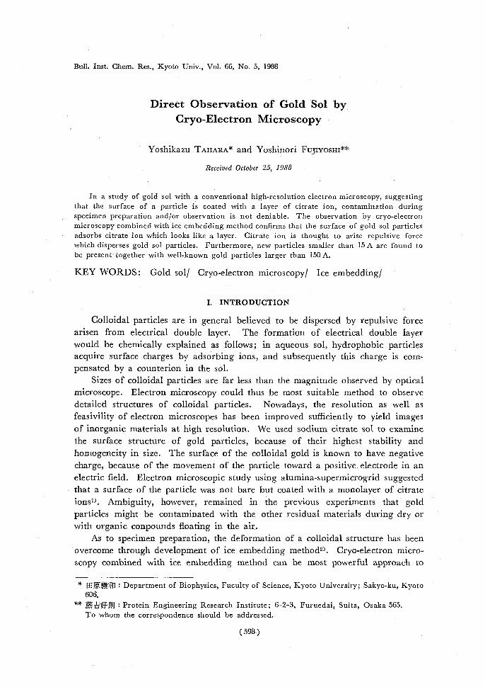
A lot of small particles with diameters below 15 A were present together with
well known gold particles. These new types of particles were identified as gold by
electron diffractogram (Fig. 3, inserted). Most of them assemble loosely together,
and some bind to larger particles as shown in Fig. 4, while a few were found to
be piesent alone.
These gold particles were too small to observed by a past conventional EM. The high resolution ciyo-EM equipped with cryo-transfer device is required to
visualize them, because noise from a supporting film, such as carbon film, will
hamper visualization of the small particles, and they were so small as to pass
through a hole of any microgrids, even an alumina-supermicrogrid5). They were
4-,4.11.~t~~{,nycy+''4i,1:4.4~#',•,ts.4..~nYSkAti~'y.'t~.,.1^,T:i}~74*
k'
Y..,~5•,.tya,...•,ly,
Fl(t'~_v:,,lCly~Yy4rf$.4:s.414i'!t,,,135'~yTS'Y~1"..4•;•`i1 , .,..,(.;,;,,t;,ti>.,7'y''Y,r1rYit~Rt`yi~~'jd/~•"PV,5.i+s'"rii`^L, 4.,
'
•,,,.;..t~42s,~+•"~".'.'~r_JUS~"'$s`"`(•-v^1c4Y',T.4:4,4V say.,,~tiay.„+~di”•+!~Y4`•,sS,~1.'F ~7`L•;^•{°~ s,..h" ~~,'ofyi@~r,-'~~"}7'•.1#!`k'-C•`C~T>.}{:"".~ tsi•~l' .:a•'(Y,§TA'~'^>t:Yr'M1S:d',r:~~``C~:ns,~Y,r44.?y.~t,y •`.°`r'i:,,~Fxtt '~i~t~'r`'s•Lat%z.a..,';,-.r':^"Ai:a}' rge,,'C4''f;~••C3'''!
t1'0.)Sty~ri~rtc'if.;, :,i~~r~.'y~`~'-~a~~a,~,,~~ht711+a.k:'~.~^L+~.~ig:~"he;,~ti`t'"~.,. •„
,~F4ur'f,!-.s~ rv,,,,r`k`..;:'e,?,:1,~rr""°,,+'.4;5Ij~..{y~}-rit:{~;j>r:'L,,,s,s, :~X '~•~,,((rr„+aY,'fy.~t.~.`nr.'~v%iw, .,w,"°~•,qr^•'~'r,;,:111.5.::''51:''''',,i t "-'1';':;j v`,,sc;,.{-G^yF#s'ku,57”.st3,I,'q~}`,Z`n':r•1- v..',-4,11=''Ss.~•".'?'C'3.'":6',4`-,191~,7 ::.`1'fi~ y4.,,Lci`yk,xS'~4 i~tgk.,N:(•y^•7 sFa,,,?D''L.5Si5.^.,_ ',i''-`e i,'' 95~,r;'l',k,-,'~'"'pw-cttr'.Pr„"~~''`k7"'7,~e',t•~,r~y..S''!'.t_ 'tG ,,x~a~;.rno~ti>~'}..',cti~~•~u'~~.i,' ,~n,•t'7+~~'y~,y.•r~^~`;'. '`r,r~ ,`X~',,'''4e~':,,sa,'a''¢y'S_'"C~,r,+4•.rofie;~,-''-.'jkN,'~'7w`wt~4«-~i-, K,. 4{ •..!`,'..„3.~,114~,Fr0.Yitlitt,/.,3,+i•.t.,.''t.~..r_4Ar,~F!.%Y `,,',,},x~4<:?-:..r_^y~t,c~-F»}i,~t,.'~titr`.b~S~` ` ,.oa-%h~..;r'ray4iX,v~,fir'?•':{F~,v,.-1`~;}q''t~.,t,_''% r~;is, v•
,,%^ }~,+•i.„:•!c.'S"^.:`',+'R,..v.4'::;,,'b.}x,.t'%3~is.,,.t.57'Z4..~..A~'CS.j,3<'+.1 :-..,,- "ti';`rZ$a't1i',:4`r.)•»ir`ii+'7!'-!r-`F•,`R'r•Y41.‘.4.,c,,:,..1-'-7~.,.'+r',,r, -- ̀ '+t.,.e .,•....8$';'..1..J,.^ -.:.s~'^t#~~L>i7 .r.."trrr~1,.~iY~~„~'a/",~~_~~~~~5;~.~'- .F` 'r7`;ii}F4r',''411illjd["!`'•ts\.,.--, •`
I.a.4'~~~,t',fyi4}5'.WS/•"'„:.','•''''3~rh`..~~yn.~.14
4:0,,,,,m.-...t. '€.i.x'ASra`jr4~iK~cFsr`,. e.°~I't7.ivy~`p„'Nff,5~8'4`.'l,J~i)`~A.;I"V::-:)'k'fg.tii~7,4V3R~4~li~tf.aw.•.{:.
C...'tk'~"zi~,0,'SS.~”ci~V4C.,,.0,4.g''3g.i~''N.4a
, Kc,3it't}Y,-e.:4+'tmy~,t4~yt^`+ryi .,rr"'
(~ c,zs`4,5W'til~rt+. 1it~*`,~kt,0 •A.?`1Z~jAIy`I~-.."-'i .41iY4fJ~kit'.11~~Y.Y.
ritz t.,,,-f''-:.r<Y'ufstfx'`,'JIA."''•1'4(>,.,i•; a'w.
~'15....~£~C~~kj~h'{~'~.r~zh_d~:~nr;v q;i+'ri?•,,,,'_,{a,k, Fig 3 Small gold particles also seen in gold sol aged for more than ten years
Inserted election diffractogram from small particles indicates that they
aic composed of gold
(601 )

Y. TAHARA and Y. F'UIIYOSHI
'iic..1l
{rC;
Fig. 4. Small particles near to or in contact with large particle.
observed in not only a freshly prepared gold sol but also in that prepared more than ten years ago (Fig. 3). Small lumps and/or thin layers were observed on the surface of gold particles as shown with arrow in Fig. 2. The similar feature was observed between two large particles (Fig. 5-a, b, c, d) as a bridge connecting two particles. These bridges are recognized by more careful observation to be constructed from materials with high and low densities, the denser in contrast would be presumably gold and less denser citrate ion. The lumps on the surface were also found to be constructed with the two.
IV. DISCUSSION
The observation of gold particles with cryo-EM revealed that the sizes of
gold sol particles are distributed into two separate peaks. However, they have been recognized so far to be single distribution of about 200 A diameter through the observation by conventional electron microscopy. The small particles below 15 A certainly coexist with larger ones, because cryo-EM is able to observe the intact structure of a colloidal sol without artificial effect on the specimen prepa-ration for electron microscopic observation- The small particles were confirmed to be gold cristallites by electron diffraction, and this eliminates a possibility that the particles are small ice crystals.
Small particles are supposed to have a tendency of dissolving into ions and/
(602 )

Direct Observation of Gold Sol by Cryo-Electron Microscopy
„r
,$/.I.<, ̀ b.I1.,-,'-t,k' ''4'vnrr'.,a'_`.,',`rti•:-t'..'•tieiry`3.:..M"'
C.,:`
;,.1'-'.`4.',,,;`1't'``'v,.<',,5'v..,.-N",„.s 4., 4 -:,11a~rr•;,'.t,:: r,4.`” 4„..,._ .. r.,.'st
'4s~.'l''''''''-, „r•-•r+.44J-i,4 , .'."','::114!;:;,,R.,,''TM+i'i. if:,,"••,,,, ^_,4.',.". '.:,J, ...,-_.',y,.",11 rf-~.4;4%,,.7F:ytt• K[^,, f.I'
.. ri'r.V'ntwiltv,.r
,: , : ,. ,,,,,,:- ' '' * ‘1. .';',.' , - .-':,= :;:„ • %,"-:', '„,-, „- , IP, ..4,R:`.i, X;,y`fV,,a.`c.,,, , ,~"gp`t7*-u.^
.0Y.:y.200A JJt
Fig 5 Various types ofadsorbate connecting two particles They are denoted by airow-heads.(a), (b) Thin layei, (c) Lumps, (e Bridge
or condensing on surfacesof largei ones. The small particles, however, at e retained stable for more than ten years or more and the major particles assembling together loosely as mentioned above. It is difficult to explain at the same time the presence and the assembly of such small particles in the sol in terms of an electrical double layer A monolayer or lumps are adsorbed on the surface of
gold sol particles and these adsorbate enhance stabilization of colloidal particles. This stabilizing model can be also applied to small particles and is supported by the preservation of a small particle adsorbed on a large particle. The adsorbate is composed of mainly citrate ion, which is tightly bonded some surface area and directs its carboxyl group towards a medium. The particles repulse one another by negative charge of the carboxyl ion. A collision together with the uncoated area makes a particle larger and causes precipitation. A probability of collision is, however, very low and sodium citrate sol is extremely stable. When the uncoated area faces to a carboxylate covering a surface of another particle, the two particles
(603)
Y. TAHARA and Y. FUJIYOSHI
tend to be tied up. There is a different way of joining two particles; when a gold ion or a crystallite combined with a carboxyl group connects to the carboxylate on another, the two particles are connected with each other in distance larger than 15 A. The fastening gold mentioned above can be detected between two particles
as shown by arrow head in Fig. 5.
ACKNOWLEDGEMENTS
The authers are grateful to Dr. K. Morikawa, for his critical reading of the manuscript.
REFERENCES
(1) M. Mabuchi, T. Takenaka, Y. Fujiyoshi and N. Uyeda, Surface Science, 119, 151 (1982). (2 ) M. Adrian, J. Dubochet, J. Lepault and A.W. McDowall, Nature, 308, 32 (1984). (3 ) Y. Fujiyoshi, N. Uyeda, H. Yamagishi, K. Morikawa, T. Mizusaki, Y. Aoki, H. Kihara
and Y. Harada, Proc. 11th Int. Congr. Electron Microsc. Kyoto, Vol. 3, p. 1829 (1986). (4 ) J. Turkevich, P.S. Steevenson and J. Hillier, Discussions Faraday Soc., 11, 58. (1951). (5 ) Y. Fujiyoshi and N. Uyeda, J. Electron Microsc., 27, 75 (1978).
(604)