Embed Size (px)
Citation preview
Value-added Pediatric Radiology
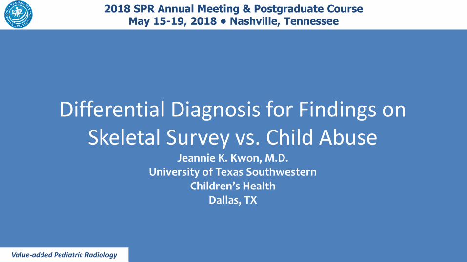
2018 SPR Annual Meeting & Postgraduate Course May 15-19, 2018 • Nashville, Tennessee
Differential Diagnosis for Findings on Skeletal Survey vs. Child Abuse
Jeannie K. Kwon, M.D. University of Texas Southwestern
Children’s Health Dallas, TX

Objectives
• Review features of entities that have findings resembling child abuse on skeletal survey
Topics • Normal variants
• Osteogenesis Imperfecta
• Skeletal dysplasias – Metaphyseal
chondrodysplasia, Schmid type
– Spondylometaphyseal dysplasia, Sutcliffe/corner fracture type
• Menkes Syndrome
• Birth injury/Iatrogenic
• Congenital syphilis
• Copper deficiency
• Scurvy
• Rickets (next talk!)

Normal Variants
• Contour changes of the metaphysis:
Step-off, Beak, Spur
– Distinguishable from metaphyseal abuse injuries
– No radiolucent separation between fragment and metaphysis
• Proximal Tibial Cortical Irregularity
• Kleinman PK, Belanger PL, Karellas A, Spevak MR. AJR Am J Roentgenol. 1991 Apr;156(4): 781-3. Normal metaphyseal radiologic variants not to be confused with findings of infant abuse.

Normal Bone bark
= perichondral collar
= subperiosteal bone collar
= ring of Lacroix
Primary spongiosa = mineralized cartilage in the developing metaphysis.
The adjacent metaphyseal margin is formed by the trabeculae
of the primary spongiosa, which have been exposed by osteoclastic
resorption (curved arrow).
Kleinman PK. Pediatr Radiol (2008) 38 (Suppl 3):S388-S394.
bone collar
bone collar extends to form spur
primary spongiosa
metaphysis resorption of trabeculae
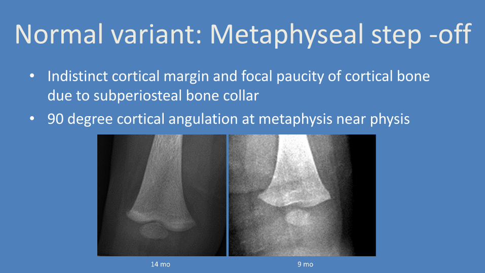
Normal variant: Metaphyseal step -off
• Indistinct cortical margin and focal paucity of cortical bone due to subperiosteal bone collar
• 90 degree cortical angulation at metaphysis near physis
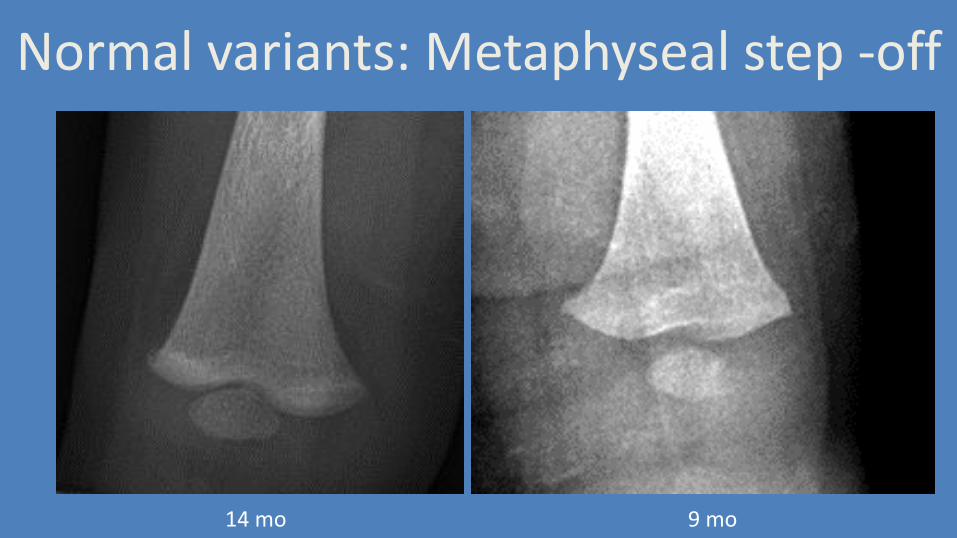
Normal variants: Metaphyseal step -off
14 mo 9 mo
Normal variant: Beak
• Medial projection of metaphysis
– Proximal humerus (16%)
– Proximal tibia (1%)
– Bilateral (77%)

Normal variant: Beak
3 mo Kleinman PK, et. Al. AJR Am J Roentgenol. 1991 Apr;156(4): 781-3.

Normal variant: Beak
14 mo
Normal variant: Spur
• Discrete linear projection of bone
• Extension of bone bark around unossified physis
• Continuous with the cortex
• Extends beyond metaphyseal margin beneath perichondrial ring

Normal variant: Spur
3 mo 2 mo, from Kleinman PK, et. Al. AJR Am J Roentgenol. 1991 Apr;156(4): 781-3.

Normal variant: Proximal Tibial Cortical Irregularity
• Medial projection of metaphysis
– 4%
– Bilateral 25%
• –May be associated with physiologic periosteal reaction

Normal variant: Proximal Tibial Cortical Irregularity
3 mo 1 mo, from Kleinman PK, et. Al. AJR Am J Roentgenol. 1991 Apr;156(4): 781-3.

Osteogenesis Imperfecta

Osteogenesis Imperfecta
• Heterogeneous group of genetic mutations controlling collagen synthesis
• Most common disease causing fracture predisposition
• Variable presentation and severity, mild to perinatal lethal – Blue sclera
– Hearing loss
– Dentinogenesis imperfecta
– Kyphoscoliosis respiratory complications
– In utero fractures
• COL1A1, COL1A2, and IFITM5 gene testing

Osteogenesis Imperfecta
Marini, J. C., A. Reich, et al. (2014). "Osteogenesis imperfecta due to mutations in non-collagenous genes: lessons in the biology of bone formation." Curr Opin Pediatr 26(4): 500-507
Mild, nondeforming
Perinatal lethal
Severe, progressively deforming

Osteogenesis Imperfecta

Osteogenesis Imperfecta
9 mo Irritability with diaper change, Older sister with unexplained fractures.

Osteogenesis Imperfecta 2 mos later Return to ED with “neck pain”

Osteogenesis Imperfecta 2 mos later Return to ED with “neck pain”
From prior skeletal survey

Osteogenesis Imperfecta 2 mos later Return to ED with “neck pain”
From prior skeletal survey
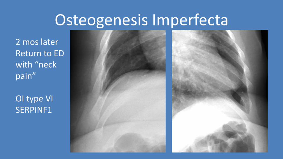
Osteogenesis Imperfecta 2 mos later Return to ED with “neck pain” OI type VI SERPINF1

Osteogenesis Imperfecta
OI type III Severe, progressively deforming COL1A2

Dysplasias

Dysplasias
• Most dysplasias with metaphyseal irregularities present with notable clinical, radiologic or laboratory findings
• Exceptions
– Metaphyseal chondrodysplasia, Schmid type
– Spondylometaphyseal dysplasia, Sutcliffe/corner fracture type
• Follow-up skeletal survey will not change

Dysplasias – Metaphyseal chondrodysplasia, Schmid type
• Autosomal dominant
• Non-familial cases present > 1yo or later
• Enlarged capital femoral epiphysis
• Pelvis almost always normal
• coxa vara
• distal ≥ proximal femoral metaphysis involved
• anterior rib changes
• Normal spine, metacarpals, phalanges

Dysplasias – Metaphyseal chondrodysplasia, Schmid type • 11 mo
• Enlarged femoral heads
• Normal pelvis
Lachman, R. S. Pediatric radiology 18.2 (1988): 93-102.
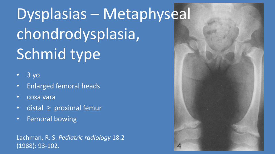
• 3 yo
• Enlarged femoral heads
• coxa vara
• distal ≥ proximal femur
• Femoral bowing
Lachman, R. S. Pediatric radiology 18.2 (1988): 93-102.
Dysplasias – Metaphyseal chondrodysplasia, Schmid type
Dysplasias – Metaphyseal chondrodysplasia, Schmid type
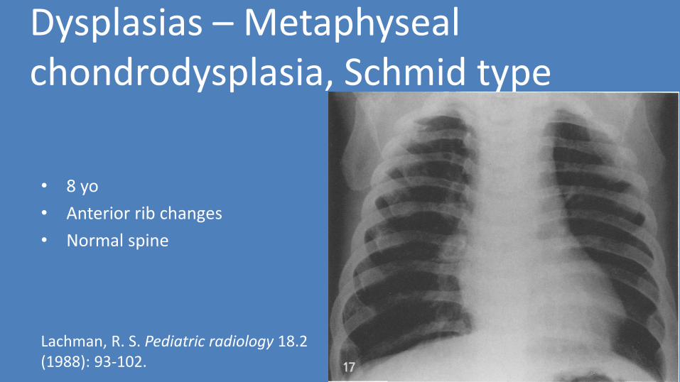
• 8 yo
• Anterior rib changes
• Normal spine
Lachman, R. S. Pediatric radiology 18.2 (1988): 93-102.
Dysplasias – SMD Sutcliffe/ Corner-fracture type

Dysplasias – SMD Sutcliffe/ Corner-fracture type
• Skeletal changes become more pronounced as patient becomes weight-bearing
• Predominant: metaphyses of long bones
• Subtle: vertebral bodies

• Predominant: long bone metaphyses – Metaphyseal irregularity
and sclerosis, "fracture-like"
– Marked bilateral coxa vara, nearly vertically oriented physes
– Tibia vara/Blount disease-like change
Dysplasias – SMD Sutcliffe/ Corner-fracture type

• Subtle:
vertebral bodies – Mild biconvex
vertebral endplates
– Very mild platyspondyly
– Odontoid hypoplasia
Dysplasias – SMD Sutcliffe/ Corner-fracture type

Menkes Syndrome

Menkes Syndrome
• AKA Kinky hair disease
• X-linked recessive
• Prevalence 1 in 100,000-250,000
• Symptoms appear in infancy; mild forms in childhood Erik N. Swartz CMAJ 2002;166:1442-1443
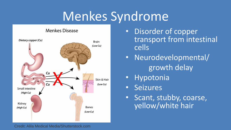
Menkes Syndrome • Disorder of copper
transport from intestinal cells
• Neurodevelopmental/ growth delay • Hypotonia • Seizures • Scant, stubby, coarse,
yellow/white hair
Credit: Alila Medical Media/Shutterstock.com

Menkes Syndrome • Metaphyseal widening, spurs
• Long bone fractures
• Wormian occipital bones
• Hydronephrosis
• Ureteral dilatation
• Bladder diverticula
• Tortuous intracranial arteries
• Cerebral and cerebellar atrophy
• Chronic subdural hemorrhages
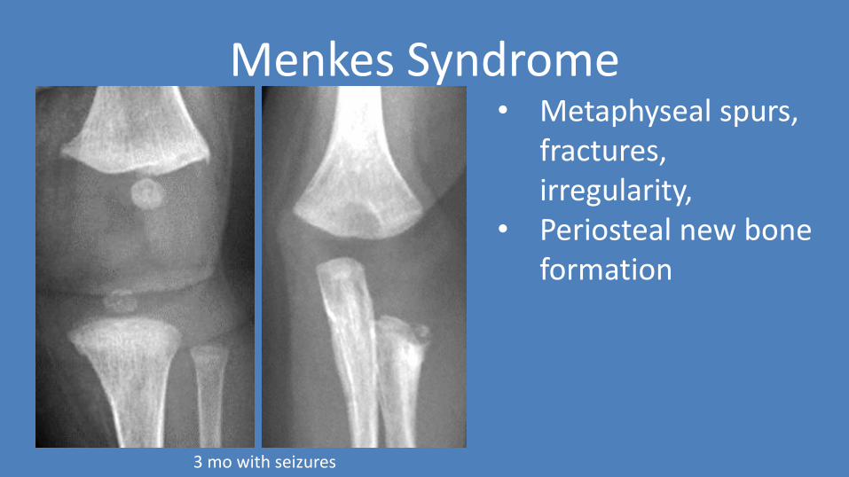
Menkes Syndrome • Metaphyseal spurs,
fractures, irregularity,
• Periosteal new bone formation
3 mo with seizures

Menkes Syndrome
3 mo: atrophy tortuous intracranial arteries Wormian bones

Birth injury/Iatrogenic

Birth injury
• Correlate with age, expected healing ~10 days
• Metaphyseal fracture, Epiphyseal separation
– Breech, armling presentation
• Long bone shaft fracture
– Caesarean section
• Clavicle fracture
– High birth weight, cephalic presentation
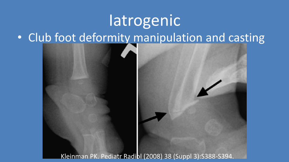
Iatrogenic • Club foot deformity manipulation and casting
Kleinman PK. Pediatr Radiol (2008) 38 (Suppl 3):S388-S394.

Iatrogenic
• VACTERL • Vertebral body anomalies • Imperforate anus • Renal agenesis • Anhydramnios • Pulmonary hypoplasia • PROM at 20 weeks • Born at 27 weeks • L knee contracture • (outside institution)

Iatrogenic

Congenital syphilis
Congenital Syphilis
• Transplacental infection by Treponema pallidum spirochete
• Disseminated throughout fetus
• 1/3 stillbirth; 1/3 contracts; 1/3 unaffected
• +/- symptomatic at birth
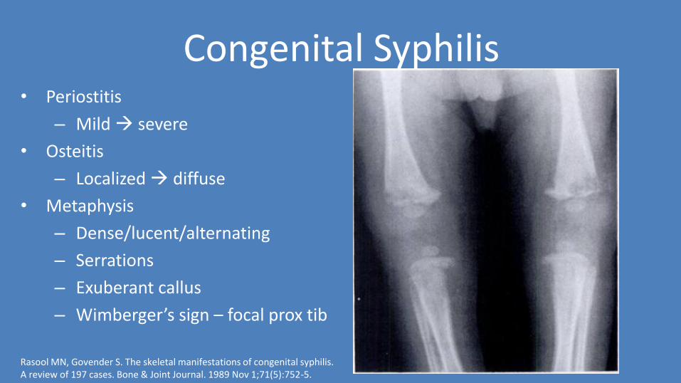
Congenital Syphilis • Periostitis
– Mild severe
• Osteitis
– Localized diffuse
• Metaphysis
– Dense/lucent/alternating
– Serrations
– Exuberant callus
– Wimberger’s sign – focal prox tib
Rasool MN, Govender S. The skeletal manifestations of congenital syphilis. A review of 197 cases. Bone & Joint Journal. 1989 Nov 1;71(5):752-5.

Congenital Syphilis
2 mo
– Periostitis
– Osteomyelitis
• Pathologic fractures
• Metaphysis
• Diaphysis
Lim HK, Smith WL, Sato Y, Choi J. Congenital syphilis mimicking child abuse. Pediatric radiology. 1995 Sep 1;25(7):560-1.

Congenital Syphilis
• Early congenital syphilis
– Prematurity
– Hepatosplenomegaly, jaundice
– Nasal chondritis, “runny nose”
– Generalized lymphadenopathy
– Maculopapular rash
• Late congenital syphilis
– Saddle nose
– Sabre shin
– Frontal bossing
– Hutchinson’s triad • Hutchinson’s teeth
• Interstitial keratitis
• CN VIII deafness

Congenital Syphilis
36 week preterm newborn, mother RPR+

Copper deficiency

Copper Deficiency
• Copper required for endochondral bone, collagen
• Rare: ~100 cases in 1987
• Full term infants body stores sufficient for 5-6 months
• Low birth weight infants have 2 months
• Modern formulas and breastfeeding should not cause
• Plasma copper < 40 μg/dl
• Ceruloplasmin < 13 mg/dl
• Other clinical findings: sideroblastic anemia, neutropenia, hypotonia, psychomotor retardation, pallor
Copper Deficiency
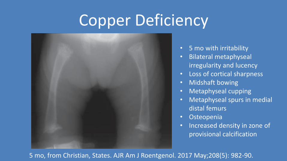
5 mo, from Christian, States. AJR Am J Roentgenol. 2017 May;208(5): 982-90.
• 5 mo with irritability • Bilateral metaphyseal
irregularity and lucency • Loss of cortical sharpness • Midshaft bowing • Metaphyseal cupping • Metaphyseal spurs in medial
distal femurs • Osteopenia • Increased density in zone of
provisional calcification

Scurvy

Scurvy
• Hypovitaminosis C
• Bleeding tendency
• Impaired collagen synthesis
• Osteoporosis
• Impaired wound healing
Scurvy
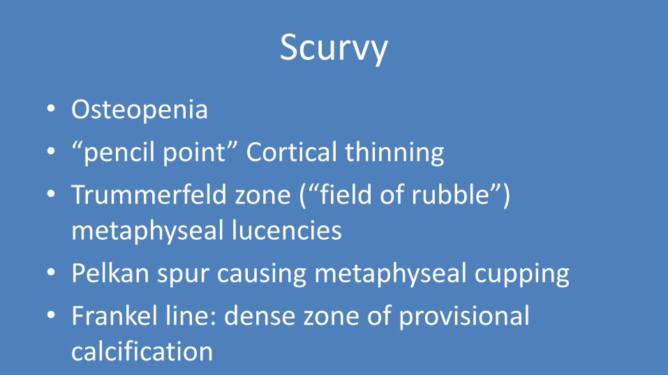
• Osteopenia
• “pencil point” Cortical thinning
• Trummerfeld zone (“field of rubble”) metaphyseal lucencies
• Pelkan spur causing metaphyseal cupping
• Frankel line: dense zone of provisional calcification

Scurvy
Noordin, Shahryar, et al. Case reports in orthopedics 2012 (2012).
Trummerfeld zone
Pelkan’s spur
Frankel line

Topics • Normal variants
• Osteogenesis Imperfecta
• Skeletal dysplasias – Metaphyseal
chondrodysplasia, Schmid type
– Spondylometaphyseal dysplasia, Sutcliffe/corner fracture type
• Menkes Syndrome
• Birth injury/Iatrogenic
• Congenital syphilis
• Copper deficiency
• Scurvy

References • Christian CW, States LJ. Medical mimics of child abuse. American Journal of Roentgenology. 2017 May;208(5):982-90.
• Kleinman PK. Problems in the diagnosis of metaphyseal fractures. Pediatric radiology. 2008 Jun 1;38(3):388.
• Kleinman PK, Belanger PL, Karellas A, Spevak MR. Normal metaphyseal radiologic variants not to be confused with findings of infant abuse. AJR. American journal of roentgenology. 1991 Apr;156(4):781-3. Dwek JR. The radiographic approach to child abuse. Clinical Orthopaedics and Related Research®. 2011 Mar 1;469(3):776-89.
• Lachman RS, Rimoin DL, Spranger J. Metaphyseal chondrodysplasia, Schmid type clinical and radiographic deliniation with a review of the literature. Pediatric radiology. 1988 Feb 1;18(2):93-102.
• Langer Jr LO, Brill PW, Ozonoff MB, Pauli RM, Wilson WG, Alford BA, Pavlov H, Drake DG. Spondylometaphyseal dysplasia, corner fracture type: a heritable condition associated with coxa vara. Radiology. 1990 Jun;175(3):761-6.
• Lim HK, Smith WL, Sato Y, Choi J. Congenital syphilis mimicking child abuse. Pediatric radiology. 1995 Sep 1;25(7):560-1.
• Marini JC, Reich A, Smith SM. Osteogenesis Imperfecta due to Mutations in Non-Collagenous Genes-Lessons in the Biology of Bone Formation. Current opinion in pediatrics. 2014 Aug;26(4):500. Noordin S, Baloch N, Salat MS, Rashid Memon A, Ahmad T. Skeletal manifestations of scurvy: a case report from Dubai. Case reports in orthopedics. 2012;2012.
• Rasool MN, Govender S. The skeletal manifestations of congenital syphilis. A review of 197 cases. Bone & Joint Journal. 1989 Nov 1;71(5):752-5.
• Stephens JR, Arenth J. Wimberger sign in congenital syphilis. The Journal of pediatrics. 2015 Dec 1;167(6):1451.