Embed Size (px)
Citation preview

Diagnostics and Drug DeliveryDOI: 10.1002/anie.200802585
Nanomedicine—Challenge and PerspectivesKristina Riehemann,* Stefan W. Schneider, Thomas A. Luger, Biana Godin,Mauro Ferrari, and Harald Fuchs*
AngewandteChemie
Keywords:nanoprobes · nanotechnology ·personalized medicine ·theranostics
K. Riehemann, H. Fuchs et al.Reviews
872 www.angewandte.org � 2009 Wiley-VCH Verlag GmbH & Co. KGaA, Weinheim Angew. Chem. Int. Ed. 2009, 48, 872 – 897

1. Introduction
The manipulation of matter locally and deliberately onthe atomic or molecular scale is an old dream of naturalscience. Starting in 1959 with the famous talk by RichardFeynman at the annual meeting of the American PhysicalSociety, where he developed the vision of manipulating andcontrolling things on a small scale, nanoscience developed,with the discovery of molecular beam epitaxy in 1968 in theBell Laboratories, the generation of nanoparticles, and theinvention of the scanning tunneling microscope, into a robustand well-accepted scientific field.[1–3] The old dream hasalready become true in the fields of nanoscience and nano-technology. New opportunities have been realized in virtuallyall branches of technology ranging from optical systems,electronic, chemical, and automotive industries, to environ-mental engineering and medicine. “Smart” surface coatings,intelligent nanoscale materials, faster electronics, unprece-dented optics, biosensors, and nanomotors are just a fewexamples from this transdisciplinary area. Although nano-technology is still in its infancy, these first practical applica-tions clearly demonstrate its enormous potential.
The field of medicine, on the other hand, faces verycomplex scientific as well as societal and ethical challenges. Inparticular because of the increased life expectancy of thepopulation, some specific diseases have been identified ashaving a very high socio-economic impact over the next fewyears. Below we will discuss some specific areas, which weconsider as promising applications of nanomedicine.
1.1. Definition
The term nanotechnology [from the Latin nanus, Greeknanos dwarf] is defined in the literature in a variety of ways,all of which have their advantages and limitations. In general,nanotechnology is concerned with dimensions and tolerancelimits of 0.1–100 nm (1 nm = 10�9 m), as well as with themanipulation of single atoms and molecules. A more specificdefinition was given in 2000 by the US National Nano-
technology Initiative: “Nanotechnology is concerned withmaterials and systems whose structures and componentsexhibit novel and significantly improved physical, chemicaland biological properties, phenomena and processes due totheir nanoscale size”. The reduction in magnitude apparentlyleads to different, and qualitatively new and advantageousproperties in the nanometer-scale materials.[4–6] A moregeneral and operational definition involves the followinginterrelated constituents: nanoscale dimensions of the wholesystem or its vital components, synthetic materials, andunique characteristics that arise because of its nanoscopicsize.[7]
Thus, nanotechnology includes the following key physicaland chemical issues:* The occurrence of novel physical properties characteristic
of the nanoscale,* analysis at the atomic and molecular scale at predefined
positions,
The application of nanotechnology concepts to medicine joins twolarge cross-disciplinary fields with an unprecedented societal andeconomical potential arising from the natural combination ofspecific achievements in the respective fields. The common basisevolves from the molecular-scale properties relevant to the twofields. Local probes and molecular imaging techniques allowsurface and interface properties to be characterized on a nanometerscale at predefined locations, while chemical approaches offer theopportunity to elaborate and address surfaces, for example, fortargeted drug delivery, enhanced biocompatibility, and neuro-prosthetic purposes. However, concerns arise in this cross-discipli-nary area about toxicological aspects and ethical implications. ThisReview gives an overview of selected recent developments and ap-plications of nanomedicine.
From the Contents
1. Introduction 873
2. Nanotechnology in MedicalDiagnostics 875
3. Nanotechnology in Therapy—Research and Development 883
4. Clinical Applications 885
5. Nanocoatings and NanostructuredSurfaces for Medical Application 890
6. Biocompatibility and Toxicity—Safety Issues Related toNanotechnology Implementation 891
7. Summary and Perspectives 892
[*] Dr. K. Riehemann, Prof. Dr. H. FuchsCenter for Nanotechnology (CeNTech) und Physikalisches InstitutWestf�lische Wilhelms-Universit�t M�nsterWilhelm-Klemm-Strasse 10, 48149 M�nster (Germany)Fax: (+ 49)251-83-33602E-mail: [email protected]: http://www.uni-muenster.de/Physik.PI/Fuchs/
Prof. Dr. S. W. SchneiderDepartment of DermatologyUniversity Medical Center Mannheim, University of HeidelbergTheodor-Kutzer-Ufer 1–3, 68135 Mannheim (Germany)
Prof. Dr. T. A. LugerKlinik und Polyklinik f�r HautkrankheitenVon-Esmarch-Strasse 58, 48149 M�nster (Germany)
Dr. B. Godin, Prof. Dr. M. FerrariDivision of NanomedicineDepartment of Biomedical EngineeringUniversity of Texas Health Science Center at HoustonHouston, TX, 77030 (USA)
NanomedicineAngewandte
Chemie
873Angew. Chem. Int. Ed. 2009, 48, 872 – 897 � 2009 Wiley-VCH Verlag GmbH & Co. KGaA, Weinheim

* control of matter at the atomic scale, that is, addressingindividual preselected atoms and molecules, and
* the generation of complex functional systems with qual-itatively novel properties (emergence).
To define the area of nanomedicine, discussed below, we firsthave to introduce a differentiation to the fields of molecularmedicine, biochemistry, as well as nanobiotechnology.
Nanomedicine means essentially applying nanotechnol-ogy to medicine. While being related in certain aspects, thefield of nano-biotechnology differs from nanomedicine, sincethe latter focuses on the applications of nanotechnologyconcepts to medical applications, while the former encom-pases all basic research at a nanoscopic level on biologicalsystems, for example, investigations on plants. Molecularmedicine, on the other hand, starts from a more conventionalbiochemical approach.
In contrast to conventional therapies—surgery, radiation,and chemotherapy—where the basic approach is to removediseased cells faster than healthy cells, nanomedicineattempts to use sophisticated approaches to either kill specificcells or repair them one cell at a time by using a biosensor todetect, for example, when a drug should be released. Thusnanomedicine needs not only to apply and adopt nano-technology concepts but will, at the same time, need tofeedback information to nanotechnology such that the twofields can cross-fertilize and develop jointly.
One goal is the design of multilevel molecular aggregatesthat have novel functional and dynamic properties that aredesirable for applications in medicine. Both the size- and site-
specific properties of these systems which are characteristic ofthe nano- and mesoscale are made use of. This approach alsooffers new possibilities towards the development of person-alized medicine, which is defined as: “the concept whichmarks the expected reform in medicine that is projected toarrive at the clinic in coming decades, harnessing genomics andproteomics technologies for tailoring the most suitable phar-macotherapy for each patient; based on individual profiling, itis also projected to allow improved treatment efficacies formany diseases”.[8] To avoid side effects and overdosing ofdrugs, efficient medications need to be established by usingselective targeting. This field is currently under intensiveinvestigation. Nanomedicine promises alternatives to molec-ular medicine by having the following general advantages:local processes at the nanometer length scale, such asdiffusion, intermixing, and sensoric response, become ultra-fast. Furthermore, nanotechnology can provide the opportu-nity of directly probing local properties. Physical andchemical processes can be controlled and intensified, theprecision enhanced, and direct access to biomarkers becomespossible. Finally, new results can be achieved in real time.These concepts together with a combination of the researchareas such as systems biology and systems medicine willcontribute significantly to form the route to personalizedmedicine.
How is personalized medicine related to nanomedicine?Similar to existing medical diagnosis and therapeutics, and asdictated by economic reasons, mass applications of newscreening and diagnostic tools in medicine have to be fast,convenient, and inexpensive. Therefore, miniaturization,
Kristina Riehemann studied biology andphysics at the University of M�nster. Shewas Junior group leader at the Institute ofMedical Biochemistry at the Centre forMolecular Biology of Inflammation, with theresearch focus “Anti-inflammatory mecha-nisms”. She then became coordinator andmanager of “Integrated functional Genom-ics”, a service unit of the “Interdisciplinarycenter of clinical research”. Currently she isgroup leader at the “Center for Nanotech-nology” (CeNTech) and Coordinator of theSino-German BMBF Project “Biocompatibil-ity of nanoparticles for medical engineering,diagnostics, and therapy”.
Harald Fuchs is Professor of ExperimentalPhysics at the University of M�nster, Ger-many, and Director of CeNTech. He is amember of the Helmholtz Association andholds three guest professorships in China. Heis a cofounder of two nanotechnology com-panies and a member of various scientificinstitutions, societies, and editorial advisoryboards. Since 2008 he has been the Germanspokesperson of the first Sino–GermanDFG/NSFC collaborative project SFB/TRR61. He was awarded the Philip MorrisResearch Prize “Challenge Future”, and is
member of the German Academy of Sciences Leopoldina and the GermanAcademy of Science and Engineering, acatech.
Thomas Luger is Professor and Chairman atthe University of M�nster. He is visitingprofessor at the Universities of Tohoku, Hir-oshima, Teikyo, Sendai, Osaka, Helsinki,Cardiff, Nottingham, Leicester, and at St.Thomas Hospital London. He is President ofthe German Dermatological Society, Editorof the journal Experimental Dermatolology,and on the Editorial Board of several scien-tific periodicals. He has received a numberof international awards and is member ofthe German Academy of Sciences Leopol-dina.
Stefan Schneider is Professor for CellularDifferentiation at the University of Man-nheim–Heidelberg and senior physician atthe University Dermatology Clinic, Man-nheim. He studied medicine in W�rzburg(Germany), Chur (Switzerland), and Izmir(Turkey), and in 1994 received his PhD(Prof. Dr. H. Oberleithner). In 1994–1997he was a DFG fellow at the Department ofPhysiology at the University clinic, W�rz-burg, as well as at Yale University (USA). In1997–2001 he carried out postdoctoralresearch and Habilitation at the Depart-
ment of Physiology in M�nster. Afterwards he moved to the Department ofDermatology in M�nster and in 2008 to the University of Mannheim–Heidelberg.
K. Riehemann, H. Fuchs et al.Reviews
874 www.angewandte.org � 2009 Wiley-VCH Verlag GmbH & Co. KGaA, Weinheim Angew. Chem. Int. Ed. 2009, 48, 872 – 897

parallelization, integration, as well as automation are man-datory. The demand for large amounts of routine in vitromeasurements on patients so as to retrieve sufficient andcomparable data dictates the development of smart inte-grated devices, such as biosensors and decentralized actua-tors, and drug-release concepts—requirements that can onlybe fulfilled with the help of nano- and microsystem technol-ogies.
Nanomedicine includes the development of nanoparticlesand nanostructured surfaces as well as nanoanalytical techni-ques for molecular diagnostics, treatment, follow-up, andtherapy of diseases (theranostics). Integrated medical nano-systems are also needed which, in the future, may performmonitoring and complex repairs in the body at the cellularlevel. Nanotechnology considers cells as a complex system ofinteracting nanomachines. Visionary concepts envisage theconstruction and control of artificial cells by using engineerednanodevices and nanostructures for medical applications(Figure 1).
2. Nanotechnology in Medical Diagnostics
Diagnostics play a key role in medicine for the successfulprevention and efficient treatment of diseases. Taking canceras an example of a widespread disease, and that is still theleading cause of death in the industrial countries, it will bedifficult to achieve a significant increase in the cure rateunless more information about the molecular mechanisms ofthe pathophysiology can be obtained, which will build the
basis for the development of new anticancer drugs.[9] Theadvantage of nanostructure-based diagnostics lies in theirpotentially higher sensitivity and selectivity compared toclassical methods.
An important area in nanotechnology is the generation ofnanoscale materials. For diagnostic purposes, quantum con-finement effects, which are characteristic of the nanometerscale, may be used. Nanoparticles may be embedded in othercrystalline or amorphous nanoscale materials to guaranteebetter functionality and bioavailability. In this area, researchinto the development of metallic and semiconductor quantumdot structures, nanoclusters, as well as nanopowders is intense.For medical applications (molecular imaging), some types ofthese particles can be used in vivo as markers in variousimaging techniques, such as IR or NMR methods to increasesignificantly the resolution and sensitivity, thus enablingearlier diagnosis of diseases. The increased resolution andsensitivity is expected to lead to cheaper clinical applicationsin therapy. Modification of the nanoparticle surfaces withchemical recognition groups allows the identification ofcomplementary groups on cell surfaces which are indicative,for example, of cancer or other severe diseases (see Figure 2).The same concept can also be applied to site-specific drugdelivery.[10–12]
2.1. In Vitro Diagnostics
The purpose of extracorporeal (in vitro) diagnostics forcells is manifold. In vitro diagnostics are necessary, forexample, to protect the blood supply for transfusion reasons,to monitor the level of drugs applied to patients, and toprovide information to assist the diagnosis and treatment ofdisease. The ultimate goal of any diagnostic procedure is anon-invasive, early, and accurate detection of the biologicaldisease markers in the process of routine screening, thusenabling the appropriate treatment regimen to be chosen.Various nanotechnology platforms have been developed toallow for the simultaneous real-time evaluation of a broadrange of disease markers by non-invasive techniques. Inter-
Mauro Ferrari is a Professor and Director atthe Center for Nanomedicine in the Depart-ment of Biomedical Engineering, Universityof Texas Health Science Center at Houston,Professor of Experimental Therapeutics, Uni-versity of Texas M.D. Anderson CancerCenter, Professor of Bioengineering, RiceUniversity, and President of the “Alliance forNanoHealth”, Houston. From 2003 to2005, he served as Expert on Nanotechnol-ogy at the NCI, providing leadership into theformulation, refinement, and approval of theNCIs Alliance for Nanotechnology in Cancer.
Biana Godin (Vilentchouk) studied andearned her PhD (in 2006) in Pharmaceut-ical Sciences from the Hebrew University ofJerusalem, Israel, specializing on the designof novel nanocarriers for improved dermaland transdermal administration of therapeu-tics. Currently, she is a postdoctoral fellow atthe “NanoMedicine Research Center” at theUniversity of Texas Health Science Center atHouston. Her primary research interest is innanotechnological solutions for advanceddrug delivery.
Figure 1. Technologies involved in the field of nanomedicine.
NanomedicineAngewandte
Chemie
875Angew. Chem. Int. Ed. 2009, 48, 872 – 897 � 2009 Wiley-VCH Verlag GmbH & Co. KGaA, Weinheim www.angewandte.org

estingly, two classes of microtechnological devices, micro-array DNA chips and microfluidic systems for lab-on-chipdiagnostics, which were developed in the 1980s, have nowbeen transferred to the nanotechnology arena. This “minia-turization” was possible in both cases because of a develop-ment in the fundamental enabling technique, namely photo-lithography. This technique now allows for lateral resolutionin the 10–100 nm range, which is three orders of magnitudelower than at the time when these approaches were firstdeveloped. As a result, the information that can be put on abiochip has increased by a factor of 106–108, thus demonstrat-ing the powerful capabilities of nanoscaling in biomedicalapplications. The use of photolithography allows the selectiveillumination and removal of photolabile groups, therebyleading to the exposure of reactive moieties. The techniquecan be used to pattern various chemical and biologicalmoieties or diverse textures very precisely on the substrate,thus enabling the surface attachment of biomolecules tospecific molecular segments, for example, single-strandedDNA for hybridization or different substrates for proteomicanalysis.[14–20]
Another goal of medical diagnostics is to analyze singlecells. Nanotechnology approaches offer the opportunity toeven investigate single molecules, and opens up the possibilityfor new methods for analysis and detection. The added valueof this approach becomes clear when it is taken intoconsideration that large amounts of primary cells are usuallymixtures of either different cell types or healthy and tumorouscells, thus making the acquisition of statistically significantresults difficult.[21] Another motivation for single-cell analysisconcerns the dilution of effects. In the case of disease, thismeans that small differences between cell types or weakeffects of drugs are not detectable using complete tissues.Biochemical methods are often inappropriate for investiga-tions because the large amount of cells needed, for example,for electrophoresis purposes, leads to the analysis not of cellsbut of tissues or cell mixtures, that is, systems, which give no
insight into the definite basic structure. The ability to describeone specific cell (type) leads to the role of this building blockin the tissue and the organism being defined, and thus thefunction of cell interaction, the effect of differentiation, anddiseases can be characterized.[22]
Different selection techniques such as cloning rings,limiting dilution, laser microdissection, live-cell catapulting,or microfabricated pallets are used for the isolation of singleadherent cells.[23–25] Fluorescent-activated cell sorting (FACS),magnetic sorting, column chromatography, panning, limitingdilution, and the isolation of cells by microfluidic approachesare commonly used for the isolation of non-adherentcells.[24,25] The analysis of those cells has until now beenperformed by classical biochemical methods such as thepolymerase chain reaction (PCR) or patch-clamp techniques.A nanotechnology alternative is now commonly used forbiochip analysis performed with photolithographic technol-ogy (see Section 2.1.1). Together with the development ofsmart surfaces, semiconductor manufacturing and combina-torial chemistry as well as bioinformatics have made signifi-cant advances in the expression analysis of single cells.[26–30]
Biochip analysis on a multicell level is now well accepted inclinical diagnostics in several fields. For example, expressionchips for the follow up of bacterial infections in the mouthhave made such significant progress that they are now used aspoint-of-care diagnostics. The modification of biochip surfa-ces by nanotechnological methods offers the possibility forever smaller probes for the analysis of RNA retrieved from asingle cell.
The success of expression profiling encouraged proteinresearchers to adopt some of the methods. As the differencesbetween the expressed form of proteins and their biochemicalappearance (for example, folding structure or secondarymodification) is remarkably high, the analysis of proteins at asingle-cell level is coming more into the focus of industrialand scientific research because the results obtained reflectmuch more the biological processes within a cell than does theexpression profiling. Different kinds of biochips with proteinarrays are available (see Section 2.1.1). The first applicationof antibody arrays were reported in 2002.[31, 32] Clinicalapplications for such protein chips include the discovery ofdisease markers for diagnosis, prognosis, and drug response,and allow the disease development and progression to betracked. Antibody arrays are suited to high-throughputmethods for the functional characterization of disease at amolecular level. Furthermore, the information gained fromthe protein array on cancer progression and tumor subtypesmay enable intervention and therapy optimization.[33–41]
Atomic force microscopy (AFM; see Section 2.1.3.1)techniques have been explored for single-cell analysis. Theyallow high-resolution in vitro investigation of cell surfacesand analysis of physical properties such as mechanicalcompliance of single cells at a single-cell level. Forcespectroscopy provides locally direct quantitative data onintra- and intermolecular forces of a single molecule.[42–48] Thisapproach was also used for manipulation, for example, as amicrodissection device. With the possibility to isolate organ-elles and to cut chromosomes in a precise way, this techniquewas applied together with subsequent PCR amplification of
Figure 2. Prostate cancer cells have taken up fluorescently labelednanoparticles (shown in red). RNA aptamers binding to the prostate-specific membrane antigen (PSMA; a well-known transmembraneprotein, which is overexpressed on prostate cancer epithelial cells)were used as the targeting molecules on the nanoparticles. The cellnuclei and cytoskeletons are stained blue and green, respectively.Similarly designed targeted nanoparticles are capable of getting insidecancer cells and releasing lethal doses of chemotherapeutic drugs todestroy the tumors. Reprinted with permission from the AmericanAssociation for the Advancement of Science (AAAS).[13]
K. Riehemann, H. Fuchs et al.Reviews
876 www.angewandte.org � 2009 Wiley-VCH Verlag GmbH & Co. KGaA, Weinheim Angew. Chem. Int. Ed. 2009, 48, 872 – 897
dissected DNA fragments for the analysis and even mechan-ical reimplantation of the isolated fragments back into itsoriginal position.[49, 50]
2.1.1. Microfluidics and Nanoarrays
A microfluidic unit can be defined as a device comprisingone or more channels with at least one dimension measuringless than 1 mm. The channels, which can have a width below afew micrometers, allow control over minute fluidic volumes inthe nanoliter and picoliter range. Microsystem technologiesdeveloped for microfluidic chips enable just about anybiological assay working at the molecular level to beincorporated onto a chip (lab-on-a-chip systems; Figure 3).These approaches not only offer the possibility to isolate andmanipulate living cells, but also to perform toxicity assays,enzyme-linked immunosorbent assays (ELISA), PCR am-plification, blood separation, or for the genotyping ofcytokine polymorphism.[51]
The flow of fluidics in a microfluidics chamber ischaracterized by the non-dimensional Reynolds number Re[Eq. (1)].
Re ¼ ð1u2Þ=ðmu=LÞ¼ 1u L=m
¼ u L=n
1 ¼ density, u ¼ velocity, m ¼ dynamic viscosity,
L ¼ characteristic length, n ¼ kinematic viscosity
ð1Þ
The Reynolds number should be less than 100 to maintainlaminar flow, which is necessary so that molecules can be
transported in a predictable manner through microchannels.Materials such as poly(dimethylsiloxane) (PDMS) enabledmicrofluidic chips to become a conventional easy-to-produceand easy-to-use technology. The ability to tailor the materialfor the analysis of single cells gave a strong push to thisresearch area.[52–54] Another important advantage of PDMS isits biocompatibility. It is assumed to be a suitable biomaterialfor biomedical devices because it causes minimal endotoxincontamination, leukocyte activation, and complement activa-tion.[55] Du et al. showed in 2006 that an antibody-basedmicrofluidics system captured more than 30% of the cancercells from a mixture of normal human glandular epithelialcells (HGEC), human cervical stromal cells (HCSC), andcervical cancer cells (HCCC).[56]
Another approach was the development of a microfluidiccell chip for monitoring allergic response. A basophilicleukemia cell line (RBL-2H3) was cultivated on a PDMSchip containing a cultivation chamber and microfluidicchannels. Molecules marked with a fluorescent dye weresecreted after allergic stimulation and observed by using aphotomultiplier tube (PMT) fitted onto a microscope. Thetechnique of photolithography was adapted from the micro-electronic industry. The lateral resolution was originally of theorder of 100 mm (or 100 000 nm). The linear spatial resolutionof lithography is now 1000 times better, that is, up to a one-million-fold increase in information density can now bepacked in “nanoarrays”. Various nanochannel structures canbe imprinted for the selective fractionating of proteins on thebasis of their molecular weight. As a result, different patternscould be produced simultaneously by treating the chip withdissimilar biological samples (Figure 4).[57]
Specific serum markers for the early diagnosis of diseasessuch as cancer are currently unavailable. Nanotechnologyoffers the possibility to evaluate a wide range of molecularmarkers simultaneously. The information obtained can behelpful for early and efficient diagnostics as well as formonitoring and selecting therapeutic strategies. Single clinicalmarkers that are now used for the diagnosis of carcinogenicconditions, for example, the prostate specific antigen (PSA),cannot provide this knowledge because of the broad varia-bility in the basal expression in individuals. Thus, ex vivodiagnosis on biological fluids such as serum, saliva, urine, ortissue exudates obtained from non- or minimally invasiveprocedures remains an unmet need.[59]
Fluidic devices based on other concepts have in themeantime become commercially available. One of these is adevice based on dielectrophoretic field cages, which give thepossibility to combine the isolation and the manipulation ofnon-adherent cells in one device. This principle was intro-duced by Fiedler et al. in 1998.[61] Certain electrode config-urations were constructed to function as a funnel, as liners tobreak aggregates of cells, or as electrical octodes to trap cellselectrostatically for manipulation. Recently it was shown thatmoderate warming induced by the electrical field can beneglected in cultures under appropriate experimental con-ditions. Moreover, possible side effects of dielectrophoreticmanipulation such as membrane polarization and Jouleheating were excluded, thus making the method appropriatefor medical applications.[60–62]
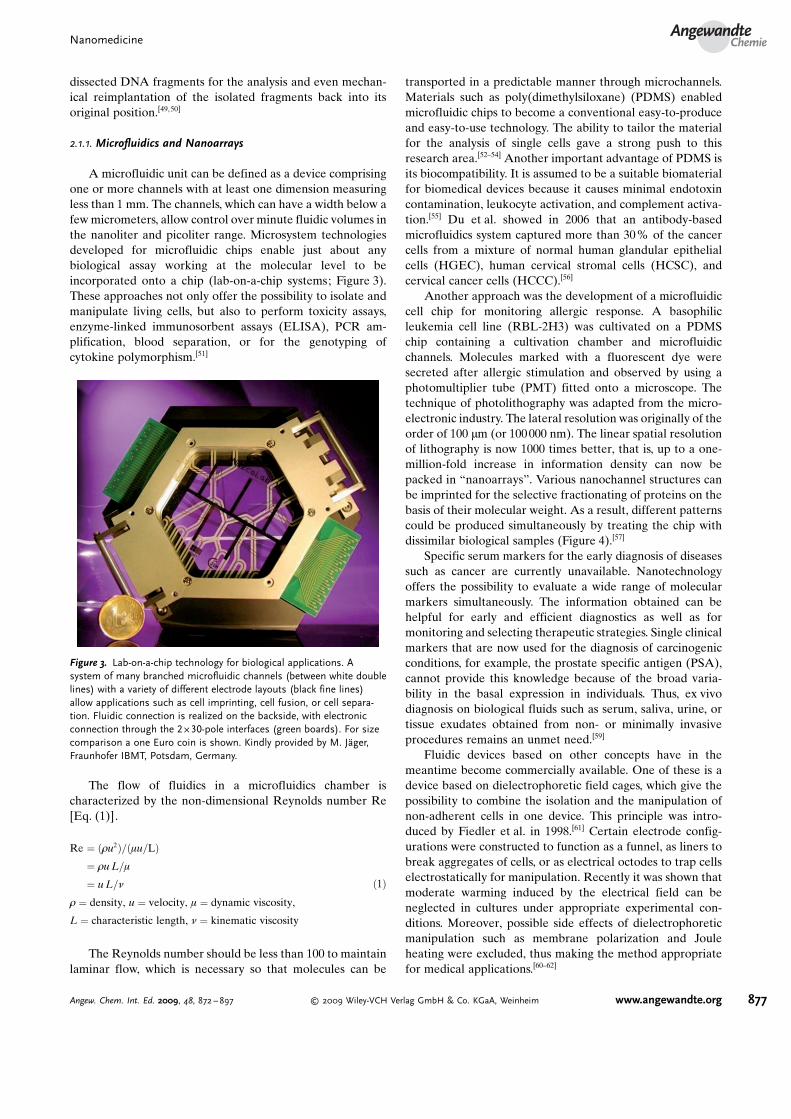
Figure 3. Lab-on-a-chip technology for biological applications. Asystem of many branched microfluidic channels (between white doublelines) with a variety of different electrode layouts (black fine lines)allow applications such as cell imprinting, cell fusion, or cell separa-tion. Fluidic connection is realized on the backside, with electronicconnection through the 2 � 30-pole interfaces (green boards). For sizecomparison a one Euro coin is shown. Kindly provided by M. J�ger,Fraunhofer IBMT, Potsdam, Germany.
NanomedicineAngewandte
Chemie
877Angew. Chem. Int. Ed. 2009, 48, 872 – 897 � 2009 Wiley-VCH Verlag GmbH & Co. KGaA, Weinheim www.angewandte.org

An urgent need was the investigation of the systemicinflammatory responses that can develop in patients followinga cardiopulmonary bypass (CPB). As a general rule, theability to clinically intervene in inflammation is limited by thelack of timely measurements on inflammatory responses;blood analysis performed in medical laboratories can takefrom several hours to days. Thus, there is a need for a systemthat can separate plasma from whole blood and measure theconcentration of the clinically relevant proteins in real time.A microfluidics device was fabricated to monitor the devel-opment of inflammation markers in real time by using aplasma analysis device.[63] Here, microfluidics offer the chanceto intervene at an early stage in an inflammatory process,which if untreated could be life-threatening. Recently, a newmicrochip was developed with an anisotropic nanofluidicstructure to separate and sort biomolecules as DNA orproteins. With an extremely tiny sieve structure, the systemcan sort through continuous streams of biological fluids andseparate proteins by size. This system thus provides anappropriate tool for the identification of small molecules forearly diagnostics and the follow up of medical treatment.[64]
One of the recent promising approaches in diagnostics isbased on the specific recognition of the biomolecularinteractions by using appropriate nanosensors. This concept,proposed by the research group of Gimzewsky, is based on thenanoscale forces and deformations produced as a result ofligand–substrate binding.[65] Micro- and nanocantilevers,devices based on this principle, deflect or change theirresonant frequencies as a result of the affinity binding ofbiomarker proteins or DNA hybridization events occurringon their free surfaces (Figure 5a). The deflections can bemonitored by lasers or detected electronically, thus enablingthe rapid and simultaneous sensing of a variety of biomarkers.
This technique allowed the detection of target oligonucleo-tides at clinically significant levels without fluorescent orradiolabeling and serum markers.[59, 65–67] Other examples ofsensor technologies based on nanofabrication are nanowiresand nanotubes.
Nanowires placed in a microfluidic system can specificallybind or absorb various analytes, thus resulting in a shift intheir conductance as a function of the electrical charges on thebound molecules (Figure 5b).[67–72] These changes can bedetected electronically and quantified precisely. Althoughthese systems are not yet in clinical practice, their multi-plexing capabilities hold promise for parallel analyses.
2.1.2. Fluorescent Labels and Imaging
Fluorescent dyes represent another important class ofin vivo imaging tools which are mainly used for the visual-ization of cells and molecules. A big disadvantage of thosedyes is their photo instability, with the fluorescent yieldrapidly fading within less than one minute. The bleaching ofthe dyes restricts the range of their applications. Inorganicquantum dots have a much higher photostability; however,selenides and sulfides, which are mostly applied for thispurpose, are cytotoxic and can, therefore, only be used fordiagnostics of biological samples outside the human body. Thebiocompatibility, high photoluminescence quantum effi-ciency, and stability against photo bleaching makes siliconquantum dots ideal candidates for replacing fluorescent dyesin biological assays. Silicon nanocrystals (NCs) can befabricated by using wet chemsitry or electron-beam lithog-raphy with reactive ion etching to give silicon nanopillars thatcan be subsequently oxidized to produce luminescent siliconcores.[73] These are so small that the addition or removal of a
Figure 4. Photolitographic techniques for manufacturing micro- and nanoarrays for DNA analysis (a) and (b) proteomics. a) A microarray with apattern of biological molecules on the surface to obtain DNA sequencing by hybridization, for example. Blue squares represent photolabilegroups, which are selectively illuminated through a mask (photolithography) and removed to expose reactive groups. Sequential application of theprocedure yields single-stranded hybridization probes with specific sequences. b) Photolithography can be used to pattern different chemicals,biological moieties, and physical textures on substrates, for the purpose of prefractionation of protein mixtures before investigation by time-of-flight mass spectrometry. Different proteomic patterns are produced by different substrate treatments. The pictures illustrate differentnanochanneled surfaces, which selectively retain proteins and proteolytic fragments. This has the effect of “focusing” the resulting protein profilesin different molecular-weight ranges. Reprinted with permission from Macmillan Publishers Ltd.[58]
K. Riehemann, H. Fuchs et al.Reviews
878 www.angewandte.org � 2009 Wiley-VCH Verlag GmbH & Co. KGaA, Weinheim Angew. Chem. Int. Ed. 2009, 48, 872 – 897

single atom changes their optical appearance significantly.Other unique properties of quantum dots are their size- andcomposition-tunable emission, broad absorption spectra, andnarrow emission spectra (Figure 6).[74, 75]
The improved luminance and photostability of quantumdots makes them appropriate for investigating cells or for thedetection of low abundance antigens.[76, 77] Tests revealed thatthey undergo degradation in vivo which leads to a quenchingof the fluorescence. Gao et al. demonstrated recently that ahydrophobic exterior protects the quantum dot from thiseffect and optimized it for medical applications. The dyesallow, for example, different types of cells to be distinguishedsimultaneously within a tumor in vivo.[78–81]
2.1.3. Local Probes and High-Resolution Imaging2.1.3.1. Chemical Probes
The profiling of mammalian cellular components bymatrix-assisted laser desorption/ionization (MALDI) time-of-flight mass spectrometry is a known way to characterizecells and tissues on a biochemical level. Further developmentsin this technology has in the meantime made it possible tocharacterize ever smaller structures—such as the proteins ofone single cell.[82–87] Benninghoven and co-workers carried outpioneering work on the application of time-of flight massspectrometry to medical questions.[88, 89] An approach tocharacterize isolated cells was first described by Colliver
Figure 5. a) Nanocantilever array: The biomarker proteins are affinity-bound to the cantilevers and cause them to deflect. The deflections can bedirectly observed with lasers. Alternatively, the shift in resonant frequencies caused by the binding can be detected electronically. As for nanowiresensors, a large number of different proteins can be sensed simultaneously in real time. b) Nanowires within a microfluidic system. The differentcolored circles represent different molecular analytes that adsorb or affinity-bind to different nanowire sensors. The binding causes a change inthe conductance of the wires, which can be electronically and quantitatively detected in real time. The working principle is that of a (biologicallygated) transistor. The charges on the binding protein disrupt electrical conduction in the underlying nanowire. The nanosized wire is required toattain high signal-to-noise ratios. Reprinted with permission from Macmillan Publishers Ltd.[58]
Figure 6. a) Size- and material-dependent emission spectra of surfactant-coated semiconductor nanocrystals. Top: The blue series representsdifferent sizes of CdSe nanocrystals with diameters of 2.1, 2.4, 3.1, 3.6, and 4.6 nm (from right to left). The green series is of InP nanocrystalswith diameters of 3.0, 3.5, and 4.6 nm. The red series is of InAs nanocrystals with diameters of 2.8, 3.6, 4.6, and 6.0 nm. Bottom: A true-colorimage of a series of silica-coated nanocrystal probes with a CdSe core and a ZnS or CdS shell in aqueous buffer. The probes were all illuminatedsimultaneously with an ultraviolet lamp. b) Cross-section of a dual-labeled sample. Reprinted with permission from the American Association forthe Advancement of Science (AAAS).[74]
NanomedicineAngewandte
Chemie
879Angew. Chem. Int. Ed. 2009, 48, 872 – 897 � 2009 Wiley-VCH Verlag GmbH & Co. KGaA, Weinheim www.angewandte.org

et al. in 1997. They used time-of-flight secondary ion massspectrometry (TOF-SIMS) to analyze single cells and providechemical information on their components. After preparingParamecium multimicronucleatum cells by freeze-fracturetechniques, TOF-SIMS analysis enabled characterization ofthe cell surface.[90] A combination of complementary techni-ques such as optical microscopy (OM), ion-induced electronemission (IIE), and secondary neutral mass spectrometry withsubsequent laser ionization (laser SNMS) was recently usedfor mapping native biomolecules within mouse kidney cells(Figure 7).[91, 92]
2.1.3.2. Scanning Probe Analysis
Since many biomedical and nanomedical processes occuron the molecular scale, the ability to image nanostructures atpredefined positions and to perform local spectroscopicstudies is becoming more and more important. Scanningprobe microscopy opened up a completely new area ofsurface-imaging technologies which complement conven-tional methods such as electron and light microscopy. Inparticular, dynamic force microscopy is well suited forinvestigating soft systems such as biological cells, and alsoallows the tracking of individual proteins and the imaging ofbiological macromolecules in liquids.[93–95] For example,cytoskeletal structures such as stress fibers can be imaged
by atomic force microscopy (AFM), and the dynamics ofnuclear pores after treatment with dexamethasone can beimaged by this technique.[96,97] The visualization of cells byAFM is possible without damaging their surface, as wasshown for renal A6 cells. Similarly, focal adhesion plaques aswell as membrane transport was successfully imaged.[98–100]
A recent experimental approach combined AFM withquantum-dot-labeled antibodies as surface markers to detectthe cystic fibrosis transmembrane conductance regulator(CFTR). This protein is frequently mutated in hereditarycystic fibrosis or not expressed in the cell membrane.Comparison of erythrocyte plasma membranes taken fromhealthy donors and CF patients revealed that erythrocytesreflect the CFTR status of the organism, and that quantifi-cation of CFTR in a blood sample could be useful in thediagnosis of CFTR-related diseases.[101] Promising develop-ments in AFM technology has enabled its utilization forin vivo imaging. As Imer et al. demonstrated, AFM technol-ogy can be used as a minimally invasive tool in clinicaldiagnostics of rheumatoid arthritis.[102]
AFM has also been proven to be a suitable method toanalyze the cell-surface morphology in intact native humanstratum corneum (SC), the outermost layer of the epidermis.The SC is composed of cornified keratinocytes (corneocytes)organized within the whole SC layer like bricks in a wall. Thesingle corneocytes are linked together by a complex matrixcomposed of lipids and proteins. Skin diseases or aging of theskin has been shown to change the composition of the SC andthe corneocyte morphology. AFM has been shown to be asuitable tool for the nanometer-scaled analysis of native SC interms of its morphology as well as quantification of thevolume and surface of single corneocytes.[103] Figure 8 showsrepresentative images of the SC of atrophic and healthy skin.
Figure 7. Top row: Optical microscopy (OM), ion-induced electronemission (IIE) and boron (10B) distribution detected by laser SNMS.The bottom row shows dispersion signals from molecular labels suchas the biological fragments C3, CN, and C3H8N which are characteristicof lipids, proteins, and nucleic acids. The samples were taken from akidney of a NMRI nude mouse and treated with a combination ofsodium mercaptoundecahydro-closo-dodecaborate (BSH) and p-boro-nophenylalanine (BPA). Area size: 120 � 120 mm2; lighter colors corre-spond to higher intensity. Reprinted with permission from Elsevier.[91]
Figure 8. AFM analysis of native human stratum corneum (SC).Comparison of atrophic skin (a and c) and healthy skin (b and d)reveals a reduced SC integrity in atrophic skin, as indicated byenlarged intercellular gaps between the individual corneocytes (whitearrows in a and b). While the surface morphology of healthy SC ischaracterized by filamentous structures forming a dense networkacross the SC (b and d), the surface of corneocytes of atrophic skin ischaracterized by a regular-shaped undulating structure (c). The blackbars (a and b) correspond to 5 mm. The black squares mark thesurface region presented as a three-dimensional image (c andd).[305]
K. Riehemann, H. Fuchs et al.Reviews
880 www.angewandte.org � 2009 Wiley-VCH Verlag GmbH & Co. KGaA, Weinheim Angew. Chem. Int. Ed. 2009, 48, 872 – 897

The changes in the composition of the SC (corneocytesand surrounding matrix) was indicated by prominent inter-celluar gaps. While the SC surface of healthy skin was coveredby a strongly pronounced filamentous network, the SCsurface of atrophic skin was characterized by a homogenousdistribution of regular-shaped undulating structures. More-over, single corneocytes flatten upon aging, as indicated by anincreased surface area of a single cell and a decreased cellheight. The application of AFM for physiological questionswas recently comprehensively reviewed in a special issues ofPflugers Archiv (European Journal of Physiology).[104]
A specialized form of the method is force sensingintegrated readout and active tip (FIRAT) analysis.[105] It ismuch faster and more sensitive than regular AFM, and ismovies can also be recorded and several physical properties ofnanostructures, such as stiffness, elasticity, and viscosity,determined simultaneously. This method may lead to amore sensitive understanding of cellular membranes thanwas possible before.
2.1.3.3. Plasmonic and Optical Techniques
A method based on surface plasmon resonance (SPR)microscopy and spectroscopy was developed by Rothenh�us-ler and Knoll in 1988 to investigate the interaction ofbiomolecules without the need for additional labels.[106] SPRis very sensitive to changes in the refractive index in thevicinity of a surface. This effect occurs when light is focused ata certain angle on the glass/metal interface of a thin metallicfilm to excite the surface plasmons—the collective oscillationsof free electrons—propagating along the film�s surface. Whenthe biomolecules immobilized at the free metal surface arebound by their ligands, an alteration of the interfacial opticalconditions occurs, which affects the propagation of theplasmons. The binding of biomolecules is measured bychanges in the refractive index. SPR microscopy offers thepossibility to measure the binding force of interactingbiomolecules. In fact, the kinetic analyses of most biomolec-ular interactions such as protein–protein, protein–lipid,protein–nucleic acid, and protein–drug is accessible by SPRtechniques. Recently, the method was used to detect theeffects of plasma exchange in the blood. It was described as anaccurate, time-saving method for measuring anti-A/B IgGtiters which can be easily standardized and used, for example,for the analysis of blood (such as during transplantations).Another development is SPR microscopy which has madehigh-throughput analysis of binding events possible.[107, 108]
Laser-optical techniques have recently experienced adramatic development in regard to nanoscopic medicine, assummarized by Peters.[109] The research group of Br�uchle hasdemonstrated that a special confocal laser optical method forsingle virus tracing (SVT) allows the direct investigation ofthe entry pathway of viruses into living cells (Figure 9). Theiranalysis method was based on fluorescence-labeled adeno-associated virus (AAV) particles.[110,111]
Biomedical information can also be retrieved from digitalholography, which allows marker-free quantitative analysis inthe cellular and subcellular range.[112–114] Holographic inter-ferometry provides information about variations in the thick-
ness/shape (with a vertical resolution of less than 8 nm) aswell as about volume changes and the micromotion of cellularsamples. The differences in dynamic processes of livinginvasive and non-invasive pancreatic tumor cell lines wasshown with this technique.[115–117] The characterization of themovement of cells by digital holography can be used as apredictive tool for the metastatic properties of a tumor.
Hell and co-workers developed a pioneering and verypromising digital imaging method. They used stimulatedemission depletion (STED) to reduce the focal spot area byabout an order of magnitude below the optical diffractionlimit, thereby resolving individual vesicles in the synapse(Figure 10). This method opens up completely new perspec-tives for high-resolution optical (far-field) imaging in nano-medicine. Although not yet used for clinical applications, thetechnique allows nanoscopic optical information within livingcells to be retrieved under physiological conditions. Suchinformation was hitherto only obtained by electron micro-scopy methods, but the cells could not be analyzed underphysiological conditions. Recently, the Hell research groupdeveloped a dual-color STED method with a resolution ofabout 25–35 nm in two channels. Nonlinear iterative(Richardson–Lucy) deconvolution leads to a further increaseof the resolution (Figure 10). The technique was applied tothe imaging of nanometer-sized features inside cells.[118–121]
The Hell research group examined neurofilaments ofneuroblastoma cells by this method. These proteins belong tothe major constituents of the axonal cytoskeleton and consistof three different subunits: the light, medium, and heavyneurofilaments. In a dual-color experiment, the light neuro-filament was stained green, whereas a-internexin, also acomponent of the mature filament, was marked in red. Thedifferent localization of the proteins are clearly shown inFigure 10.[121] Thus, STED provides complementary informa-tion to electron microscopy, with the added value of allowinginvestigations on living cells.
Also recently, Juette et al. demonstrated optical resolu-tion of samples under 100 nm by using biplane-fluorescencephotoactivation localization microscopy (BP-FPLAM). This
Figure 9. Trajectories of single AAV-Cy5 particles on entry into a livingcervical cancer cell line (HeLa). The traces showing single diffusingvirus particles were recorded at different times. They describe variousstages of AAV infection, for example, diffusion in solution (1 and 2),touching the cell membrane (2), penetration of the cell membrane (3),diffusion in the cytoplasm (3 and 4), penetration of the nuclearenvelope (4), and diffusion in to the nucleoplasm (4). Reprinted withpermission from the AAAS.[111]
NanomedicineAngewandte
Chemie
881Angew. Chem. Int. Ed. 2009, 48, 872 – 897 � 2009 Wiley-VCH Verlag GmbH & Co. KGaA, Weinheim www.angewandte.org

far-field technique allowed the generation of images with 30 �30 � 75 nm resolution over a depth of several micrometers.[122]
Complementary to the advanced optical techniquesdeveloped over the years, various electron microscopytechniques play an important role in imaging a biologicalspecimen and can provide an enormous amount of usefulinformation. In recent years, numerous fascinating high-resolution structures were obtained by cryo-electron micro-scopy (cryo-EM). The technique is currently being developedto enable a comprehensive three-dimensional analysis ofcomplex structures, including viruses and molecular land-scapes within whole cells. This will pave the way for a “visualproteomic”, which aims to complement and extend massspectrometry based methods, and to provide a quantitativedescription of the macromolecular interactions that underliecellular functions.[123–125]
2.2. In Vivo Diagnostics
The evolution of nanotechnology and the need forpersonalized medicine have provided the impetus to developpoint-of-care diagnostics with higher sensitivity, specificity,and reliability. In vivo diagnostics provide data instantane-ously from the patient and allows disease development andtherapy to be tracked. The “find, fight, and follow” concept(“theranostics”) of early diagnosis, therapy, and follow-up willtake a new turn with developments in nanotechnology.Appropriate contrast agents for imaging a single cell(“find”), delivery of therapeutic drugs (“fight”), and mon-itoring of the therapeutic development (“follow”) are keyissues of future medical care.
Advancement in this research area will also relyon imaging single molecules and on implantabledevices. The aim of molecular imaging is to createdetection agents that can also deliver and monitortherapy. In particular, the detection of diseases atan earlier stage is a central goal. Nanotechnologyoffers a unique possibility to produce new biosen-sors and medical imaging techniques with highersensitivity and precision of recognition. This goalcan be reached, for example, through the develop-ment of new nanoparticles for more specific andmore sensitive imaging. In addition, the miniatur-izing of biosensors gives a chance for the implanta-tion of diagnostic devices which send continuousinformation to a monitor outside of the body (forexample, to detect the amount of cholesterol inblood). Such devices will result in a big improve-ment in the living conditions of people who needpermanent medical monitoring.[126]
2.2.1. Targeted Imaging
Optical and electronic effects originating fromthe size of the nanoparticles are not observable inmacroscopic samples of the same materials. Devel-opments in this area include quantum dots, metallicand semiconductor nanoclusters, and nanopow-
ders.[127] Some of these particles can be used within thehuman body as markers in nuclear imaging techniques (forexample, magnetic resonance imaging). These particlesenhance the resolution and sensitivity dramatically whileenabling earlier diagnosis of disease.[75, 128] As a consequence,cheaper clinical measures can also be applied in therapy.Functionalized nanoparticles exhibit vectorial character (seeSection 3). They can specifically identify complementarygroups on cell surfaces that are indicative of diseases. As anexample, superparamagnetic iron oxide nanoparticles(SPION) linked to a phosphorothioate-modified oligodeoxy-nucleotide (sODN) complementary to c-fos mRNA (SPION-cfos) were developed to trace neurodegenerative diseases bymagnetic resonance (MR) techniques.[12]
A well-established application of cells labeled with super-paramagnetic iron oxide (SPIO), or ultrasmall superpara-magnetic iron oxide (USPIO), in combination with magneticresonance imaging (MRI) is the tracking of immune cells(monocytes/macrophages) during the development of aninflammation. This method is used for the diagnosis of, forexample, cardiovascular diseases or multiple sclerosis. Addi-tionally, these iron oxide particles can pass through the blood–brain barrier by using macrophages as carriers, which offersthe possibility for the investigation of, for example, neuro-degenerative brain diseases.[129–133] MRI with nanoparticletracers can also be applied to the detection of apoptosis,angiogenesis, and tissue infiltration during the developmentof cancer. Other applications of targeted imaging use SPIOparticles for stem-cell tracking, multimodal perfluorocarbonnanoparticles for visualization of angiogenesis, liposomes fortargeting atheroma components, and microbubbles for imag-ing transplant rejection.[134–138]
Figure 10. Comparison of fluorescence imaging techniques: a) Confocal, b) STED, andc) Richardson–Lucy deconvolved STED images of neurofilaments (green: light subunits, red:a-internexin). d) In contrast to the confocal image, STED reveals three well-separated a-internexin strands of the axon. e) Structures of the light subunits exhibit a full-width at halfmaximum (FWHM) value of <40 nm. Note the different organization of the light subunitsand a-internexin. Reprinted with permission from Ref. [121].
K. Riehemann, H. Fuchs et al.Reviews
882 www.angewandte.org � 2009 Wiley-VCH Verlag GmbH & Co. KGaA, Weinheim Angew. Chem. Int. Ed. 2009, 48, 872 – 897

In elaborate systems, diagnostic particles have to displaydifferent specific properties and functions, such as magneticbehavior, stimulated optical emission, and targeted binding(see Section 3). However, multiple functionalities embeddedinto a single system could inhibit each other, thereby leadingto a loss of the desired function. For example, nanobodiesused for targeting may inhibit the attachment of dyes to thesystem. On the other hand, nanoparticles offer a bettersurface-to-volume ratio, and consequently smaller particlespresent more of their reactive sites at the surface. Quantumdots belong to this class of system.[139]
Targeted imaging techniques are currently developed byclose collaboration between physicists, medical specialists,biochemists, and chemists as well as engineers. This approachwill also benefit the development of positron emissiontomography (PET) and nuclear magnetic resonance imaging(MRI).[140, 141] Together with computer tomography (CT) andsingle photon emission CT (SPECT), these clinical imagingtechniques belong to the rapidly developing area of molecularimaging techniques that give ever finer details of tissuesin vivo. For example, bioactive radiotracer molecules arerequired to visualize organs by PET. The application of[18F]fluorodeoxyglucose (18F-FDG) for the detection ofdifferent types of cancer is well established in thisfield.[142–145] The tracer must be appropriately chosen for therelevant application, for example, for the detection of aninflammation or a specific cancer. Thus, the true power of thisfunctional imaging relies on the availability of tracers that arespecific to the biological question.[146] The challenge fornanotechnology is to develop tracers for new applications, forexample, for the in vivo detection of gene expression.
Although the materials developed for MRI applicationhave a size mostly far beyond the nanoscale, this methodstrongly depends on the development of new nanosizedcontrast agents which may significantly improve its range ofapplication and resolution power. For example, Au3Cu hollownanoclusters with an average diameter of (48.9� 19.1) nmand a shell thickness of (5.8� 1.8) nm have been devel-oped.[147] These bimetallic agents enhance the contrast ofblood vessels and offer great potential for use as intravascularcontrast agents in MR angiography. Colloidal magneticnanoparticles represent another group of agents for thevisualization of organs by magnetic resonance. They combinea small size with strong magnetism, have a high biocompat-ibility, and can bind to the desired receptors through an activefunctional group. When coupled to cancer-targeting anti-bodies, nanocrystals show huge advantages for monitoringin vivo targeting events in human cancer cells implanted inlive mice. Other MRI contrast agents are gadolinium-baseddendrimers which can be effective at a very low concentra-tion. A number of different dendrimers of different sizes exist,which target different organs.[148, 149] Winter et al. character-ized an iodinated oil nanoparticle for imaging atheroscleroticplaques by CT.[150] With a size of about 160 nm, the nano-particles used in these experiments are not within thelimitations of the strict definition of “nano” (up to 100 nm),but this was one of the first studies to describe specifictargeted nanometer-scale agents for CT.
3. Nanotechnology in Therapy—Research andDevelopment
One advantage of nanovectors—nanoparticles capable oftransporting and delivering one or more bioactive molecules,including therapeutic agents and imaging contrast enhanc-ers—for biomedical applications is their ability to overcomevarious biological barriers and to localize into the targettissue. The nanovectors currently used and investigated canbe classified into three main groups or “generations”(Figure 11).[151]
The first generation (Figure 11 a) comprises a passivedelivery system that localizes into the target site. In the case ofa tumor as a target tissue and liposomes as the nanovectors,the mechanism of action leads to an enhanced permeationand retention (EPR) effect, which drives the system to homein on the tumor through the fenestrations in the adjacentneovasculature.[152] These systems are generally decorated ontheir surface by a “stealth” layer (for example, polyethyleneglycol, PEG) which prevents their uptake by phagocytic bloodcells, thus substantially prolonging their circulationtime.[153–155] The most well-known representatives of thisgeneration in clinical use are liposomes. Other systems inthis category include metal nanoparticles, for use in diagnos-tics, and albumin–paclitaxel nanoparticles, which wereapproved in early 2005 for use in metastatic breast
Figure 11. a) First-generation nanovectors (for example, currently clin-ical used liposomes) comprise a container and an active principle.They localize in the tumor by enhanced permeation and retention(EPR), or through the enhanced permeability of the tumor neovascula-ture. b) Second-generation nanovectors possess the ability to targettheir therapeutic action through antibodies and other biomolecules,remote activation, or responsiveness to the environment. c) Third-generation nanovectors (such as multistage agents) are capable ofmore complex functions, such as time-controlled deployment of multi-ple waves of active nanoparticles across different biological barriersand different subcellular targets. Reprinted with permission fromMacmillan Publishers Ltd.[58]
NanomedicineAngewandte
Chemie
883Angew. Chem. Int. Ed. 2009, 48, 872 – 897 � 2009 Wiley-VCH Verlag GmbH & Co. KGaA, Weinheim www.angewandte.org
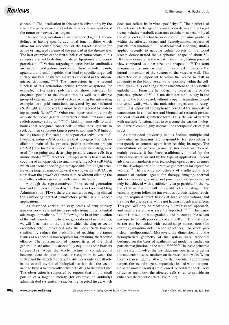
cancer.[156] The localization in this case is driven only by thesize of the particles and is not related to specific recognition ofthe tumor or neovascular targets.
The second generation of nanovectors (Figure 11b) aredefined as having specific additional functionalities whichallow for molecular recognition of the target tissue or foractive or triggered release of the payload at the disease site.The best examples of the first subclass of nanovectors in thiscategory are antibody-functionalized liposomes and nano-particles.[157–159] Various targeting moieties besides antibodiesare under investigation worldwide. These include ligands,aptamers, and small peptides that bind to specific target-cellsurface markers or surface markers expressed in the diseasemicroenvironment.[160–162] The nanovectors in the secondsubclass of this generation include responsive systems, forexample, pH-sensitive polymers or those activated byenzymes specific to the disease site, as well as a diversegroup of externally activated vectors. Among the interestingexamples are gold nanoshells activated by near-infrared(NIR) light, and iron oxide nanoparticles triggered by switch-ing magnetic fields.[163, 164] Other techniques used to remotelyactivate the second generation vectors include ultrasound andradiofrequency stimulus.[58, 165–167] Linking nanoshells to anti-bodies that recognize cancer cells enables these systems toseek out their cancerous targets prior to applying NIR light orheating them up. For example, nanoparticles activated with 2’-fluoropyrimidine–RNA aptamers that recognize the extrac-ellular domain of the prostate-specific membrane antigen(PSMA), and loaded with docetaxel as a cytostatic drug, wereused for targeting and destroying prostate cancer cells in amouse model.[168, 169] Another new approach is based on thecoupling of nanoparticles to small interfering RNA (siRMA),which can silence specific genes responsible for malignancies.By using targeted nanoparticles, it was shown that siRNA canslow down the growth of tumors in mice without eliciting theside effects often associated with cancer therapies.
Although the representatives of the second generationhave not yet been approved by the American Food and DrugAdministration (FDA), there are numerous ongoing clinicaltrials involving targeted nanovectors, particularly in cancerapplications.
As described earlier, the easy access of drug-deliverynanovectors to cells and tissue provides tremendous potentialadvantage in medicine.[58,151] Following the brief introductionof the wide variety of the first two generations of nanovectors,we will focus here on the barriers which the drug or vectorencounter when introduced into the body. Such barrierssignificantly reduce the probability of reaching the targettissues at a concentration required for obtaining therapeuticefficacy. The construction of nanoparticles of the thirdgeneration are aimed to successfully negotiate these barriers(Figure 11 c). When the whole picture is considered, itbecomes clear that the molecular recognition between thevector and the affected or target tissue plays only a small rolein the overall myriad of biological barriers that the vectorneed to bypass to efficiently deliver the drug to the target site.This observation is supported by reports that only a smallportion of a targeted moiety (for example, an antibody)administrated systemically reaches the targeted tissue, which
does not reflect its in vitro specificity.[170] The plethora ofobstacles which the agent encounters on its way to the targettissue includes metabolic clearance and chemical instability ofthe drug, endoepithelial barriers, osmotic pressure gradientswithin the affected tissue, and hemodynamical aspects ofparticle margination.[151, 171] Mathematical modeling studiesapplied recently to nanoparticulate objects in the bloodstream demonstrated that a spherical shape of about 50–100 nm in diameter is the worst from a margination point ofview compared to other sizes and shapes.[171–174] The termmargination dynamics is used in this context to describe thelateral movement of the vectors to the vascular wall. Thischaracteristic is important to allow the vector to drift inproximity to the blood vessel walls—possibly within the cell-free layer—thus enabling firmer attachment to the vascularendothelium. From the hemodynamic forces acting on theparticles, spheres of 50–100 nm diameter tend to stay in thecenter of the blood vessel, without proper margination towardthe vessel walls, where the molecular targets can be recog-nized. It is important to emphasize here that the majority ofnanovectors in clinical use and biomedical research possessthe least favorable geometric form. Thus, the use of vectorswith multiple functionalities to overcome the various biolog-ical barriers could highly improve the therapeutic efficacy ofdrugs.
As mentioned previously in this Section, multiple andsequential mechanisms are responsible for preventing atherapeutic or contrast agent from reaching its target. Thecontribution of particle geometry has been overlooked,mainly because it has been traditionally limited by thefabrication/synthesis and by the type of application. Recentadvances in nanofabrication technology open up new avenuesfor the development of alternative geometries for injectablevectors.[175] The carrying and delivery of a sufficiently largeamount of various agents for therapy, imaging, thermalablation, remote guidance, and possibly other functions canonly be achieved with a sufficiently large particle. In theory,the ideal nanovector will be capable of circulating in thevascular system following intravenous administration, reach-ing the required target tissues at high concentrations, andtreating the disease site, while not having any adverse effects.This goal will only be reached by a “multistage” approach,and such a system was recently reported.[176, 177] The nano-vector is based on biodegradable and biocompatible siliconmicroparticles with pores sizes of up to 50 nm. This first stagecarrier can be loaded with second-stage nanocarriers (forexample, quantum dots, carbon nanotubes, iron oxide par-ticles, nanoliposomes). Moreover, the dimensions and thehemispherical geometry of the system were rationallydesigned on the basis of mathematical modeling studies onparticle margination in the blood.[171, 178,179] The basic principleof the system involves the first stage microparticles targetingthe molecular disease markers on the vasculature walls. Whenthese carriers tightly attach to the vascular endotheliumtargets, the second stage nanoparticles loaded with therapeu-tic or diagnostic agent(s) are released to facilitate the deliveryof active agent into the affected cells so as to provide anenhanced therapeutic effect (Figure 12).
K. Riehemann, H. Fuchs et al.Reviews
884 www.angewandte.org � 2009 Wiley-VCH Verlag GmbH & Co. KGaA, Weinheim Angew. Chem. Int. Ed. 2009, 48, 872 – 897

Exciting applications of nanotechnology have also beenreported in regenerative medicine. In clinical research,regenerative medicine includes the manipulation of stemcells by nanoparticles and nanostructured surfaces as well astissue engineering to treat organs lost as a result of diseaseand trauma. This includes skin substitution after burn injuries,the reversal of paralysis or blindness through spinal cord orretina regeneration, heart regeneration after infarcts, andminimization of stroke dysfunction through neuron repair.The nanomaterials support the reconstitution of healthytissues. Results obtained by Stupp and co-workers indicatethat the regeneration within the central nervous system can bereached by applying self-organized nanofibers. An amphi-philic peptide (IKVAV) which self-assembles into a nano-network and recognizes a3b1 integrin was used for thispurpose (Figure 13). The induced signaling appears to stim-ulate the axons to grow longer and promotes neurondevelopment. In parallel, the inhibition of axon regenerationby scar-forming astrocytes was blocked. In a similar approachheparin-coated nanoparticles promotes angiogenesis.[180–182]
4. Clinical Applications
As described in Section 3,nanomedicine has enteredmany different fields includ-ing tissue engineering andtargeted drug delivery. Clin-ical application is fairlybroad, but mainly focuses oncancer. Known to be a causeof the development of dis-eases such as cancer, arthro-sclerosis, and age-related ill-nesses, chronic inflammationtakes a central position inclinical investigations. Themechanisms of this correla-tion have been reviewed byseveral authors, who dis-cussed how the immunestatus in humans affects therisk of cancer development inan etiology-dependentmanner. The molecularmachinery underlying thedevelopment of chronicinflammation makes it anexpanding area of researchfor nanomedicine.[183–187]
Therapies for chronicinflammation address cell-mediated or humoral immun-ity by blocking mediatorssuch as interleukines (IL) ortargeting receptors (for anoverview of immunological
mechanisms see Refs. [188–197]). The classical treatment ofchronic inflammation is based on drugs such as glucocorti-coids, cyclosporine A, sulfasalazine/5-aminosalicylic acid (5-AZA), or calcinneurin inhibitors. Immunotherapies by meansof antibodies, such as anti-CD20 or anti-CTLA4, specific forcertain cells are also used. These commonly used therapiesspecifically or unspecifically suppress the cellular or humoralimmune response, thus causing a variety of—sometimes life-threatening—side effects, such as hyperglycemia (steroiddiabetes), osteoporosis, lymphopenia, sepsis, liver failure,hepatitis, skin atrophy, or adrenal insufficiency. Calcineurininhibitors are important regulators of IL-2 and activators of T-helper cells, and are thus an alternative to unspecificimmunosuppressants. However, potentially severe sideeffects such as infection and sepsis were also reportedfollowing systemic application of calcineurin inhibitors.Furthermore, administration routes are often problematicand inefficient (for example, drug degradation may occurduring oral administration). Similar problems of low effi-ciency, severe side effects, and inefficient application routeswere identified a while ago in classical cancer treatment.Therefore, successful efforts have been made in this field todevelop targeted drug delivery and diagnostic approaches,
Figure 12. Mechanism of action of multistage (3rd generation) nanovectors. Top left: Rationally designedstage one nanoparticles marginate to the vessel wall and adhere to the endothelium. Top right: Stage onenanoparticles release a reagent to break down tight junctions and the basement membrane. Finally, stagetwo particles—in this case, liposomes—will be released. Bottom: the stage two liposomes interact with thetarget cell membrane, and then deliver the intended payload—in this case, siRNA. Reprinted withpermission from Ref. [151].
NanomedicineAngewandte
Chemie
885Angew. Chem. Int. Ed. 2009, 48, 872 – 897 � 2009 Wiley-VCH Verlag GmbH & Co. KGaA, Weinheim www.angewandte.org

and bring them to clinical application. Nanosized drug-delivery systems for the treatment of chronic inflammationcan improve on the already existing application of the drug interms of reduced side effects, enhanced efficacy, betterbioavailability, and reduced health-care costs. Anotheradvantage of nanocarriers is the capacity for medical exploi-tation of highly toxic, poorly soluble, and unstable com-pounds.[198, 199]
Nanoscale drug- or gene-delivery systems are supra- andsupermolecular aggregates of simple components with vari-ous sizes, shapes, and composition. These characteristics holdtrue for the majority of the nanoscale particles applied innanomedicine. In general, the carrier is characterized bycertain parameters such as a high drug or gene loadingcapacity, or are superparamagnetic as in the case of iron oxidenanoparticles. Independently of the composition, nanovectorsare usually further modified depending on their individualapplication, such as surface decoration with polyethyleneglycol (PEG) for intravenous injection to prevent earlyclearance and to increase blood circulation time.[58,200]
4.1. Lipid Vehicles
Liposomes are the most clinically established nanometer-scale systems used for drug delivery. Biocompatibility,biodegradability, and flexibility of size and surface manipu-lations are the important features that liposomes offercompared to other nanoparticulate delivery systems. Lip-osomal nanotherapeutics for cancer treatment have been onthe market for more than a decade, whereas other liposomaldrugs are in various stages of clinical development. Intro-duced to increase the solubility of hydrophobic chemother-apeutics and to enable trapping of drug molecules with a highpotency, liposomes have been shown to be effective inreducing systemic side effects and toxicity, as well as inattenuating drug clearance.[201, 202] Some examples of availabledrugs that have higher efficacy and lower toxicity thannonliposomal preparations are: liposomal amphotericin B(brand names: AmBisome, Amphotec, Abelcet), stealthliposomal doxorubicin (brand names: Doxol/Caelyx), lip-osomal daunorubicine (brand names: DaunoXomo), andliposomal cytosine b-arabinoside (brand name: DepoCyt).These are just some representative examples to demonstratethe great impact of nanomedicine in current therapies.[203] Anenormous number of diverse synthetic, semisynthetic, andnatural polymers are now available, particularly those pre-pared from biodegradable polymers such as poly(lactic acid)(PLA), poly(d,l-lactide-co-glycolide) (PLGA), poly(e-capro-lactone), gelatin, and chitosan. These systems have far-reaching clinical applications. PLGA nanoparticles are anestablished biodegradable and biocompatible carrier system.Polymeric micelles based on block copolymers that formthermo- and pH-sensitive or enzyme-sensitive structures haveraised interest for delivery applications, in particular ofhydrophobic compounds. These systems are preferablydesigned in such a way that they allow for self-assembly inthe presence of the drug to be incorporated. This willsignificantly facilitate their applicability in a clinical environ-ment.
4.1.1. Liposomal Drug Carriers in Chemotherapy4.1.1.1. Doxorubicin
Doxorubicin is an anticancer drug that is widely used forthe treatment of different types of tumors such as breastcancer, Kaposi sarkoma, and ovarian cancer. Doxorubicin is ahighly toxic compound affecting not only tumor tissue butalso heart and kidney, a fact that limits its therapeuticapplications. Therefore, intense research was done to estab-lish a more compatible formulation of doxorubicin. Thedevelopment of doxorubicin enclosed in liposomes culmi-nated in an approved nanomedical drug-deliverysystem.[204, 205] Liposomal formulation result in a reduceddelivery of doxorubicin to the heart and renal system, whilethe accumulation in tumor tissue is elevated.[206, 207] Nano-vectors of this type accumulate in tumors because of the EPReffect, that is, the characteristic hyperpermeability of tumortissue which results in a selective delivery of the drug to thetumors.[208, 209] The cutoff size of the blood–tumor barrierdepends on the location of the tumor and the modulation of
Figure 13. Functionalized nanoparticles for nerve regeneration: a) Sche-matic representation of an IKVAV-containing amphiphilic peptide;b) SEM image of a self-assembled network of IKVAV amphiphiles;c) supported by a nanofiber network, progenitor cells differentiated tofunctioning neurons instead of the scar-forming astrocyte. Reprintedwith permission from the AAAS.[182]
K. Riehemann, H. Fuchs et al.Reviews
886 www.angewandte.org � 2009 Wiley-VCH Verlag GmbH & Co. KGaA, Weinheim Angew. Chem. Int. Ed. 2009, 48, 872 – 897

the microenvironment, but is usually between 300 and800 nm, which corresponds to the size of liposomal carri-ers.[210] Particles larger than 200 nm activate, however, thecomplement system and provoke clearance by phagocytosis.Fast clearances of nanomaterials by phagocyte activityprevent long circulation of the carrier and thus inhibit thelong-term controlled release of the load. The circulationbehavior of liposomes was improved by modification of theliposomal surface with PEG.[198] PEG reduces the clearance ofthe liposome by phagocytes in the liver and spleen consid-erably, since opsonization of the liposomal surface is stronglyhindered.[211] A reduced clearance increases the circulationperiod of the carrier in the blood and prolongs the drugrelease, thereby enhancing the probability of the EPRphenomena. Interestingly, a lipid composition itself isunable to modulate the clearance of PEGylated liposomes,as opposed to non-PEGylated liposomes.[212,213]
More recent studies revealed an increased clearance rateof PEGylated liposomal carrier upon multiple injec-tions.[214–216] Here, clearance is presumed to be mainlygoverned by liver and spleen macrophages and dependenton a soluble heat-labile serum factor (or factors) that primesthe so-called enhanced clearance effect. The enhancedclearance effect diminishes with time and seems to be relatedto the life time of the macrophages that come directly incontact with the injected liposomes.[214, 217] Therefore, theinjection intervals of such liposomes should be adapted to thelife time of the macrophages.
A disadvantage of liposomal drug delivery is the release ofthe drug into the extracellular fluid since liposomes usuallycannot enter the cells.[214] A more specific targeting of theliposomal drug carriers or a specific cellular uptake istherefore envisaged to reduce the toxicity and increase theeffectiveness of the drug (second and third generations ofnanovectors).
In contrast to an indirect targeting governed by the EPRphenomena, an improved tumor-specific drug delivery can beachieved by coupling antibodies to the surface of liposomes.The advantages of these immunoliposomes are the potentialcellular uptake by the target tissue accompanied by anincreased toxicity to the tumor cells, and a reduced clearancerate since the delivery to the kidney and spleen is reduced. Forexample, anti-2C5 monoclonal antibodies were coupled to aliposomal surface so as to transfer the encased doxorubicin tobrain tumors. This antibody was shown to bind specifically tohuman astrocytoma cell surfaces in vivo.[218] The antibody isdirected against nucleosomes localized on living tumor cellsurfaces originating from apoptotic neighboring tumorcells.[219] Another approach to treat human brain tumorsin vivo is the application of sulfatide-containing liposomes(SCL), which bind to certain glycoproteins upregulated intumor cells. Anti-CD19-labeling of liposomes was shown toimprove the targeting to murine B-cell lymphoma cells andthe intracellular release of liposomal doxorubicin.[220] Theseexamples show that vectorial, that is, site-directed, drugtransport and release will revolutionize the therapy of braintumors, since the present therapy is of limited success due toinsufficient drug delivery. However, the toxicity does notdepend only on the targeting but was proven previously to be
strongly related to the release characteristic of the injectedliposomal formulation.[220, 221]
Other approaches are currently under investigation toenhance the specificity of the drug transport. A recent studyreported on thermosensitive liposomes that release doxoru-bicin when heated. Specific release of the antitumor drug wasachieved by selective heating of the targeted tumor. Hyper-thermia was induced in this case by heated water deliveredin vivo through small catheters.[222]
4.1.1.2. AmBisome/Amphotericin B
AmBisome is a liposomal formulation of an antifungalagent amphotericin B, which is recommended for differentfungal infections and as an empirical therapy for presumedfungal infection in febrile neutropenic patients. It can also beused for treatment of visceral leishmaniasis. AmBisome iscomposed of very rigid, small unilamellar liposomes with amean diameter of under 100 nm, with amphotericin B inter-calated within the membrane. Such liposomes are known tohave long circulation times and accumulate in the requiredareas. In preclinical and clinical studies, AmBisome showedless toxicity and fewer side effects than amphotericin B, butretained the full spectrum of antifungal activity.[223] Therefore,in contrast to classical amphotericin B therapies, it can beused in patients suffering from kidney damage. It was shownin animal experiments that AmBisome did not distributeevenly throughout the kidney tissue, but rather tended tolocalize near the areas of fungal infection. Moreover,AmBisome was found to be attached to the fungal wall andpenetrate inside. In summary, liposomal amphotericin Baccumulates at the infection sites, shows higher stability, andfewer side effects and toxicity than the free drug. Thesustained release of amphotericin B from AmBisome mayalso serve as a prophylaxis, as shown in mice challenged withHistoplasma capsulatum.[224]
4.2. Polymer-Based Delivery
Natural polymers such as proteins or polysaccharides tendto be internalized and degraded rapidly, thus enabling amoderate intracellular release of the drug or gene.[225] Bloodcirculation time or clearance is controlled by surface modi-fication or the formation of polymer conjugates with poly-thylenglycol (PEG).[226] A current example of clinically usedpolymeric nanoparticles are paclitaxel albumin bound nano-particles (brand name: Abraxane) for the treatment ofpatients with breast cancer resistant to conventional ther-apy.[227, 228] These nanoparticles are water-dispersible, there-fore avoiding the use of Cremophor (a solvent commonlyused to solubilize and formulate free paclitaxel, a veryhydrophobic drug). Cremophor was reported to cause allergicreactions, thus limiting the drug dosing.[229–231] Abraxanedemonstrates a significantly higher response rate, a longertime to tumor progression, and absence of hypersensitivityreactions.[227] However, a severe side effect was recentlyreported, which demonstrates the ongoing debate on the
NanomedicineAngewandte
Chemie
887Angew. Chem. Int. Ed. 2009, 48, 872 – 897 � 2009 Wiley-VCH Verlag GmbH & Co. KGaA, Weinheim www.angewandte.org

safety and drug metabolism of nanoparticles (see alsoSection 6).[232]
A promising anticancer treatment based on passivetargeting of drug-polymer conjugates was suggested by theresearch group of Duncan. As in the case of the firstgeneration nanovectors, it makes use of the fact that neo-vascular systems close to tumors are permeable for certainparticle sizes, in contrast to those supplying healthy tissue.The research group of Vicent reported on the coupling ofapoptosis-inducing anticancer agents to nanoparticles for theenhancement of efficacy.[233, 234]
Figure 14 shows the possibilities of coupling drugs topolymers. This also demonstrates that the generation andoptimization of nanovectors forms an important interdiscipli-nary area between chemistry, biochemistry, and medicine.
The coupling of proteins and drugs to synthetic polymers,especially PEG, increases their plasma residence, reducesprotein immunogenicity, and can increase their therapeuticrange. Several PEGylated enzymes (such as l-asparaginase)and cytokines (including interferon-a and granulocytecolony-stimulating factor) have now entered routine clinicaluse.[233, 234]
4.3. Metal Nanoparticles4.3.1. Nanocrystalline Silver for Wound Care
Silver, mostly in the form of nitrate or sulfadiazine salts, isa well-studied antimicrobial agent and a common compoundfor wound treatment.[235–237] Wound healing can be subdivided
into distinct phases.[238] Soon after injury and coagulation,wound healing is characterized by cell invasion by leucocyteswhich causes inflammation. This inflammation phase cleansthe wound and a microbial infection is prevented. Woundhealing proceeds, after the inflammation has ceased, by tissueremodeling and maturation of the new tissue. Latent micro-bial infection could cause the inflammatory phase to beprolonged, thereby causing chronic nonhealing wounds.Medical treatment of chronic wounds with dressings contain-ing silver salts significantly reduce the bacterial load of thewound and allow complete healing.[239] Silver is a promisingalternative to antibiotics since multiresistance against anti-biotics develops progressively. A recent study does notindicate an increased development of resistance upon silverusage.[240–242] In comparison to antibiotics, which potentiallyaffect wound tissue, the toxicity of silver is not specific,although the antimicrobially active doses of silver are low(nm–mm range) and is commonly tolerated well.[242] Theadvantage of nanocrystalline silver over silver salts is not onlydue to an increased antimicrobial activity but also due to itsantiinflammatory properties.[243] However, the mechanism ofaction remains to be elucidated. The application of nano-crystalline silver during wound management demonstratesthe entrance of nano-biotechnology to medical therapy. Thesetypes of dressings are currently applied in cases of first- andsecond-degree burns and in several types of chronic non-healing wounds.
4.3.2. Magnetic Nanoparticles for Diagnosis and Therapy
Iron or iron oxide nanoparticles have a great potential forvarious nano-biomedical applications including drug delivery.The nanoparticles can be coated with hydrophilic polymerssuch as PEG or dextrane to prevent or to increase the cellularclearance of the particle, respectively.[244] Cell-specific trans-port is also possible by coating with antibodies, receptor-specific peptides, or aminosilane. In addition to these moregeneral properties of nanocarrier systems, the superparamag-netic character of the particles and their dimensions of 2–20 nm pave the way for applications other than drug delivery.
Current research and applications of iron-containingnanoparticles mainly involve in vitro cell labeling and cellseparation, in vivo drug delivery, magnetic resonance imaging(MRI; see Section 2), and hyperthermia.[245] The most popularapplication of iron nanoparticles in medicine is hyperthermia,which is the destruction of tumors by locally over-heating thetissue. Hyperthermia is an effective and specific anticancertreatment, since an increased temperature of the treatedtissue up to 44 8C is less well tolerated by cancer cells than byhealthy cells. This approach is usually applied in combinationwith other traditional therapies such as chemotherapy. Hyper-thermia treatment by iron oxide nanoparticles is induced byexposure of the particles to an alternating magneticfield.[203, 246] A local accumulation of nanoparticles allows fortissue-specific hyperthermia that preferentially addresses thetumor tissue (Figure 15).
The benefits over classical cancer therapies are minimalinvasiveness, accessibility of hidden tumors, and very few sideeffects. Conventional heating of a tissue (microwaves, laser
Figure 14. Polymer conjugates of anticancer drugs: a) Paclitaxel (PTX)is linked to the carrier polyglutamate (PGA) through an ester bond.The main drug release occurs after polymer degradation by thelysosomal enzyme cathepsin B; b) conjugate of camptothecin (CPT)and a linear b-cyclodextrin-based PEG polymer (CDP). Pharmacokineticand preclinical studies have demonstrated that this conjugate exhibitsa longer plasma half-life and better distribution to the tumor tissuethan CPT alone. Reprinted with permission from Ref. [234].
K. Riehemann, H. Fuchs et al.Reviews
888 www.angewandte.org � 2009 Wiley-VCH Verlag GmbH & Co. KGaA, Weinheim Angew. Chem. Int. Ed. 2009, 48, 872 – 897

light etc.) results in the healthy tissue surrounding the tumoralso being destroyed. However, targeted paramagnetic par-ticles provide a powerful tool for highly localized energyabsorption and heating of mainly the cancerous cells. Severalkinds of nanoparticles differing in material, composition, andsize are available for this purpose; for example, hyperthermiacan also be applied by using magnetite cationic liposomes(MCLs) as carrier systems combined with heat-shock pro-teins. However, the low-levels of side effects means thetreatment is well accepted by patients.[203, 247–249]
Superparamagnetic iron oxide particles have also beenused for MRI studies.[245] Dextran-coated particles (brandnames: Resovist, Feridex) are commercially available andhave mainly been used for in vivo MRI of liver tumor tissue.The dextran coating increases the intracellular deposition ofthe particles into the cancer cells, thereby enabling diagnosisand monitoring of the progression of the tumor. Iron particlesare cleared by the liver macrophages, and enter the retic-uloendothelial system to join the physiological iron pool.
4.4. Nanoshells
In other hyperthermal concepts, gold–silica nanoshellsconsisting of a spherical dielectrical nanoparticle surroundedby an ultrathin conductive metal layer are used. The nano-particles absorb light in the NIR region, thus guaranteeingthat an optimal optical transmission through the tissue. Amoderate exposure to extracorporeal near-infrared light(820 nm, 4 Wcm�2) resulted in a heating of the tumor tissueand irreversible tissue damage, as evident by coagulation, cellshrinkage, and loss of nuclear staining.[164] The big advantageof nanoshells is their tuneable plasmon resonance from thevisible to the infrared regime by varying the composition anddimensions of the layers.
Nanoshells have not only been investigated for thetreatment of cancer but also for diagnostic purposes, such asacquiring higher resolution images in optical coherencetomography (OCT). The OCT applications reach from
ophthalmology up to the reconstruction of whole brainsections. Some other noteworthy NIR imaging applicationsare confocal imaging, iridotomy, and photothermal coagula-tion, all of which take advantage of the increased trans-parency of the tissue within this region.[250]
4.5. Non-injectable Nanovectors
The most preferred way of introducing drugs into thebody is orally; therefore, the pharmaceutical industry investsmuch effort in to the development of appropriate deliverysystems that can be improved by nanotechnology. Nano-sphere carriers derived from hydrogels—highly stable organiccompounds that swell when their environment becomes moreacidic—have been successfully formulated into controlled-release tablets and capsules, which release active compoundsin a pH-dependent manner.
Nanoparticles can also provide an efficient delivery toolfor drugs that have to bypass the blood–brain barrier, such aschemotherapeutic agents for brain malignancies, antiepilep-tics, and anesthetics (for example, Dalargin). Nanoparticlescoated with polysorbate 80 and loaded with doxorubicin(5 mgkg�1) achieved very high levels of 6 mgg�1 of the drug inbrain tissue, while all the controls, including uncoated nano-particles and doxorubicin solutions mixed with polysorbate,were below the analytical detection limit.[251] Another newlydesigned delivery system is based on chitosan coupled toantibodies through a PEG linker. These immune nanoparti-cles have, on the one hand, the ability to interact with thenegative charges of the brain endothelium through thecationic (with a full positive charge) chitosan, and, on theother hand, an affinity for the transferrin receptor by themonoclonal antibody OX26, which makes them perfectlydesigned to cross the blood–brain barrier. Nanospheresloaded with the peptide Z-DEVD-FMK, an inhibitor ofcaspase-3, were also investigated. Inhibition of this enzyme isknown to increase neuronal cell survival following cerebralischemia.[252]
Implantable drug-delivery systems improved by nano-technology are often preferred to the use of injectable drugs,because the latter frequently display side effects. For example,the blood concentration may increase rapidly, but decreasesslowly over time. This can diminish drug efficacy as the drugconcentration falls below the therapeutically relevant level. Incontrast, implantable time-release systems may help inminimizing peak plasma levels, thereby reducing the risk ofadverse reactions and the frequency of re-dosing, thusimproving patient compliance. The benefits of nanotechnol-ogy in this regards are exemplified by biodegradable poroussilicon (pSi) materials. These nanostructured materials storean active compound or second stage nanoparticles effectivelyin nanosized pockets that release minute amounts of drug asthe silicon dissolves. pSi is currently being explored for tissueengineering and ophthalmic delivery.[253, 254]
Nanotechnology also refines transdermal delivery, a safeand non-invasive method of administering drugs. The trans-port of large-molecular-weight proteins (such as vaccines) isrelatively inefficient when the substance is applied directly
Figure 15. Comparison of a healthy and a tumor cell incubated withnanoparticles. The phase-contrast light microscopy image shows aprostate carcinoma cell and a fibroblast cell. While the tumor cell (left)shows a high level of pigmentation because of the uptake of a largenumber of nanoparticles, the adjacent fibroblast cell shows lowerlevels of pigmentation, that is, no or lower levels of particle uptake.Reprinted with permission from Elsevier.[246]
NanomedicineAngewandte
Chemie
889Angew. Chem. Int. Ed. 2009, 48, 872 – 897 � 2009 Wiley-VCH Verlag GmbH & Co. KGaA, Weinheim www.angewandte.org

onto bare skin. Recent evidence has shown that this barriercan be overcome by using properly structured nanoparti-cles.[255]
Finally, nanotechnology can also be used for the removalof toxins. Colloidal dispersions have already been shown toremove potentially lethal compounds from the bloodstream,including high concentrations of lipophilic therapeutics,illegal drugs, and chemical and biological agents.[256, 257]
5. Nanocoatings and Nanostructured Surfaces forMedical Application
Knowledge about the nanostructuring of surfaces devel-ops rapidly with the development of nanoparticles. The mainresearch into the nanostructuring of surfaces focuses on theoptimization of the interaction of prostheses, such as artificialjoints, with the organism, with the aim of producing materialswhich have a close connection to the body tissues, whileavoiding side effects, such as chronic inflammations orallergies. Nanostructuring of a surface coating controlsproperties such as charge, conductivity, roughness, porosity,wettability, friction, physical and chemical reactivity, andcompatibility with the organism. There is a growing need forsmart surfaces which show a high biocompatibility, particu-larly in the area of artificial organs and prosthetics.[258–263]
Another potential application of nanotechnology residesin the possibility of mimicking a variety of compoundmaterials and self-organized systems found ubiquitously innature. Complex structures such as complete cells, as well assubstructures such as folded proteins and molecular motors,represent the kind of self-organized nanomachines thatcurrently cannot be prepared in a synthetic way. Nature,however, has made use of informed dynamic molecularsystems and demonstrated that self-organized complexmolecular systems are indeed extremely successful. Whensuccessful, these concepts will be partly transferred intosynthetic systems in the future, and their implementation maylead to new developments that cannot be achieved byconventional large-scale manufacturing processes.
Nanostructuring can be done physically, chemically, or byself-assembly. The most popular naturally occurring exampleis probably the surface structure of the leaves of a lotusflower. The special structured surface, with a typical bimodalsize distribution in the micrometer and sub-micrometerregime leads to self-cleaning behavior. Another example isthe manipulation of cell behavior by changing the surfacestructure while keeping their chemical composition approx-imately the same. This was recently demonstrated by theresearch group of Spatz. A different growth and attachmentbehavior of fibroblasts has been observed by altering thedistance between functionalized gold particles attached to asurface (Figure 16).[264–266] In another approach Sun et al. usedN-isobutyryl-l(d)-cysteine (NIBC) enantiomers to success-fully alter the adsorption characteristics of surfaces.[267]
The understanding of these effects opens up ways for thegeneration of surfaces with nonfouling properties, and also ofsurfaces with optimized template structures for specific cellgrowth. For example, the nanostructuring of titanium alloy by
hydroxyapatite coatings apparently results in enhancedmechanical properties and in promoting the proliferation ofosteoblast cells.[268] This finding is interesting for the develop-ment of implant materials.
Changing the surface to a nanotubular structure resultedin artificial joints that were better incorporated withoutinducing chronic inflammation.[269, 270] The tailored surfaceshave the advantage of mimicking the surface of naturalstructures, not only by the coating of inorganic materials butalso by adhering proteins or peptides to mimic naturalconditions. The next step in this development is its applicationto the field of bionics. The exchange between life forms andsynthetic constructs is a most promising attempt becauseevolution has already selected appropriate materials andprocesses. Besides prosthetics, this approach is most promis-ing for neural applications.
Nanostructured surfaces with specific well-defined prop-erties have also found applications in diagnostics. The study ofcells often fails because of unfavorable interaction betweenthe device and the cells. This is especially the case whenimmune cells are investigated. Receptors on the surface ofleucocytes interact unspecifically with artificial structures,which results in an unwanted activation or differentiation ofthe cells. Here, surfaces are needed that do not induce anykind of activation after contact. This is important for allmicrofluidic devices and surfaces used for biochips andproteomics.[271–273]
By using responsive molecular systems it is possible toswitch between different states (such as superhydrophobicityand superhydrophilicity) by external stimuli (for example,electrical or optical fields, pH value) applied to the function-
Figure 16. Phase-contrast optical micrographs of 3T3 fibroblasts onpolyethylene glycol diacrylate (PEGDA) 700 hydrogels. a) Cells on anon-RGD-functionalized gold nanoparticle pattern. b–d) Cells on cyclo-(-RGDfK-)-functionalized gold particles after 24 h in culture; cyclo-(-RGDfK-) patches are separated by various distances b) 40 nm,c) 80 nm, and d) 100 nm. e) Dense cell layer on a PEG support after14 days in culture. The bottom part of the sample was patterned withgold nanoparticles functionalized with the cyclo(-RGDfK-) peptidespaced 40 nm apart. Reprinted with permission from Ref. [266].
K. Riehemann, H. Fuchs et al.Reviews
890 www.angewandte.org � 2009 Wiley-VCH Verlag GmbH & Co. KGaA, Weinheim Angew. Chem. Int. Ed. 2009, 48, 872 – 897

alized surfaces. These coatings are of interest for diagnosticpurposes with miniaturized lab-on-a-chip systems or for thecoating of artificial blood vessels and implants, thus mimick-ing biological systems.
6. Biocompatibility and Toxicity—Safety IssuesRelated to Nanotechnology Implementation
The generation of small particles may be a major issuewith respect to toxicology. The potentially high reactivityarising from the large surface-to-volume ratio of nanoparti-cles compared to bulk systems means there is a latent risk forall new nanosystems, which must be carefully considered.While the existing laws for new chemicals and pharmaceuticalmaterials seems to be currently sufficient for these types ofmaterials, each new nanoparticle system has to be inves-tigated carefully with respect to its potential side effectswithin the human body and the environment. Fortunately, thepublic and scientific awareness of nanotechnology is high andthere is an increasing intensity of discussion on these ethicaland societal issues. The clinical application of nanotechnologyalso requires a number of regulatory guidelines to ensure theappropriate use of new medical devices and drugs originatingfrom nanoscience.[274–277] The potential of molecular diagnos-tics and analysis based on nanotechnology and nanomedicinealso deserves attention from the political side. This not onlyincludes in regard to the above-mentioned toxicologicalaspects but also to the question of the improvement in thequality of life in the cases of severe diseases, cost-effectivetreatment of patients, the artificial extension of our naturalsenses, neural-electronic interface systems etc., which mightbe only available for a limited number of people.
The toxicological risk for human health includes effectsduring interaction with medical devices. According to thedefinition of the EU Medical Devices Directive, “medicaldevices” comprise tools for:* diagnosis, monitoring, treatment, or alleviation of or
compensation for an injury or handicap;* investigation, replacement, or modification of the anatomy
or of a physiological process;* control of conception which does not achieve its principal
intended action in or on the human body by pharmaco-logical, immunological, or metabolic means, but whichmay be assisted in its function by such means.
If one considers nanoparticles as “medical devices”, theycould have a broad range of applications in the humanbody.[278, 279] A drawback is their potential toxicity and theirpossible incompatibility, which may result in the generation ofdisorders such as inflammation, immunoreaction, or cancer.Mechanisms of those effects are not well-studied yet, butmight be due to an enhanced hydrophobic interaction withbiological material or an increased generation of free radicalsby surface catalysis.[198] Recent experimental data have shownthat inhalation of nanoparticles with a size below 100 nm fromair pollution can lead to the induction of pulmonaryinflammation. It has been demonstrated that in this case theindividual expression of glutathione S-transferase (GST)
determines the extent of the inflammation. Interestingly, thephysiological task of GST is the detoxification of reactiveoxygen species, thus indicating the generation of reactiveoxygen species on the particle surfaces.[280, 281] Comparabledata were obtained in relation to the inhalation of single-walled carbon nanotubes (SWCNs). SWCNs have beenshown to elicit inflammation in the lungs of mice, whilethey cause small, focal interstital fibrotic lesions in ratalveolars.[282, 283]
The discussion on the risk of carbon nanotubes recentlyreceived new input from Poland et al. They reported that thiskind of nanoparticle acts, according to their needle-likestructure, in the same way as asbestos, thus indicating thesame risk for handlers.[284] In other investigations, highlypurified carbon nanotubes seemed not to possess short-termtoxicity, and can be considered biocompatible with cardio-myocytes in culture. The long-term negative effects that wereevidenced were suggested to be due to physical rather thanchemical interactions. This effect was investigated by theresearch group of Krug, who demonstrated that these nano-particles induce no acute cytotoxicity or inflammatorymarkers such as nitric oxide or interleucine-8. The observedside effects were associated with metal traces in the commer-cially available nanotubes.[285] The cytotoxicity of severalkinds of nanoparticles was recently reviewed by Lewinskiet al. , who showed in an impressive way the manifold ofinteractions between foreign bodies and cells.[286]
Nanoparticles that enter organisms and are not excretedaccumulate in the cells and tissues, thus developing a stillunknown potential of causing diseases at these sites over thelong term. It has been shown that nanomaterials can enter thehuman body by several means. Accidental or involuntarycontact during production or use is most likely to happen viathe lung, from where a rapid translocation through the bloodstream is possible to other vital organs, as demonstrated inanimal models.[287] On the cellular level, it has been shownthat nanoparticles can act as gene vectors.[288] Carbon blacknanoparticles have been implicated in interfering with cellsignaling.[289,290]
Nanoparticles used for oral drug delivery have been foundaccumulated in the liver, and excessive immune responsesmay cause permanent damage there.[291] This accumulation incells is also well documented in the cases of pulmonaryfibrosis caused by asbestos fibers (asbestosis) and silicosis, adisease that comes from breathing in silica or quartzdust.[292–294]
It has since been shown that a (high) concentration ofnanoparticles may result in the transformation of cells into thetumorous state, thus causing cancer. Investigations on hepaticand renal tissues affected by cryptogenic granulomatosis byscanning electron microscopy (SEM) and X-ray microanalysiswith an energy-dispersive (EDS) detector showed a correla-tion between the presence of inert, non-biodegradable,exogenous micro- and nanoparticles and diseases that tradi-tional histopathology could not account for (Figure 17).[295]
It is well known that debris produced by the wear of hipprostheses could induce an inflammatory reaction and a localforeign-body granulomatous reaction. In addition, the migra-tion and dissemination of debris in other parts of the body, far
NanomedicineAngewandte
Chemie
891Angew. Chem. Int. Ed. 2009, 48, 872 – 897 � 2009 Wiley-VCH Verlag GmbH & Co. KGaA, Weinheim www.angewandte.org

from their origin, has been documented, which has thepossibility of causing further pathologies. No efficient gastro-intestinal barrier for inert particles with a diameter below20 mm is so far known. On more than one occasion, the sourceof such minute foreign bodies was identified as dentalmaterials such as porcelain or over-worn alloys such as gold/ruthenium. The migration of barium sulfate particles, a verycommon contrast medium used in gastroendoscopy, into livertissue (cells) was a further indication that small particles, inprinciple, may cross the intestinal barrier.[159,296]
Nanoparticles used for drug delivery are exposed tobiomolecules in the lung, the gastrointestinal tract, or to theendothelial barrier. The contact may result in the uptake ofnanoparticles through endocytosis (mediated by receptors),membrane penetration in the case of hydrophobic particles,or by transmembrane channels in the case of very smallnanoparticles (< 5 nm).[297] A strategy to prevent cellularinternalization and, therefore, uncontrolled cytotoxicity ofnanoparticles with sizes below 100 nm is by surface modifi-cation with hydrophilic polymers.[244]
In an aqueous environment, different types of biomole-cules as well chemicals such as pesticides adsorb to nano-particles. Adsorbed molecules dictate biological interactions,especially bioavailability, and the activation of cells. Anexample of the interaction of nanoparticles with biomoleculesis the binding of C60 fullerenes to antibodies. Recent reportsattribute the cytotoxic effect of C60 fullerene to lipidperoxidation. In organisms, all extracellular proteins such ascomplement proteins or antibodies can adsorb onto nano-particles. The proteins possibly change their conformation,and as a consequence their reactivity, during the adsorption,thereby resulting in an autoimmune response.[298, 299]
To investigate the potential risks of nanotechnology, toolsand methods have been developed and adapted to performhigh-throughput and standardized testing of the interactionbetween nanoparticles and, for example, biological barriers.
An established method of proving the integrity of biologicalbarriers is the measurement of the transepithelial electricalresistance (TER). This method was adopted for determiningthe toxicity of nanoparticles and was developed for routineapplication.[300–303] The initial results show, for example, noinitial effect of silica-based nanoparticles on Madin Darbycanine kidney (MDCK) cells, but a decrease of the TER after150 h was observed, thus indicating disrupture of the cellmonolayer.[304] Theses studies show the importance of long-term studies in the investigation of the toxicity of nano-particles. The studies will be extended over a broaderspectrum of nanoparticles, which will help to understand theinteractions between nanoparticles and biological systems inmore detail.
These few examples demonstrate that the effects ofnanotechnology on human health could be double edged,similar to conventional drug exposure, but possibly based oncompletely different mechanisms. Many of the investigatedsystems, so far seem to exhibit relatively few short-term risks.Nevertheless, since all new technologies may bear hiddenrisks, systematic risk assessment in parallel to the technolog-ical development has to be done to keep the potential hazardsas low as possible.
7. Summary and Perspectives
The potential applications of nanotechnology for diag-nosis, prevention, and treatment of diseases are currently verybroad. Practical application of nanomedicine requires, there-fore, besides creativity and visionary power, simpleapproaches, and systematic development.
In this Review we have provided an overview on somefascinating developments in the area of nanomedical researchand applications. Since the field is currently expanding at avery fast pace, we could not include all aspects of presentnanomedicine in detail. Our aim was mainly to demonstratethe highly transdisciplinary character of nanomedicine and togive a view on developments and research topics in chemistry,biology, physics, and engineering that can revolutionizeclinical therapies and diagnostics. Nanotechnology hasalready made an important impact on clinical applications,which are expected to grow exponentially during the next fewyears.
Nanomedicine relies on:* chemical knowledge to provide required modifications to
the nanovector surface and to enable conjugation of thedrug/contrast agent and to improve the biocompatabilityof implants;
* detailed understanding of disease biology and pathophysi-ology to enable efficient targeting and therapy;
* awareness of the physical properties of multilevel complexnanosystems to be able to finely engineer and manipulatematter for the design of new nanoscale detection and drug-delivery systems.
The main focus of clinical nanomedical applications iscurrently on the treatment and efficient diagnosis of cancer.To efficiently detect malignancies, for example, molecular
Figure 17. a) SEM image of a section of a granulomatous liver showingtwo small particles and a cluster of nanodebris. b,c) EDS spectrashowing that the debris has different compositions, and probablydifferent origins. Reprinted with permission from Elsevier.[295]
K. Riehemann, H. Fuchs et al.Reviews
892 www.angewandte.org � 2009 Wiley-VCH Verlag GmbH & Co. KGaA, Weinheim Angew. Chem. Int. Ed. 2009, 48, 872 – 897

changes to the cells must be detected as early as possible. Thismeans that extremely sensitive techniques have to be used.Nanotechnological concepts have, for example, the potentialto address single cells and so meet this challenge. Theknowledge currently being gained in this field will provide thebasis for establishing personalized medicine.
We gratefully acknowledge the graphic material from ourcolleagues. We thank K. Hardes for support during thepreparation of this Review, B. Schneider for designing thecover page, and C. Gorzelanny for helpful discussions andsuggestions. K. Riehemann and H. Fuchs thank the “FederalMinistry of Research and Technology” (BMBF; FKZ0312025A), and S. W. Schneider thanks DFG (SPP13113;Schn 474/2-1) and the “Interdisziplin�re Medizinische For-schung” (IMF, M�nster) for financial support. M. Ferrari andB. Godin acknowledge the support from the State of TexasEmerging Technology Fund, NIH, NCI (R01A128797),Department of Defense (W81XWH-07-2-0101), and NASA(NNJ06HE06A), and would also like to thank MatthewLandry for graphic design.
Received: June 3, 2008
[1] R. P. Feynman in Nanotechnology: Research and Perspectives(Ed.: J. Lewis), MIT Press, Boston, 1992, p. 347.
[2] H. Gleiter, Prog. Mater. Sci. 1989, 33, 223.[3] G. Binnig, H. Rohrer, C. Gerber, E. Weibel, Phys. Rev. Lett.
1982, 49, 57.[4] National Nanotechnology Initiative. Leading to the Next
Industrial Revolution. A Report by the Interagency WorkingGroup on Nanoscience, Engineering and Technology. 2000.Washington, DC, Committee on Technology, National Scienceand Technology Council.
[5] G. Oberdorster, E. Oberdorster, J. Oberdorster, Environ.Health Perspect. 2007, 115, A290.
[6] W. Vogelsberger, J. Phys. Chem. B 2003, 107, 9669.[7] M. Ferrari, Nat. Nanotechnol. 2006, 1, 8.[8] D. Gurwitz, G. Livshits, Eur. J. Hum. Genet. 2006, 14, 376.[9] SEER Cancer Statistics Review; 1975 – 2003. (Eds.: L. A. G.
Ries, D. Harkins, M. Krapcho, A. Mariotto, B. A. Miller, E. J.Feuer, L. Clegg, M. P. Eisner, M. J. Horner, N. Howlader, M.Hayat, B. F. Hankey, B. K. Edwards), National Cancer Insti-tute, Bethesda, MD, 2004.
[10] D. K. Pettit, W. R. Gombotz, Trends Biotechnol. 1998, 16, 343.[11] G. Tosi, L. Costantino, B. Ruozi, F. Forni, M. A. Vandelli,
Expert Opin. Drug Delivery 2008, 5, 155.[12] C. H. Liu, S. Huang, J. Cui, Y. R. Kim, C. T. Farrar, M. A.
Moskowitz, B. R. Rosen, P. K. Liu, FASEB J. 2007, 21, 3004.[13] O. C. Farokhzad, S. Jon, A. Khademhosseini, T. N. Tran, D. A.
Lavan, R. Langer, Cancer Res. 2004, 64, 7668.[14] D. K. Maurya, W. Y. Ng, K. A. Mahabadi, Y. N. Liang, I.
Rodriguez, Biotechnol. J. 2007, 2, 1381.[15] V. N. Truskett, M. P. Watts, Trends Biotechnol. 2006, 24, 312.[16] K. B. Lee, J. H. Lim, C. A. Mirkin, J. Am. Chem. Soc. 2003, 125,
5588.[17] A. Bruckbauer, D. Zhou, L. Ying, Y. E. Korchev, C. Abell, D.
Klenerman, J. Am. Chem. Soc. 2003, 125, 9834.[18] J. Zimmermann, M. Rabe, D. Verdes, S. Seeger, Langmuir 2008,
24, 1053.[19] D. Geho, M. M. Cheng, K. Killian, M. Lowenthal, S. Ross, K.
Frogale, J. Nijdam, N. Lahar, D. Johann, P. Herrmann, G.
Whiteley, M. Ferrari, E. Petricoin, L. Liotta, BioconjugateChem. 2006, 17, 654.
[20] A. J. Nijdam, C. M. Ming-Cheng, D. H. Geho, R. Fedele, P.Herrmann, K. Killian, V. Espina, E. F. Petricoin III, L. A.Liotta, M. Ferrari, Biomaterials 2007, 28, 550.
[21] D. Hanahan, R. A. Weinberg, Cell 2000, 100, 57.[22] J. V. Sweedler, E. A. Arriaga, Anal. Bioanal. Chem. 2007, 387,
1.[23] Y. Wang, G. Young, M. Bachman, C. E. Sims, G. P. Li, N. L.
Allbritton, Anal. Chem. 2007, 79, 2359.[24] D. Patel, Separating cells, Springer, Berlin, 2001.[25] C. E. Sims, M. Bachman, G. P. Li, N. L. Allbritton, Anal.
Bioanal. Chem. 2007, 387, 5.[26] S. Sharma, R. W. Johnson, T. A. Desai, Langmuir 2004, 20, 348.[27] A. Kikuchi, T. Okano, J. Controlled Release 2005, 101, 69.[28] J. A. Ferguson, F. J. Steemers, D. R. Walt, Anal. Chem. 2000, 72,
5618.[29] F. J. Steemers, J. A. Ferguson, D. R. Walt, Nat. Biotechnol. 2000,
18, 91.[30] R. Tomellini, U. Faure, O. Panzer, “Nanotechnology for
Health: Vision Paper and Basis for a Strategic ResearchAgenda for NanoMedicine”, European Technology Platformon NanoMedicine, September 2005, http://cordis.europa.eu/nanotechnology/nanomedicine.htm.
[31] X. Zhu, M. Gerstein, M. Snyder, Genome Biol. 2006, 7, R110.[32] Y. Lin, R. Huang, N. Santanam, Y. G. Liu, S. Parthasarathy,
R. P. Huang, Cancer Lett. 2002, 187, 17.[33] K. Usui-Aoki, K. Shimada, H. Koga, Mol. Biosyst. 2007, 3, 36.[34] K. Kato, M. Toda, H. Iwata, Biomaterials 2007, 28, 1289.[35] D. Nedelkov, K. A. Tubbs, R. W. Nelson, Electrophoresis 2006,
27, 3671.[36] H. Koga, M. Kyo, K. Usui-Aoki, K. Inamori, Electrophoresis
2006, 27, 3676.[37] M. Sanchez-Carbayo, Clin. Chem. 2006, 52, 1651.[38] M. Watanabe, W. Guo, S. Zou, S. Sugiyo, R. Dubner, K. Ren,
Neurosci. Lett. 2005, 382, 128.[39] B. B. Haab, Mol. Cell. Proteomics 2005, 4, 377.[40] S. S. Ivanov, A. S. Chung, Z. L. Yuan, Y. J. Guan, K. V. Sachs,
J. S. Reichner, Y. E. Chin, Mol. Cell. Proteomics 2004, 3, 788.[41] S. P. Lal, R. I. Christopherson, C. G. dos Remedios, Drug
Discovery Today 2002, 7, S143.[42] G. Neuert, C. Albrecht, E. Pamir, H. E. Gaub, FEBS Lett. 2006,
580, 505.[43] M. Grandbois, W. Dettmann, M. Benoit, H. E. Gaub, J.
Histochem. Cytochem. 2000, 48, 719.[44] W. A. Linke, A. Grutzner, Pflugers Arch. 2008, 456, 101.[45] W. A. Linke, M. C. Leake, Phys. Med. Biol. 2004, 49, 3613.[46] Y. Oberd�rfer, S. Schrot, H. Fuchs, E. Galinski, A. Janshoff,
Phys. Chem. Chem. Phys. 2003, 5, 1876.[47] A. Janshoff, M. Neitzert, Y. Oberdorfer, H. Fuchs, Angew.
Chem. 2000, 112, 3346; Angew. Chem. Int. Ed. 2000, 39, 3212.[48] L. A. Chtcheglova, J. Waschke, L. Wildling, D. Drenckhahn, P.
Hinterdorfer, Biophys. J. 2007, 93, L11.[49] S. Thalhammer, S. Langer, M. R. Speicher, W. M. Heckl, J. B.
Geigl, Chromosome Res. 2004, 12, 337.[50] S. Thalhammer, R. W. Stark, S. Muller, J. Wienberg, W. M.
Heckl, J. Struct. Biol. 1997, 119, 232.[51] B. Wandelt, P. Cywinski, G. D. Darling, B. R. Stranix, Biosens.
Bioelectron. 2005, 20, 1728.[52] J. A. Vickers, M. M. Caulum, C. S. Henry, Anal. Chem. 2006, 78,
7446.[53] T. H. Park, M. L. Shuler, Biotechnol. Prog. 2003, 19, 243.[54] S. Takayama, E. Ostuni, P. LeDuc, K. Naruse, D. E. Ingber,
G. M. Whitesides, Nature 2001, 411, 1016.[55] M. B. Gorbet, E. L. Yeo, M. V. Sefton, J. Biomed. Mater. Res.
1999, 44, 289.
NanomedicineAngewandte
Chemie
893Angew. Chem. Int. Ed. 2009, 48, 872 – 897 � 2009 Wiley-VCH Verlag GmbH & Co. KGaA, Weinheim www.angewandte.org

[56] Z. Du, N. Colls, K. H. Cheng, M. W. Vaughn, L. Gollahon,Biosens. Bioelectron. 2006, 21, 1991.
[57] Y. Matsubara, Y. Murakami, M. Kobayashi, Y. Morita, E.Tamiya, Biosens. Bioelectron. 2004, 19, 741.
[58] M. Ferrari, Nat. Rev. Cancer 2005, 5, 161.[59] A. Majumdar, Disease Markers 2002, 18, 167.[60] C. Reichle, K. Sparbier, T. Muller, T. Schnelle, P. Walden, G.
Fuhr, Electrophoresis 2001, 22, 272.[61] S. Fiedler, S. G. Shirley, T. Schnelle, G. Fuhr, Anal. Chem. 1998,
70, 1909.[62] M. S. Jaeger, T. Mueller, T. Schnelle, J. Phys. D 2007, 40, 95.[63] S. Yang, A. Undar, J. D. Zahn, ASAIO J. 2005, 51, 585.[64] J. Fu, R. B. Schoch, A. L. Stevens, S. R. Tannenbaum, J. Han,
Nat. Nanotechnol. 2007, 2, 121.[65] J. Fritz, M. K. Baller, H. P. Lang, H. Rothuizen, P. Vettiger, E.
Meyer, H. Guntherodt, C. Gerber, J. K. Gimzewski, Science2000, 288, 316.
[66] M. Su, S. Li, V. P. Dravid, Appl. Phys. Lett. 2003, 82, 3562.[67] G. Wu, R. H. Datar, K. M. Hansen, T. Thundat, R. J. Cote, A.
Majumdar, Nat. Biotechnol. 2001, 19, 856.[68] M. Yue, H. Lin, D. E. Dedrick, S. Satyanarayana, A. Majumdar,
A. S. Bedekar, J. W. Jenkins, S. Sundaram, J. Microelectromech.Syst. 2004, 13, 290.
[69] Y. Cui, Q. Wei, H. Park, C. M. Lieber, Science 2001, 293, 1289.[70] J. Kong, N. R. Franklin, C. Zhou, M. G. Chapline, S. Peng, K.
Cho, H. Dai, Science 2000, 287, 622.[71] A. T. Woolley, C. Guillemette, C. C. Li, D. E. Housman, C. M.
Lieber, Nat. Biotechnol. 2000, 18, 760.[72] J. E. Koehne, H. Chen, A. M. Cassell, Q. Ye, J. Han, M.
Meyyappan, J. Li, Clin. Chem. 2004, 50, 1886.[73] J. Valenta, R. Juhasz, J. Linnros, Appl. Phys. Lett. 2002, 80, 1070.[74] M. Bruchez, Jr., M. Moronne, P. Gin, S. Weiss, A. P. Alivisatos,
Science 1998, 281, 2013.[75] A. P. Alivisatos, W. Gu, C. Larabell, Annu. Rev. Biomed. Eng.
2005, 7, 55.[76] J. Zheng, A. A. Ghazani, Q. Song, S. Mardyani, W. C. Chan, C.
Wang, Lab Hematol. 2006, 12, 94.[77] S. C. De Rosa, L. A. Herzenberg, L. A. Herzenberg, M.
Roederer, Nat. Med. 2001, 7, 245.[78] X. Gao, Y. Cui, R. M. Levenson, L. W. Chung, S. Nie, Nat.
Biotechnol. 2004, 22, 969.[79] X. Gao, L. W. Chung, S. Nie, Methods Mol. Biol. 2007, 374, 135.[80] M. Stroh, J. P. Zimmer, D. G. Duda, T. S. Levchenko, K. S.
Cohen, E. B. Brown, D. T. Scadden, V. P. Torchilin, M. G.Bawendi, D. Fukumura, R. K. Jain, Nat. Med. 2005, 11, 678.
[81] R. K. Jain, M. Stroh, Nat. Biotechnol. 2004, 22, 959.[82] S. S. Rubakhin, J. D. Churchill, W. T. Greenough, J. V. Swee-
dler, Anal. Chem. 2006, 78, 7267.[83] E. B. Monroe, J. C. Jurchen, B. A. Koszczuk, J. L. Losh, S. S.
Rubakhin, J. V. Sweedler, Anal. Chem. 2006, 78, 6826.[84] U. Distler, J. Souady, M. Hulsewig, I. Drmic-Hofman, J. Haier,
A. Denz, R. Grutzmann, C. Pilarsky, N. Senninger, K.Dreisewerd, S. Berkenkamp, M. A. Schmidt, J. Peter-Katalinic,J. Muthing, Mol. Cancer Ther. 2008, 7, 2464.
[85] S. Y. Vakhrushev, M. F. Snel, J. Langridge, J. Peter-Katalinic,Carbohydr. Res. 2008, 343, 2172.
[86] S. Berkenkamp, F. Kirpekar, F. Hillenkamp, Science 1998, 281,260.
[87] F. Hillenkamp, M. Karas, Methods Enzymol. 1990, 193, 280.[88] N. Bourdos, F. Kollmer, A. Benninghoven, M. Ross, M. Sieber,
H. J. Galla, Biophys. J. 2000, 79, 357.[89] P. Cullen, M. Fobker, K. Tegelkamp, K. Meyer, F. Kannenberg,
A. Cignarella, A. Benninghoven, G. Assmann, J. Lipid Res.1997, 38, 401.
[90] T. L. Colliver, C. L. Brummel, M. L. Pacholski, F. D. Swanek,A. G. Ewing, N. Winograd, Anal. Chem. 1997, 69, 2225.
[91] H. F. Arlinghaus, C. Kriegeskotte, M. Fartmann, A. Wittig, W.Sauerwein, D. Lipinsky, Appl. Surf. Sci. 2006, 252, 6941.
[92] H. Nygren, P. Malmberg, C. Kriegeskotte, H. F. Arlinghaus,FEBS Lett. 2004, 566, 291.
[93] D. Ebeling, H. Holscher, J. Appl. Phys. 2007, 102, 114310.[94] A. Schirmeisen, H. Holscher, B. Anczykowski, D. Weiner,
M. M. Schafer, H. Fuchs, Nanotechnology 2005, 16, S13.[95] A. Schirmeisen, B. Anczykowski, H. Fuchs in Handbook of
Nanotechnology (Ed.: B. Bushan), Springer, Berlin, 2007,p. 737.
[96] C. Riethmuller, T. E. Schaffer, F. Kienberger, W. Stracke, H.Oberleithner, Ultramicroscopy 2007, 107, 895.
[97] V. Shahin, L. Albermann, H. Schillers, L. Kastrup, C. Schafer,Y. Ludwig, C. Stock, H. Oberleithner, J. Cell. Physiol. 2005, 202,591.
[98] J. Gorelik, Y. Zhang, A. I. Shevchuk, G. I. Frolenkov, D.Sanchez, M. J. Lab, I. Vodyanoy, C. R. Edwards, D. Klenerman,Y. E. Korchev, Mol. Cell. Endocrinol. 2004, 217, 101.
[99] C. M. Franz, D. J. Muller, J. Cell Sci. 2005, 118, 5315.[100] A. Kueng, C. Kranz, A. Lugstein, E. Bertagnolli, B. Mizaikoff,
Angew. Chem. 2005, 117, 3485; Angew. Chem. Int. Ed. 2005, 44,3419.
[101] T. Lange, P. Jungmann, J. Haberle, S. Falk, A. Duebbers, R.Bruns, A. Ebner, P. Hinterdorfer, H. Oberleithner, H. Schillers,Mol. Membr. Biol. 2006, 23, 317.
[102] R. Imer, M. Stolz, N. F. de Rooij, U. Aebi, N. F. Friederich, R.Kilger, R. Gottardi, R. Raiteri, D. Wirz, A. U. Daniels, U.Staufer, Nanomedicine 2006, 2, 282.
[103] C. Gorzelanny, T. Goerge, E. M. Schnaeker, K. Thomas, T. A.Luger, S. W. Schneider, Exp. Dermatol. 2006, 15, 387.
[104] “Atomic force microscopy enters physiology”: H. Oberleithner,Pflugers Arch. 2008, 456.
[105] A. G. Onaran, M. Balantekin, W. Lee, W. L. Hughes, B. A.Buchine, R. O. Guldiken, Z. Parlak, C. F. Quate, F. L. Deger-tekin, Rev. Sci. Instrum. 2006, 77, 023501.
[106] B. Rothenh�usler, W. Knoll, Nature 1988, 332, 615.[107] K. Yurugi, S. Kimura, E. Ashihara, H. Tsuji, A. Kawata, Y.
Kamitsuji, R. Hishida, M. Takegawa, H. Egawa, T. Maekawa,Transfus. Med. 2007, 17, 97.
[108] C. T. Campbell, G. Kim, Biomaterials 2007, 28, 2380.[109] R. Peters, Small 2006, 2, 452.[110] T. Endress, M. Lampe, J. A. Briggs, H. G. Krausslich, C.
Br�uchle, B. Muller, D. C. Lamb, Eur. Biophys. J. 2008, 37,1291.
[111] G. Seisenberger, M. U. Ried, T. Endress, H. Buning, M. Hallek,C. Br�uchle, Science 2001, 294, 1929.
[112] P. Marquet, B. Rappaz, P. J. Magistretti, E. Cuche, Y. Emery, T.Colomb, C. Depeursinge, Opt. Lett. 2005, 30, 468.
[113] C. Depeursinge, T. Colomb, Y. Emery, J. Kuhn, F. Charriere, B.Rappaz, P. Marquet, Conf. Proc. IEEE Eng Med. Biol. Soc.2007, 2007, 6244.
[114] B. Kemper, G. von Bally, Appl. Opt. 2008, 47, A52.[115] B. Kemper, D. Carl, J. Schnekenburger, I. Bredebusch, M.
Schafer, W. Domschke, G. von Bally, J. Biomed. Opt. 2006, 11,34005.
[116] W. Avenhaus, B. Kemper, S. Knoche, D. Domagk, C. Poremba,G. von Bally, W. Domschke, Lasers Med. Sci. 2005, 19, 223.
[117] D. Carl, B. Kemper, G. Wernicke, G. von Bally, Appl. Opt. 2004,43, 6536.
[118] W. H. Richardson, J. Opt. Soc. Am. 1972, 62, 55.[119] S. W. Hell, Science 2007, 316, 1153.[120] K. I. Willig, S. O. Rizzoli, V. Westphal, R. Jahn, S. W. Hell,
Nature 2006, 440, 935.[121] L. Meyer, D. Wildanger, R. Medda, A. Punge, S. O. Rizzoli, G.
Donnert, S. W. Hell, Small 2008, 4, 1095.
K. Riehemann, H. Fuchs et al.Reviews
894 www.angewandte.org � 2009 Wiley-VCH Verlag GmbH & Co. KGaA, Weinheim Angew. Chem. Int. Ed. 2009, 48, 872 – 897

[122] M. F. Juette, T. J. Gould, M. D. Lessard, M. J. Mlodzianoski,B. S. Nagpure, B. T. Bennett, S. T. Hess, J. Bewersdorf, Nat.Methods 2008, 5, 527.
[123] C. V. Robinson, A. Sali, W. Baumeister, Nature 2007, 450, 973.[124] J. O. Ortiz, F. Forster, J. Kurner, A. A. Linaroudis, W. Bau-
meister, J. Struct. Biol. 2006, 156, 334.[125] M. Beck, F. Forster, M. Ecke, J. M. Plitzko, F. Melchior, G.
Gerisch, W. Baumeister, O. Medalia, Science 2004, 306, 1387.[126] S. K. Arya, A. K. Prusty, S. P. Singh, P. R. Solanki, M. K.
Pandey, M. Datta, B. D. Malhotra, Anal. Biochem. 2007, 363,210.
[127] S. Kinge, M. Crego-Calama, D. N. Reinhoudt, ChemPhysChem2008, 9, 20.
[128] J. M. Nam, C. S. Thaxton, C. A. Mirkin, Science 2003, 301, 1884.[129] C. H. Liu, S. Huang, J. Cui, Y. R. Kim, C. T. Farrar, M. A.
Moskowitz, B. R. Rosen, P. K. Liu, FASEB J. 2007, 21, 3004.[130] H. H. Chang, J. M. Moura, Y. L. Wu, C. Ho, IEEE Trans. Med.
Imaging 2008, 27, 1095.[131] G. Stoll, M. Bendszus, Neuroscience 2008, in press.[132] J. Kim, D. I. Kim, S. K. Lee, D. J. Kim, J. E. Lee, S. K. Ahn, Acta
Radiol. 2008, 49, 580.[133] S. Jander, M. Schroeter, A. Saleh, Stroke 2007, 38, 642.[134] F. G. Blankenberg, J. Nucl. Med. 2008, 49 Suppl 2, 81S.[135] T. Barrett, M. Brechbiel, M. Bernardo, P. L. Choyke, J. Magn.
Reson. Imaging 2007, 26, 235.[136] H. E. Daldrup-Link, T. Henning, T. M. Link, Eur. Radiol. 2007,
17, 743.[137] D. Maluf, A. Cotterell, B. Clark, T. Stravitz, H. M. Kauffman,
R. A. Fisher, Transplant. Proc. 2005, 37, 2195.[138] A. M. Morawski, G. A. Lanza, S. A. Wickline, Curr. Opin.
Biotechnol. 2005, 16, 89.[139] U. Pison, T. Welte, M. Giersig, D. A. Groneberg, J. Pharm.
Chem. 2006, 533, 341.[140] K. Kopka, O. Schober, S. Wagner, Basic Res. Cardiol. 2008, 103,
131.[141] F. Buther, L. Stegger, M. Dawood, F. Range, M. Schafers, R.
Fischbach, T. Wichter, O. Schober, K. P. Schafers, J. Nucl. Med.2007, 48, 1060.
[142] L. H. Jamil, K. R. Gill, M. B. Wallace, Curr. Opin. Gastro-enterol. 2008, 24, 530.
[143] P. C. Tumeh, C. G. Radu, A. Ribas, J. Nucl. Med. 2008, 49, 865.[144] R. J. Wong, J. Surg. Oncol. 2008, 97, 649.[145] K. Bouchelouche, P. Oehr, Curr. Opin. Oncol. 2008, 20, 321.[146] V. Sossi, T. J. Ruth, J. Neural Transm. 2005, 112, 319.[147] C. H. Su, H. S. Sheu, C. Y. Lin, C. C. Huang, Y. W. Lo, Y. C. Pu,
J. C. Weng, D. B. Shieh, J. H. Chen, C. S. Yeh, J. Am. Chem. Soc.2007, 129, 2139.
[148] Y. M. Huh, Y. W. Jun, H. T. Song, S. Kim, J. S. Choi, J. H. Lee, S.Yoon, K. S. Kim, J. S. Shin, J. S. Suh, J. Cheon, J. Am. Chem.Soc. 2005, 127, 12387.
[149] H. Kobayashi, M. W. Brechbiel, Adv. Drug Delivery Rev. 2005,57, 2271.
[150] P. M. Winter, H. P. Shukla, S. D. Caruthers, M. J. Scott, R. W.Fuhrhop, J. D. Robertson, P. J. Gaffney, S. A. Wickline, G. M.Lanza, Acad. Radiol. 2005, 12 Suppl 1, S9.
[151] J. Sakamoto, A. Annapragada, P. Decuzzi, M. Ferrari, ExpertOpin. Drug Delivery 2007, 4, 359.
[152] J. W. Park, Breast Cancer Res. 2002, 4, 95.[153] B. Romberg, W. E. Hennink, G. Storm, Pharm. Res. 2008, 25,
55.[154] A. Gabizon, F. Martin, Drugs 1997, 54 Suppl 4, 15.[155] J. M. Harris, R. B. Chess, Nat. Rev. Drug Discovery 2003, 2, 214.[156] W. J. Gradishar, Expert Opin. Pharmacother. 2006, 7, 1041.[157] D. Goren, A. T. Horowitz, S. Zalipsky, M. C. Woodle, Y.
Yarden, A. Gabizon, Br. J. Cancer 1996, 74, 1749.[158] R. Langer, Nature 1998, 392, 5.
[159] L. Brannon-Peppas, J. O. Blanchette, Adv. Drug Delivery Rev.2004, 56, 1649.
[160] T. M. Allen, Nat. Rev. Cancer 2002, 2, 750.[161] J. Kang, M. S. Lee, J. A. Copland, III, B. A. Luxon, D. G.
Gorenstein, Bioorg. Med. Chem. Lett. 2008, 18, 1835.[162] G. R. Souza, D. R. Christianson, F. I. Staquicini, M. G. Ozawa,
E. Y. Snyder, R. L. Sidman, J. H. Miller, W. Arap, R. Pasqua-lini, Proc. Natl. Acad. Sci. USA 2006, 103, 1215.
[163] R. Duncan, Nat. Rev. Drug Discovery 2003, 2, 347.[164] L. R. Hirsch, R. J. Stafford, J. A. Bankson, S. R. Sershen, B.
Rivera, R. E. Price, J. D. Hazle, N. J. Halas, J. L. West, Proc.Natl. Acad. Sci. USA 2003, 100, 13549.
[165] L. Douziech-Eyrolles, H. Marchais, K. Herve, E. Munnier, M.Souce, C. Linassier, P. Dubois, I. Chourpa, Int. J. Nanomed.2007, 2, 541.
[166] A. Schroeder, Y. Avnir, S. Weisman, Y. Najajreh, A. Gabizon,Y. Talmon, J. Kost, Y. Barenholz, Langmuir 2007, 23, 4019.
[167] W. L. Monsky, J. B. Kruskal, A. N. Lukyanov, G. D. Girnun, M.Ahmed, G. S. Gazelle, J. C. Huertas, K. E. Stuart, V. P. Torch-ilin, S. N. Goldberg, Radiology 2002, 224, 823.
[168] O. C. Farokhzad, J. Cheng, B. A. Teply, I. Sherifi, S. Jon, P. W.Kantoff, J. P. Richie, R. Langer, Proc. Natl. Acad. Sci. USA2006, 103, 6315.
[169] O. C. Farokhzad, J. M. Karp, R. Langer, Expert Opin. DrugDelivery 2006, 3, 311.
[170] A. A. Epenetos, D. Snook, H. Durbin, P. M. Johnson, J. Taylor-Papadimitriou, Cancer Res. 1986, 46, 3183.
[171] P. Decuzzi, S. Lee, B. Bhushan, M. Ferrari, Ann. Biomed. Eng.2005, 33, 179.
[172] P. Decuzzi, F. Gentile, A. Granaldi, A. Curcio, F. Causa, C.Indolfi, P. Netti, M. Ferrari, Int. J. Nanomed. 2007, 2, 689.
[173] M. Ferrari, Small 2008, 4, 20.[174] P. Decuzzi, M. Ferrari, Biomaterials 2008, 29, 377.[175] M. Ferrari, Nat. Nanotechnol. 2008, 3, 131.[176] E. Tasciotti, X. Liu, R. Bhavane, K. Plant, A. D. Leonard, B. K.
Price, M. M.-C. Cheng, P. Decuzzi, J. M. Tour, F. Robertson, M.Ferrari, Nat. Nanotechnol. 2008, 3, 151.
[177] Targeted Delivery for Nanoparticles: Microcontainers couldimprove cancer treatment by carrying nanoparticles directly totumors. Technology review: Nanotechnology, 10-4-0008.
[178] P. Decuzzi, M. Ferrari, Biomaterials 2006, 27, 5307.[179] F. Gentile, C. Chiappini, D. Fine, R. Bhavane, M. S. Pellucio,
M. C. Cheng, X. Liu, M. Ferrari, P. Decuzzi, J. Biomech. 2008,41, 2312.
[180] V. M. Tysseling-Mattiace, V. Sahni, K. L. Niece, D. Birch, C.Czeisler, M. G. Fehlings, S. I. Stupp, J. A. Kessler, J. Neurosci.2008, 28, 3814.
[181] K. Rajangam, H. A. Behanna, M. J. Hui, X. Han, J. F. Hulvat,J. W. Lomasney, S. I. Stupp, Nano Lett. 2006, 6, 2086.
[182] G. A. Silva, C. Czeisler, K. L. Niece, E. Beniash, D. A.Harrington, J. A. Kessler, S. I. Stupp, Science 2004, 303, 1352.
[183] L. M. Coussens, Z. Werb, Nature 2002, 420, 860.[184] D. Sarkar, P. B. Fisher, Cancer Lett. 2006, 236, 13.[185] S. H. Itzkowitz, X. Yio, Am. J. Physiol. Gastrointest. Liver
Physiol. 2004, 287, G7.[186] “Inflammation is a secret killer: The surprising link between
inflammation and asthma, heart attacks, cancer, Alzheimer�sand other diseases”: C. Gorman, A. Park, Time, 23 February2004.
[187] K. E. de Visser, A. Eichten, L. M. Coussens, Nat. Rev. Cancer2006, 6, 24.
[188] C. A. Janeway, Jr., R. Medzhitov, Annu. Rev. Immunol. 2002,20, 197.
[189] C. A. Janeway, Jr., Microbes. Infect. 2001, 3, 1167.[190] A. K. Abbas, C. A. Janeway, Jr., Cell 2000, 100, 129.[191] C. Nathan, Nature 2002, 420, 846.
NanomedicineAngewandte
Chemie
895Angew. Chem. Int. Ed. 2009, 48, 872 – 897 � 2009 Wiley-VCH Verlag GmbH & Co. KGaA, Weinheim www.angewandte.org

[192] P. R. Taylor, L. Martinez-Pomares, M. Stacey, H. H. Lin, G. D.Brown, S. Gordon, Annu. Rev. Immunol. 2005, 23, 901.
[193] S. Gordon, Nat. Rev. Immunol. 2003, 3, 23.[194] S. Gordon, Cell 2002, 111, 927.[195] A. Walther, K. Riehemann, V. Gerke, Mol. Cell 2000, 5, 831.[196] K. Riehemann, B. Behnke, K. Schulze-Osthoff, FEBS Lett.
1999, 442, 89.[197] K. Schulze-Osthoff, D. Ferrari, K. Riehemann, S. Wesselborg,
Immunobiology 1997, 198, 35.[198] D. F. Emerich, C. G. Thanos, Biomol. Eng. 2006, 23, 171.[199] O. C. Farokhzad, R. Langer, Adv. Drug Delivery Rev. 2006, 58,
1456.[200] V. P. Torchilin, Adv. Drug Delivery Rev 2006, 58, 1532.[201] G. Blume, G. Cevc, Biochim. Biophys. Acta Biomembr. 1990,
1029, 91.[202] V. P. Torchilin, Nat. Rev. Drug Discovery 2005, 4, 145.[203] V. Wagner, A. Dullaart, A. K. Bock, A. Zweck, Nat. Biotech-
nol. 2006, 24, 1211.[204] E. A. Forssen, Z. A. Tokes, Biochem. Biophys. Res. Commun.
1979, 91, 1295.[205] S. A. Abraham, D. N. Waterhouse, L. D. Mayer, P. R. Cullis,
T. D. Madden, M. B. Bally, Methods Enzymol. 2005, 391, 71.[206] G. Berry, M. Billingham, E. Alderman, P. Richardson, F. Torti,
B. Lum, A. Patek, F. J. Martin, Ann. Oncol. 1998, 9, 711.[207] T. Safra, F. Muggia, S. Jeffers, D. D. Tsao-Wei, S. Groshen, O.
Lyass, R. Henderson, G. Berry, A. Gabizon, Ann. Oncol. 2000,11, 1029.
[208] R. K. Jain, Adv. Drug Delivery Rev. 2001, 46, 149.[209] H. Maeda, J. Wu, T. Sawa, Y. Matsumura, K. Hori, J. Controlled
Release 2000, 65, 271.[210] S. K. Hobbs, W. L. Monsky, F. Yuan, W. G. Roberts, L. Griffith,
V. P. Torchilin, R. K. Jain, Proc. Natl. Acad. Sci. USA 1998, 95,4607.
[211] H. Song, J. Zhang, Z. Han, X. Zhang, Z. Li, L. Zhang, M. Fu, C.Lin, J. Ma, Cancer Chemother. Pharmacol. 2006, 57, 591.
[212] J. H. Senior, Crit. Rev. Ther. Drug Carrier Syst. 1987, 3, 123.[213] R. M. Schiffelers, I. A. Bakker-Woudenberg, S. V. Snijders, G.
Storm, Biochim. Biophys. Acta Biomembr. 1999, 1421, 329.[214] P. Laverman, M. G. Carstens, O. C. Boerman, E. T. Dams, W. J.
Oyen, N. van Rooijen, F. H. Corstens, G. Storm, J. Pharmacol.Exp. Ther. 2001, 298, 607.
[215] G. J. Charrois, T. M. Allen, J. Pharmacol. Exp. Ther. 2003, 306,1058.
[216] T. Ishida, K. Atobe, X. Wang, H. Kiwada, J. Controlled Release2006, 115, 251.
[217] E. T. Dams, P. Laverman, W. J. Oyen, G. Storm, G. L. Scher-phof, J. W. van Der Meer, F. H. Corstens, O. C. Boerman, J.Pharmacol. Exp. Ther. 2000, 292, 1071.
[218] B. Gupta, V. P. Torchilin, Cancer Immunol. Immunother. 2007,56, 1215.
[219] L. Z. Iakoubov, V. P. Torchilin, Cancer Detect. Prev. 1998, 22,470.
[220] T. M. Allen, D. R. Mumbengegwi, G. J. Charrois, Clin. CancerRes. 2005, 11, 3567.
[221] L. D. Mayer, L. C. Tai, D. S. Ko, D. Masin, R. S. Ginsberg, P. R.Cullis, M. B. Bally, Cancer Res. 1989, 49, 5922.
[222] A. M. Ponce, B. L. Viglianti, D. Yu, P. S. Yarmolenko, C. R.Michelich, J. Woo, M. B. Bally, M. W. Dewhirst, J. Natl. CancerInst. 2007, 99, 53.
[223] J. Adler-Moore, R. T. Proffitt, J. Antimicrob. Chemother. 2002,49 Suppl 1, 21.
[224] A. Garcia, J. P. Adler-Moore, R. T. Proffitt, Antimicrob. AgentsChemother. 2000, 44, 2327.
[225] V. R. Sinha, A. Trehan, J. Controlled Release 2003, 90, 261.[226] G. Kaul, M. Amiji, Pharm. Res. 2005, 22, 951.[227] N. K. Ibrahim, N. Desai, S. Legha, P. Soon-Shiong, R. L.
Theriault, E. Rivera, B. Esmaeli, S. E. Ring, A. Bedikian,
G. N. Hortobagyi, J. A. Ellerhorst, Clin. Cancer Res. 2002, 8,1038.
[228] N. Desai, V. Trieu, Z. Yao, L. Louie, S. Ci, A. Yang, C. Tao, T.De, B. Beals, D. Dykes, P. Noker, R. Yao, E. Labao, M.Hawkins, P. Soon-Shiong, Clin. Cancer Res. 2006, 12, 1317.
[229] H. Gelderblom, J. Verweij, K. Nooter, A. Sparreboom, Eur. J.Cancer 2001, 37, 1590.
[230] E. K. Rowinsky, R. C. Donehower, N. Engl. J. Med. 1995, 332,1004.
[231] L. van Zuylen, M. O. Karlsson, J. Verweij, E. Brouwer, P.de Bruijn, K. Nooter, G. Stoter, A. Sparreboom, CancerChemother. Pharmacol. 2001, 47, 309.
[232] J. Lee Villano, D. Mehta, L. Radhakrishnan, Invest. New Drugs2006, 24, 455.
[233] R. Duncan, Nat. Rev. Cancer 2006, 6, 688.[234] M. J. Vicent, AAPS. J 2007, 9, E200.[235] D. J. Stickler, Curr. Opin. Infect. Dis. 2000, 13, 389.[236] R. Warriner, R. Burrell, Adv. Skin Wound Care 2005, 18 Suppl
1, 2.[237] J. Stephen-Haynes, L. Toner, Br. J. Community Nurs. 2007, 12,
(3) Suppl, S6.[238] P. Martin, S. J. Leibovich, Trends Cell Biol. 2005, 15, 599.[239] R. G. Sibbald, A. C. Browne, P. Coutts, D. Queen, Ostomy
Wound. Manage. 2001, 47, 38.[240] R. Strohal, M. Schelling, M. Takacs, W. Jurecka, U. Gruber, F.
Offner, J. Hosp. Infect. 2005, 60, 226.[241] D. J. Leaper, Int. Wound. J. 2006, 3, 282.[242] S. L. Percival, P. G. Bowler, D. Russell, J. Hosp. Infect. 2005, 60,
1.[243] J. B. Wright, K. Lam, A. G. Buret, M. E. Olson, R. E. Burrell,
Wound Repair Regen. 2002, 10, 141.[244] A. K. Gupta, M. Gupta, Biomaterials 2005, 26, 3995.[245] D. L. Huber, Small 2005, 1, 482.[246] A. Jordan, R. Scholz, K. Maier-Hauff, M. Johannsen, P. Wust, J.
Nadobny, H. Schirra, H. Schmidt, S. Deger, S. Loening, W.Lanksch, R. Felix, J. Magn. Magn. Mater. 2001, 225, 118.
[247] P. Fortina, L. J. Kricka, D. J. Graves, J. Park, T. Hyslop, F. Tam,N. Halas, S. Surrey, S. A. Waldman, Trends Biotechnol. 2007, 25,145.
[248] A. Ito, F. Matsuoka, H. Honda, T. Kobayashi, Cancer Immunol.Immunother. 2004, 53, 26.
[249] M. Johannsen, U. Gneveckow, K. Taymoorian, B. Thiesen, N.Waldofner, R. Scholz, K. Jung, A. Jordan, P. Wust, S. A.Loening, Int. J. Hyperthermia 2007, 23, 315.
[250] B. W. Pogue, C. Willscher, T. O. McBride, U. L. Osterberg,K. D. Paulsen, Med. Phys. 2000, 27, 2693.
[251] J. R. Kreuter, Int. Congr. Ser. 2005, 1277, 85.[252] A. Yesim, M. Yemisci, K. Andrieux, R. N. Gursoy, M. J.
Alonso, E. Fernandez-Megia, R. Novoa-Carballal, E. Quinoa,R. Riguera, M. F. Sargon, H. H. Celik, A. S. Demir, A. A.Hincal, T. Dalkara, Y. Capan, P. Couvreur, Bioconjugate Chem.2005, 16, 1503.
[253] L. Canham, Proceedings of the 1st Annual InternationalConference on Microtechnologies in Medicine and Biology,2000, 109.
[254] K. E. Orosz, S. Gupta, M. Hassink, M. Abdel-Rahman, L.Moldovan, F. H. Davidorf, N. I. Moldovan, Mol. Vision 2004,10, 555.
[255] B. W. Barry, Nat. Biotechnol. 2004, 22, 165.[256] D. W. Lee, J. Flint, T. Morey, D. Dennis, R. Partch, R. Baney, J.
Pharm. Sci. 2005, 94, 373.[257] E. M. Renehan, F. K. Enneking, M. Varshney, R. Partch, D. M.
Dennis, T. E. Morey, Reg. Anesth. Pain Med. 2005, 30, 380.[258] F. Zeifang, M. Grunze, G. Delling, H. Lorenz, C. Heisel, G.
Tosounidis, D. Sabo, H. G. Simank, J. H. Holstein, Med. Sci.Monit. 2008, 14, BR35.
K. Riehemann, H. Fuchs et al.Reviews
896 www.angewandte.org � 2009 Wiley-VCH Verlag GmbH & Co. KGaA, Weinheim Angew. Chem. Int. Ed. 2009, 48, 872 – 897

[259] U. Schmelmer, A. Paul, A. Kuller, M. Steenackers, A. Ulman,M. Grunze, A. Golzhauser, R. Jordan, Small 2007, 3, 459.
[260] A. Welle, M. Grunze, D. Tur, J. Colloid Interface Sci. 1998, 197,263.
[261] J. P. Spatz, B. Geiger, Methods Cell Biol. 2007, 83, 89.[262] C. Mohrdieck, F. Dalmas, E. Arzt, R. Tharmann, M. M.
Claessens, A. R. Bausch, A. Roth, E. Sackmann, C. H. Schmitz,J. Curtis, W. Roos, S. Schulz, K. Uhrig, J. P. Spatz, Small 2007, 3,1015.
[263] T. Steinberg, S. Schulz, J. P. Spatz, N. Grabe, E. Mussig, A. Kohl,G. Komposch, P. Tomakidi, Nano. Lett. 2007, 7, 287.
[264] Y. T. Cheng, D. E. Rodak, Appl. Phys. Lett. 2005, 86, 144101.[265] A. Marmur, Langmuir 2004, 20, 3517.[266] S. V. Graeter, J. Huang, N. Perschmann, M. Lopez-Garcia, H.
Kessler, J. Ding, J. P. Spatz, Nano. Lett. 2007, 7, 1413.[267] T. Sun, D. Han, K. Riehemann, L. Chi, H. Fuchs, J. Am. Chem.
Soc. 2007, 129, 4853.[268] M. Sato, A. Aslani, M. A. Sambito, N. M. Kalkhoran, E. B.
Slamovich, T. J. Webster, J. Biomed. Mater. Res. Part A 2008, 84,265.
[269] K. C. Popat, L. Leoni, C. A. Grimes, T. A. Desai, Biomaterials2007, 28, 3188.
[270] H. Li, K. A. Khor, V. Chow, P. Cheang, J. Biomed. Mater. Res.Part A 2007, 82, 296.
[271] P. Bajaj, D. Akin, A. Gupta, D. Sherman, B. Shi, O. Auciello, R.Bashir, Biomed. Microdevices 2007, 9, 787.
[272] A. S. Zuruzi, M. S. Ward, N. C. MacDonald, Nanotechnology2005, 16, 1029.
[273] D. M. Cao, T. Wang, B. Feng, W. J. Meng, K. W. Kelly, ThinSolid Films 2001, 398, 553.
[274] M. Powell, M. Griffin, S. Tai, Environmental Management 2008,42(3), 426.
[275] D. G. Rickerby, J. Nanosci. Nanotechnol. 2007, 7, 4618.[276] J. S. Tsuji, A. D. Maynard, P. C. Howard, J. T. James, C. W. Lam,
D. B. Warheit, A. B. Santamaria, Toxicol. Sci. 2006, 89, 42.[277] V. L. Colvin, Nat. Biotechnol. 2003, 21, 1166.[278] H. Wang, N. Eliaz, Z. Xiang, H. P. Hsu, M. Spector, L. W.
Hobbs, Biomaterials 2006, 27, 4192.[279] T. Furuzono, M. Masuda, M. Okada, S. Yasuda, H. Kadono, R.
Tanaka, K. Miyatake, ASAIO J. 2006, 52, 315.[280] F. D. Gilliland, Y. F. Li, A. Saxon, D. Diaz-Sanchez, Lancet
2004, 363, 119.[281] A. Nel, T. Xia, L. Madler, N. Li, Science 2006, 311, 622.[282] A. A. Shvedova, E. R. Kisin, R. Mercer, A. R. Murray, V. J.
Johnson, A. I. Potapovich, Y. Y. Tyurina, O. Gorelik, S.Arepalli, D. Schwegler-Berry, A. F. Hubbs, J. Antonini, D. E.Evans, B. K. Ku, D. Ramsey, A. Maynard, V. E. Kagan, V.
Castranova, P. Baron, Am. J. Physiol. Lung Cell Mol. Physiol.2005, 289, L698.
[283] J. B. Mangum, E. A. Turpin, A. Antao-Menezes, M. F. Cesta, E.Bermudez, J. C. Bonner, Part. Fibre Toxicol. 2006, 3, 15.
[284] C. A. Poland, R. Duffin, I. Kinloch, A. Maynard, W. A. H.Wallace, A. Seaton, V. Stone, S. Brown, W. MacNee, K.Donaldson, Nat. Nanotechnol. 2008, in press.
[285] K. Pulskamp, S. Diabat�, H. F. Krug, Toxicol. Lett. 2007, 168,58.
[286] N. Lewinski, V. Colvin, R. Drezek, Small 2008, 4, 26.[287] A. Nemmar, H. Vanbilloen, M. F. Hoylaerts, P. H. Hoet, A.
Verbruggen, B. Nemery, Am. J. Respir. Crit. Care Med. 2001,164, 1665.
[288] M. Chorny, I. Fishbein, I. S. Alferiev, O. Nyanguile, R. Gaster,R. J. Levy, Mol. Ther. 2006, 14, 382.
[289] D. M. Brown, V. Stone, P. Findlay, W. MacNee, K. Donaldson,Occup. Environ. Med. 2000, 57, 685.
[290] V. Stone, M. Tuinman, J. E. Vamvakopoulos, J. Shaw, D. Brown,S. Petterson, S. P. Faux, P. Borm, W. MacNee, F. Michaelangeli,K. Donaldson, Eur. Respir. J. 2000, 15, 297.
[291] P. Jani, G. W. Halbert, J. Langridge, A. T. Florence, J. Pharm.Pharmacol. 1990, 42, 821.
[292] Y. Zhang, Z. Hu, M. Ye, Y. Pan, J. Chen, Y. Luo, Y. Zhang, L.He, J. Wang, Eur. J. Pharm. Biopharm. 2007, 66, 268.
[293] M. E. Akerman, W. C. Chan, P. Laakkonen, S. N. Bhatia, E.Ruoslahti, Proc. Natl. Acad. Sci. USA 2002, 99, 12617.
[294] S. Chong, K. S. Lee, M. J. Chung, J. Han, O. J. Kwon, T. S. Kim,Radiographics 2006, 26, 59.
[295] A. M. Gatti, F. Rivasi, Biomaterials 2002, 23, 2381.[296] A. M. Gatti, Biomaterials 2004, 25, 385.[297] P. H. Hoet, I. Bruske-Hohlfeld, O. V. Salata, J. Nanobiotechnol.
2004, 2, 12.[298] H. Benyamini, A. Shulman-Peleg, H. J. Wolfson, B. Belgorod-
sky, L. Fadeev, M. Gozin, Bioconjugate Chem. 2006, 17, 378.[299] E. Oberdorster, Environ. Health Perspect. 2004, 112, 1058.[300] a) J. Wegener, D. Abrams, W. Willenbrink, H. J. Galla, A.
Janshoff, Biotechniques 2004, 37, 590, b) J. Wegener, D.Abrams, W. Willenbrink, H. J. Galla, A. Janshoff, Biotechniques2004, 37, 592, c) J. Wegener, D. Abrams, W. Willenbrink, H. J.Galla, A. Janshoff, Biotechniques 2004, 37, 597.
[301] J. Wegener, A. Hakvoort, H. J. Galla, Brain Res. 2000, 853, 115.[302] D. Hoheisel, T. Nitz, H. Franke, J. Wegener, A. Hakvoort, T.
Tilling, H. J. Galla, Biochem. Biophys. Res. Commun. 1998, 244,312.
[303] J. Wegener, M. Sieber, H. J. Galla, J. Biochem. Biophys.Methods 1996, 32, 151.
[304] K. Riehemann et al., unpublished results.[305] S. W. Schneider, unpublished results.
NanomedicineAngewandte
Chemie
897Angew. Chem. Int. Ed. 2009, 48, 872 – 897 � 2009 Wiley-VCH Verlag GmbH & Co. KGaA, Weinheim www.angewandte.org


![Nature Reviews Drug Discovery Volume 13 Issue 10 2014 [Doi 10.1038%2Fnrd4391]](https://img.dokumen.tips/doc/110x75/563db864550346aa9a934556/nature-reviews-drug-discovery-volume-13-issue-10-2014-doi-1010382fnrd4391.jpg)