Embed Size (px)
Citation preview
O P I N I O N
Integrating molecular diagnostics into anticancer drug discoveryIstván Peták, Richárd Schwab, László Őrfi, László Kopper and György Kéri
Abstract | in the 1990s, the breast cancer drug trastuzumab (Herceptin; Genentech/roche) — an antibody specific for human epidermal growth factor receptor 2 (Her2; also known as erBB2) — was approved based on trials in which Her2 expression levels were used to select patients in clinical trials. This provided support for analogous efforts for drugs that target the epidermal growth factor receptor (eGFr). However, the development of these drugs, such as cetuximab (erbitux; Bristol–Myers Squibb/Lilly) and gefitinib (iressa; AstraZeneca), has revealed that eGFr expression is an insufficient and unreliable biomarker to select patients for eGFr-targeted therapies in both lung and colon cancer. indeed, evidence on patient populations that are likely to respond to such therapies, on the basis of specific mutations in proteins of the targeted pathway, has only recently been clinically validated and incorporated into some of the drug labels. This article highlights lessons learned from the development of the first drugs targeting the eGFr family and discusses strategies to decrease the risk of failure in clinical development by more effectively integrating molecular diagnostics into anticancer drug discovery.
Anticancer drug discovery and development has been transformed in recent years by the concept that targeting a single protein, often encoded by an oncogene, that has a hypothet-ical role in tumour biology could be effective in treating cancers that overexpress the target protein. However, with a few notable excep-tions, such as the success of imatinib (Glivec/Gleevec; Novartis) in the treatment of chronic myelogenous leukaemia, the first generation of molecularly targeted anticancer drugs were clinically less successful than anticipated, with a number of high-profile failures1–4.
These failures are related to several aspects of anticancer drug discovery and development that are considered in this article, including tumour cell biology, medicinal chemistry, molecular diagnostics and clinical development. First, histopatho-logical tumour classes, particularly epithelial cancers, are heterogeneous at the molecular level. For example, it seems that mutations in the KRAS and epidermal growth factor receptor (EGFR) oncogenes might be mutually exclusive in lung adenocarcinomas, and cancers associated with these different mutations may have different optimal treat-ments5–7. Second, many of the first generation of molecularly targeted small-molecule drugs (in particular, kinase inhibitors) were selected for clinical development because
of their specificity for the target protein. However, we now know that they are less selective than desired and that the off-target profile could also be important in their clinical activity. Indeed, multi-target drugs could be more desirable than highly selective agents. This might be because simultaneous inhibition of many signalling pathways is necessary for activity in most tumour types, or because a more relevant target might be among the inhibited ‘off-targets’ of the agent. Third, the ability of diagnostic tests — which have been developed to detect solely the presence of the putative molecular targets — to predict therapeutic response has not been sufficiently validated in preclinical and clinical studies. In particular, tests based on quantitative evaluation of protein expression by immunohistochemistry (IHC) have frequently been unreliable (TABLE 1).
Owing to such issues, there have often been two types of deficiencies in clinical development strategies for molecularly targeted drugs. First, in some cases in which the hypothetical target is known to be present in the majority of the targeted tumour types, there has been no prior plan for biomarker studies in the clinical trials, and so this analysis has had to be done retrospectively in non-optimal circumstances8–12. Second, in some cases, the patient population has been
pre-selected by a single, clinically untested diagnostic method, which hypothetically detects the hypothetical target13,14. Both deficiencies lead to a situation in which the real target patient population cannot be reliably identified, and so some patients (‘false negatives’) cannot access an effective therapy, whereas other patients (‘false positives’) unnecessarily receive the drug.
This article considers lessons learned from the development of several agents targeting the eGFr family that have so far received regulatory approval. It discusses strategies to avoid the deficiencies men-tioned above, which will decrease the risk of failure in clinical development and respond to the growing need to demonstrate cost-effectiveness for targeted anticancer drugs.
Lessons from EGFR-targeted drugsepithelial cancers (for example, lung cancer, colon cancer and breast cancer) are respon-sible for the majority of cancer incidence and cancer-related mortality. The eGFr family comprises four closely related tyrosine kinase receptors: eGFr (also known as Her1 and erBB1), Her2 (also known as erBB2 and NeU), Her3 (also known as erBB3) and Her4 (also known as erBB4). They have a crucial role in the physiological regulation of proliferation, differentiation and survival of epithelial cells and have been implicated in oncogenesis in epithelial cancers. The extracellular domain of these cell surface receptors is an ideal target for antibody therapies and the intracellular kinase domain can be targeted by small-molecule drugs15.
HER2-targeted drugs: trastuzumab. In 1998, trastuzumab (Herceptin; Genentech/roche), a monoclonal antibody (mAb) specific for Her2, became the first approved drug targeting a member of the eGFr family. Its considerable success has provided encouragement for the development of further targeted drugs. Her2 is over-expressed in 25–30% of invasive ductal breast cancers16, and high Her2 expression is associated with increased proliferation of tumour cells and poor prognosis17. The developers of trastuzumab calculated that, without pre-selection of Her2-positive patients, it would have required too many patients and too much follow-up time to demonstrate a statistically significant clinical benefit with a Her2-targeted therapy. To select Her2-positive patients, the obvious and most attractive method was IHC, which directly detects the target protein, is fast and cost-efficient and can be per-formed in standard pathology laboratories18.
P e r s P e c t i v e s
NATUre revIewS | Drug DiscOvery vOLUMe 9 | jULy 2010 | 523
© 20 Macmillan Publishers Limited. All rights reserved10
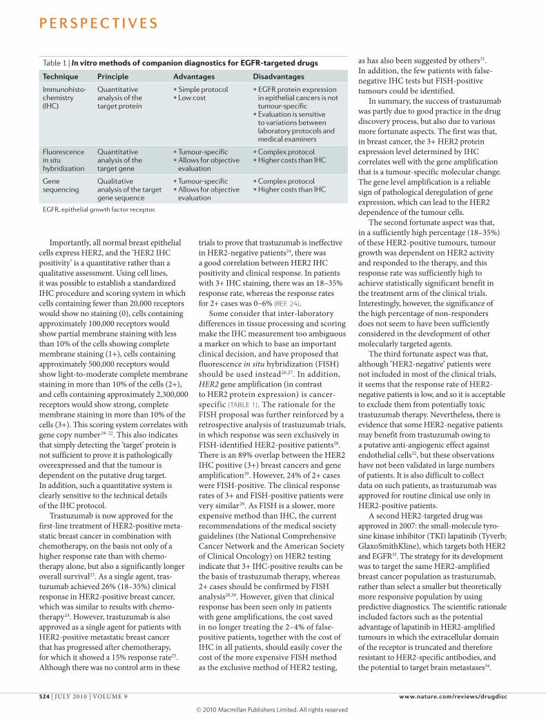
Importantly, all normal breast epithelial cells express Her2, and the ‘Her2 IHC positivity’ is a quantitative rather than a qualitative assessment. Using cell lines, it was possible to establish a standardized IHC procedure and scoring system in which cells containing fewer than 20,000 receptors would show no staining (0), cells containing approximately 100,000 receptors would show partial membrane staining with less than 10% of the cells showing complete membrane staining (1+), cells containing approximately 500,000 receptors would show light-to-moderate complete membrane staining in more than 10% of the cells (2+), and cells containing approximately 2,300,000 receptors would show strong, complete membrane staining in more than 10% of the cells (3+). This scoring system correlates with gene copy number18–22. This also indicates that simply detecting the ‘target’ protein is not sufficient to prove it is pathologically overexpressed and that the tumour is dependent on the putative drug target. In addition, such a quantitative system is clearly sensitive to the technical details of the IHC protocol.
Trastuzumab is now approved for the first-line treatment of Her2-positive meta-static breast cancer in combination with chemotherapy, on the basis not only of a higher response rate than with chemo-therapy alone, but also a significantly longer overall survival23. As a single agent, tras-tuzumab achieved 26% (18–35%) clinical response in Her2-positive breast cancer, which was similar to results with chemo-therapy24. However, trastuzumab is also approved as a single agent for patients with Her2-positive metastatic breast cancer that has progressed after chemotherapy, for which it showed a 15% response rate25. Although there was no control arm in these
trials to prove that trastuzumab is ineffective in Her2-negative patients24, there was a good correlation between Her2 IHC positivity and clinical response. In patients with 3+ IHC staining, there was an 18–35% response rate, whereas the response rates for 2+ cases was 0–6% (REF. 24).
Some consider that inter-laboratory differences in tissue processing and scoring make the IHC measurement too ambiguous a marker on which to base an important clinical decision, and have proposed that fluorescence in situ hybridization (FISH) should be used instead26,27. In addition, HER2 gene amplification (in contrast to Her2 protein expression) is cancer-specific (TABLE 1). The rationale for the FISH proposal was further reinforced by a retrospective analysis of trastuzumab trials, in which response was seen exclusively in FISH-identified Her2-positive patients28. There is an 89% overlap between the Her2 IHC positive (3+) breast cancers and gene amplification29. However, 24% of 2+ cases were FISH-positive. The clinical response rates of 3+ and FISH-positive patients were very similar29. As FISH is a slower, more expensive method than IHC, the current recommendations of the medical society guidelines (the National Comprehensive Cancer Network and the American Society of Clinical Oncology) on Her2 testing indicate that 3+ IHC-positive results can be the basis of trastuzumab therapy, whereas 2+ cases should be confirmed by FISH analysis28,30. However, given that clinical response has been seen only in patients with gene amplifications, the cost saved in no longer treating the 2–4% of false-positive patients, together with the cost of IHC in all patients, should easily cover the cost of the more expensive FISH method as the exclusive method of Her2 testing,
as has also been suggested by others31. In addition, the few patients with false-negative IHC tests but FISH-positive tumours could be identified.
In summary, the success of trastuzumab was partly due to good practice in the drug discovery process, but also due to various more fortunate aspects. The first was that, in breast cancer, the 3+ Her2 protein expression level determined by IHC correlates well with the gene amplification that is a tumour-specific molecular change. The gene level amplification is a reliable sign of pathological deregulation of gene expression, which can lead to the Her2 dependence of the tumour cells.
The second fortunate aspect was that, in a sufficiently high percentage (18–35%) of these Her2-positive tumours, tumour growth was dependent on Her2 activity and responded to the therapy, and this response rate was sufficiently high to achieve statistically significant benefit in the treatment arm of the clinical trials. Interestingly, however, the significance of the high percentage of non-responders does not seem to have been sufficiently considered in the development of other molecularly targeted agents.
The third fortunate aspect was that, although ‘Her2-negative’ patients were not included in most of the clinical trials, it seems that the response rate of Her2-negative patients is low, and so it is acceptable to exclude them from potentially toxic trastuzumab therapy. Nevertheless, there is evidence that some Her2-negative patients may benefit from trastuzumab owing to a putative anti-angiogenic effect against endothelial cells32, but these observations have not been validated in large numbers of patients. It is also difficult to collect data on such patients, as trastuzumab was approved for routine clinical use only in Her2-positive patients.
A second Her2-targeted drug was approved in 2007: the small-molecule tyro-sine kinase inhibitor (TKI) lapatinib (Tyverb; GlaxoSmithKline), which targets both Her2 and eGFr33. The strategy for its development was to target the same Her2-amplified breast cancer population as trastuzumab, rather than select a smaller but theoretically more responsive population by using predictive diagnostics. The scientific rationale included factors such as the potential advantage of lapatinib in Her2-amplified tumours in which the extracellular domain of the receptor is truncated and therefore resistant to Her2-specific antibodies, and the potential to target brain metastases34.
Table 1 | In vitro methods of companion diagnostics for EGFR-targeted drugs
technique Principle Advantages Disadvantages
immunohisto-chemistry (iHc)
Quantitative analysis of the target protein
•Simple protocol•Low cost
•eGFr protein expression in epithelial cancers is not tumour-specific
•evaluation is sensitive to variations between laboratory protocols and medical examiners
Fluorescence in situ hybridization
Quantitative analysis of the target gene
•Tumour-specific•Allows for objective
evaluation
•complex protocol•Higher costs than iHc
Gene sequencing
Qualitative analysis of the target gene sequence
•Tumour-specific•Allows for objective
evaluation
•complex protocol•Higher costs than iHc
eGFr, epithelial growth factor receptor.
P e r s P e c t i v e s
524 | jULy 2010 | vOLUMe 9 www.nature.com/reviews/drugdisc
© 20 Macmillan Publishers Limited. All rights reserved10
Overall, the success of trastuzumab, as well as that of imatinib, helped create a false perception that, if there is a validated target for an anticancer drug, all that is needed for success in clinical development is to select patients on the basis of target presence and/or overexpression, or that patient selection is not necessary if the target is present and/or overexpressed in the majority of patients. This perception was a key factor in the failures and controversies associated with several other drugs designed to target eGFr.
EGFR-specific mAbs: cetuximab and panitumumab. eGFr is the major growth factor receptor of epithelial cells and has been a promising target of cancer therapies since the discovery of its growth-stimulating effect on A431 human epithelial cancer cells35. According to several reports, eGFr is ‘overexpressed’ in 60–80% of colon and lung cancers36. Cetuximab (erbitux; Bristol–Myers Squibb/Lilly) was the first humanized eGFr-specific mAb developed to treat cancer, and was granted accelerated approval by the US Food and Drug Administration (FDA) for the treatment of colorectal cancer in 2004. The preclinical and clinical develop-ment of cetuximab followed an analogous approach to that of trastuzumab, in this case selecting patients that had tumours expressing eGFr instead of Her2 (REF. 37). A standardized IHC test was developed (the PharmDx kit, developed by Dako) as the clinical trial assay to select patients with colon carcinoma expressing eGFr in at least 1% of the cancer cells in their tumour tissue sample. So, the marketing approval of cetux-imab was based on clinical trials in which the efficacy of the drug was demonstrated in patients with colon carcinomas that stained positive for eGFr with this IHC test. The response rate was 8.8% as a mono-therapy in irinotecan-resistant patients and 22.9% in combination with irinotecan13,14.
However, retrospective analysis of the trials showed that the overall response rate did not correlate with the level of eGFr, in contrast to the case with trastuzumab and Her2. Patients with 1% IHC positivity had similar response rates to patients with 100% staining13,14. A control group of patients with negative IHC staining was not included in these trials. Nevertheless, these studies were considered acceptable by many researchers and by the FDA as a basis for cetuximab to be approved only for patients who are positive for this particular IHC test. This is also despite a surprising report indicating that 25% of IHC-negative patients can benefit from cetuximab therapy38.
These controversies — in contrast to the case with trastuzumab — triggered a search for more reliable diagnostic biomarkers. retrospective analyses have revealed such biomarkers that statistically significantly predict response to cetuximab. Moroni et al. found that 8 out of 9 responders had a high EGFR copy number determined by FISH compared with only 1 out of 21 non-responders (p < 0.0001)39. Lievre et al. found high EGFR copy number in 3 out of 11 responders compared with none in the non-responders among 30 patients with colon cancer40. It seems from these studies that FISH analysis or another molecular diag-nostic method that detects high EGFR copy number could be suitable for a positive selec-tion of patients who are likely to respond to cetuximab. Data from one study indicated that only 11.5% of colorectal cancers (n = 147) have increased gene copy number41. However, the response rates were higher than 11.5%, and so markers are needed that do not exclude patients who might benefit from cetuximab therapy despite normal EGFR copy numbers.
As the level of either the target protein or gene were not sufficient biomarkers for cetuximab response, investigators started to analyse the problem from a signal transduction perspective, focusing on the downstream signalling molecule KrAS. Approximately 36–50% of colon carcinomas harbour oncogenic mutant KRAS, which keeps KrAS constitutively active independ-ently of signals from upstream growth factor receptors such as eGFr42. Lievre et al. found that, of the 11 out of 30 patients who responded to cetuximab, none had KRAS mutations, in contrast to 13 out of 19 of the therapy-resistant tumours (p = 0.0003)40. Patients with wild-type KRAS also lived longer (p = 0.016; 16.3 versus 6.9 months). Meta-analysis of the studies of Moroni and Lievre reveals that 91.3% of patients with KRAS mutations did not respond to therapy, whereas 50% of patients with wild-type KRAS were responders (p = 0.001). The only patient with a complete response had wild-type KRAS and the highest EGFR copy number (greater than 20). In summary, 10% of colon carcinoma patients who have increased EGFR copy number had nearly 100% response to cetuximab and 60% of patients who have wild-type KRAS had 50% response (FIGS 1,2,3).
In a Phase II trial known as OPUS and a Phase III trial known as CrySTAL, cetuximab was combined with first-line chemotherapy (fluorouracil, leucovorin and oxaliplatin, or irinotecan, fluorouracil and
leucovorin, respectively)43,44. In these trials, clinical benefit was restricted to the wild-type KRAS population. Surprisingly, in both trials there was a trend towards a worse response to chemotherapy plus cetuximab than to chemotherapy alone in patients with KRAS mutations, which suggests that cetuximab might cause harm in this patient population.
However, the results above indicating the importance of KRAS status in response to cetuximab were not generally accepted in clinical use until recently, because they were based on retrospective analysis of clinical samples and preclinical research, and not on prospective randomized clinical trials. Clinicians therefore continued to use a test that was approved by the FDA based on a clinical trial that had design flaws — namely that the diagnostic test itself was not control-led and randomized. Four years after the first reports on the importance of KRAS mutations in cetuximab response, this molecular test has now been widely accepted by the scientific community and regulatory authorities. The european Medicines Agency (eMA) integrated KRAS testing as a mandatory condition for the administration of cetuximab in May 2008, and the same requirement was introduced by the FDA in july 2009. Unfortunately, IHC testing for eGFr expres-sion is still required, which could continue to unnecessarily exclude patients from treatment.
The efficacy of cetuximab was also investigated in patients with non-small-cell lung cancer (NSCLC). In a Phase III trial known as FLeX, the addition of cetuximab to platinum-based chemotherapy margin-ally, but significantly, increased the overall survival from 10.1 months to 11.3 months45. The patient selection was again only based on eGFr IHC positivity. This small survival advantage was sufficient for the FDA to register cetuximab for this new indication, but the eMA’s Committee for Medicinal Products for Human Use adopted a negative opinion on its approval. This decision indi-cates that regulators may no longer consider mathematically significant but very modest improvements in survival to be sufficient for market approval. The solution is to identify the subgroup of patients in which clinically meaningful benefit can be shown using improved biomarkers.
In the SwOG 0342 Phase II study of cetuximab in patients with NSCLC, both progression-free survival (6 months versus 3 months) and overall survival (16 months versus 8 months) were significantly longer in patients shown to be eGFr-positive by FISH. However, in this trial, all patients received cetuximab in combination with
P e r s P e c t i v e s
NATUre revIewS | Drug DiscOvery vOLUMe 9 | jULy 2010 | 525
© 20 Macmillan Publishers Limited. All rights reserved10
chemotherapy, and so a potential correlation between cetuximab response and high EGFR copy number could not be evaluated46. A biomarker-based evaluation of the Phase III BMS 099 trial, which investigated a taxane and carboplatin with or without cetuximab as first-line therapy, did not find longer survival or progression-free survival in patients with FISH-identified eGFr-positive tumours who were treated with cetuximab than in those who were treated with chemo-therapy alone47. Interestingly, there was also no correlation between cetuximab response and the presence of KRAS mutations in this trial, in agreement with preliminary reports on the biomarker analysis of the FLeX study48. The failure of the two most logical biomarkers (EGFR copy number and KRAS mutation) to predict the response to cetuximab in NSCLC might be related to a mechanism that is independent of the eGFr pathway or simply to poor efficacy in the selected patient population.
At the end of 2006, the FDA also approved a fully human eGFr-specific mAb, panitu-mumab (vectibix; Amgen) for the treatment of patients with colon carcinoma. The pivotal open-label, randomized study (involving 463 patients) for this drug used the same IHC kit and inclusion criteria of 1% eGFr positivity as was the case with cetuximab49. The overall response rate in the trial was 8% as mono-therapy in previously chemotherapy-resistant patients, and the mean progression-free survival was 96 days in the panitumumab arm compared with 60 days in the control arm, in which patients received best supportive care. There was no correlation between the clinical benefit and the level of eGFr expression, similarly to cetuximab, but again an eGFr IHC test must be positive for the indication of panitumumab, as there are no data on the efficacy of panitumumab in IHC-identified eGFr-negative patients49. retrospective biomarker analysis revealed that — similarly to cetuximab — patients with KRAS muta-tions do not benefit from panitumumab therapy, and therefore the FDA have now included this test in the drug label. In europe, given the evolving data on the importance of KRAS mutations, the eMA approved pani-tumumab only for patients with wild-type KRAS tumours, in December 2007.
EGFR-specific TKIs: gefitinib and erlotinib. As 50–80% of NSCLC cases reportedly express the eGFr protein37, it was expected that an efficient eGFr inhibitor would have anticancer activity in the majority of patients with NSCLC (with no selection needed), which stimulated considerable efforts to
Nature Reviews | Drug Discovery
EGFR HER2
KRAS PIK3CA
BRAF AKT
MEK P53
PTEN
mTOR
MET
Proliferation Apoptosis
10% CNV+ 0% M+40% CNV+ 10% M+
MET CNV+ or M+
HER2 M+
?
BRAF M+
KRAS M+PIK3CA M+PTEN CNV–
EGFR M+
EGFR CNV+
3% CNV+ 3% M+22% CNV+ 2% M+
10% CNV+ 3% M+4% CNV+ 12% M+
Colon cancerNSCLC
10% M+4% M+
40% CNV–40–70% CNV–40% M+
20% M+
5% M+5% M+
50% M+40% M+
a
b
Positive markersNegative markers
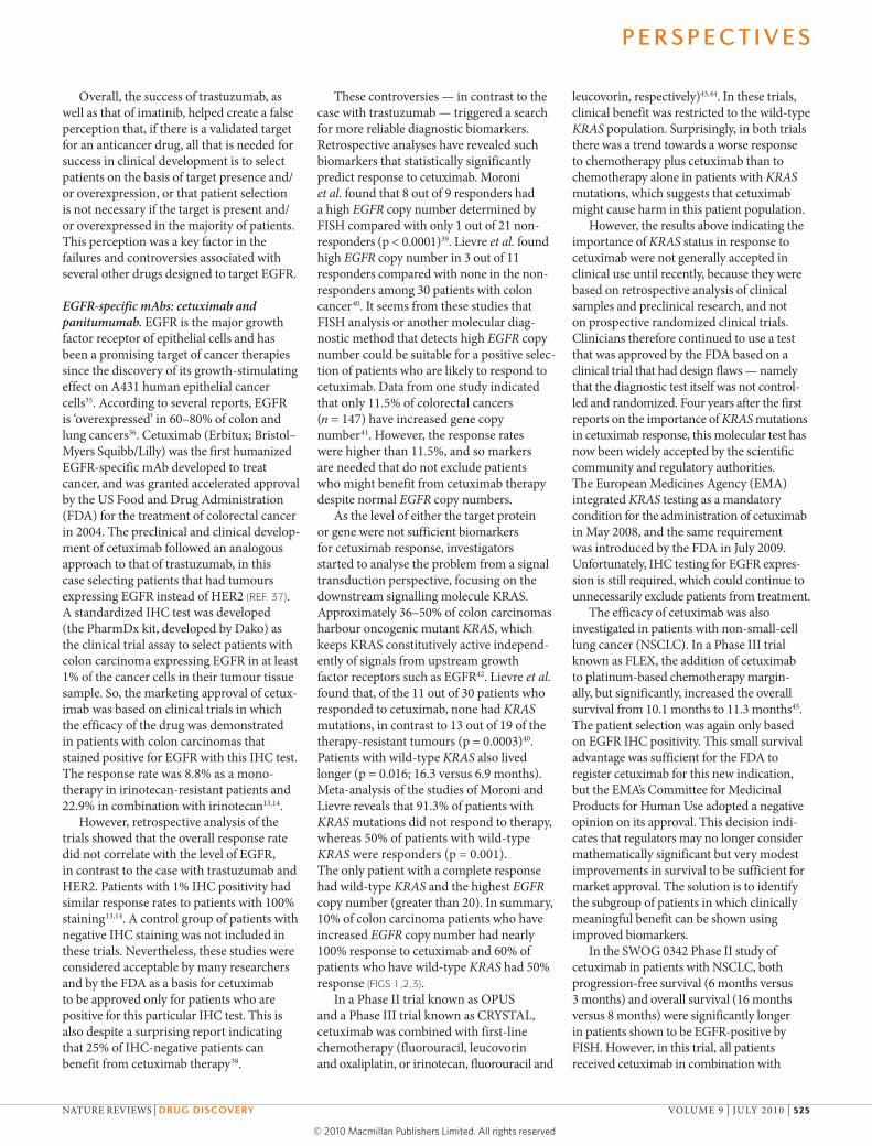
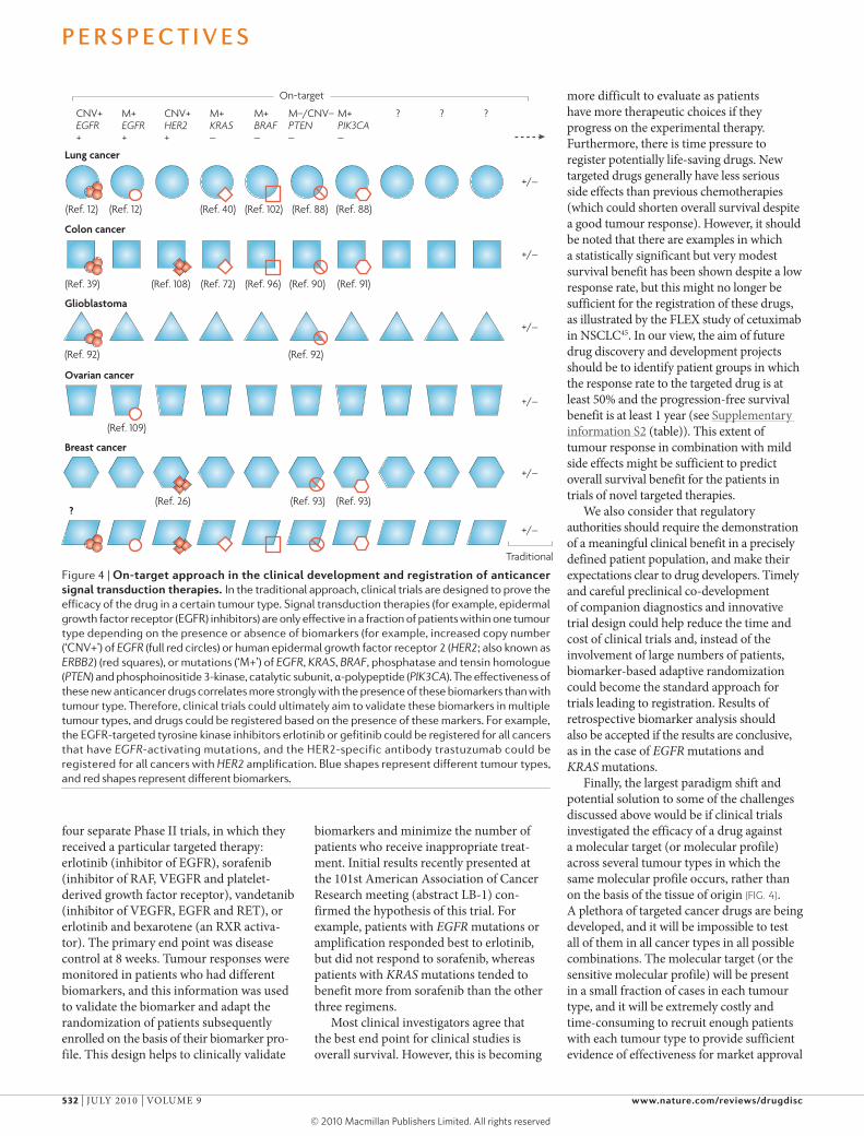
Figure 1 | Molecular mechanism of egFr signal-dependence in epithelial cancers. epidermal growth factor receptor (eGFr) inhibitors are only effective if the proliferation and/or survival of the cancer cells are dependent on constitutive eGFr signalling. The level of the oncogene dependence and the predicted efficacy of related targeted therapy can be illustrated by a pyramid (a), in which the upper part shows positive markers and the lower part shows negative markers. Mutations of genes that encode growth factor receptors and components of the downstream signal transduction path-ways (b) determine the dependence on eGFr activity. The frequency of copy number variations (increase: ‘cNv+’ or loss: ‘cNv–’) or mutations (‘M+’) of these genes in colon cancer (shown in blue) or in non-small-cell lung cancer (NScLc; shown in pink) is indicated. Activating mutations of the EGFR gene are associated with the highest objective response rate to eGFr-targeted tyrosine kinase inhibi-tors (TKis) in lung cancer74, but also in one case of ovarian cancer109. High gene copy number is a predic-tive marker of good response to eGFr-targeted TKi therapy in lung cancer74 and glioblastomas92, and eGFr-specific antibody therapy in colon cancer39–41. Alternative signalling from activated receptor tyrosine kinases, such as mutant human epidermal growth factor receptor 2 (Her2; also known as erBB2)97 or amplified MeT99, decreases the eGFr dependence and therapeutic response. The other important cause of resistance to eGFr inhibitors is the receptor-independent activation of the KrAS–rAF pathway40,63,72,86 or the phosphoinositide 3-kinase (Pi3K)–phosphatase and tensin homologue (PTeN)–protein kinase B (AKT)88–91 pathway by mutations in, or hypermethylation of, the genes coding for the signal transduction proteins downstream of the receptor. Genes responsible for the regulation of apoptosis, such as P53, may also be important in the clinical response to novel therapeutics96. MeK, mitogen-activated protein kinase–extracellular signal-regulated kinase kinase; mTOr, mammalian target of rapamycin; PiK3cA, phosphoinositide 3-kinase, catalytic subunit, α-polypeptide.
P e r s P e c t i v e s
526 | jULy 2010 | vOLUMe 9 www.nature.com/reviews/drugdisc
© 20 Macmillan Publishers Limited. All rights reserved10
develop such drugs37. The first two eGFr TKIs to reach clinical trials were gefitinib (Iressa; AstraZeneca) and erlotinib (Tarceva; roche), which have a similar chemical structure and both compete with ATP for binding to the catalytic domain of eGFr50.
response rates to gefitinib as a second- or third-line monotherapy for patients with NSCLC were only 18.4% and 11.8%, respectively, in the IDeAL1 and IDeAL2 Phase II trials51,52. Nevertheless, the data were sufficient to obtain accelerated approval by the FDA in May 2003. However, gefitinib failed to achieve a significant overall survival benefit in subsequent Phase III trials both in combination with chemotherapy (in a trial known as INTACT I-II)2,3 and as mono-therapy (in a trial known as ISeL)53. Therefore, gefitinib was withdrawn from the US market except for patients already taking and responding to the therapy, and the application for the european marketing authorization was also withdrawn in 2005 (REF. 54).
Meanwhile, erlotinib also failed to provide a survival benefit as a third-line treatment in combination with chemotherapy for patients with NSCLC in two Phase III trials, TALeNT55 and TrIBUTe56. However, in the placebo-controlled Phase III trial known as Br.21 in patients with locally advanced or metastatic NSCLC who had failed at least one chemotherapy regimen, there was a
moderate but significant (6.7 months versus 4.7 months) survival benefit from erlotinib, although the response rate was also small (8.9%)57. On the basis of the significant survival benefit, in November 2004 the FDA approved erlotinib for the second- and third-line therapy of NSCLC patients, without a link to a biomarker.
In summary, both gefitinib and erlotinib produced a treatment response in a surpris-ingly small population of NSCLC patients, although these patients often experienced complete remission and prolonged tumour-free survival57 (see Supplementary informa-tion S1 (box)). A search therefore began for a reliable biomarker to identify these patients. A second question was why erlotinib was more successful in the placebo-controlled Phase III Br.21 trial than was gefitinib in the similar ISeL trial.
In the spring of 2004, three independent groups reported that, based on retrospective biomarker analysis, activating mutations in the tyrosine kinase domain of EGFR were associ-ated with good clinical response to eGFr-targeted TKIs9–11. All of these papers reported 100% response in EGFR-mutant patients, in contrast to only a few percent in the wild-type patients. These reports were followed by numerous independent studies that confirmed these results. Overall, in the 19 reports, the average response rate in the EGFR-mutant
patients was 82%, compared with 11.5% in the wild-type patients58. Nine of these studies also provided data that overall survival is significantly longer in EGFR-mutant patients treated with an eGFr-targeted TKI (average of >2 years) than in wild-type patients (average of 8 months)58. The sensitizing mutations clustered in exons 18, 19 and 21 of EGFR6,59 and were associated with adenocar-cinoma histology, non-smoking history, Asian race and female gender6,59. The frequencies of mutations in lung adenocarcinomas were 22–67% in Asia, 3–25% in North America and 10–24% in South europe7,9–11,58,60–62. Thus, the frequency of mutations corresponds to the frequency of objective tumour responses to eGFr-targeted TKIs.
retrospective biomarker analysis of the Br.21 trial also found a higher response rate in EGFR-mutant patients, although this was only 16% compared with 7% in wild-type patients, and there was no correlation between survival benefit and mutation status12. However, the tests were not initially confirmed in independ-ent PCr reactions, and a re-analysis reported a response rate of 27% in patients with mutant EGFR, although this is still less than the average reported in the literature63.
with regard to other potential biomarkers in Br.21, survival among patients with eGFr protein overexpression (50–55%) was longer in the erlotinib group than in the placebo group, but there was no survival advantage among patients with IHC-identified eGFr-negative tumours. The p value, however, for the interaction between IHC and treatment response was 0.25, which indicates a low statistical power. The FISH analysis provided stronger predictive value, but was still not significant (p value for interaction = 0.1)12.
On the basis of these results, as noted above, erlotinib received market authoriza-tion in the United States without being linked to a biomarker. Interestingly, in europe, the market authorization includes a note: “In eGFr-negative cases, the efficacy is not proven”. It is not specified, however, how eGFr positivity should be determined. The easiest test is IHC, but this has the lowest predictive value — far from being significant according to the Br.21 study. The concern is that, without patient selec-tion or by favouring the cheaper and more frequently positive biomarker, oncologists are increasingly hesitant to indicate this treatment for their patients owing to the modest clinical responses of IHC-positive patients. Furthermore, there is evidence that patients with activating mutations and gene amplifications are not all IHC positive64 (FIGS 1,2). In vitro results also suggest that the
Nature Reviews | Drug Discovery
a Colon cancer b NSCLC
KRAS M+KRAS M+
IHC+
IHC+
RAFM+
HER2M+
METM+
PIK3CA M+PTEN CNV–
EGFR CNV+EGFR CNV+
RR 9–14%RR 20–30%
EGFR M+
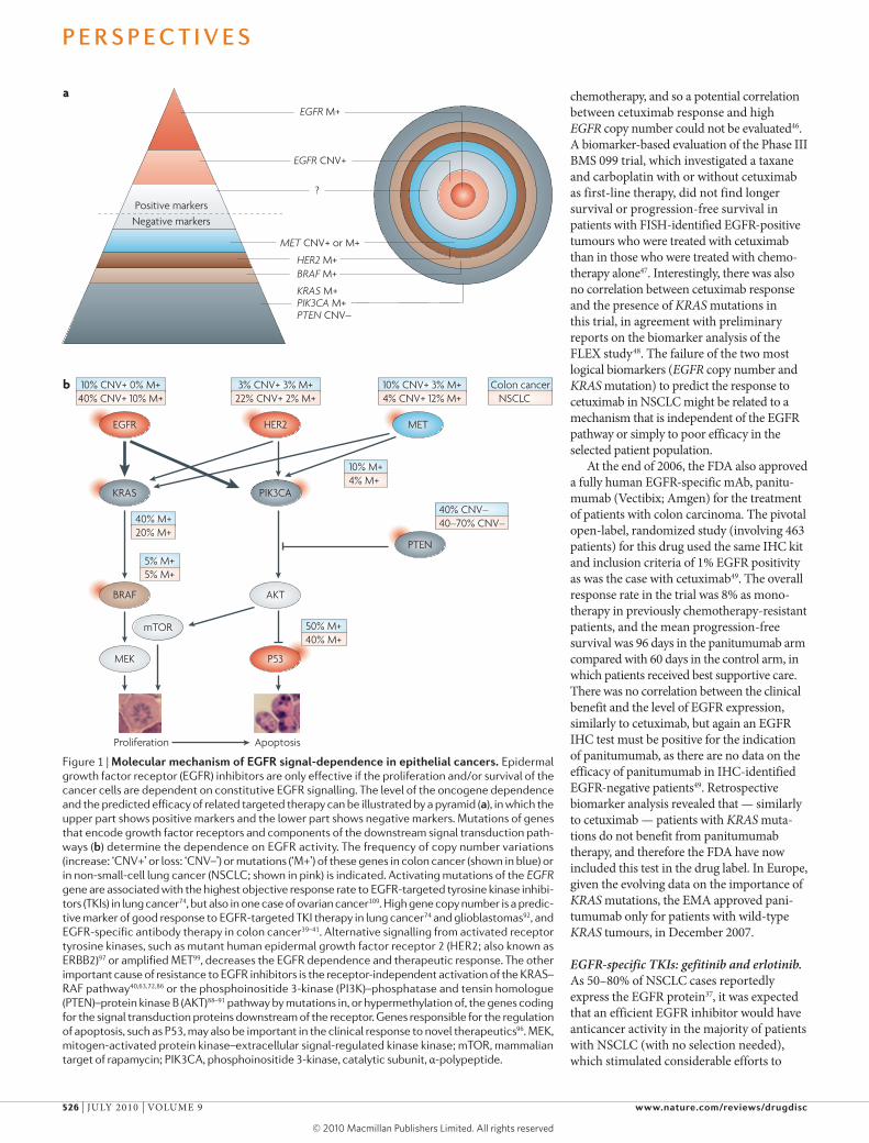
Figure 2 | Prevalence of predictive biomarkers of response to egFr inhibitors. The epidermal growth factor receptor (eGFr)-specific antibody therapy cetuximab in colon cancer (a) and the eGFr-targeted tyrosine kinase inhibitors erlotinib or gefitinib in lung cancer (b) only induce an objective response in a fraction of patients (green circles). Genetic alterations responsible for differences in drug sensitivity are similar in both tumour types, but the prevalence of these mutations is different, which is indicated by the size of the circles. Mutations (‘M+’) in KRAS40,63, RAF86, human epidermal growth factor receptor 2 (HER2; also known as ERBB2)97, MET99 or in the phosphoinositide 3-kinase (Pi3K)–phosphatase and tensin homologue (PTeN) pathway predict resistance88–91, whereas high copy numbers of the EGFR gene determined by fluorescence in situ hybridization (orange circles; ‘cNv+’) analysis in colon cancer39–41 or activating EGFR mutations in lung cancer predict objective response in most of the cases64,74. eGFr immunohistochemistry (yellow circles; ‘iHc+’) is an unreliable predictor of response rate (rr) in both tumour types, as it does not correlate with tumour response38,64,74.
P e r s P e c t i v e s
NATUre revIewS | Drug DiscOvery vOLUMe 9 | jULy 2010 | 527
© 20 Macmillan Publishers Limited. All rights reserved10
molecular genetic alterations can sensitize cancer cells to eGFr-targeted TKIs inde-pendently of the level of protein expression65. Therefore, using IHC-identified eGFr expression to select patients might exclude those who benefit most from erlotinib treatment.
The data described indicated that EGFR mutation testing has considerable potential as a biomarker for response to eGFr-targeted TKIs in NSCLC. However, critics noted that these studies were based on retro-spective analysis, and furthermore that they compared the clinical outcome of EGFR-mutant and wild-type patients all treated with eGFr-targeted TKIs and so could not clearly show whether EGFR-mutant patients have a better prognosis regardless of the type of treatment. The breakthrough in the appli-cation of EGFR mutation analysis to guide treatment in NSCLC did not come until recently. In july 2009, the eMA approved the use of gefitinib for patients with NSCLC carrying activating EGFR mutations, in all treatment lines. This decision was based on the biomarker analysis of a randomized Phase III trial known as IPASS66.
IPASS evaluated the efficacy, safety and tolerability of gefitinib versus carbo-platin and paclitaxel as first-line treatment in a clinically selected population of Asian patients. The trial enrolled 1,217 patients from 9 Asian countries who were light smokers or had never smoked, and had been diagnosed with stage IIIB–Iv adenocarcinoma NSCLC. Although the trial reached its target of proving the supe-riority of gefitinib in this clinically selected population, the biomarker analysis revealed that this advantage is restricted to patients with EGFR mutations. EGFR mutations were found in 60% of the 437 patients whose tumour samples were available for analysis. The progression-free survival in EGFR-mutant patients was 9.5 months with gefitinib treatment versus 6.3 months with carboplatin–paclitaxel treatment, whereas in mutation-negative patients, the progression-free survival in the gefitinib arm was only 1.5 months compared with 5.5 months when treated with chemotherapy66.
The approval of gefinitib for EGFR-mutant NSCLC patients in all treatment lines was also based on the results of another Phase III
trial known as INTereST, which showed that gefitinib is not inferior to docetaxel in previously treated patients67. The biomarker analysis found greater progression-free survival and response rates in EGFR-mutant and in high EGFR copy number patients than in patients with wild-type EGFR, but this difference between the biomarker subgroups was not observed with regard to overall survival, probably because many patients were subsequently treated with both drugs68. Although EGFR-mutant patients seemed to have a better prognosis independent of the type of treatment, they benefit much more from gefitinib than chemotherapy. It is also striking that chemotherapy is more effective than gefitinib in patients with wild-type EGFR. These data indicate that an EGFR mutation test is crucial in optimizing the treatment of NSCLC with gefitinib.
Questions for the future include: why erlotinib provided a survival benefit in an unselected population; whether patients with wild-type EGFR benefit more from erlotinib than from chemotherapy (in contrast to gefitinib); whether erlotinib will also be registered for the first-line therapy of EGFR-mutant NSCLC; and which drug is better for EGFR-mutant patients. One possible explanation for differences between erlotinib and gefitinib is that, although they have similar eGFr-inhibiting activity, their pharmacokinetics are different69. In addition, erlotinib is used at the maximum tolerated dose, thereby providing higher plasma levels than with gefitinib, which is used at a dose 4–5 times lower than the maximum tolerated dose70. Furthermore, both gefitinib and erlotinib have lower half-maximal inhibitory concentration (IC50) values in biochemical assays with mutant EGFR than with wild-type EGFR71. So, considering their in vivo free plasma levels and these IC50 values, it can be speculated that gefitinib levels are only sufficient to block mutant eGFr, but not wild-type eGFr. By contrast, erlotinib also inhibits wild-type eGFr, which might explain why it could achieve some therapeutic effect (mostly stable disease) in another subgroup of patients carrying wild-type eGFr in whom this protein had a role in disease (for example, owing to gene amplification) (FIG. 3). However, the wide-spread inhibition of wild-type eGFr could also explain the almost twice as frequent skin and gastrointestinal side effects observed in patients receiving erlotinib compared with those receiving gefitinib57.
Nature Reviews | Drug Discovery
Biological and pharmacologicalsensitivity to EGFR inhibitors
Biologicalsensitivity to EGFR inhibitors
Biologicalresistance to EGFR inhibitors
CR–PR13,9% (N = 145)
SD30–35%
EGFR inhibitors
EGFRM+ 14%
EGFRCNV+ 40%
HER2CNV+20%
METM+
12%
KRASM+
20%
PTENM+
40%
BRAFM+
5%
PIK3CAM+
4%
HER2M+
4%
METinhibitors
HER2inhibitors
AKT and mTORinhibitors
BRAF inhibitorsMEK inhibitors
PD50–60%
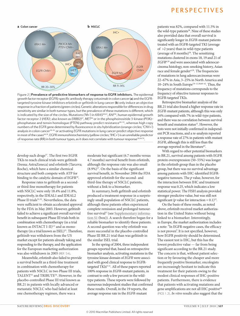
Figure 3 | Personalized therapy for lung adenocarcinomas. The upper horizontal bar represents genes affected by copy number variation (cNv) or mutations and their frequency. The middle bar indicates the target of potential signal transduction therapies associated with the particular molecular subtype. The lower bar indicates the clinical responses to epidermal growth factor receptor (eGFr)-targeted tyrosine kinase inhibitors (TKis) (as determined in the Br.21 trial) and the possible association with the different biomarkers57. Activating mutations of EGFR increase the dependence on eGFr and lower the threshold of effective drug concentration of the eGFr-targeted TKi74. cNv gain of eGFr only increases the biological sensitivity to monoclonal antibodies (mAbs) targeting eGFr46 and eGFr-targeted TKis12. cNv gain of Her2 increases the sensitivity to eGFr-targeted TKis100, but decreases response to eGFr-specific mAbs97. Activation of downstream signalling molecules confers resistance to eGFr inhibitors, but may provide an ideal target for other therapies40,63,72,86,97,99. conversely, activation of eGFr decreases the effectiveness of drugs targeting downstream signalling molecules102. AKT, protein kinase B; cr, complete response; Her2, human epidermal growth factor receptor 2 (also known as erBB2); MeK, mitogen-activated protein kinase–extracellular signal-regulated kinase kinase; mTOr, mammalian target of rapamycin; PD, progressive disease; Pr, partial response; PiK3cA, phosphoinositide 3-kinase, catalytic subunit, α-polypeptide; PTeN, phosphatase and tensin homologue; SD, stable disease.
P e r s P e c t i v e s
528 | jULy 2010 | vOLUMe 9 www.nature.com/reviews/drugdisc
© 20 Macmillan Publishers Limited. All rights reserved10

Finally, similarly to cetuximab for the treatment of colon carcinomas, the presence of mutant KRAS is correlated with a lack of response to eGFr-targeted TKIs in the treat-ment of lung cancer72 (FIGS 1,2). However, KRAS mutation is not a sufficient negative marker for erlotinib use, as its frequency is only 17–20% in adenocarcinomas72; there-fore ~80% of patients would in theory be treated based on this criterion, but only 50% of patients in clinical trials showed some evidence of disease control (objective response plus stable disease) due to erlotinib treatment57. In addition, a recent study did not find a significant difference in the effect of erlotinib on progression-free survival for NSCLC patients with wild-type EGFR and KRAS, and those with wild-type EGFR and mutant KRAS73. This means additional negative markers are needed to exclude patients who do not benefit from eGFr-targeted TKI treatment, which are discussed below (FIG. 3).
Post-marketing studies of EGFR inhibitors. The IHC tests to detect eGFr and Her2 discussed above were approved based on trials that were uncontrolled from a diagnostics perspective, with response rates of around 9–30% in the selected population. Given that 25 independent reports found an average response rate of 72% and a time to progression of more than 1 year in NSCLC patients with EGFR mutations, but only an average 10% response rate and 2-month time to progression in EGFR wild-type patients58, it is surprising that for 5 years EGFR muta-tion testing was viewed as scientifically interesting but not ready for clinical use74. In this case, there seems to be an inconsist-ency in the standards by which the evidence associated with identification of patients who do or do not respond to a targeted therapy is judged.
For example, a single retrospective study of EGFR mutation status in archived tissues from patients from the Br.21 trial of erlo-tinib was considered sufficient to make the conclusion, “there is no need for mutation testing”12. However, the importance of many other independent studies that reported that patients with EGFR mutations respond well to erlotinib have been downplayed as preliminary and retrospective; the concept of EGFR mutation only as an overall good prognostic marker instead of a marker for response is readily accepted from the same study, whereas previous studies that had a different conclusion are disregarded6. In response, new studies have been published to counter this report75,76.
Methodological problems in the molecular diagnostic studies in paraffin-embedded tissues have been another pitfall in biomarker studies. This was at least the case in the initial retrospective biomarker analysis of the Br.21 trial of erlotinib12,63. recent recommendations of the european Association of Pathologists on KRAS molecular diagnostics suggested that not only the methods, but most importantly the laboratories, have to be accredited to perform this type of test77. Owing to the many potential errors from the sample preparation (for example, lack of changing the blade on the microtome between each sample), the influence of chemical contami-nants on the performance of DNA poly-merases in PCr reactions and the stringency of quality control in general, even standard-ized, certified in vitro diagnostic assays do not remove the need for ‘clinical’ validation of the laboratories. The question is not only whether the results of a clinical trial can be explained by the biomarker analysis of one central laboratory, but which laboratory can most accurately predict the clinical effective-ness of a targeted anticancer drug in the relevant patient subgroups; for example, 100% prospective association between the clinical response to erlotinib and gefitinib and the presence of activating mutations in EGFR detected by a laboratory can provide this type of validation64.
In summary, trastuzumab seems to be the most successful of the marketed drugs targeting the eGFr family. However, little is known about its possible off-target effects, and the predictive value of the companion diagnostic is far from perfect. This is an emerging problem, because recent results that have indicated the positive effects of 1-year adjuvant trastuzumab treatment in all Her2-positive patients78 could place a large cost burden on health-care systems. There is therefore a need for better diagnostics for more precise patient selection. In the case of cetuximab and panitumumab, KRAS testing has now become more widely used in the clinic, but IHC testing for eGFr still has to be performed. IHC testing for eGFr was also used to select NSCLC patients in a trial that showed that adding cetuximab to chemotherapy resulted in a statistically significant improvement in survival, but registration was not approved by the eMA owing to the modest absolute benefit45. After a 5-year delay, gefitinib has been approved in europe for the treat-ment of EGFR-mutant patients. erlotinib is approved for the second-line treatment of all NSCLC patients in both the United
States and in europe, although in Hungary, for example, the state-owned health insur-ance only covers the treatment of patients with wild-type KRAS or mutant EGFR. This provides a precedent of a payer using a biomarker that is not in the label of the drug to increase the cost-effectiveness of a therapy.
Preclinical biomarker integrationIn cancer cells, the expression of hundreds of genes can be different to that in normal cells. The key question is which genes are driving the cancer and promoting cancer cell survival, and which are accompanying signals. If we can identify the factors respon-sible for cancer cell survival in particular cancers (many of which are kinases), these should be excellent candidates for targeted therapy. Genes that are mutated or amplified in cancer cells are more likely to be survival factors than those which are only overex-pressed. The identification of such mutated or amplified genes could form the basis for biomarkers. A combination of target-driven and chemistry-driven drug discovery with biomarker identification and linkage to companion diagnostics could be used to provide more personalized targeted therapy. By “chemistry-driven drug discovery”, we mean selecting drug candidates on the basis of cellular assays, and subsequent selectivity profiling and off-target identification (BOX 1), aided by pharmacophore models and the pool of inhibitors with known selectivity79–81.
For the identification of potential predic-tive biomarkers that might form the basis of companion diagnostics for both small-molecule inhibitors and mAbs, the lessons learned with the first generation of eGFr inhibitors can be applied. The most impor-tant lesson is that the predictive marker of signal transduction therapies (BOX 2) is usually not equivalent to the expression of the target protein. Growth or survival of the particular tumour has to be dependent on the activity of the targeted signal transduc-tion molecule. recent genome-wide cancer genetics studies have identified hundreds of mutated genes, but only a few important ‘cancer driver’ signal transduction path-ways82. The challenge for predictive diagnostics is to determine which pathway is the main driver of the malignant phenotype in the particular tumour sample and which drug is able to block this pathway. Activating mutations of EGFR seem to be single cancer drivers in the absence of other mutated oncogenes, which explains the high sensitivity of tumours harbouring these mutations to eGFr inhibitors83. Over-activation of
P e r s P e c t i v e s
NATUre revIewS | Drug DiscOvery vOLUMe 9 | jULy 2010 | 529
© 20 Macmillan Publishers Limited. All rights reserved10
eGFr owing to mutations or increased gene copy number have been proved to be the best positive predictor of eGFr signalling dependence and therapeutic response to eGFr-targeted therapies (mAbs or TKIs) in colon and lung cancer38,40,64.
In the absence of a key single driver oncogene, cancer typically involves the activation of several oncogenes82. In the case of inhibitors that specifically target a single receptor tyrosine kinase, activation of alternative growth factor receptors by amplification or activating mutations, or receptor-independent activation of down-stream signalling molecules, almost always lead to drug resistance both in vitro and in a clinical setting84, seemingly regardless of the tumour type and the signal transduction target85. For example, eGFr inhibitors are ineffective in KRAS-mutant cells40,63 and RAF-mutant colon cancer86, and KrAS inhibitors are ineffective in RAF-mutant cells87. Furthermore, loss of the phosphatase and tensin homologue (PTEN) tumour suppressor gene or activating mutations in phosphoinositide 3-kinase, catalytic sub-unit, α-polypeptide (PIK3CA) increases the eGFr-independent activity of the protein kinase B pathway, which predicts resistance to eGFr inhibitors in lung cancer88,89, in colon carcinomas90,91 and in glioblastomas92, similarly to trastuzumab resistance in
Her2-positive breast cancer93. In addition, not only can anticancer cytotoxicity medi-ated by DNA-damaging agents be dependent on the executors of active cell death (mostly apoptosis), such as functional tumour pro-tein P53 (REF. 94) and FAS95, but so can that mediated by signal transduction therapeutics such as gefitinib96.
Activation of alternative growth factor receptors also confers resistance to drugs targeting single tyrosine kinase receptors. For example, HER2 amplification or overex-pression of insulin-like growth factor 1 have been implicated in cetuximab resistance in colon cancer97, and activating mutations in HER2 have been implicated in gefitinib resistance in lung cancer98. The amplifica-tion of MET leads to resistance to gefitinib in lung cancer99. However, amplification of HER2 increases gefitinib sensitivity through the activation of eGFr100.
The development of predictive biomarkers can be integrated into preclinical anticancer drug discovery by using multiple cell lines to model typical molecular types of cancers. For example, the A431 cell line was first used for the discovery of eGFr inhibitors in bioassays as eGFr was discovered in these cells, in which EGFR is amplified and overexpressed101. However, most lung cancer cell lines express much lower EGFR levels, and some carry activating mutations and/or
drug resistance mutations in EGFR as well as mutations in genes encoding downstream signalling molecules such as KrAS, rAF and PTeN. Therefore, screening against many cell lines of the same histological origin may give information about the general effectiveness of the lead compound while providing a system to model drug sensitivity and resistance and develop molecular diagnostics.
This strategy should also be used for screening other targeted compounds. For example, novel inhibitors of MeK (mitogen-activated protein kinase–extracellular signal-regulated kinase) kinases are ineffective in all cell lines carrying EGFR mutations, effective in all cell lines carrying RAF mutations, but only effective in half of the cell lines with KRAS mutations102 (FIG. 1). The cancer specifi-city of the compounds could be determined using normal cell culture as controls, or syn-genic synthetic lethality screens to identify compounds that are more effective in the presence of cancer-specific genetic changes103.
Clinical biomarker integrationAspects of clinical trial design can intention-ally or unintentionally reveal or obscure the importance of biomarkers. First, increasing the number of patients enrolled in the treatment and control arms can ensure a statistically significant difference between the groups, even if only a subset of patients responds to therapy. For example, if a clinical trial of trastuzumab had been carried out enrolling four times more unselected patients with breast cancer than was neces-sary to prove efficacy in patients pre-selected by Her2 testing, the results would presum-ably have been sufficient to support the use of trastuzumab in treating all breast cancer patients without testing for a biomarker. Conversely, even if a novel targeted drug entering clinical trials is well characterized for target specificity and molecular mecha-nism of action, limiting patient recruitment in clinical trials on the basis of the detection of the target protein by IHC or by a diag-nostic test that has not been validated in a controlled preclinical or clinical experiment, and without the information of the full target profile (selectivity panel) of the drug, risks the unjustified restriction of patient access to the drug. This might be more harmful than not selecting patients at all.
recruitment into the trial of only those patients that have tumour samples for detailed retrospective molecular profiling may provide more useful information to establish a validated companion diagnostic test. If there is a preclinically validated com-panion diagnostic available, the clinical trial
Box 1 | Characterizing the target profile of small-molecule kinase inhibitors
Themostobviouswaytocharacterizethetargetprofileofakinaseinhibitoristoperformkinaseassaysonalargepanelofkinases.Otherapproachesarebasedontheproteomicidentificationofkinasestowhichthedrugcandidatebindswithhighaffinity.Forexample,ourrecentlydeveloped‘targetfishing’technology(developedinclose
collaborationwiththeMaxPlanckInstituteofBiochemistry(MPI))reliesonthecouplingofasmall-moleculekinaseinhibitortoamatrixthroughalinker.Theaimistogenerateanaffinitychromatographymaterialthatexploitsthestrongbindingaffinitiesbetweenkinaseinhibitorsandtheirmoleculartargets.Thetechnologyisstraightforwardinprinciple,butrequiresspecializedchemistrytolinkthekinaseinhibitorandmaintainitsbiologicalactivityandasensitiveproteomictechnology(providedbytheMPI)toidentifytheboundproteins82,113–117.Oncecoupledandprovenactive,immobilizedligandsareprocessedinanaffinitychromatographyapproachtoidentifyalltherelevanttargetsandoff-targetsfromacrudeextract,whichbindtothismatrix.Allbindersaresubsequentlycloned,expressedandpurified(kinasesandnon-kinases).Theaffinitiesofthefreeligandsforthevariousenzymesaredeterminedinrelevantin vitroassays.Thus,quantitativebindingcanbedeterminedandeventhebindingtonon-kinasescanbeverified.Asaproof-of-conceptexperimentforthetechnology,weusedimmobilizedgefitinibtoidentifyitsoff-targetsandtoobtainabetterunderstandingofthepharmacologyofgefitinib115.Massspectrometry(MS) analysisoftheidentifiedproteinspotsandaparallelliquidchromatography–MS/MS analysisnotonlypermitteddetectionoftheinternalcontrol,epidermalgrowthfactorreceptor,asexpected,butidentifiedmorethan20additionalgefitinibtargets.Othertargetfishingtechnologieshavealsobeendescribedintheliterature118,whichusea
chemicalproteomicsapproachtoprofiletheinteractionofsmallmoleculeswithendogenouslyexpressedproteinkinasesandpurine-bindingproteins.Thissubproteomeiscapturedbyimmobilizednonselectivekinaseinhibitors(kinobeads),andtheboundproteinsarequantifiedinparallelbymassspectrometryusingisobarictagsforrelativeandabsolutequantification(iTRAQ).Thetechniqueisbasedonacompetitivebindingassayandwillidentifyonlytheproteinsthatwerepulleddownbythebroad-spectrumkinaseinhibitorsonthekinobeads119.
P e r s P e c t i v e s
530 | jULy 2010 | vOLUMe 9 www.nature.com/reviews/drugdisc
© 20 Macmillan Publishers Limited. All rights reserved10
design can be optimized by selecting patients with this diagnostic method, but a biomarker-negative control arm should be included for the clinical validation of the companion diagnostics.
There are major technical and logistical challenges to integrating molecular biomar-ker tests into clinical trials and into routine clinical work104. First, molecular pathology laboratories and their diagnostic technolo-gies need to be sufficiently sensitive and robust to give a diagnosis from the majority of tissue samples. A guideline paper from the Society of european Pathologists recom-mends that only laboratories that can pro-vide results with a success rate of more than 95% and an accuracy of more than 97%, within 2 weeks, should perform KRAS muta-tion testing77. Quality control investigations should be carried out regularly to accredit laboratories for companion diagnostics. However, such investigations in Germany and Hungary, for example, have revealed that only a few laboratories can meet these standards at present, and therefore most oncology centres do not have such a labora-tory in the same institute (see the KrAS information website).
A second question is whether the companion diagnostic test should be auto-matically integrated into the work flow of pathology laboratories. This would shorten the time to obtain the result of companion diagnostics, but may result in many unnec-essary tests being performed. For example, the current typical practice in the case of colon carcinoma is that, after diagnostic work that includes staging with computed
tomography scans and histological diagnosis from endoscopic or surgical biopsy, a decision is made as to whether the patient should receive chemotherapy or only surgical treatment. If the patient has only liver metastasis, first-line eGFr-targeted (cetuximab) treatment should be consid-ered105, and the oncologist sends a request to the central molecular pathology laboratory accredited to perform KRAS testing. The molecular pathology laboratory then sends a request to the primary pathology depart-ment to send the specimen to the laboratory. After the test, the sample is sent back to the pathology laboratory (if there is tissue left) and diagnosis of the KRAS test is sent to the oncologist. The challenge is to organize this process within 2 weeks, which is considered the clinically acceptable time frame to start the therapy.
Besides the considerations for biomarker validation, molecular mechanisms of action should be given greater consideration in the clinical trials of future combination thera-pies. The accepted approach in Phase III trials is to compare the current standard treatment to the new therapy combined with the standard treatment, which theoretically minimizes the patients’ risk of receiving less effective treatment than the best treatment that is currently available. However, knowl-edge of mechanism of action of the drugs and preclinical experiments can predict that some combinations could be antagonistic, thus potentially harming patients and risking the success of the experimental drug. For example, eGFr inhibitors induce cell cycle blockade, which increases the resistance
to chemotherapy106. This effect may have contributed to the failure of erlotinib in combination with gemcitabine–cisplatin55 or paclitaxel–carboplatin56 and gefitinib in combination with gemcitabine–cisplatin2 or paclitaxel–carboplatin3 in Phase III studies.
In addition, only randomized trials with two or more arms can uncover the difference between the prognostic (general clinical behaviour) and predictive (treatment-specific) value of molecular biomarkers. For example, the INTereST study found a higher response rate to both gefitinib (42.1%) and docetaxel (21.1%) in EGFR-mutant NSCLC patients than in NSCLC patients with wild-type EGFR (6.6% and 9.8% respectively)68. This indicates either that EGFR mutations are positive prognostic markers irrespective of the type of treatment or that EGFR-mutant tumours are more responsive to both treatments. The design of the trial enabled an important question to be answered — namely, which treatment is most beneficial to EGFR-mutant patients — as there was a significantly higher response to gefitinib than to docetaxel, and this was also reflected by significantly longer progression-free survival in gefitinib-treated EGFR-mutant patients. Conversely, in accordance with other reports and understanding of the eGFr signalling pathway, there was 0% response to gefitinib in KRAS-mutant patients. However, the response to docetaxel was also low in this patient population (3.7%) and there was no difference in progression-free survival and overall survival between KRAS-mutant patients treated with gefitinib or docetaxel. These results might indicate that KRAS-mutant patients do not benefit significantly less from gefitinib than from docetaxel, but potentially also have a worse prognosis and poor response to chemotherapy in general.
Preclinical hypothesis-based combinations and predictive biomarkers might be most effectively investigated in adaptive clinical trials. A good example is the BATTLe trial at the MD Anderson Cancer Center107. In this trial, patients with NSCLC who had failed at least one prior chemotherapy regi-men were enrolled in an umbrella trial in which they gave their consent for a biopsy from their tumour. The tumour sample was analysed for 11 pre-specified biomarkers and, based on the results, patients were classified into four groups (EGFR mutant, or high copy number type; KRAS–BRAF mutants; vascular endothelial growth factor receptor (VEGFR)-overexpressing type; and retinoid X receptor (rXr)–cyclinD activator). The patients were then randomized into
Box 2 | Signal transduction therapy
Theapprovalofmolecularlytargeteddrugssuchastrastuzumabandimatinibisoftenconsideredtorepresenta‘paradigmshift’inanticancerdrugdevelopment.Nevertheless,ithasbeenarguedthatclassicalanticanceragentssuchasthetopoisomeraseIinhibitorcamptothecin,thetopoisomeraseIIinhibitoretoposideorthethymidylatesynthaseinhibitor5-fluorouracilaremolecularlytargeted,makingthedistinctionartificial.Indeed,individualpolymorphismsintheseenzymesandenzymesresponsibleforthemetabolismofthesedrugscandetermineindividualdrugsensitivityandmoleculardiagnosticscanbeusedtopersonalizetherapy112.However,itisimportanttodistinguishthesetherapeuticagentsfromnovelsignaltransduction
therapies.TheclassicalanticancerdrugstargetthemolecularapparatusoftheexecutionofcellularfunctionssuchasmitosisorDNArepair,whereasthesignaltransductiontherapiestargetthepathologicalsignaltransductionmechanismsthatareresponsibleforthetumour-specificpathologicalregulationofthesecellularfunctions.Therefore,forsignaltransductiontherapies,itisoftennotenoughtodetectthepresenceofthedrug-sensitivevariantofatargetprotein;theproliferationorthesurvivaloftheparticulartumourmustbedependent ontheabnormalactivityoftheparticulartargetprotein(FIG. 1).Inotherwords,thesignaltransductiontherapytargetsthesignallingpathway.Forexample,evenifthegeneencodingepidermalgrowthfactorreceptor(EGFR)isoverexpressed,ifdownstreammutantKRASmaintainstheEGFR-independentautonomicproliferationsignal,EGFRinhibitorsarenoteffective.Thisdifferencemakesthedevelopmentofcompaniondiagnosticsforsignaltransductiontherapiesmoreimportantandmorecomplicated.
P e r s P e c t i v e s
NATUre revIewS | Drug DiscOvery vOLUMe 9 | jULy 2010 | 531
© 20 Macmillan Publishers Limited. All rights reserved10
four separate Phase II trials, in which they received a particular targeted therapy: erlotinib (inhibitor of eGFr), sorafenib (inhibitor of rAF, veGFr and platelet-derived growth factor receptor), vandetanib (inhibitor of veGFr, eGFr and reT), or erlotinib and bexarotene (an rXr activa-tor). The primary end point was disease control at 8 weeks. Tumour responses were monitored in patients who had different biomarkers, and this information was used to validate the biomarker and adapt the randomization of patients subsequently enrolled on the basis of their biomarker pro-file. This design helps to clinically validate
biomarkers and minimize the number of patients who receive inappropriate treat-ment. Initial results recently presented at the 101st American Association of Cancer research meeting (abstract LB-1) con-firmed the hypothesis of this trial. For example, patients with EGFR mutations or amplification responded best to erlotinib, but did not respond to sorafenib, whereas patients with KRAS mutations tended to benefit more from sorafenib than the other three regimens.
Most clinical investigators agree that the best end point for clinical studies is overall survival. However, this is becoming
Nature Reviews | Drug Discovery
Lung cancer
Colon cancer
Glioblastoma
Ovarian cancer
Breast cancer
?
+/–
+/–
+/–
+/–
+/–
+/–
Traditional
(Ref. 12) (Ref. 12)
(Ref. 39) (Ref. 108) (Ref. 72) (Ref. 96) (Ref. 90) (Ref. 91)
(Ref. 92) (Ref. 92)
(Ref. 109)
(Ref. 26) (Ref. 93) (Ref. 93)
(Ref. 40) (Ref. 102) (Ref. 88) (Ref. 88)
CNV+EGFR+
M+EGFR+
CNV+HER2+
M+KRAS–
M+BRAF–
M–/CNV–PTEN–
M+PIK3CA–
? ? ?
On-target
Figure 4 | On-target approach in the clinical development and registration of anticancer signal transduction therapies. in the traditional approach, clinical trials are designed to prove the efficacy of the drug in a certain tumour type. Signal transduction therapies (for example, epidermal growth factor receptor (eGFr) inhibitors) are only effective in a fraction of patients within one tumour type depending on the presence or absence of biomarkers (for example, increased copy number (‘cNv+’) of EGFR (full red circles) or human epidermal growth factor receptor 2 (HER2; also known as ERBB2) (red squares), or mutations (‘M+’) of EGFR, KRAS, BRAF, phosphatase and tensin homologue (PTEN) and phosphoinositide 3-kinase, catalytic subunit, α-polypeptide (PIK3CA). The effectiveness of these new anticancer drugs correlates more strongly with the presence of these biomarkers than with tumour type. Therefore, clinical trials could ultimately aim to validate these biomarkers in multiple tumour types, and drugs could be registered based on the presence of these markers. For example, the eGFr-targeted tyrosine kinase inhibitors erlotinib or gefitinib could be registered for all cancers that have EGFR-activating mutations, and the Her2-specific antibody trastuzumab could be registered for all cancers with HER2 amplification. Blue shapes represent different tumour types, and red shapes represent different biomarkers.
more difficult to evaluate as patients have more therapeutic choices if they progress on the experimental therapy. Furthermore, there is time pressure to register potentially life-saving drugs. New targeted drugs generally have less serious side effects than previous chemotherapies (which could shorten overall survival despite a good tumour response). However, it should be noted that there are examples in which a statistically significant but very modest survival benefit has been shown despite a low response rate, but this might no longer be sufficient for the registration of these drugs, as illustrated by the FLeX study of cetuximab in NSCLC45. In our view, the aim of future drug discovery and development projects should be to identify patient groups in which the response rate to the targeted drug is at least 50% and the progression-free survival benefit is at least 1 year (see Supplementary information S2 (table)). This extent of tumour response in combination with mild side effects might be sufficient to predict overall survival benefit for the patients in trials of novel targeted therapies.
we also consider that regulatory authorities should require the demonstration of a meaningful clinical benefit in a precisely defined patient population, and make their expectations clear to drug developers. Timely and careful preclinical co-development of companion diagnostics and innovative trial design could help reduce the time and cost of clinical trials and, instead of the involvement of large numbers of patients, biomarker-based adaptive randomization could become the standard approach for trials leading to registration. results of retrospective biomarker analysis should also be accepted if the results are conclusive, as in the case of EGFR mutations and KRAS mutations.
Finally, the largest paradigm shift and potential solution to some of the challenges discussed above would be if clinical trials investigated the efficacy of a drug against a molecular target (or molecular profile) across several tumour types in which the same molecular profile occurs, rather than on the basis of the tissue of origin (FIG. 4). A plethora of targeted cancer drugs are being developed, and it will be impossible to test all of them in all cancer types in all possible combinations. The molecular target (or the sensitive molecular profile) will be present in a small fraction of cases in each tumour type, and it will be extremely costly and time-consuming to recruit enough patients with each tumour type to provide sufficient evidence of effectiveness for market approval
P e r s P e c t i v e s
532 | jULy 2010 | vOLUMe 9 www.nature.com/reviews/drugdisc
© 20 Macmillan Publishers Limited. All rights reserved10
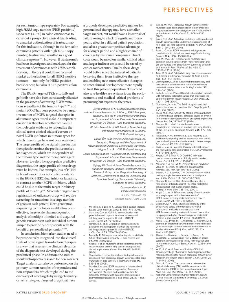
for each tumour type separately. For example, high HER2 copy number (FISH positivity) is too rare (3–5%) in colon carcinomas to carry out a prospective clinical trial leading to a market authorization of trastuzumab for this indication, although in the few colon carcinoma patients with high HER2 copy number, trastuzumab resulted in a good clinical response108. However, if trastuzumab had been investigated and marketed for the treatment of carcinomas with HER2 ampli-fication, in theory it could have received market authorization for all Her2-positive tumours — not only for Her2-positive breast cancer, but also Her2-positive colon carcinoma.
The eGFr-targeted TKIs erlotinib and gefitinib have also been consistently effective in the presence of activating EGFR muta-tions regardless of the tumour type74,109, and mutant KRAS has been proved to be a nega-tive marker of eGFr-targeted therapies in all tumour types tested so far. An important question is therefore whether we can use the molecular evidence to rationalize the clinical use or clinical trials of current or novel eGFr inhibitors in tumour types for which these drugs have not been registered. The target profile of the signal transduction therapies determines the predictive molecu-lar diagnostics, which are independent of the tumour type and the therapeutic agent. However, to select the appropriate predictive diagnostics, the target profile of these drugs must be known. For example, loss of PTeN in breast cancer does not confer resistance to the eGFr–Her2 dual inhibitor lapatinib, in contrast to other eGFr inhibitors, which could be due to the multi-target inhibitory profile of this drug110. Molecular target-based registration of anticancer drugs will require screening for mutations in a large number of genes in each patient. Next-generation sequencing technologies might allow cost-effective, large-scale pharmacogenetic analysis of multiple inherited and acquired genetic variations in each individual tumour sample to provide cancer patients with the benefit of personalized genomics82,111.
In conclusion, biomarker studies need to be prospectively integrated into the clinical trials of novel signal transduction therapies in a way that assesses the clinical relevance of the diagnostic test developed during the preclinical phase. In addition, the studies should retrospectively search for new markers. Target analysis can also be performed on the primary tumour samples of responders and non-responders, which might lead to the discovery of new targets by using chemistry-driven strategies. Targeted drugs that have
a properly developed predictive marker for personalized therapy may have a smaller target market, but would have a lower risk of failure owing to a lack of significant thera-peutic effect in a diluted patient population, and also a greater competitive advantage for a longer period and a higher chance of coverage by insurance companies. Direct costs would be saved on smaller clinical trials and larger indirect costs could be saved by earlier market entry. Finally, these drugs would better serve the interest of patients by saving them from ineffective therapy and enabling new, more effective therapies to enter clinical development more rapidly to treat this patient population. This could also save health-care systems from the socio-economic and medical–ethical problems of promising but expensive therapies.
István Peták is at KPS Medical Biotechnology and Healthcare Services Ltd, 5 Ribary, 1022 Budapest,
Hungary, and the Ist Department of Pathology and Experimental Cancer Research, Semmelweis University, 26 Üllői út, 1085 Budapest, Hungary.
Richárd Schwab is at KPS Medical Biotechnology and Healthcare Services Ltd, 5 Ribary,
1022 Budapest, Hungary.
László Örfi is at Vichem Chemie Research Ltd, Budapest, Hungary, and the Department of
Pharmaceutical Chemistry, Semmelweis University, Högyes E. u. 9., 1092 Budapest, Hungary.
László Kopper is at the Ist Department of Pathology and Experimental Cancer Research, Semmelweis
University, 26 Üllői út, 1085 Budapest, Hungary.
György Kéri is at Vichem Chemie Research Ltd, Budapest, Hungary, and the Pathobiochemistry
Research Group of the Hungarian Academy of Sciences, Department of Medical Chemistry and
Pathobiochemistry, Semmelweis University, Budapest, H-1085, Hungary.
Correspondence to I.P. e-mail: [email protected]
doi:10.1038/nrd3135 Published online 7 June 2010
1. Morgillo, F. & Lee, H. Y. Lonafarnib in cancer therapy. Expert Opin. Investig. Drugs 6, 709–719 (2006).
2. Giaccone, G. et al. Gefitinib in combination with gemcitabin and cisplatin in advanced non-small cell lung cancer: a phase III trial — INTACT1. J. Clin. Oncol. 22, 777 (2004).
3. Herbst, R. S. et al. Gefitinib in combination with paclitaxel and carboplatin in advanced non-small cell lung cancer: a phase III trial — INTACT2. J. Clin. Oncol. 22, 785–794 (2004).
4. Twombly, R. Failing survival advantage in crucial trial, future of Iressa is in jeopardy. J. Natl Cancer Inst. 97, 249–250 (2005).
5. Kosaka, T. et al. Mutations of the epidermal growth factor receptor gene in lung cancer: biological and clinical implications. Cancer Res. 64, 8919–8923 (2004).
6. Shigematsu, H. et al. Clinical and biological features associated with epidermal growth factor receptor gene mutations in lung cancers. J. Natl Cancer Inst. 97, 339–346 (2005).
7. Marchetti, A. et al. EGFR mutations in non-small-cell lung cancer: analysis of a large series of cases and development of a rapid and sensitive method for diagnostic screening with potential implications on pharmacologic treatment. J. Clin. Oncol. 23, 857–865 (2005).
8. Bell, D. W. et al. Epidermal growth factor receptor mutations and gene amplification in non-small-cell lung cancer: molecular analysis of the IDEAL/INTACT gefitinib trials. J. Clin. Oncol. 31, 8081–8092 (2005).
9. Lynch, T. J. et al. Activating mutations in the epidermal growth factor receptor underlying responsiveness of non-small-cell lung cancer to gefitinib. N. Engl. J. Med. 350, 2129–2139 (2004).
10. Paez, J. G. et al. EGFR mutations in lung cancer: correlation with clinical response to gefitinib therapy. Science 304, 1497–1500 (2004).
11. Pao, W. et al. EGF receptor gene mutations are common in lung cancers from “never smokers” and are associated with sensitivity of tumors to gefitinib and erlotinib. Proc. Natl Acad. Sci. USA 101, 13306–13311 (2004).
12. Tsao, M. S. et al. Erlotinib in lung cancer — molecular and clinical predictors of outcome. N. Engl. J. Med. 353, 133–144 (2005).
13. Cunningham, D. et al. Cetuximab monotherapy and cetuximab plus irinotecan in irinotecan-refractory metastatic colorectal cancer. N. Engl. J. Med. 351, 337–345 (2004).
14. Saltz, L. B. et al. Phase II trial of cetuximab in patients with refractory colorectal cancer that expresses the epidermal growth factor receptor. J. Clin. Oncol. 22, 1201–1208 (2004).
15. Normanno, N. et al. The ErbB receptors and their ligands in cancer: an overview. Curr. Drug Targets 3, 243–257 (2005).
16. Press, M. F. et al. Sensitivity of HER-2/neu antibodies in archival tissue samples: potential source of error in immunohistochemical studies of oncogene expression. Cancer Res. 54, 2771–2777 (1994).
17. Slamon, D. J. et al. Human breast cancer: correlation of relapse and survival with amplification of the HER-2/neu oncogene. Science 235, 177–182 (1987).
18. Navolanic, P. M., Steelman, L. S. & McCurey, J. A. EGFR family signaling and its association with breast cancer development and resistance to chemotherapy. Int. J. Oncol. 22, 237–252 (2003).
19. Ross, J. S. et al. Targeted therapy in breast cancer: the HER-2/neu gene and protein. Mol. Cell. Proteomics 4, 379–398 (2004).
20. Hayes, D. F. & Thor, A. D. C-erbB-2 in breast cancer: development of a clinically useful marker. Semin. Oncol. 29, 231–145 (2002).
21. Masood, S. & Bui, M. M. Prognostic and predictive value of HER2/neu oncogene in breast cancer. Microsc. Res. Tech. 59, 102–108 (2002).
22. Schnitt, S. J. & Jacobs, T. W. Current status of HER2 testing: caught between a rock and a hard place. Am. J. Clin. Pathol. 116, 806–810 (2001).
23. Slamon, D. J. et al. Use of chemotherapy plus a monoclonal antibody against HER2 for metastatic breast cancer that overexpresses HER2. N. Engl. J. Med. 344, 783–792 (2001).
24. Vogel, C. L. et al. Efficacy and safety of trastuzumab as a single agent in first-line treatment of HER2-overexpressing metastatic breast cancer. J. Clin. Oncol. 20, 719–726 (2002).
25. Cobleigh, M. A. et al. Multinational study of the efficacy and safety of humanized anti-HER2 monoclonal antibody in women who have HER2-overexpressing metastatic breast cancer that has progressed after chemotherapy for metastatic disease. J. Clin. Oncol. 17, 2639–2648 (1999).
26. Mass, R. D., Press, M. F., Anderson, S., Murphy, M. & Slamon, D. Improved survival benefit from Herceptin (Trastuzumab) in patients selected by fluorescence in situ hybridization (FISH). Proc. ASCO. 20, 22a Abstract 85 (2001).
27. Ogura, H., Akiyama, F., Kasumi, F., Kazui, T. & Sakamoto, G. Evaluation of HER-2 status in breast carcinoma by fluorescence in situ hybridization and immunohistochemistry. Breast Cancer 10, 234–240 (2003).
28. Wolff, A. C. et al. American Society of Clinical Oncology/College of American Pathologists guideline recommendations for human epidermal growth factor receptor 2 testing in breast cancer. J. Clin. Oncol. 25, 118–145 (2007).
29. Mass, R. D. et al. The concordance between the clinical trials assay (CTA) and fluorescence in situ hybridization (FISH) in the Herceptin pivotal trials. Proc. Am. Soc. Clin. Oncol. 19, 75A (2000).
30. National Comprehensive Cancer Network. NCCN Clinical Practice Guidelines in Oncology. v.2.2008: Breast Cancer (2008).
P e r s P e c t i v e s
NATUre revIewS | Drug DiscOvery vOLUMe 9 | jULy 2010 | 533
© 20 Macmillan Publishers Limited. All rights reserved10
31. Fornier, M., Risio, M., Van Poznak, C. & Seidman, A. HER-2 testing and correlation with efficacy in Trastuzumab therapy. Oncology 16, 1340–1358 (2003).
32. Izumi, Y., Xu, L., di Tomaso, E., Fukumura, D. & Jain, R. K. Tumour biology: herceptin acts as an anti-angiogenic cocktail. Nature 416, 279–280 (2002).
33. Geyer, C. E. et al. Lapatinib plus capecitabine for HER2-positive advanced breast cancer. N. Engl. J. Med. 355, 2733–2743 (2006).
34. Scaltriti, M. et al. Expression of p95HER2, a truncated form of the HER2 receptor, and response to anti-HER2 therapies in breast cancer. J. Natl Cancer Inst. 99, 628–638 (2007).
35. Kawamoto, T. et al. Growth stimulation of A431 cells by epidermal growth factor: identification of high-affinity receptors for epidermal growth factor by an anti-receptor monoclonal antibody. Proc. Natl Acad. Sci. USA 80, 1337–1341 (1983).
36. El-Rayes, B. F. & LoRusso, P. M. Targeting the epidermal growth factor receptor. Br. J. Cancer 91, 418–424 (2004).
37. Mendelsohn, J. & Baselga, J. Epidermal growth factor receptor targeting in cancer. Semin. Oncol. 33, 369–385 (2006).
38. Chung, K. Y., Shia, J., Kemeny, N. E. & Saltz, L. B. Cetuximab shows activity in colorectal cancer patients with tumors that do not express the epidermal growth factor receptor by immunohistochemistry. J. Clin. Oncol. 23, 1803–1810 (2005).
39. Moroni, M. et al. Gene copy number for epidermal growth factor receptor (EGFR) and clinical response to antiEGFR treatment in colorectal cancer: a cohort study. Lancet Oncol. 6, 279–286 (2005).
40. Lievre, A. KRAS mutation status is predictive of response to cetuximab therapy in colorectal cancer. Cancer Res. 66, 3992–3995 (2006).
41. Shia, J. et al. Epidermal growth factor receptor expression and gene amplification in colorectal carcinoma: an immunohistochemical and chromogenic in situ hybridization study. Mod. Pathol. 18, 1350–1356 (2005).
42. Andreyev, H. J., Norman, A. R., Cunningham, D., Oates, J. R. & Clarke, P. A. Kirsten ras mutations in patients with colorectal cancer: the multicenter ‘‘RASCAL’’ study. J. Natl Cancer Inst. 90, 675–684 (1998).
43. Bokemeyer, C. et al. Fluorouracil, leucovorin, and oxaliplatin with and without cetuximab in the first-line treatment of metastatic colorectal cancer. J. Clin. Oncol. 27, 663–671 (2009).
44. Van Cutsem, E. et al. Cetuximab and chemotherapy as initial treatment for metastatic colorectal cancer. N. Engl. J. Med. 360, 1408–1417 (2009).
45. Pirker, R. et al. Cetuximab plus chemotherapy in patients with advanced non-small-cell lung cancer (FLEX): an open-label randomised phase III trial. Lancet 373, 1525–1531 (2009).
46. Hirsch, F. R. et al. Increased EGFR gene copy number detected by fluorescent in situ hybridization predicts outcome in non-small-cell lung cancer patients treated with cetuximab and chemotherapy. J. Clin. Oncol. 26, 3351–3357 (2008).
47. Khambata-Ford, S. et al. K-Ras mutations (MT) and EGFR-related markers as potential predictors of cetuximab benefit in 1st line advanced NSCLC: results from the BMS099 study. J. Thorac Oncol. 3, S304 (2008).
48. O’Byrne, K. J. et al. Molecular and clinical predictors of outcome for cetuximab in non-small cell lung cancer (NSCLC): data from the FLEX study. J. Clin. Oncol. 27, 15S Abstract 8007 (2009).
49. Gibson, T. B., Ranganathan, A. & Grothey, A. Randomized phase III trial results of panitumumab, a fully human anti-epidermal growth factor receptor monoclonal antibody, in metastatic colorectal cancer. Clin. Colorectal Cancer 1, 29–31 (2006).
50. Fry, D. W. Mechanism of action of erbB tyrosine kinase inhibitors. Exp. Cell Res. 10, 131–139 (2003).
51. Fukuoka, M. et al. Multi-institutional randomized phase II trial of gefitinib for previously treated patients with advanced non-small-cell lung cancer (The IDEAL 1 Trial). J. Clin. Oncol. 15, 2237–2246 (2003).
52. Kris, M. G. et al. Efficacy of gefitinib, an inhibitor of the epidermal growth factor receptor tyrosine kinase, in symptomatic patients with non-small cell lung cancer: a randomized trial. JAMA 22, 2149–2158 (2003).
53. Thatcher, N. et al. Gefitinib plus best supportive care in previously treated patients with refractory advanced non-small-cell lung cancer: results from a randomised, placebo-controlled, multicentre study (Iressa Survival Evaluation in Lung Cancer). Lancet 366, 1527–1537 (2005).
54. US Food and Drug Administration. FDA public health advisory: new labeling and distribution program for gefitinib (Iressa). Washington, DC. 17 June 2005.
55. Gatzemeier, U. et al. Phase III study of erlotinib in combination with cisplatin and gemcitabine in advanced non-small-cell lung cancer: the Tarceva Lung Cancer Investigation Trial. J. Clin. Oncol. 25, 1545–1552 (2007).
56. Herbst, R. S. et al. TRIBUTE: a phase III trial of erlotinib hydrochloride (OSI-774) combined with carboplatin and paclitaxel chemotherapy in advanced non-small-cell lung cancer. J. Clin. Oncol. 23, 5892–5899 (2005).
57. Shepherd, F. A. et al. National Cancer Institute of Canada Clinical Trials Group. Erlotinib in previously treated non-small-cell lung cancer. N. Engl. J. Med. 353, 123–132 (2005).
58. Uramoto, H. & Mitsudomi, T. Which biomarker predicts benefit from EGFR-TKI treatment for patients with lung cancer? Br. J. Cancer 96, 857–863 (2007).
59. Tokumo, M. et al. The relationship between epidermal growth factor receptor mutations and clinicopathologic features in non-small cell lung cancers. Clin. Cancer Res. 11, 1167–1173 (2005).
60. Cortes-Funes, H. et al. Epidermal growth factor receptor activating mutations in Spanish gefitinib-treated non-small-cell lung cancer patients. Ann. Oncol. 16, 1081–1086 (2005).
61. Eberhard, D. A. et al. Mutations in the epidermal growth factor receptor and in KRAS are predictive and prognostic indicators in patients with non-small-cell lung cancer treated with chemotherapy alone and in combination with erlotinib. J. Clin. Oncol. 23, 5900–5909 (2005).
62. Sequist, L. V. et al. Epidermal growth factor receptor mutation testing in the care of lung cancer patients. Clin. Cancer Res. 12, 4403–4408 (2006).
63. Zhu, C. Q. et al. Role of KRAS and EGFR as biomarkers of response to erlotinib in National Cancer Institute of Canada clinical trials group study BR.21. J. Clin. Oncol. 26, 4268–4275 (2008).
64. Pinter, F. et al. Epidermal growth factor receptor (EGFR) high gene copy number and activating mutations in lung adenocarcinomas are not consistently accompanied by positivity for EGFR protein by standard immunohistochemistry. J. Mol. Diagn. 10, 160–168 (2008).
65. Helfrich, B. A. et al. Antitumor activity of the epidermal growth factor receptor (EGFR) tyrosine kinase inhibitor gefitinib (ZD1839, Iressa) in non-small cell lung cancer cell lines correlates with gene copy number and EGFR mutations but not EGFR protein levels. Clin. Cancer Res. 12, 7117–7125 (2006).
66. Mok, T. S. et al. Gefitinib or carboplatin-paclitaxel in pulmonary adenocarcinoma. N. Engl. J. Med. 361, 947–957 (2009).
67. Kim, E. S. et al. Gefitinib versus docetaxel in previously treated non-small-cell lung cancer (INTEREST): a randomised phase III trial. Lancet 372, 1809–1818 (2008).
68. Douillard, J. Y. et al. Molecular predictors of outcome with gefitinib and docetaxel in previously treated non-small-cell lung cancer: data from the randomized phase III INTEREST trial. J. Clin. Oncol. 28 Dec 2009 (doi: 10.1200/JCO.2009.24.3030).
69. Li, J. et al. CYP3A phenotyping approach to predict systemic exposure to EGFR tyrosine kinase inhibitors. J. Natl Cancer Inst. 98, 1714–1723 (2006).
70. Hidalgo, M. et al. Phase I and pharmacologic study of OSI-774, an epidermal growth factor receptor tyrosine kinase inhibitor, in patients with advanced solid malignancies J. Clin. Oncol. 19, 3267–3279 (2001).
71. Varkondi, E. et al. Biochemical assay-based selectivity profiling of clinically relevant kinase inhibitors on mutant forms of EGF receptor. J. Recept. Signal. Transduct. Res. 28, 295–306 (2008).
72. Riely, G. J., Marks, J. & Pao, W. KRAS mutations in non-small cell lung cancer. Proc. Am. Thorac. Soc. 6, 201–205 (2009).
73. Jackman, D. M. et al. Impact of epidermal growth factor receptor and KRAS mutations on clinical outcomes in previously untreated non-small cell lung cancer patients: results of an online tumor registry of clinical trials. Clin. Cancer Res. 15, 5267–5273 (2009).
74. Bonomi, P. D., Buckingham, L. & Coon, J. Selecting patients for treatment with epidermal growth factor tyrosine kinase inhibitors. Clin. Cancer Res. 13, 4606–4612 (2007).
75. Lim, K. H. et al. Lack of prognostic value of EGFR mutations in primary resected non-small cell lung cancer. Med. Oncol. 24, 388–393 (2007).
76. Kim, Y. T. et al. Molecular changes of epidermal growth factor receptor (EGFR) and KRAS and their impact on the clinical outcomes in surgically resected adenocarcinoma of the lung. Lung Cancer 59, 111–118 (2008).
77. van Krieken, J. H. et al. KRAS mutation testing for predicting response to anti-EGFR therapy for colorectal carcinoma: proposal for an European quality assurance program. Virchows Arch. 453, 417–431 (2008).
78. Viani, G. A., Afonso, S. L., Stefano, E. J., De Fendi, L. I. & Soares, F. V. Adjuvant trastuzumab in the treatment of her-2-positive early breast cancer: a meta-analysis of published randomized trials. BMC Cancer 7, 153 (2007).
79. Keri, G. et al. Drug discovery in the kinase inhibitory field using the nested chemical library™ technology. ASSAY Drug Dev. Techn. 3, 543–551 (2005).
80. Vulpetti, A. & Bosotti, R. Sequence and structural analysis of kinase ATP pocket residues. Farmaco. 59, 759–765 (2004).
81. Szántai-Kis, C. et al. Prediction oriented QSAR modelling of EGFR inhibition. Curr. Med. Chem. 13, 277–287 (2006).
82. Ding, L. et al. Somatic mutations affect key pathways in lung adenocarcinoma. Nature 455, 1069–1075 (2008).
83. Wood, L. D. et al. The genomic landscapes of human breast and colorectal cancers. Science 16, 1108–1113 (2007).
84. Keri, G. et al. Signal transduction therapy with rationally designed kinase inhibitors. Curr. Signal Transd. 1, 67–95 (2006).
85. Jain, K. K. Personalised medicine. Curr. Opin. Mol. Ther. 4, 548–558 (2002).
86. Cappuzzo, F. et al. Primary resistance to cetuximab therapy in EGFR FISH-positive colorectal cancer patients. Br. J. Cancer 99, 83–89 (2008).
87. Ikehara, N. et al. BRAF mutation associated with dysregulation of apoptosis in human colorectal neoplasms. Int. J. Cancer 115, 943–950 (2005).
88. Endoh, H., Yatabe, Y., Kosaka, T., Kuwano, H. & Mitsudomi, T. PTEN and PIK3CA expression is associated with prolonged survival after gefitinib treatment in EGFR-mutated lung cancer patients. J. Thorac. Oncol. 1, 629–634 (2006).
89. Noro, R. et al. PTEN inactivation in lung cancer cells and the effect of its recovery on treatment with epidermal growth factor receptor tyrosine kinase inhibitors. Int. J. Oncol. 31, 1157–1163 (2007).
90. Frattini, M. et al. PTEN loss of expression predicts cetuximab efficacy in metastatic colorectal cancer patients. Br. J. Cancer 97, 1139–1145 (2007).
91. Jhawer, M. et al. PIK3CA mutation/PTEN expression status predicts response of colon cancer cells to the epidermal growth factor receptor inhibitor cetuximab. Cancer Res. 68, 1953–1961 (2008).
92. Mellinghoff, I. K. et al. Molecular determinants of the response of glioblastomas to EGFR kinase inhibitors. N. Engl. J. Med. 353, 2012–2024 (2006).
93. Berns, K. et al. A functional genetic approach identifies the PI3K pathway as a major determinant of trastuzumab resistance in breast cancer. Cancer Cell 4, 395–402 (2007).
94. Petak, I., Tillman, D. M. & Houghton, J. A. p53 dependence of Fas induction and acute apoptosis in response to 5-fluorouracil-leucovorin in human colon carcinoma cell lines. Clin. Cancer Res. 6, 4432–4441 (2000).
95. Petak, I. & Houghton, J. A. Shared pathways: death receptors and cytotoxic drugs in cancer therapy. Pathol. Oncol. Res. 7, 95–106 (2001).
96. Rho, J. K. et al. p53 enhances gefitinib-induced growth inhibition and apoptosis by regulation of Fas in non-small cell lung cancer. Cancer Res. 67, 1163–1169 (2007).
97. Cohen, S. J., Cohen, R. B. & Meropol, N. J. Targeting signal transduction pathways in colorectal cancer — more than skin deep. J. Clin. Oncol. 23, 5374–5385 (2005).
98. Wang, S. E. et al. HER2 kinase domain mutation results in constitutive phosphorylation and activation of HER2 and EGFR and resistance to EGFR tyrosine kinase inhibitors. Cancer Cell 10, 25–38 (2006).
P e r s P e c t i v e s
534 | jULy 2010 | vOLUMe 9 www.nature.com/reviews/drugdisc
© 20 Macmillan Publishers Limited. All rights reserved10
99. Engelman, J. A. et al. MET amplification leads to gefitinib resistance in lung cancer by activating ERBB3 signaling. Science 316, 1039–1043 (2007).
100. Gandhi, J. et al. Alterations in genes of the EGFR signaling pathway and their relationship to EGFR tyrosine kinase inhibitor sensitivity in lung cancer cell lines. PLoS One 4, 4576 (2009).
101. Wakeling, A. E. et al. Specific inhibition of epidermal growth factor receptor tyrosine kinase by 4-anilinoquinazolines. Breast Cancer Res. Treat. 38, 67–73 (1996).
102. Pratilas, C. A. et al. Genetic predictors of MEK dependence in non-small cell lung cancer. Cancer Res. 68, 9375–9383 (2008).
103. Kaelin, W. G. Jr. The concept of synthetic lethality in the context of anticancer therapy. Nature Rev. Cancer 9, 689–698 (2005).
104. Pao, W. et al. Integration of molecular profiling into the lung cancer clinic. Cancer Res. 15, 5317–5322 (2009).
105. Saif, M. W. Secondary hepatic resection as a therapeutic goal in advanced colorectal cancer. World J. Gastroenterol. 15, 3855–3864 (2009).
106. Morelli, M. P. et al. Sequence-dependent antiproliferative effects of cytotoxic drugs and epidermal growth factor receptor inhibitors. Ann. Oncol. 4, 61–66 (2005).
107. Kim, E. S. et al. Phase II randomized study of biomarker-directed treatment for non-small cell lung cancer (NSCLC): the BATTLE (Biomarker-Integrated Approaches of Targeted Therapy for Lung Cancer Elimination) clinical trial program. J. Clin. Oncol. 27, 15S Abstract 8024 (2009).
108. Ramanathan, R. K. et al. Low overexpression of HER-2/neu in advanced colorectal cancer limits the usefulness of trastuzumab (Herceptin) and irinotecan as therapy. A phase II trial. Cancer Invest. 22, 858–865 (2004).
109. Schilder, R. J. Phase II study of gefitinib in patients with relapsed or persistent ovarian or primary peritoneal carcinoma and evaluation of epidermal growth factor receptor mutations and immuno histochemical expression: a Gynecologic Oncology Group Study. Clin. Cancer Res. 11, 5539–5548 (2005).
110. Xia, W. et al. Lapatinib antitumor activity is not dependent upon phosphatase and tensin homologue deleted on chromosome 10 in ErbB2-overexpressing breast cancers. Cancer Res. 67, 1170–1175 (2007).
111. Guffanti. A. et al. A transcriptional sketch of a primary human breast cancer by 454 deep sequencing. BMC Genomics. 10, 163 (2009).
112. Abraham, J., Earl, H. M., Pharoah, P. D. & Caldas, C. Pharmacogenetics of cancer chemotherapy. Biochim. Biophys. Acta 1766, 168–183 (2006).
113. Jänsch, L. et al. Proteomics analysis of protein kinases by target class-selective prefractionation and tandem mass spectrometry. Mol. Cell Proteomics 6, 537–547 (2007).
114. Godl, K. et al. Proteomic characterization of the angiogenesis inhibitor SU6668 reveals multiple impacts on cellular kinase signalling. Cancer Res. 65, 6919–6926 (2005).
115. Brehmer, D. et al. Cellular targets of gefitinib. Cancer Res. 65, 379–382 (2005).
116. Daub, H. et al. Kinase-selective enrichment enables quantitative phosphoproteomics of the kinome across the cell cycle. Mol. Cell 31, 438–448 (2008).
117. Sharm, K. et al. Proteomics strategy for quantitative protein interaction profiling in cell extracts. Nature Methods 6, 741–744 (2009).
118. Bantscheff, M., Scholten, A. & Heck, A. J. Revealing promiscuous drug-target interactions by chemical proteomics. Drug Discov. Today 14, 1021–1029 (2009).
119. Bantscheff, M. et al. Quantitative chemical proteomics reveals mechanisms of action of clinical ABL kinase inhibitors. Nature Biotechnol. 25, 994–996 (2007).
AcknowledgementsThis work was supported by grants (KKKII-05/2005, NKFP_07_A2-NANODRUG, TECH_08_A2-STEMKILL, NAP_BIO_06-FLU_DRUG and OTKA-T046665).
Competing interests statementThe authors declare competing financial interests: see web version for details.
DATABASESentrez Gene: http://www.ncbi.nlm.nih.gov/geneEGFR | KRASUniProtKB: http://www.uniprot.orgeGFr | erBB2 | erBB3 | erBB4 | Her2
FURTHER INFORMATIONistván Peták’s homepage: http://www.kps.huKrAs information: http://www.kras-info.com
SUPPLEMENTARY INFORMATIONsee online article: S1 (box) | S2 (table)
All liNks Are Active iN the ONliNe PDF
P e r s P e c t i v e s
NATUre revIewS | Drug DiscOvery vOLUMe 9 | jULy 2010 | 535
© 20 Macmillan Publishers Limited. All rights reserved10

Copyright of Nature Reviews Drug Discovery is the property of Nature Publishing Group and its content may
not be copied or emailed to multiple sites or posted to a listserv without the copyright holder's express written
permission. However, users may print, download, or email articles for individual use.