Embed Size (px)
Citation preview
Developmental Healing of
Cartilage - Uses Growth Hormone to Recapitulate the Cascades of
Development to Regenerate Articular Cartilage
The Science of Intra-Articular Growth
Hormone(IAGH)
Allan R. Dunn, M.D. Miami, Florida

I HAVE NO CONFLICTS OF INTEREST AND RECEIVE NO MONETARY REWARDS FOR THIS PAPER
GOOD MORNING
I want to thank those who made it possible for me to present my new method which recapitulates cascades of develop-ment to regenerate real articular cartilage. What you are about to see is science-based and completely biological. The method is demonstrated in animal studies which were translated to clinical treatments which use HGH and is called IAGH.

What you are about to learn
• A new science called developmental healing (DHC) of cartilage.
• A new paradigm for treating various joint problems.
• The first part of this talk is about basic science.
• The second part is about the translation of the results of basic science studies to the clinics.
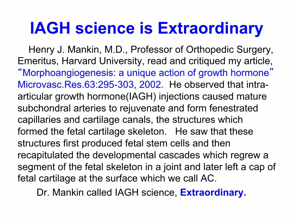
IAGH science is Extraordinary Henry J. Mankin, M.D., Professor of Orthopedic Surgery, Emeritus, Harvard University, read and critiqued my article, “Morphoangiogenesis: a unique action of growth hormone” Microvasc.Res.63:295-303, 2002. He observed that intra-articular growth hormone(IAGH) injections caused mature subchondral arteries to rejuvenate and form fenestrated capillaries and cartilage canals, the structures which formed the fetal cartilage skeleton. He saw that these structures first produced fetal stem cells and then recapitulated the developmental cascades which regrew a segment of the fetal skeleton in a joint and later left a cap of fetal cartilage at the surface which we call AC. Dr. Mankin called IAGH science, Extraordinary.

What is articular cartilage?
Articular cartilage is a growth plate which is a remnant of the fetal cartilage skeleton. It remains as a cap of fetal cartilage at the ends of bones in adults after 98 % of the fetal skeleton has morphed into bone. This fetal cartilage growth plate which we call articular cartilage responds to growth hormone. This is the basis for IAGH.

The problem: It has been impossible to regenerate real articular cartilage(AC) because you must first re-grow a segment of the fetal skeleton in an adult joint. The author’s discovery of develop-mental healing of cartilage has solved this age-old problem. IAGH injections produce fenestrated capillaries(FC) and cartilage canals(CC) which in turn produce stem cells to form the fetal cartilage segment(FCS) and AC. IAGH---FC---CC---FCS---AC

Architecture of Articular cartilage
• Parallel vertical columns of chondro-cytes with arcades at the surface.
• Chondrocytes are produced from stem cells produced by fenestrated
capillaries and cartilage canals. • No inflammation is involved and no
scar is produced.


Fibrocartilage with clumps of cells, clefts and fissures.

Structures and Cascades which regrow articular cartilage
• Fenestrated capillaries, cartilage canals and Glomeruloids all provide stem cells.
• Developmental cascades continue the process and leave a cap of real articular cartilage at the surface.
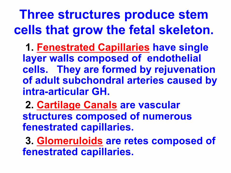
Three structures produce stem cells that grow the fetal skeleton. 1. Fenestrated Capillaries have single
layer walls composed of endothelial cells. They are formed by rejuvenation of adult subchondral arteries caused by intra-articular GH.
2. Cartilage Canals are vascular structures composed of numerous fenestrated capillaries.
3. Glomeruloids are retes composed of fenestrated capillaries.

Fenestrated capillaries form this cartilage canal
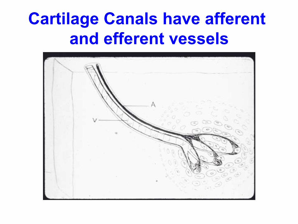
Cartilage Canals have afferent and efferent vessels

Glomeruloids are retes made of Fenestrated Capillaries

Chondrotubules enclose and guide all chondrocytes in matrix.
• Chondrocytes do not lie free in matrix
as presented in textbooks. This “raisins in jello” look is scientifically incorrect.
• All chondrocytes are enclosed in chondrotubules which arise from the vascular structures.
Chondrotubules
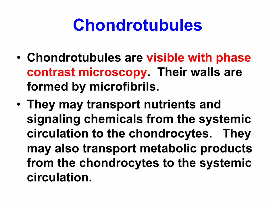
• Chondrotubules are visible with phase contrast microscopy. Their walls are formed by microfibrils.
• They may transport nutrients and signaling chemicals from the systemic circulation to the chondrocytes. They may also transport metabolic products from the chondrocytes to the systemic circulation.

Normal articular surface seen with light microscopy – raisins in jello
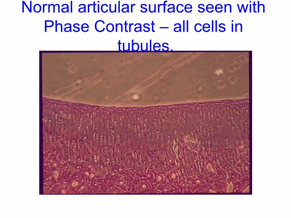
Normal articular surface seen with Phase Contrast – all cells in
tubules.
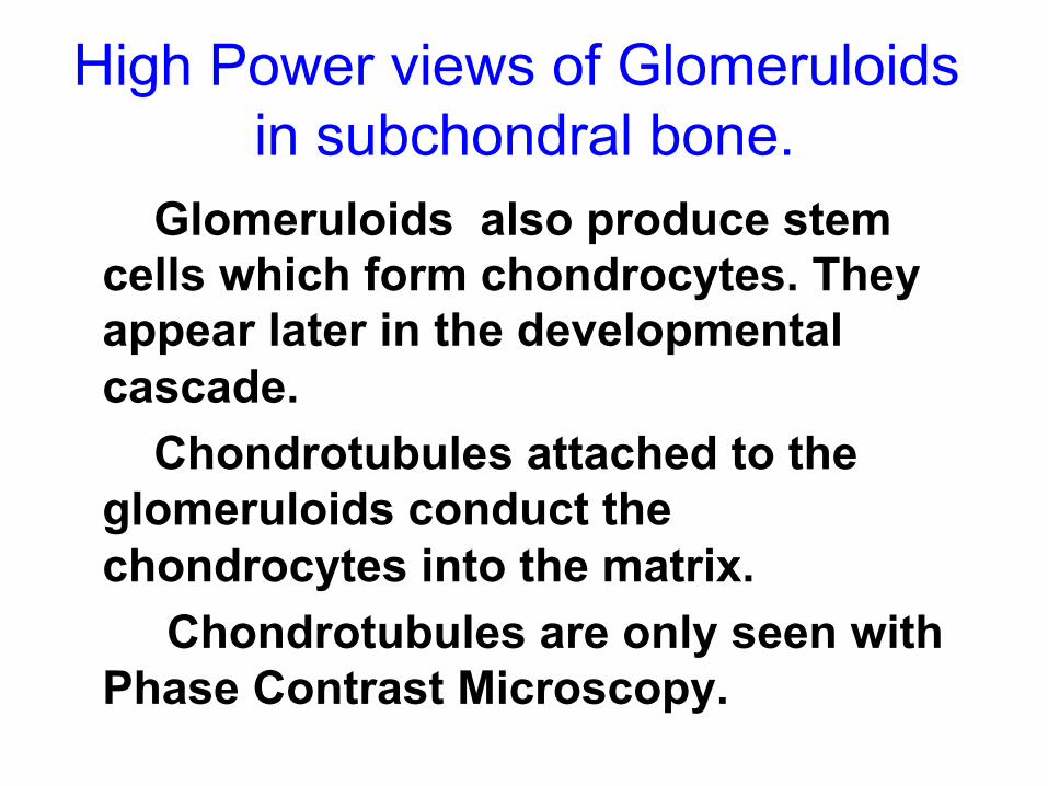
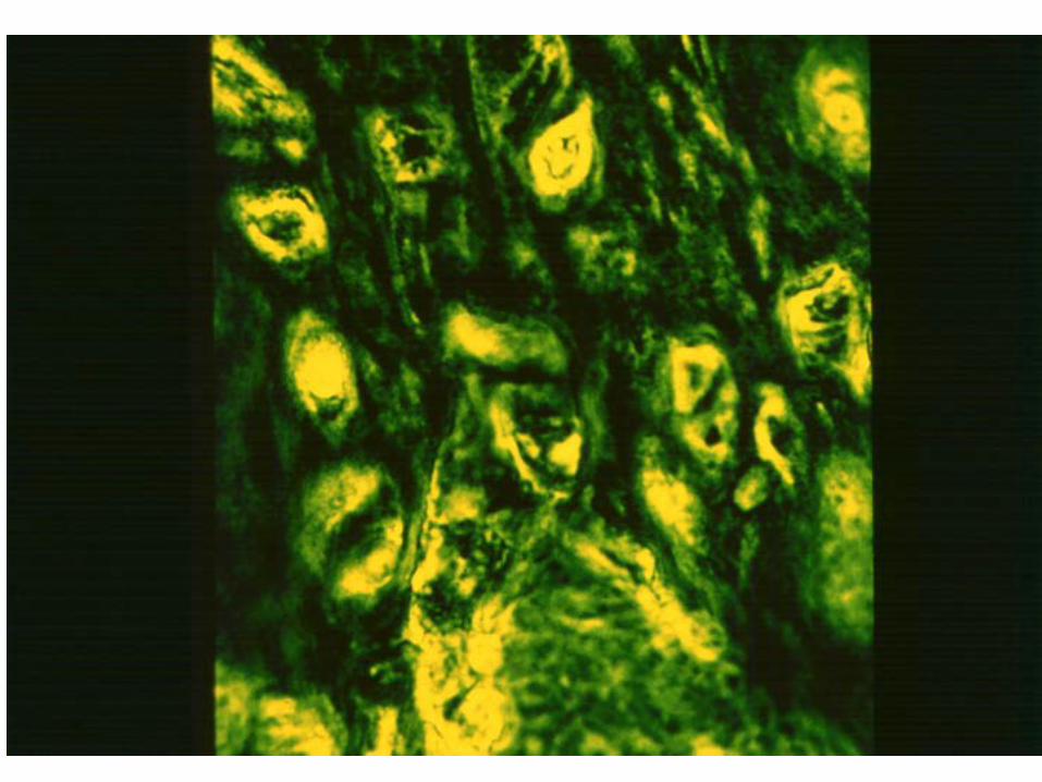
High Power views of Glomeruloids in subchondral bone.
Glomeruloids also produce stem cells which form chondrocytes. They appear later in the developmental cascade.
Chondrotubules attached to the glomeruloids conduct the chondrocytes into the matrix.
Chondrotubules are only seen with Phase Contrast Microscopy.

Glomeruloids in subchondral bone. Light microscopy.

Glomeruloids in subchondral bone. Phase Contrast.
There are two levels of maturity
in a joint 1. Mature areas. 2. Immature areas.

Mature and immature areas in an adult knee Joint
Two Series of Experiments demonstrating the cascades of
regeneration of articular cartilage.
1. Two series of non-operated rabbits – 1965 and 1980. 2. One series of rabbits with surgical debridement of the trochlear surface.

Series of non-operated
mature rabbits. • Control group: • Ten knees injected with 1 ml. of Hank’s buffer, pH 8.2 with no hormone. • Experimental group: • Ten knees injected with 1.2 mgm of purified bovine growth hormone dissolved in 1 ml. of Hank’s buffer pH 8.2.

Non-operated mature joints injected with plain buffer.
Control Group • When the joint is injected with buffer without
hormone nothing happens. • There is a continuous tide-mark. • The osteones are unchanged. • There are vertical parallel columns of cells
formed by a fibrillar microstructure which ends in arcades (normal AC).
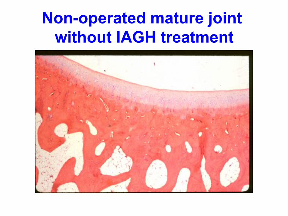
Non-operated mature joint without IAGH treatment
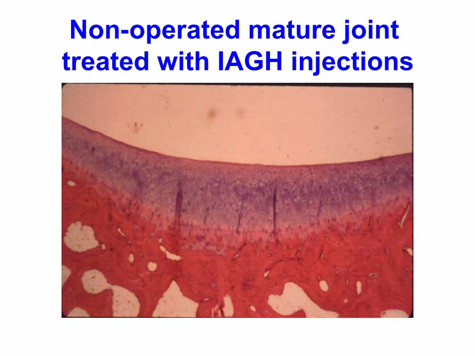
Non-operated mature joint treated with IAGH injections

Non-operated mature joints injected with IAGH.
Experimental Group
Growth hormone has these effects: • The cartilage layer becomes twice as thick
as the control. • The matrix becomes hyper-chromatic. • The tide-mark is no longer continuous and
cellular columns pierce it frequently. • The upper layer of subchondral bone
dematurates and returns to its earlier cartilagenous state, a form of rejuvenation.
NOW TO LEARN ABOUT HEALING.
Healing is complex. Most doctors have never heard of prenatal develop-mental healing(PNDH). I lectured to 250 doctors in the AAOS in New Orleans 2010. When I asked for a show of hands by any who knew anything about PNDH , no hands were raised! PNDH needs to be promoted.

2 Forms of Healing. • 1. Post-natal healing is most familiar. It always forms a scar, i.e. appendix scar. • 2. Pre-natal developmental healing
(PNDH)was discovered and studied by the fetal surgical group at Moffitt Hospital, Univ. Calif. San Francisco 25 + years ago and has resulted in 100’s of peer reviewed articles about PNDH of soft tissue healing.

Post-natal(adult) Healing of Soft Tissue
1. Post-natal healing of soft tissue is familiar because all traumatic and surgical
wounds heal this way. Post-natal healing commences with bleeding and clot formation followed by the in-pouring of inflammatory cell. Later there is an influx of fibroblasts which produce scar.
Summary: Post-natal healing always relies on inflammation and always produces scar.
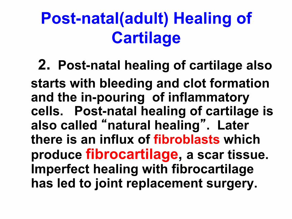
Post-natal(adult) Healing of Cartilage
2. Post-natal healing of cartilage also starts with bleeding and clot formation
and the in-pouring of inflammatory cells. Post-natal healing of cartilage is also called “natural healing”. Later there is an influx of fibroblasts which produce fibrocartilage, a scar tissue. Imperfect healing with fibrocartilage has led to joint replacement surgery.
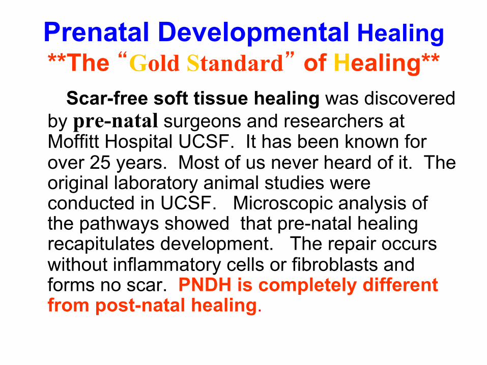
Prenatal Developmental Healing **The “Gold Standard” of Healing**
Scar-free soft tissue healing was discovered by pre-natal surgeons and researchers at Moffitt Hospital UCSF. It has been known for over 25 years. Most of us never heard of it. The original laboratory animal studies were conducted in UCSF. Microscopic analysis of the pathways showed that pre-natal healing recapitulates development. The repair occurs without inflammatory cells or fibroblasts and forms no scar. PNDH is completely different from post-natal healing.

Pre-natal Soft Tissue Healing: Pre-natal soft tissue repair performed
in the first half of gestation is scar-free developmental healing. There are no inflammatory cells, no fibroblasts, and therefore no scar.
A means to translate this pre-natal soft tissue “gold standard” to post-natal clinical surgery has not yet been discovered. Therefore, we have to accept dermal scars. I feel sorry for the plastic surgeons.

Pre-natal Cartilage Healing: Pre-natal cartilage repair is also
developmental healing. There are no inflammatory cells, no fibroblasts and no Fibrocartilage (no scar tissue).
In contrast to the inability to translate pre-natal soft tissue healing to adults, pre-natal cartilage healing has been translated to adult treatment upon the discovery of IAGH. I am happy for the orthopedic surgeons.

Fenestrated capillaries form this cartilage canal
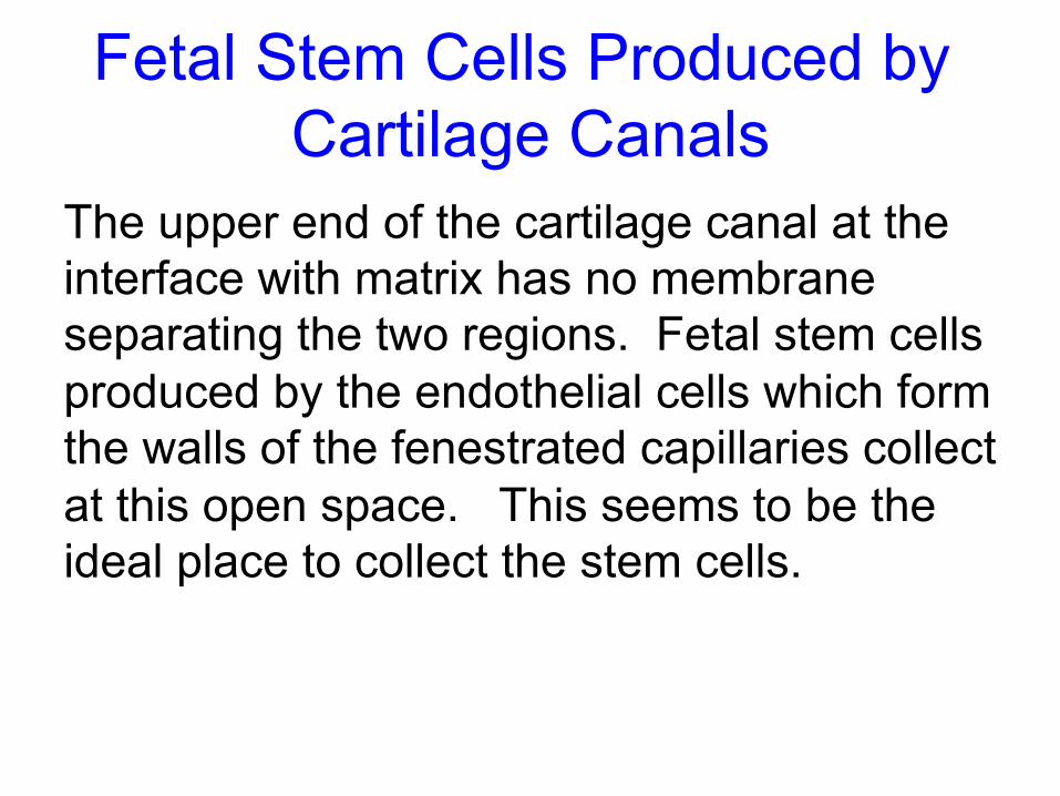
Fetal Stem Cells Produced by Cartilage Canals
The upper end of the cartilage canal at the interface with matrix has no membrane separating the two regions. Fetal stem cells produced by the endothelial cells which form the walls of the fenestrated capillaries collect at this open space. This seems to be the ideal place to collect the stem cells.
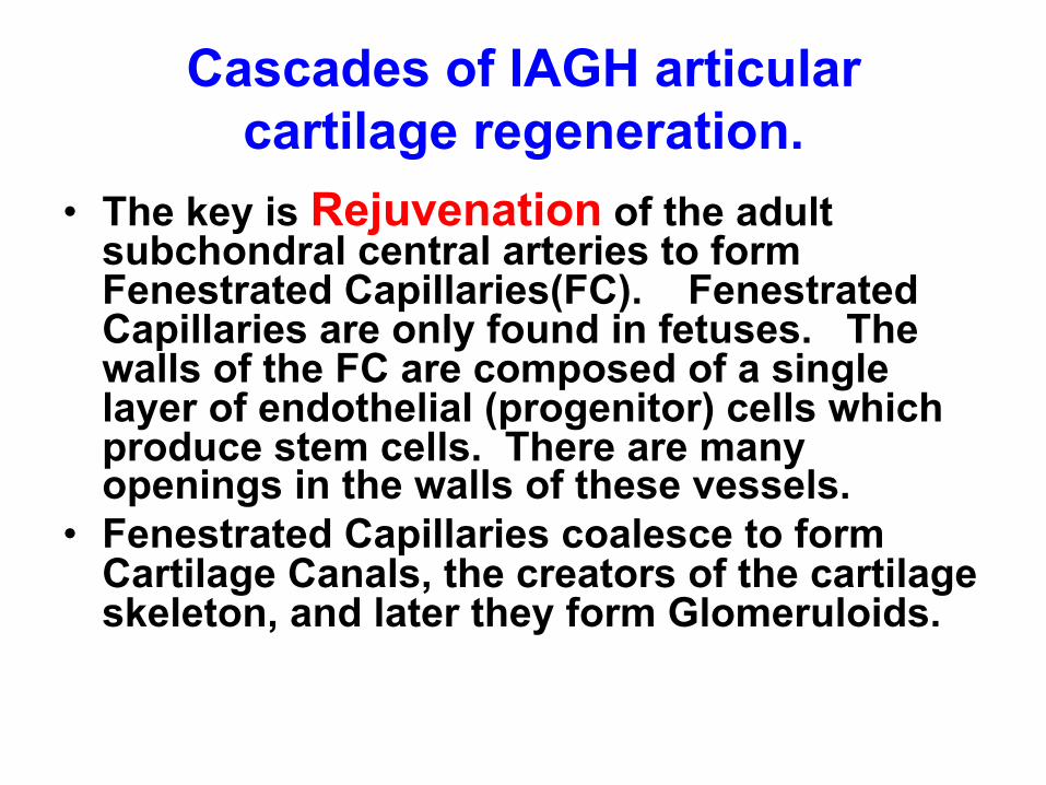
Cascades of IAGH articular cartilage regeneration.
• The key is Rejuvenation of the adult subchondral central arteries to form Fenestrated Capillaries(FC). Fenestrated Capillaries are only found in fetuses. The walls of the FC are composed of a single layer of endothelial (progenitor) cells which produce stem cells. There are many openings in the walls of these vessels.
• Fenestrated Capillaries coalesce to form Cartilage Canals, the creators of the cartilage skeleton, and later they form Glomeruloids.

Cascades Observed in surgically Debrided Knees in Adult Rabbits
• The following are a series of photomicro-graphs from an experiment on adult rabbits. They demonstrate rejuvenation of subchondral vessels and the formation of a segment of the fetal cartilage skeleton in an adult joint. The new cartilage is hyper-chromatic suggesting strong metabolic activity. There are no inflam-matory cells and no fibrocartilage.

Vascular rejuvenation begins soon after surgical debridement and
IAGH injection

Magnification of an osteone enlarging by rapid decalcification 24 hours after surgery and IAGH.
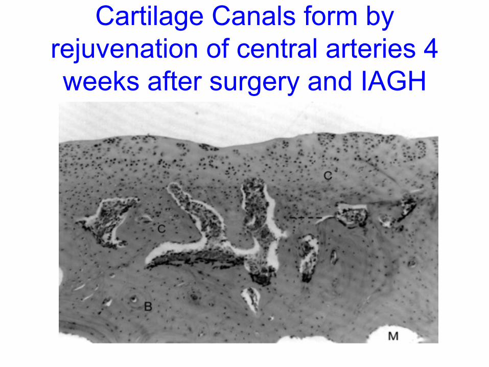
Cartilage Canals form by rejuvenation of central arteries 4 weeks after surgery and IAGH

Cartilage Canals
• Cartilage canals form two weeks after surgery and injection with GH.
• This program focuses on the middle cartilage canal.
• There is no membrane at the interface between the cartilage canal and matrix.
• No inflammatory cells are present and active in the process – it is PNDH!

Cartilage Canal seen with Light Microscopy (“raisins in jello”)

All Chondrocytes are in Chondro-tubules seen with Phase Contrast.

7 weeks post surgery and IAGH injection. No inflammatory cells
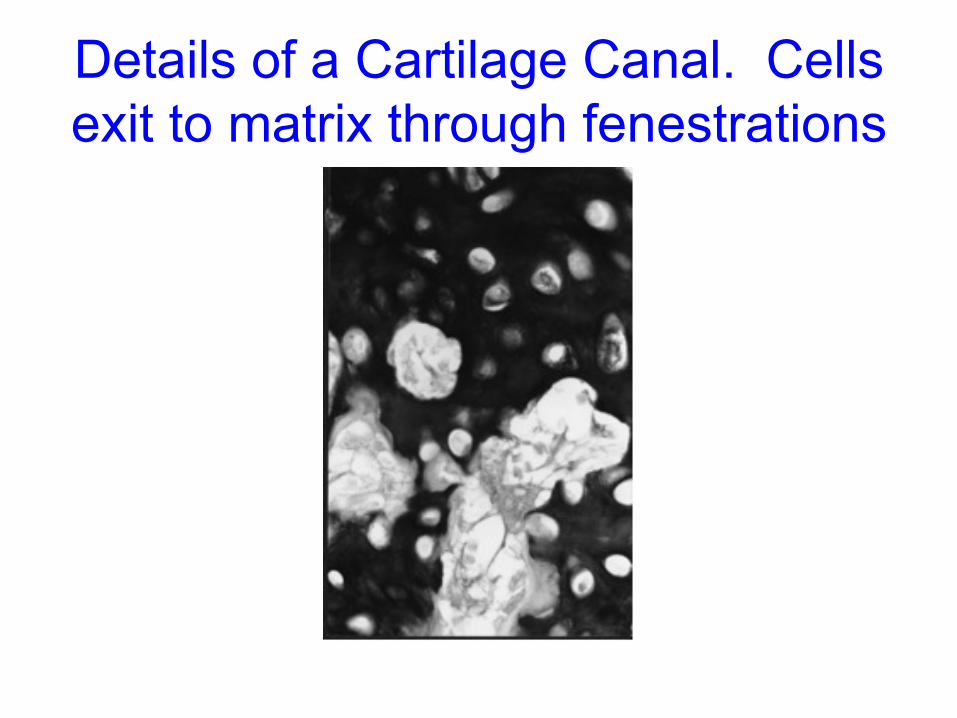
Details of a Cartilage Canal. Cells exit to matrix through fenestrations

Useless Fibrocartilage(103 days) grows without IAGH.

IAGH Produced Surface – Pre-natal Developmental Healing(103 days)

Another IAGH Surface(103 days.) No inflammation, clefts or fissures.
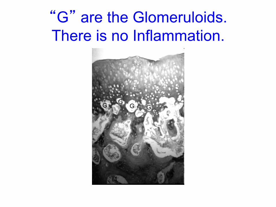
“G” are the Glomeruloids. There is no Inflammation.
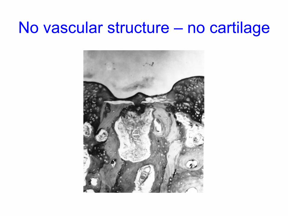
No vascular structure – no cartilage

Summary of production of Fetal Stem Cells
• Mature vascular structures in subchondral bone can be rejuvenated to become fenestrated capillaries as a reaction to local injection of growth hormone.
• Endothelial cells which compose the walls of fenestrated capillaries then produce fetal stem cells.
• The stem cells collect at the open interface between the vascular structures and matrix where they can be harvested.
• Testing can determine that these fetal stem cells are pluripotent and can be signaled to become a variety of tissues, making them a vital source for use in medical science.
• The process is rapid, economical, and avoids all religious and political objections.

Treatment with IAGH • Clinical examples of IAGH treatment –
knees, hips, ankles. • Dosages of Human Growth Hormone
(rHGH) used to regrow articular cartilage.
• Finally, I will explain the benefits of IAGH treatment, and why it should be your first choice for arthritic and infected joints.
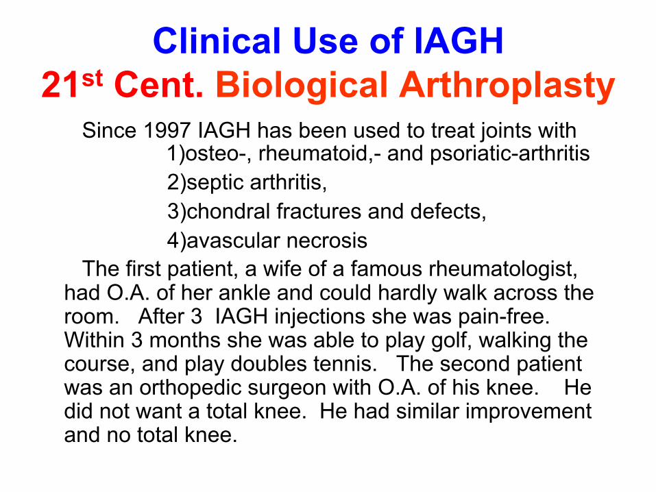
Clinical Use of IAGH 21st Cent. Biological Arthroplasty Since 1997 IAGH has been used to treat joints with
1)osteo-, rheumatoid,- and psoriatic-arthritis 2)septic arthritis, 3)chondral fractures and defects, 4)avascular necrosis The first patient, a wife of a famous rheumatologist,
had O.A. of her ankle and could hardly walk across the room. After 3 IAGH injections she was pain-free. Within 3 months she was able to play golf, walking the course, and play doubles tennis. The second patient was an orthopedic surgeon with O.A. of his knee. He did not want a total knee. He had similar improvement and no total knee.
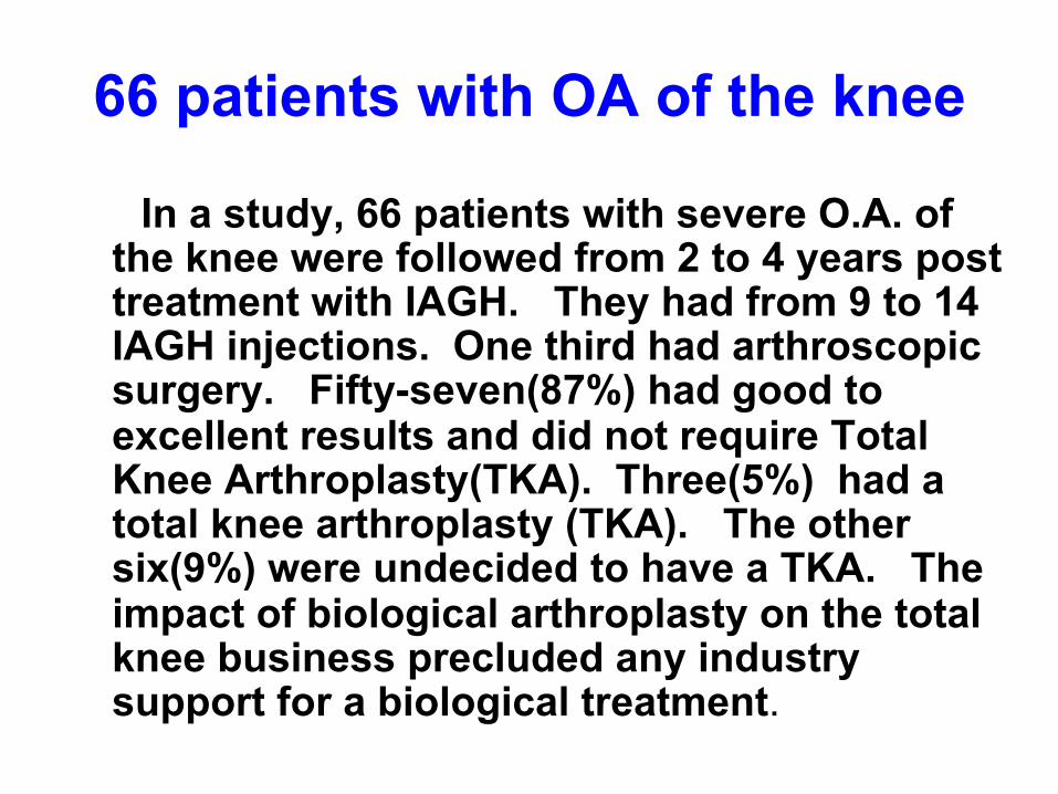
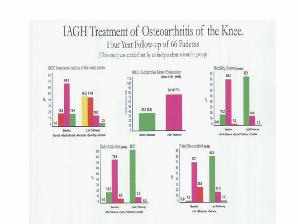
66 patients with OA of the knee
In a study, 66 patients with severe O.A. of the knee were followed from 2 to 4 years post treatment with IAGH. They had from 9 to 14 IAGH injections. One third had arthroscopic surgery. Fifty-seven(87%) had good to excellent results and did not require Total Knee Arthroplasty(TKA). Three(5%) had a total knee arthroplasty (TKA). The other six(9%) were undecided to have a TKA. The impact of biological arthroplasty on the total knee business precluded any industry support for a biological treatment.

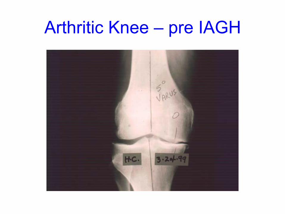
Arthritic knee – pre IAGH

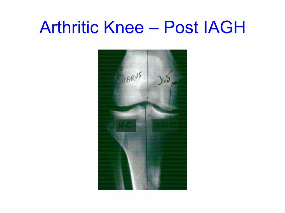
Arthritic Knee – post IAGH
Arthritic Knee – pre IAGH
Arthritic Knee – Post IAGH
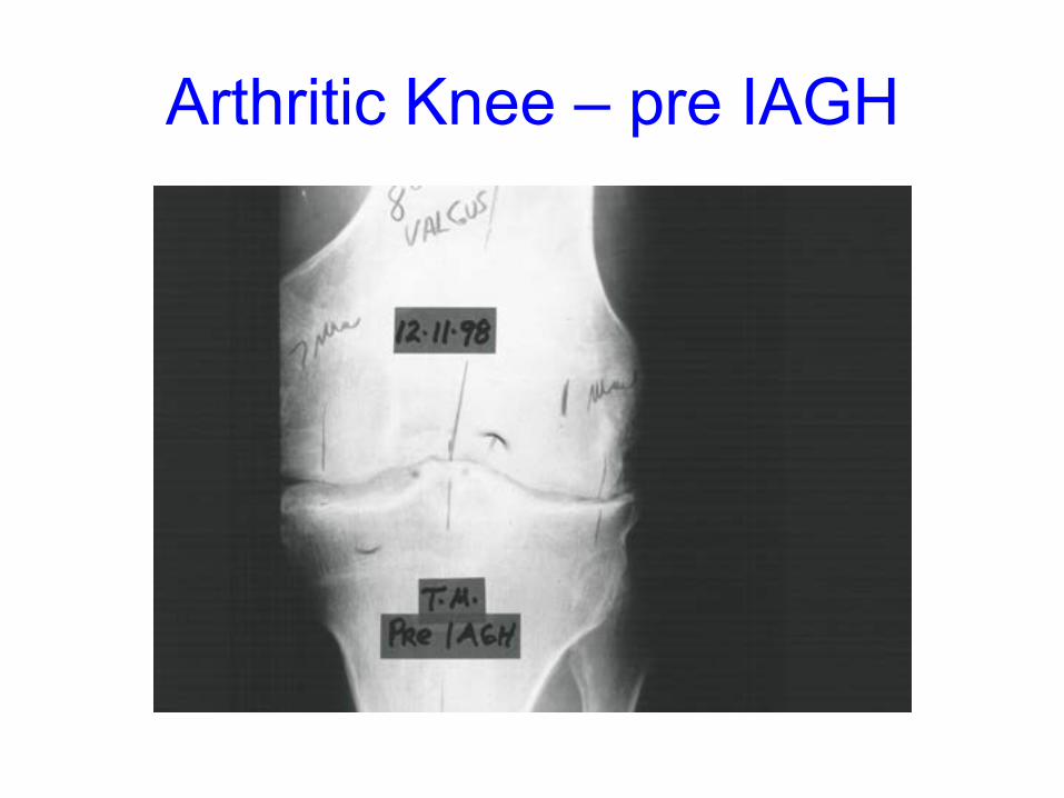
Arthritic Knee – pre IAGH

Arthritic Knee – Post IAGH

Pre – IAGH 10/28/10

Post IAGH 10/6/11
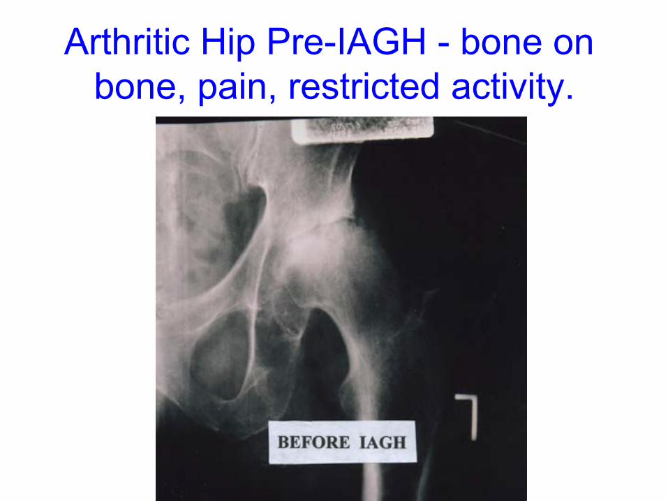
Arthritic Hip Pre-IAGH - bone on bone, pain, restricted activity.

Arthritic Hip Post IAGH – 2.5 to 4.0 mm. joint space, no pain, active.
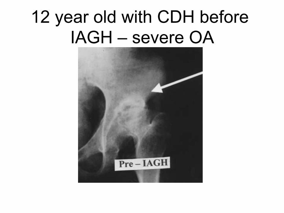
12 year old with CDH before IAGH – severe OA

Same patient after IAGH treatment – no pain


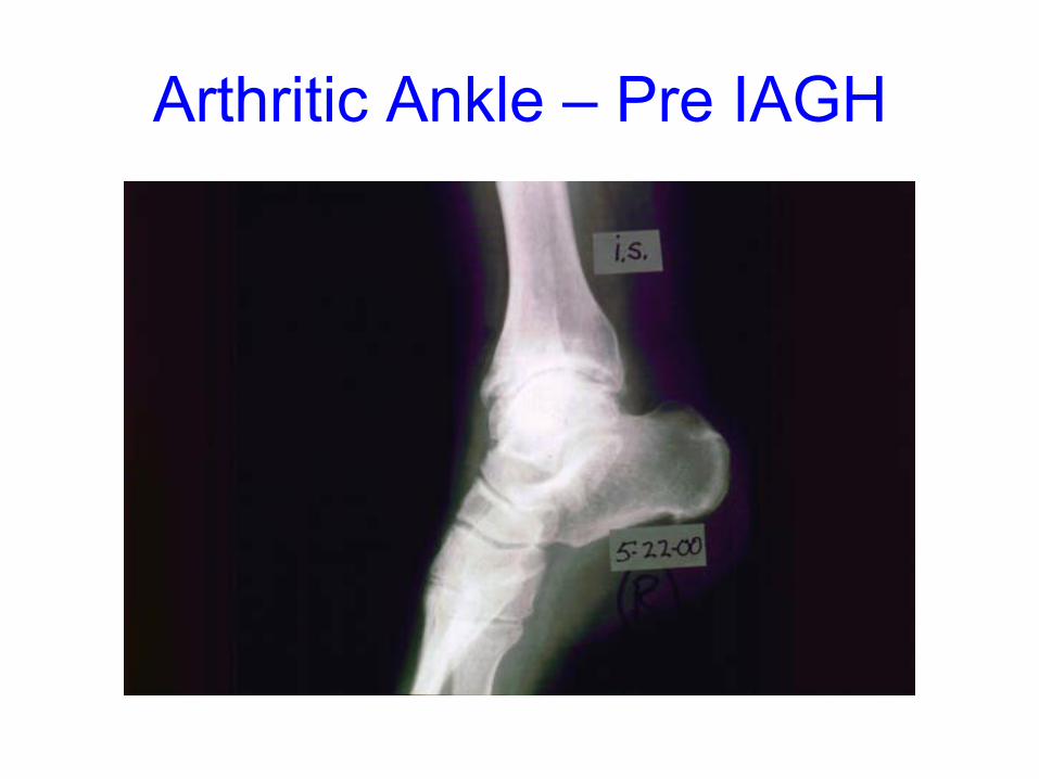
Arthritic Ankle – pre IAGH

Arthritic Ankle – Post IAGH

Severely infected open fracture Pre – IAGH – Pain, Limited Activity.
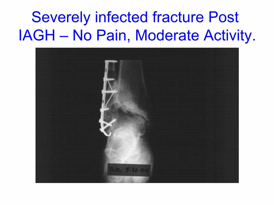
Severely infected fracture Post IAGH – No Pain, Moderate Activity.

Severe infected open fracture Pre – IAGH - Lateral view

Severe infected open fracture Post IAGH – lateral view
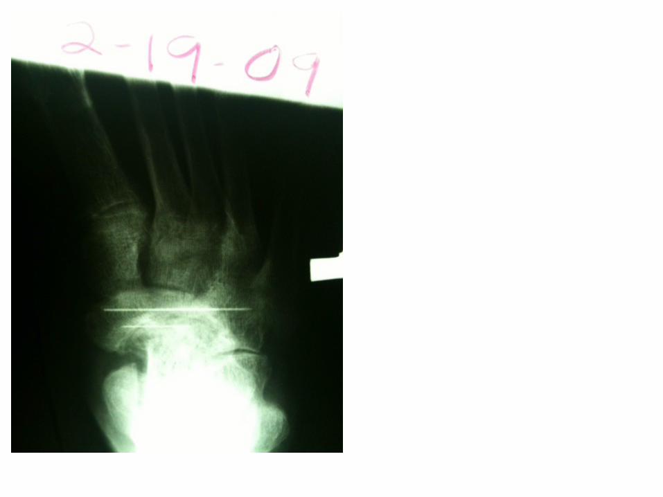
Arthritic Ankle – Pre IAGH

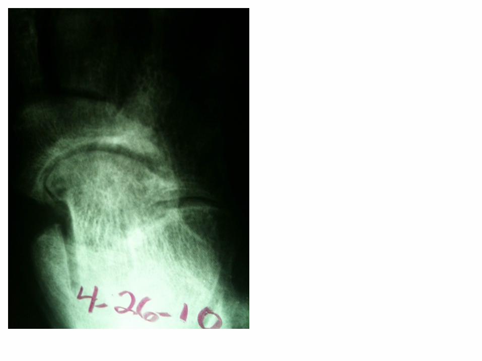
Arthritic Ankle – Post IAGH


Arthritic Sub-talar joint – Pre IAGH Pain and limited activities.
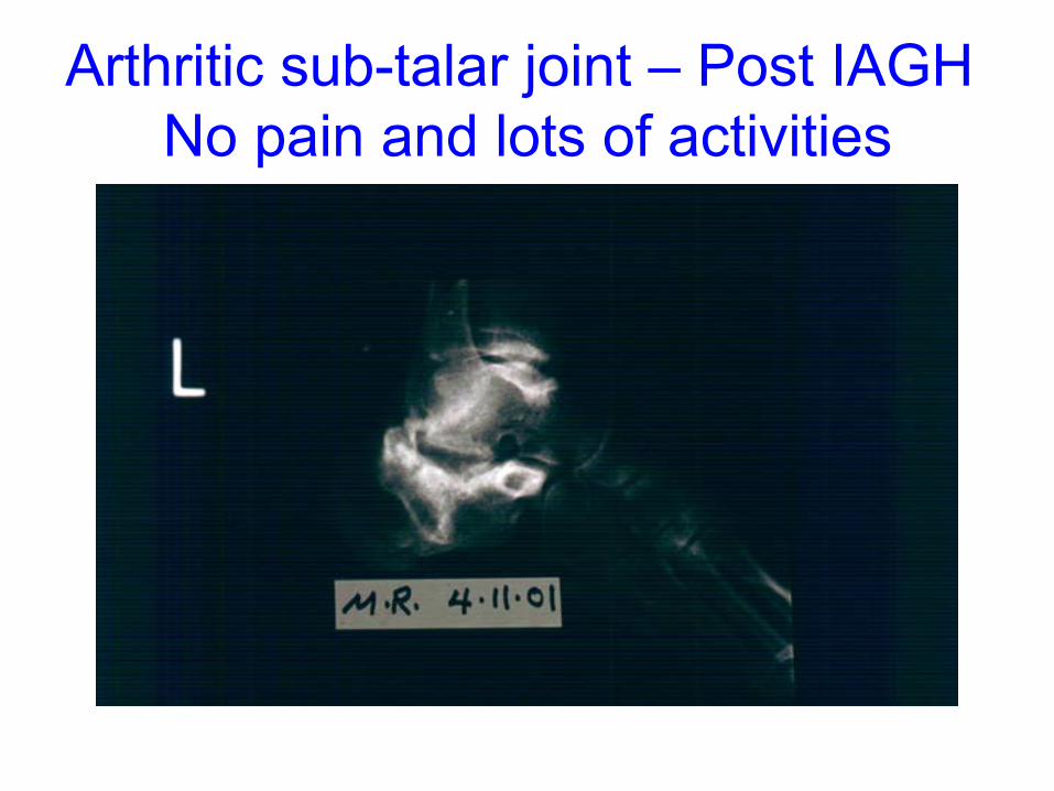
Arthritic sub-talar joint – Post IAGH No pain and lots of activities


Curing AVN of the Talus 1 First injury age 16 was OCD of the talus treated with crutches. Age 30 some pain treated with NSAIDS. Age 50 increasing pain diagnosed as AVN talar body. Offered allograft but refused. Patient came to Florida for IAGH. Had ankle scope. Needed deep currettement to reach bleeding points. 1.2 cm. cavity left empty.

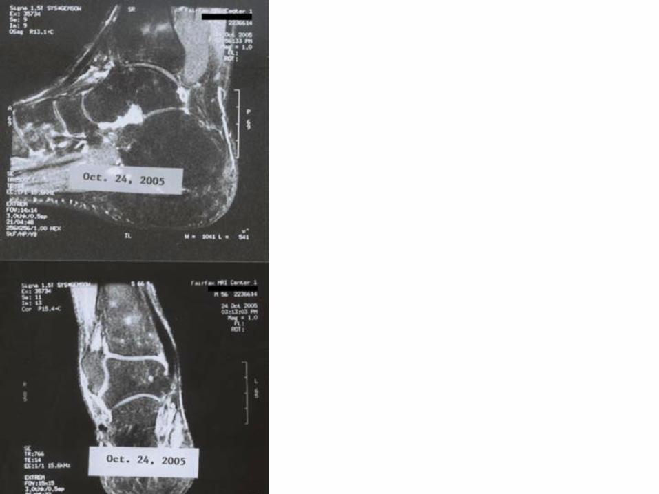
Curing AVN of the Talus 2 Received five weekly injections and 3 monthly IAGH injections 5 mgm each. No weight bearing on the treated side for three months. After 3 months wore a patellar-tendon bearing brace with free ankle motion for 6 months. All pain disappeared. Repeat MRI’s demonstrated complete healing. Returned to full activity.


Dosages of rHGH The dosage of Human Growth Hormone
(HGH) varies according to the size of the joint. Dose varies from 2.5 mgm for small joints to 10 mgm or more for hips. Injections are given weekly for 3 to 12 or more weeks. Injections must be intra-articular, never in the surrounding soft tissue.
Hip, mid-tarsal, sub-talar and base of thumb joints require X-ray control to ensure entry into the joint.

Results of IAGH Treatment Joint % success Knees 87 Ankles and sub-talar 95 Mid-tarsal 95 Shoulders 75 Elbows 95 Wrists 95 Base of thumb 75 Hips (method improving) less than 50 %

Side Effects from IAGH
Side effects are minimal. The most common is occasional mild pain at the injection site lasting a few hours.
With IAGH, there have been none of the complications associated with total joint surgery: No swelling, effusions, infections, no DVT, no pulmonary embolism. No deaths. No patient has gotten worse from IAGH treatment.

Evaluation and testing prior to IAGH
Every patient has X-rays and MRI of the joint to be treated. They also have routine blood tests, and tests for endocrine levels. Women have mammograms, CA-125, estrogen levels and CEA. Men have testosterone levels and PSA. Dr. Dunn examines each patient. He evaluates all the imaging and blood tests. Abnormalities require additional evaluation. 0verweight patients are required to lose weight before treatment. Lower extremity joints must be non weight bearing for the entire treatment period plus an additional 4 weeks. There is no immobilization. Knee, hip and ankle patients are required to walk in shoulder-deep water and swim daily. All patients are given range of motion resistive exercises to do T.I.D. at home. Patients must not smoke, and must avoid second hand smoke. They are permitted one small alcoholic beverage only on week-ends.

Other treatments
• Injections of hyaluronic acid (Synvisc, Euflexxa, etc.) do not grow cartilage, but may reduce pain for up to 6 months.
• PRP (platelet rich plasma)is popular, but has little in the way of scientific studies to support its effects. Does not grow cartilage
• I A injections of pure IGF-1 cause retinal damage and permanent blindness.
• Prolotherapy does not grow cartilage.
Lower Cost of IAGH
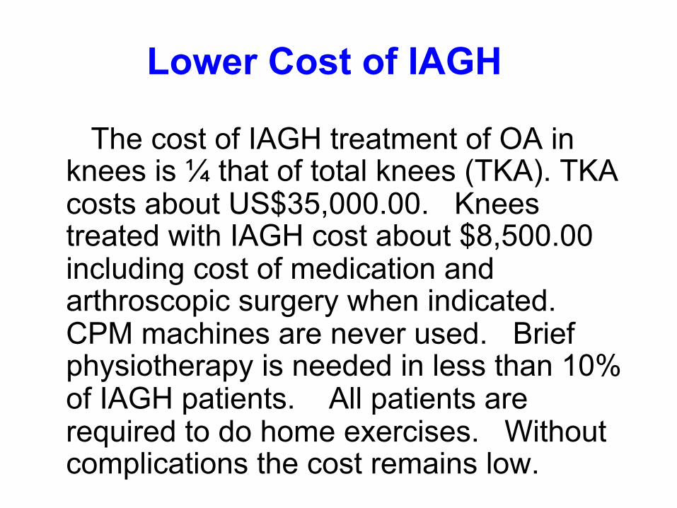
The cost of IAGH treatment of OA in knees is ¼ that of total knees (TKA). TKA costs about US$35,000.00. Knees treated with IAGH cost about $8,500.00 including cost of medication and arthroscopic surgery when indicated. CPM machines are never used. Brief physiotherapy is needed in less than 10% of IAGH patients. All patients are required to do home exercises. Without complications the cost remains low.

IAGH should be the Treatment of Choice
When viewed from the perspective of efficacy, safety, and cost, IAGH should be the first treatment for all arthritic joints. It is 21st Century biological medicine. IAGH should be tried before considering total joint replacement ( a 20th Cent. treatment) or arthrodesis (a 19th Cent. treatment).
The addition of joint distraction has not improved the results when used together with IAGH injections.

Alternative Medicine? No. 21st Century Medicine-Yes.
IAGH - Biological Arthroplasty is the cutting edge of 21st Century medicine.

IAGH- a Safe Effective Treatment
I hope more and more orthopedic surgeons will use IAGH as initial treatment for arthritic joints, septic joints, and AVN as they begin to appreciate the safety, efficacy, and economy of IAGH, .
I invite all doctors interested in IAGH to contact me to learn more. [email protected]

A Parting Thought
The old adage that, once destroyed, articular cartilage can never be restored is now
*History!* Thanks to IAGH.
Allan R. Dunn, M.D.
Miami, Florida
Thank you again for allowing me to tell you
about IAGH.